Embed Size (px)
Citation preview

MEDICAL ILLUSTRATION
Successful Treatment of Discoid Lupus Erythematosus with Chloroquine
Laurentius A. Pramono, Resultanti, Suryo Anggoro, Sandra Langow, Rudy Hidayat, Bambang Setyohadi, Harry IsbagioDepartment of Internal Medicine, Faculty of Medicine, Universitas Indonesia – Cipto Mangunkusumo Hospital, Jakarta, Indonesia.
Correspondence mail:Department of Internal Medicine, Faculty of Medicine University of Indonesia-Cipto Mangunkusumo Hospital.Jl. Diponegoro no. 71, Jakarta 10430, Indonesia. email: [email protected].
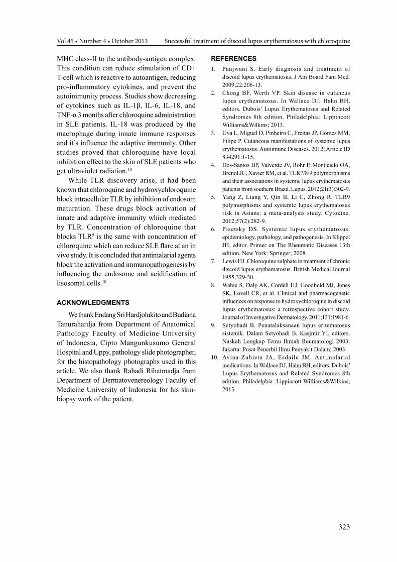
Figure 1. Discoid LE in the scalp before treatment with chloroquine
Figure 2. Discoid LE in the scalp after 2 months treatment with chloroquine
Figure 3. Histopathology of the skin shows discoid LE (400 times)
321Acta Medica Indonesiana - The Indonesian Journal of Internal Medicine

Laurentius A. Pramono Acta Med Indones-Indones J Intern Med
Discoid lupus erythematosus (DLE) is a chronic cutaneus lupus with no internal organ involvement.1,2 There are several conditions lead to manifestation of cutaneus lupus, such as genetic, environment, and drug-induced. Partial deficiencies of C2 and C4 complement reported in chronic cutaneus lupus, including discoid lupus and panniculitis lupus. Genetic polimorphism that lead to this diseases are pro-inflammatory cytokines, tyrosine kinase-2, Fc receptors II (FcRII), T-cell receptor loci, adhesion molecules, antioxidant enzymes, and apoptotic genes. Ultraviolet rays can trigger chronic cutaneus lupus and smoking habbit is closely related to discoid lupus.2
This is a case of a 37 year-old woman who suffers from itching, hair loss, and alopecia in several location (multiple) in her scalp since 6 years ago. She had been given several cream and medication from several physicians but none of them help her problem. The description of the lesions are alopecia in several spots (multiple), atrophic scars, numular, discrete, and firm border in its lesion (Figure 1).
There are no systemic problems complained by the patient, such as fever, fatigue, musculoskeletal pain, fotosensitivity, malar rash, dyspnea, pleuritic pain, urinary problem, and edema. From the physical examination, there were no abnormalities except the scalp. From the laboratory examination, there were no hematologic abnormality, proteinuria, and raised of ureum and creatinin level. The ANA (antinuclear antibody) was positive 1/320 (coarse speckled), but the complement level (C3 and C4) was normal (C3 113 and C4 26) and also anti-ds DNA was normal (<2,6). From the chest x-ray, there are no pleural effusions.
The patient underwent skin biopsy at the department of dermatology, and from the histopathologic features, there were hyperkeratocis and atrophy in the epidermis, followed by thickening in the basement membrane, and also follicular plugging. This feature is diagnosed histopathologically as discoid lupus erythematosus (Figure 3). After compiling data from clinical symptoms, laboratories, and histopatology examination, the patient was diagnosed with discoid lupus
erythematosus. We gave the patient chloroquine (250 mg) once daily (after we checked her retina with an ophthalmologist) and the dermatologist gave her desonide cream as topical medication. After 2 months of treatment, the lesions in the scalp showed improvement, atrophic scar and hair loss begin to diminish, and itching symptom reduced (Figure 2). The medication was continued.
Discoid lupus is one of many clinical manifestations of lupus.3 The manifestation of lupus is influenced by the expression of toll-like receptors (TLR).4-6 This condition explains why patient can get systemic (SLE) or localized (cutaneus) lupus. DLE may develop to SLE with the rate of 20% in 20 years.2 Other literature says 1% to 5% of DLE develop to SLE.1 The diagnosis of discoid lupus was based on clinical features. Histopathology examination confirm the diagnosis; with the characteristic of “it is that of a lichenoid tissue reaction with changes at the dermo-epidermal junction that include thickening of the basement membrane and vacuolar degeneration of the basal cells along with perivascular and peri-appendageal inflammatory cell infiltration of a variable degree in the reticular dermis. Hyperkeratosis is more evident and follicular plugging may be seen in more mature lesions”.1
Antimalarials were first reported as effective agents for discoid lupus in a case series published by British Medical Journal (BMJ) in 1955.7
Antimalarial agents such as chloroquine and hydroxychloroquine have important effect for discoid lupus and SLE affecting to the skin.1,2,8
It is the first-line systemic therapy for DLE.1 Several effects of antimalarials in cutaneus lupus are (1) sunblocking and sunscreen effect as chloroquine can bind to the melanin, (2) immunosupressant by binding to lysosomal membrane to interrupt α and β chain metabolism at HLA class-II, and (3) anti-inflammation by reducing IL-1, IL-6, and TNF-α release from macrophage and IL-2 and IFN-γ release from T-cell.9,10
Antimalarials such as hydroxychloroquine and chloroquine increase intracytoplasm pH. With the increase in pH, chloroquine blocks the process and gathering of the self-peptide with
322
Vol 45 • Number 4 • October 2013 Successful treatment of discoid lupus erythematosus with chloroquine
MHC class-II to the antibody-antigen complex. This condition can reduce stimulation of CD+ T-cell which is reactive to autoantigen, reducing pro-inflammatory cytokines, and prevent the autoimmunity process. Studies show decreasing of cytokines such as IL-1β, IL-6, IL-18, and TNF-α 3 months after chloroquine administration in SLE patients. IL-18 was produced by the macrophage during innate immune responses and it’s influence the adaptive immunity. Other studies proved that chloroquine have local inhibition effect to the skin of SLE patients who get ultraviolet radiation.10
While TLR discovery arise, it had been known that chloroquine and hydroxychloroquine block intracellular TLR by inhibition of endosom maturation. These drugs block activation of innate and adaptive immunity which mediated by TLR. Concentration of chloroquine that blocks TLR9 is the same with concentration of chloroquine which can reduce SLE flare at an in vivo study. It is concluded that antimalarial agents block the activation and immunopathogenesis by influencing the endosome and acidification of lisosomal cells.10
ACknowLEDgmEnTS
We thank Endang Sri Hardjolukito and Budiana Tanurahardja from Department of Anatomical Pathology Faculty of Medicine University of Indonesia, Cipto Mangunkusumo General Hospital and Uppy, pathology slide photographer, for the histopathology photographs used in this article. We also thank Rahadi Rihatmadja from Department of Dermatovenereology Faculty of Medicine University of Indonesia for his skin-biopsy work of the patient.
REFEREnCES1. Panjwani S. Early diagnosis and treatment of
discoid lupus erythematosus. J Am Board Fam Med. 2009;22:206-13.
2. Chong BF, Werth VP. Skin disease in cutaneus lupus erythematosus. In Wallace DJ, Hahn BH, editors. Dubois’ Lupus Erythematosus and Related Syndromes 8th edition. Philadelphia: Lippincott Williams&Wilkins; 2013.
3. Uva L, Miguel D, Pinheiro C, Freitas JP, Gomes MM, Filipe P. Cutaneous manifestations of systemic lupus erythematosus. Autoimune Diseases. 2012; Article ID 834291:1-15.
4. Dos-Santos BP, Valverde JV, Rohr P, Monticielo OA, Brenol JC, Xavier RM, et al. TLR7/8/9 polymorphisms and their associations in systemic lupus erythematosus patients from southern Brazil. Lupus. 2012;21(3):302-9.
5. Yang Z, Liang Y, Qin B, Li C, Zhong R. TLR9 polymorphisms and systemic lupus erythematosus risk in Asians: a meta-analysis study. Cytokine. 2012;57(2):282-9.
6. Pisetsky DS. Systemic lupus erythematosus: epidemiology, pathology, and pathogenesis. In Klippel JH, editor. Primer on The Rheumatic Diseases 13th edition. New York: Springer; 2008.
7. Lewis HJ. Chloroquine sulphate in treatment of chronic discoid lupus erythematosus. British Medical Journal 1955;329-30.
8. Wahie S, Daly AK, Cordell HJ, Goodfield MJ, Jones SK, Lovell CR, et al. Clinical and pharmacogenetic influences on response to hydroxychloroqine in discoid lupus erythematosus: a retrospective cohort study. Journal of Investigative Dermatology. 2011;131:1981-6.
9. Setyohadi B. Penatalaksanaan lupus eritematosus sistemik. Dalam Setyohadi B, Kasjmir YI, editors. Naskah Lengkap Temu Ilmiah Reumatologi 2003. Jakarta: Pusat Penerbit Ilmu Penyakit Dalam; 2003.
10. Avina-Zubieta JA, Esdaile JM. Antimalarial medications. In Wallace DJ, Hahn BH, editors. Dubois’ Lupus Erythematosus and Related Syndromes 8th edition. Philadelphia: Lippincott Williams&Wilkins; 2013.
323


![Atypical Presentations of Cutaneous Leishmaniasis: A Real ......[Figure 5], erysipeloid [Figure 6 and 7], psoriasiform [Figure 8], pseudotumoral, discoid lupus-like, squamous cell](https://img.dokumen.tips/doc/110x75/610115ca5befcb26727dacd5/atypical-presentations-of-cutaneous-leishmaniasis-a-real-figure-5-erysipeloid.jpg)