Embed Size (px)
Citation preview

Abilene Christian UniversityDigital Commons @ ACU
Honors College ACU Student Research, Theses, Projects, andDissertations
12-2017
Sub-cloning Genes of the Mevalonate Pathwayfrom Enterococcus faecalis into pDUET andpET28Madison Anne HarrisAbilene Christian University
Follow this and additional works at: https://digitalcommons.acu.edu/honors
This Thesis is brought to you for free and open access by the ACU Student Research, Theses, Projects, and Dissertations at Digital Commons @ ACU.It has been accepted for inclusion in Honors College by an authorized administrator of Digital Commons @ ACU.
Recommended CitationHarris, Madison Anne, "Sub-cloning Genes of the Mevalonate Pathway from Enterococcus faecalis into pDUET and pET28" (2017).Honors College. 31.https://digitalcommons.acu.edu/honors/31

1
Sub-cloning Genes of the Mevalonate Pathway from
Enterococcus faecalis into pDUET and pET28
An Honors College Project Thesis
Presented to
The Department of Chemistry and Biochemistry
Abilene Christian University
In Partial Fulfillment
of the Requirements for
Honors Scholar
by
Madison Anne Harris
December 2017

2
Copyright 2017
Madison Anne Harris
ALL RIGHTS RESERVED

3

4
ABSTRACT
All organisms are capable of synthesizing isopentenyl pyrophosphate (IPP), a precursor to many
different biomolecules such as ubiquinone Q8, cholesterol, and β-carotene. Two pathways, the
mevalonate and non-mevalonate pathway, synthesize IPP. Unlike most eubacteria that use the
non-mevalonate pathway, low-G+C gram-positive cocci bacteria solely use the mevalonate
pathway. This pathway differs from mammals at the rate-limiting step of HMG-CoA reductase
and is a potential target for future antibiotics against nosocomial infections caused by
Enterococcus faecalis. Unique to enterococci is a fusion protein (encoded by mvaE) made of the
first enzyme (acetoacetyl-CoA thiolase) and the third enzyme (3-hydroxy-3-methylglutaryl-CoA
reductase) of the pathway. This research focuses on the first three enzymes of the pathway and
relies on a series of sub-cloning. First, to isolate thiolase from the fusion protein, sub-cloning
mvaC, the gene for thiolase, into pET28 was attempted. Secondly, sub-cloning of the gene for
the fusion protein (mvaE) and gene for HMG-CoA synthase (mvaS), which is the second enzyme
in the pathway, into pDUET was attempted. Each sub-cloning reaction employed a double
restriction digest, gel electrophoresis, DNA purification, ligation of insert and vector,
transformation, and mini-preparation of plasmid DNA. Despite repeated attempts and
troubleshooting, results were inconclusive. This research has led to the refining of the sub-
cloning protocol using the pDUET vector, and future research is needed to understand more
about the fusion protein in Enterococcus faecalis.

5
TABLE OF CONTENTS
INTRODUCTION .......................................................................................................................... 6
MATERIALS AND METHODS .................................................................................................. 11
Materials. ................................................................................................................................... 11
Double Restriction Digest. ........................................................................................................ 11
Gel Electrophoresis. .................................................................................................................. 11
DNA Purification. ..................................................................................................................... 12
Phenol/Chloroform Extraction. ................................................................................................. 12
Dephosphorylation. ................................................................................................................... 12
Ligation. .................................................................................................................................... 12
Transformation. ......................................................................................................................... 13
Isolation of ligated plasmid. ...................................................................................................... 13
Mini-preparation of DNA.......................................................................................................... 13
Nanodrop. .................................................................................................................................. 13
RESULTS ..................................................................................................................................... 14
Genes cut from pET28. ............................................................................................................. 14
Vectors pDUET and pET28. ..................................................................................................... 15
Sub-cloning mvaC into pET28. ................................................................................................. 15
Sub-cloning mvaE and mvaS into pDUET. ............................................................................... 18
mvaE ...................................................................................................................................... 18
mvaS....................................................................................................................................... 19
DISCUSSION ............................................................................................................................... 20
ACKNOWLEDGMENTS ............................................................................................................ 23
REFERENCES ............................................................................................................................. 24

6
INTRODUCTION
Enterococcus faecalis is a gram-positive bacterium that causes nosocomial infections and is
problematic as some strains are antibiotic resistant (1). New methods to target E. faecalis are
needed. One possible target is the bacteria’s biosynthesis of isopentenyl diphosphate (IPP), a
five-carbon precursor to many different molecules such as steroids, cholesterol, and coenzyme Q
(1, 2).
IPP is synthesized in one of two ways, the mevalonate pathway (Fig. 1) or the non-mevalonate
pathway (1, 2). Unlike most bacteria, which synthesize IPP by the non-mevalonate pathway,
low-G+C gram-positive cocci such as the Enterococcus species, Staphylococcus species, and
Streptococcus species synthesize IPP by the mevalonate pathway (2). This is similar to
eukaryotes, archaea, and fungi (2). Previous studies comparing DNA sequences suggest that low-
G+C gram-positive cocci acquired the mevalonate pathway through horizontal gene transfer with
primitive eukaryotes (2).
The mevalonate pathway begins with acetyl-CoA and produces IPP after several enzymatic
reactions. The first three enzymes of the pathway, acetoacetyl-CoA thiolase, 3-hydroxy-3-
methylglutyral-CoA (HMG-CoA) synthase, and HMG-CoA reductase are of particular interest.
The rate-limiting step for this pathway is at HMG-CoA reductase. Previous studies have
determined two different classes of HMG-CoA reductase, based on the kinetic properties and
structure of the enzyme (2). While eukaryotes employ class I HMG-CoA reductase, E. faecalis,
S. aureus, and S. pneumonia all use class II. Class I HMG-CoA reductase is more sensitive to
statin drugs than class II reductase (2). This is a key point in developing a new antibiotic that can

7
target HMG-CoA reductase class II in gram-positive cocci and not target HMG-CoA class I in
mammals.
Figure 1: The Mevalonate Pathway (4).

8
Importantly, the mevalonate pathway is essential for gram-positive cocci bacteria to live, as
determined in a gene-knockout study with S. pneumonia (2). Bactericidal effects were observed
when the gene for HMG-CoA reductase (mvaA) was knocked out and a supply of mevalonate
was not provided. E. faecalis is inferred to behave in the same manner because it is a gram-
positive cocci bacterium and solely uses the mevalonate pathway (2). Combining the fact that the
mevalonate pathway is believed to be essential to E. faecalis and differs from that in mammals, it
is a potential target for new antibiotics.
Unique to Enterococci is a fusion protein comprising the first and third enzymes of its
mevalonate pathway, thiolase (encoded by mvaC) and reductase (encoded by mvaA) (2). The two
enzymes are encoded on the same RNA transcript, with one start codon and one stop codon.
Their transcript is translated into one large fusion protein, encoded by mvaE (Fig. 2). Little is
known about the fusion protein, so it is of interest to research. Previously, reductase was isolated
from Enterococcus faecalis and it was determined to be a dimer (4). However, isolated reductase
presented different kinetic properties than when part of the fusion protein (4). Thiolase has yet to
be isolated from the fusion protein, and the proper folding of the fused protein in E. faecalis is of
interest to research.
mvaS mvaA
Figure 2. Depiction of the fusion protein in Enterococcus faecalis that is encoded by the gene mvaE.
mvaE contains the genes for thiolase (mvaC) and reductase (mvaA). The gene that encodes synthase
is mvaS (2).
9
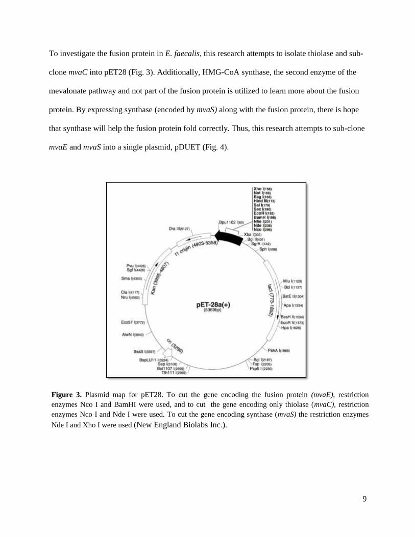
To investigate the fusion protein in E. faecalis, this research attempts to isolate thiolase and sub-
clone mvaC into pET28 (Fig. 3). Additionally, HMG-CoA synthase, the second enzyme of the
mevalonate pathway and not part of the fusion protein is utilized to learn more about the fusion
protein. By expressing synthase (encoded by mvaS) along with the fusion protein, there is hope
that synthase will help the fusion protein fold correctly. Thus, this research attempts to sub-clone
mvaE and mvaS into a single plasmid, pDUET (Fig. 4).
Figure 3. Plasmid map for pET28. To cut the gene encoding the fusion protein (mvaE), restriction
enzymes Nco I and BamHI were used, and to cut the gene encoding only thiolase (mvaC), restriction
enzymes Nco I and Nde I were used. To cut the gene encoding synthase (mvaS) the restriction enzymes
Nde I and Xho I were used (New England Biolabs Inc.).

10
Figure 4. The plasmid map for pDUET used to sub-clone both mvaE and mvaS (New England
Biolabs Inc.).

11
MATERIALS AND METHODS
Materials. Vectors used include pDUET and pET28 (New England Biolabs Inc.). E. coli DH5
alpha competent cells (New England Biolabs Inc.) were used. Enzymes used include Nco I, Nde
I, Bam HI, Xho I, T4 bacteriophage ligase, RNAase (New England Biolabs Inc). Buffers used
include buffer 3.1, CutSmart buffer, 6x loading dye, and ligase buffer (New England Biolabs
Inc.), as well as freshly prepared 0.5 X TBE (Sigma-Aldrich). Other material included the Sigma
GenElute Gel Extraction kit (Sigma-Aldrich), DNA ladder (GoldBio), Luria-Bertani media
(Fischer), agar (Fischer), and 1% ethidium bromide (Fischer-BioReagents).
Double Restriction Digest. For mvaE, buffer 3.1 and restriction enzymes Nco I and Bam HI
were used to cut pDUET and pET28 with mvaE. Restriction enzymes Nde I and Xho I were used
to cut pDUET and pET28 with mvaS, and the cutsmart buffer was used. To cut pET28 and mvaC
from the fusion protein, Nde I, Nco I, and buffer 3.1 were used. The total volume of each double
restriction digest was 50 μL. Each double restriction digest incubated at 37 ˚C from 15 minutes
to 4 hours. Typical run time was 1 hour (9).
Gel Electrophoresis. Gel electrophoresis was used to obtain the desired fragments of cut DNA
following the restriction digest. A 0.7 % agarose gel was used with 0.5x TBE buffer and a 1 %
solution of ethidium bromide. Loading dye was added to each sample to a final concentration of
1X. Gel wells were individually loaded with the DNA ladder and the maximum amount of
sample. Electrophoresis ran typically at 100 volts for 1 hour. Bands were viewed with UV light.
Desired DNA fragments were excised (10).

12
DNA Purification. Excised bands were weighed and then purified from the gel with the Sigma
GenElute Gel Extraction kit. The kit provided the Column Preparation Solution, Gel
Solubilization Solution, Wash Solution Concentrate G, Elution Solution, GenElute binding
columns, and collection tubes for the experiment. The procedure was followed exactly as
described by the kit (5).
Phenol/Chloroform Extraction. If gel electrophoresis was omitted for the vectors, a phenol
chloroform extraction was used to remove the protein from the nucleic acids. The protocol for
this was obtained from the Molecular Cloning Laboratory Manual (8). ACS grade Chloroform
was used, as well as a 1:1 ratio of Phenol:Chloroform in TB. The aqueous layer was removed
three total times.
Dephosphorylation. Dephosphorylation of cut pDUET was carried out following the procedure
from the Molecular Cloning Laboratory Manual (7). Calf intestinal alkaline phosphatase (CIP)
was used to remove the 5’ phosphate group along with a 10x CIP buffer, and incubated for 30
minutes at 37 ˚C.
Ligation. The insert (mvaS, mvaE, or mvaC) and corresponding vector (pDUET or pET28) were
ligated together. The quantity of vector and insert used was determined by the concentration and
the New England Biolabs online generator for 2:1, 3:1, and 5:1 ratios of insert:vector. T4 DNA
bacteriophage ligase and ligase buffer were used. Ligation conditions included incubation at
room temperature for four hours, 37 ˚C for one hour, or stored in the refrigerator overnight. The
most frequent ligation condition was at 37 ˚C for one hour (11).

13
Transformation. E. coli DH5 alpha competent cells were transformed with ligation samples,
and were plated onto Luria-Bertani (LB) media with the necessary antibiotic. Ampicillin plates
(50 μg/mL) were used for experiments with pDUET, and kanamycin plates (10 μg/mL) were
used for pET28. Incubated overnight (about 17 hours) at 37 ˚C.
Isolation of ligated plasmid. Transformed colonies that grew overnight were stabbed at random
and grown in liquid LB media with the appropriate antibiotic. Three to five colonies from each
plate were stabbed to account for mutations. The colonies were grown overnight with shaking at
37oC in LB with 50 μg/mL of ampicillin for pDUET, and 10 μg/mL of kanamycin for pET28.
Mini-preparation of DNA. After incubating at 37 ˚C overnight, DNA for each sample was
mini-prepped to remove the DNA from the competent E. coli cells. Protocol for the small-scale
preparation of plasmid DNA was obtained from the Molecular Cloning Laboratory Manual (6).
Concentrations to make solutions 1-3 were provided in the lab procedure, with solution 2
prepared fresh for each mini-preparation. The final DNA was redissolved in a TE buffer of pH
8.0, and RNAase (0.5 mg/ml) was added to remove any RNA from the sample.
Nanodrop. To check the DNA concentration after a mini-preparation as well as a purification of
DNA, the Nanodrop was used. DNA was noted by a significant peak at 260 nm and was
differentiated from protein by looking at the 260:280 ratio. A peak at any other wavelength
indicated a contamination.

14
RESULTS
Genes cut from pET28. Each gene was successfully cut out of the plasmid pET28 (Fig. 3).
Restriction enzymes Nco I and Bam HI successfully cut mvaE from pET28. The 2,500 base pair
mvaE was seen on the gel. Nde I and Nco I successfully cut mvaC, about 1,500 base pairs, and
Nde I and Xho I successfully cut mvaS, about 1,100 base pairs (Fig. 5).
2.5 kB
1.5 kB
1.0 kB
Figure 5. A) Double Restriction Digest to cut inserts from pET28. The green box highlights mvaE; the
blue box highlights mvaS, and the yellow box highlights mvaC. B) DNA ladder used to identify to correct
DNA fragment by looking at base pair length (12). The two brightest bands are 3,000 bps and 1,000 bps.
A B

15
The restriction digest was typically run for an hour at 37 ˚C. A short (15 minutes) and long (1
hour) restriction digest were originally attempted, with the short digest cutting the insert equally
as well as the long digest. Thereafter, Nco I began to have problems cutting the DNA, so new
enzyme was ordered from New England Biolabs Inc. Each subsequent restriction digest ran for
an hour and successfully cut the appropriate gene from pET28 as described above.
Vectors pDUET and pET28. Unlike the successful restriction digests with each gene, cutting
plain plasmid, pDUET and pET28, were not always cut with both enzymes. Sometimes the
restriction digest with the two appropriate enzymes for pDUET and pET28 was successful.
However, this was not consistent.
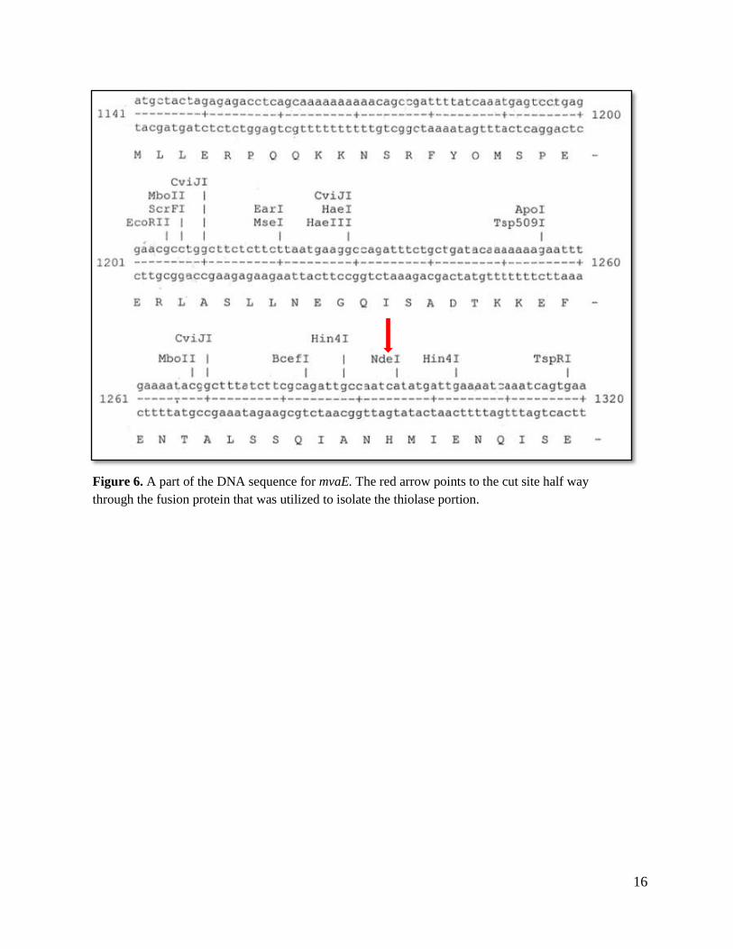
Sub-cloning mvaC into pET28. To isolate thiolase from the fusion protein, mvaE was cut with
Nco I (at the beginning of the sequence) and Nde I, which was in the middle of the sequence
(Fig. 6). As previously mentioned, mvaC was successfully cut from the fusion protein. pET28
appeared to be cut by the restriction enzymes; however, they have not been successfully ligated
together.
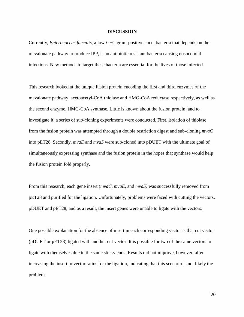
The results of the ligation of mvaC with cut pET28 showed about 5-10 colonies for each
insert:vector ratio, 2:1, 3:1, and 5:1 (Fig. 7). Some colonies grew on the control plate, although
fewer than the experimental plates. After growing the colonies overnight, mini-prepping the
DNA, and running a diagnostic restriction digest, it was determined that the colonies that grew
did not contain the mvaC insert (Fig. 8).
16
Figure 6. A part of the DNA sequence for mvaE. The red arrow points to the cut site half way
through the fusion protein that was utilized to isolate the thiolase portion.
17
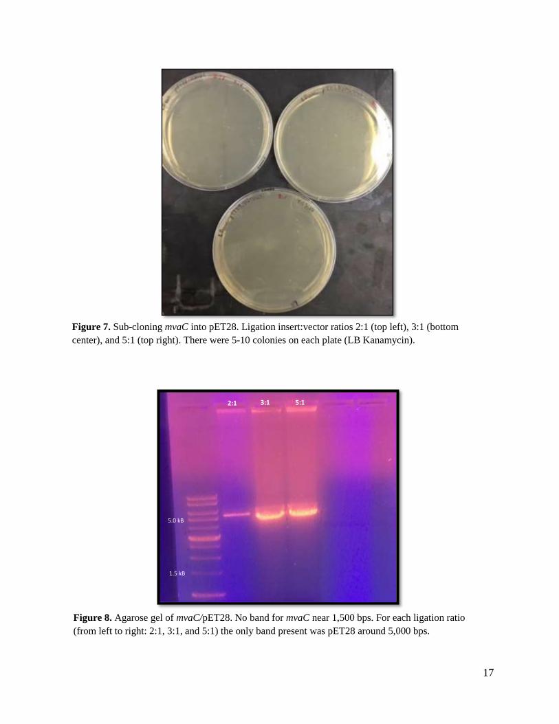
Figure 7. Sub-cloning mvaC into pET28. Ligation insert:vector ratios 2:1 (top left), 3:1 (bottom
center), and 5:1 (top right). There were 5-10 colonies on each plate (LB Kanamycin).
Figure 8. Agarose gel of mvaC/pET28. No band for mvaC near 1,500 bps. For each ligation ratio
(from left to right: 2:1, 3:1, and 5:1) the only band present was pET28 around 5,000 bps.
1.5 kB
2:1 3:1 5:1
5.0 kB

18
Sub-cloning mvaE and mvaS into pDUET. pDUET was used in the reaction with mvaE and
mvaS, as it has multiple cloning sites. For both experiments with pDUET, the ligation was not
successful.
mvaE. To sub-clone mvaE into pDUET, the plasmid was cut with Nco I and Bam HI. As
described above, a new supply of Nco I was ordered, and the reaction was allowed to progress
for 1 hour rather than 15 minutes. Under these conditions, mvaE was consistently cut from
pET28, while results for the restriction digest with pDUET were inconsistent. Purifying mvaE
and what appeared to be cut pDUET, were ligated together for a short time (1 hour at 37
˚Celsius) and long time (4 hours at room temperature). Colonies grew for both the short and long
ligation, and there was an absence of growth on the cut control (Fig. 9). However, a diagnostic
restriction digest revealed that mvaE failed to insert into pDUET (Fig. 10).
Figure 9. LB Ampicillin plates with sub-cloned mvaE in pDUET. No growth on cut control (top left).
Colonies grew for both short ligation time (top right), and the long ligation (bottom center).

19
This experiment was repeated with a phenol:chloroform extraction of the vector as well as
dephosphorylation of the 5’ end of pDUET, as described in the methods section. Following these
changes, mvaE still did not ligate with pDUET.
mvaS. Similar problems were experienced with sub-cloning mvaS into pDUET. Purified
mvaS and cut pET28 with restriction enzymes Nde I and Xho I failed to ligate together. Colonies
never grew for this set of experiments.
Figure 10. Agarose gel of short ligation time for mvaE/pDUET plasmids. No band for the insert
gene, mvaE at 2,500 bps. pDUET fragment is shown at 5,000 base pairs.
5.0 kB
2.5 kB
20
DISCUSSION
Currently, Enterococcus faecalis, a low-G+C gram-positive cocci bacteria that depends on the
mevalonate pathway to produce IPP, is an antibiotic resistant bacteria causing nosocomial
infections. New methods to target these bacteria are essential for the lives of those infected.
This research looked at the unique fusion protein encoding the first and third enzymes of the
mevalonate pathway, acetoacetyl-CoA thiolase and HMG-CoA reductase respectively, as well as
the second enzyme, HMG-CoA synthase. Little is known about the fusion protein, and to
investigate it, a series of sub-cloning experiments were conducted. First, isolation of thiolase
from the fusion protein was attempted through a double restriction digest and sub-cloning mvaC
into pET28. Secondly, mvaE and mvaS were sub-cloned into pDUET with the ultimate goal of
simultaneously expressing synthase and the fusion protein in the hopes that synthase would help
the fusion protein fold properly.
From this research, each gene insert (mvaC, mvaE, and mvaS) was successfully removed from
pET28 and purified for the ligation. Unfortunately, problems were faced with cutting the vectors,
pDUET and pET28, and as a result, the insert genes were unable to ligate with the vectors.
One possible explanation for the absence of insert in each corresponding vector is that cut vector
(pDUET or pET28) ligated with another cut vector. It is possible for two of the same vectors to
ligate with themselves due to the same sticky ends. Results did not improve, however, after
increasing the insert to vector ratios for the ligation, indicating that this scenario is not likely the
problem.

21
Specifically for the pDUET/mvaE experiment, it is possible that the vector pDUET re-ligated
with itself. Due to very similar, sticky ends cut by Nco I and Bam HI, it is possible that one
pDUET vector re-ligated (Fig. 11). The sticky ends were the same length, and when ligated
together have only two mismatched base pairs. Prevention of this problem was attempted by
dephosphorylating the 5’ end of pDUET; however, this did not improve the results. Additional
trials of this research are needed to understand if the sticky ends of pDUET are ligating with
themselves.
Because of the difficulty reproducing our results of cutting each plasmid (pET28 and pDUET),
another explanation is that the restriction enzymes did not initially cut the vectors, as they
should. For pET28, there was some growth (about 5 colonies) on the cut control plate suggesting
that it is likely both enzymes failed to cut pET28. The same problem could explain the results for
pDUET. After running a four-hour long double restriction digest for each sub-cloning
experiment (pDUET/mvaE, pDUET/mvaS, and pET28/mvaC), enzymes Nco I and/or Nde I
seemed to be having trouble cutting the plasmids.
A B C
Figure 11. Restriction sites for the restriction enzyme BamHI (A) and Nco I (B), and the possibility of
the sticky ends of pDUET re-ligating with itself without the insert gene (C).

22
Additional restriction digests with fresh enzymes and various digest conditions are necessary to
improve the cutting of pDUET and pET28. With successful restriction digests, there is hope to
sub-clone mvaE and mvaS into pDUET and mvaC into pET28. A second idea to express mvaS
and mvaE simultaneously is to sub-clone mvaS into pET21 and mvaE in pET28 to grow together
in a transformation. The two vector plasmids, pET21 and pET28 have different antibiotic
resistance, so the only cells that should grow will have genes for both synthase and the fusion
protein. After each gene has been sub-cloned into its corresponding vector, future research is
needed on the expression of the genes to study the structure and function of the fusion protein as
well as thiolase alone. More understanding of the structure and function of thiolase and the
fusion protein may provide useful knowledge for future drug targets on the mevalonate pathway
in E. faecalis.

23
ACKNOWLEDGMENTS
From the Welch Foundation, I am thankful for their funding of this research (Grant No. R-0021).
I would also like to thank the Department of Chemistry and Biochemistry at Abilene Christian
University for providing the Summer Research Internship that initiated my research project, as
well as the resources, technology, and means to conducting this research up to the present time.
A special thank you to Dr. Sutherlin, for allowing me the opportunity to work alongside her in
her research, teaching me, and pushing me to develop a scientific mind. Finally yet importantly, I
would like to thank my advisory committee Dr. Sarah Lee, Dr. Qiang Xu, and Dr. Kim Pamplin,
and the Honors College for the opportunity and guidance in writing an honors thesis.

24
REFERENCES
1. Held, M. et al. 2002. Enterococcus faecalis Acetoacetyl-Coenzyme A Thiolase/3-Hydroxy-
3-Methylglutaryl-Coenzyme A Reductase, a Dual-Function Protein of Isopentenyl
Diphosphate Biosynthesis. J. Bacteriol. 184: 2116-2122.
2. Imogen Wilding, E. et al. 2000. Identification, Evolution, and Essentiality of the
Mevalonate Pathway for Isopentenyl Diphosphate Biosynthesis in Gram-Positive Cocci. J.
Bacteriol. 182: 4319–4327.
3. Sutherlin, A. et al. 2002. Enterococcus faecalis 3-Hydroxy-3-Methylglutaryl Coenzyme A
Synthase, an Enzyme of Isopentenyl Diphosphate Biosynthesis. J. Bacteriol. 184: 4065–
4070.
4. Sutherlin, A. The Mevalonate Pathway of Isopentenyl Pyrophosphate Biosynthesis in
Enterococcus faecali, Ph.D Thesis, Purdue University, 2003.
5. GenElute Gel Extraction Kit. Sigma-Aldrich. St. Louis, MO. Catalog number NA1111.
6. Sambrooke, J. et al. 1989. “Small-scale Preparations of Plasmid DNA.” Molecular Cloning:
A Laboratory Manual. Cold Spring Harbor Laboratory Press, USA. 2nd
edition. (1.25-1.28).
7. Sambrooke, J. et al. 1989. “Dephosphorylation of Linearized Plasmid DNA.” Molecular
Cloning: A Laboratory Manual. Cold Spring Harbor Laboratory Press, USA. 2nd
edition.
(1.60-1.61).
8. Sambrooke, J. et al. 1989. “Extraction with Pehnol:Chloroform.” Molecular Cloning: A
Laboratory Manual. Cold Spring Harbor Laboratory Press, USA. 2nd
edition. (E.3-E.4)
9. New England Biolabs. 2013. “Double Digest Protocol with Standard Restriction Enzymes.”
New England Biolabs Inc. [Online]. https://www.neb.com/protocols/2014/05/07/double-
digest-protocol-with-standard-restriction-enzymes. (Accessed November 6, 2017).

25
10. Sambrooke, J. et al. 1989. “Separation of Restriction Fragments of GGel Electrophoresis.”
Molecular Cloning: A Laboratory Manual. Cold Spring Harbor Laboratory Press, USA. 2nd
edition. (6.2-6.13).
11. Sambrooke, J. et al. 1989. “Ligation of Cohesive Termini.” Molecular Cloning: A
Laboratory Manual. Cold Spring Harbor Laboratory Press, USA. 2nd
edition. (1.68-1.69).
12. Gold Bio. 2014. “DNA Ladder.” Gold Biotechnology, UAS. [Online].
https://www.goldbio.com/product/14/1-kb-dna-ladder. (Accessed November 6, 2017).


![Cold shock induction of recombinant Arctic environmental genes · Sub-cloning of genes into the pCold-II-based vectors was done by RF-cloning [22, 23], as described below. Several](https://img.dokumen.tips/doc/110x75/5f0c76ec7e708231d43588d8/cold-shock-induction-of-recombinant-arctic-environmental-genes-sub-cloning-of-genes.jpg)