Embed Size (px)
Citation preview

Studying Ascl1-GSX2 interactions
Juliana MadziaMentor: Kaushik Roychoudhury
PI: Kenny Campbell

Transcription Factor Regulation of Progenitor Maturation
• During development, Ventral telencephalic progenitors differentiate and give rise to neurons, oligodendrocytes, and astrocytes.
• The early progenitors divide to form more progenitors or give rise to intermediate progenitors that mature into neurons or glia
• Presence of GSX2 in the LGE progenitors keep them from differentiating
• Presence of GSX1 and Ascl1 in the progenitors help them differentiate
• GSX2 appears to be upstream of Ascl1 in LGE progenitors

BackgroundWhile performing a variety of candidate protein-
protein interaction tests, we found that in the yeast, Ascl1 (Mash1) binds directly to GSX2, but not GSX1
We could co-precipitate Mash1 while pulling down GSX2, both from in vitro synthesized protein mixture as well as from E12.5 embryo telencephalon.
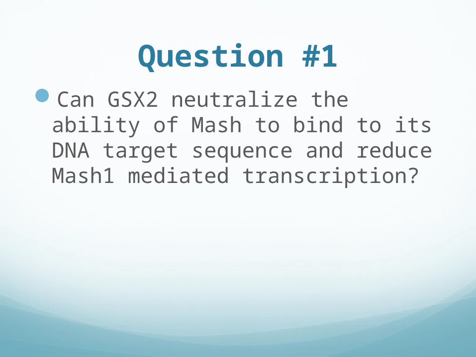
Question #1Can GSX2 neutralize the ability of
Mash to bind to its DNA target sequence and reduce Mash1 mediated transcription?

Luciferase assay
Basic ConstructB
indin
g si
te
Prom
oter
Reporter
(Firefly lu
ciferase
)

Luciferase Assay
Mash OnlyB
indin
g si
te
Prom
oter
Reporter
(Firefly lu
ciferase
)Mash1

Luciferase Assay
Bin
din
g si
te
Prom
oter
Reporter
(Firefly lu
ciferase
)Mash1
Gsx2
Mash and GSX2

Kaushik’s old data on high affinity e-box (In HEK 293T
cells)
No Mash,
No GSX
0ng 6ng 12ng 25ng 50ng 100ng 200ng No Mash, 100
GSX2
0
10
20
30
40
50
60
70
80
90
100
12.5ng Mash
Fold
in
crease
over
contr
ol
Increasing GSX2

Kaushik’s old data on Delta N E-box (Low affinity E-box) in P-19 cells

Experimental Procedure
1. Plated 2x10⁴ NIH3t3 fibroblast cell line in wells of 48 well plates
2. Incubated in a 5% CO2 incubator at 37°C for 12 hours
3. Transfected cells with different combinations of Delta enhancer Luciferase construct, Ascl1, Gsx2, Beta actin promoter driven renilla luciferase and empty pCDNA6V5 plasmid.
4. Incubated another 48 hours in 5% CO2, 37°C
5. Lysed cells, transferred lysate to luminometer and quantified luminiscence
6. Recorded firefly luciferase and renilla luciferase activity
0 50 100 150 200 250 3000
5
10
15
20
25
30
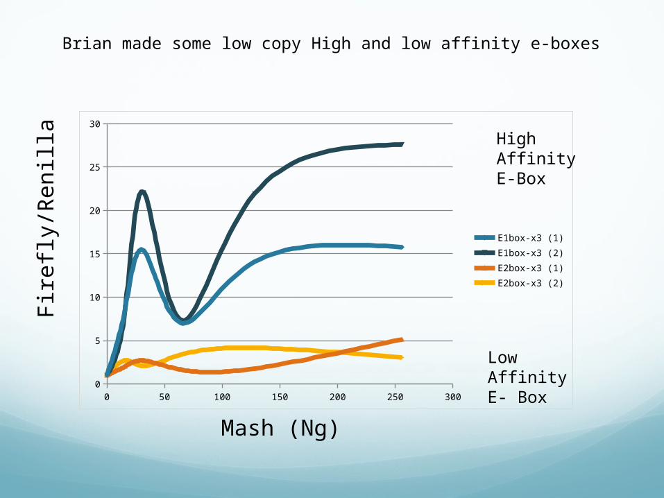
E1box-x3 (1)E1box-x3 (2)E2box-x3 (1)E2box-x3 (2)
High Affinity E-Box
Low Affinity E- Box
Brian made some low copy High and low affinity e-boxes
Mash (Ng)
Fire
fly/R
en
illa

0 20 40 60 80 100 120 1400
0.5
1
1.5
2
2.5Fi
refly/r
enill
a
0 20 40 60 80 100 120 1400
0.5
1
1.5
2
2.5
3
3.5
4
4.5
Fire
fly/r
enill
a
Ng of Mash
Delta M Long
0 20 40 60 80 100 120 1400
0.5
1
1.5
2
2.5
3
3.5
4
4.5
5
Ng Mash
Ng of Mash
Fire
fly/r
enill
a

Results
Delta L Delta L+ Mash
DeltaL + Mash+ Gsx2
0
0.2
0.4
0.6
0.8
1
1.2
1.4
Delta
ML
DML+16
Mas
h
DML+16
Mas
h+50
ng G
sx2
DML+16
Mas
h+20
0ng
Gsx2
0
0.5
1
1.5
2
2.5
3
3.5
4
64ng Mash116ng Mash1
Error bar = 1 Standard Deviation of three independent cultures
6.5 fold
14 fold

Question #2
• Do Mash1 and GSX2 physically interact in mammalian cells?

Proximity Ligation Assay
A pair of oligonucleotide labeled secondary antibodies (PLA probes) generates a signal only when the two PLA probes have bound in close proximity
The signal from each detected pair of PLA probes is visualized as an individual fluorescent spot
PLA signals can be quantified (counted) and assigned to a specific subcellular location based on microscopy images

1st PrincipleA. The samples are incubated with primary
antibodies that bind to the protein(s) to be detected.

2nd PrincipleSecondary antibodies conjugated with
oligonucleotides (PLA probe MINUS and PLA probe PLUS) are added to the reaction and incubated.
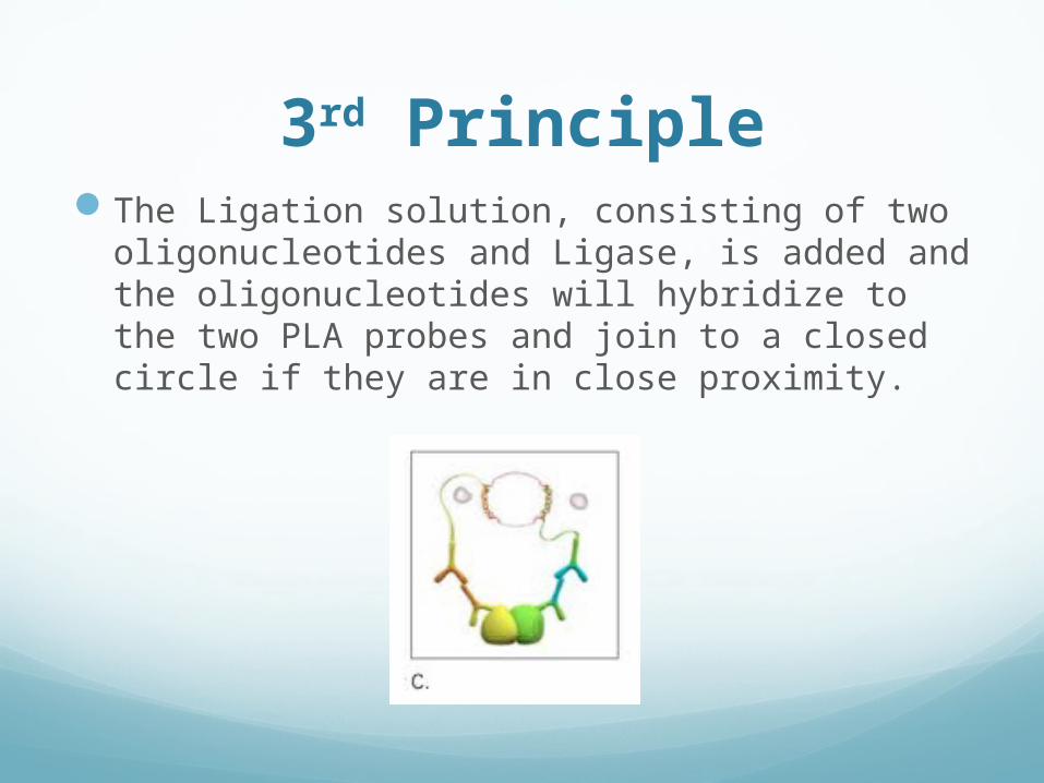
3rd PrincipleThe Ligation solution, consisting of two
oligonucleotides and Ligase, is added and the oligonucleotides will hybridize to the two PLA probes and join to a closed circle if they are in close proximity.

4th Principle The Amplification solution is added together with Polymerase.
The oligonucleotide arm of one of the PLA probes acts as a primer for a rolling-circle amplification reaction using the ligated circle as a template, generating a concatemeric (repeated sequence) product. The fluorescently labeled oligonucleotides will hybridize to the RCA product. The signal is easily visible as a distinct fluorescent spot and analyzed by fluorescence microscopy.
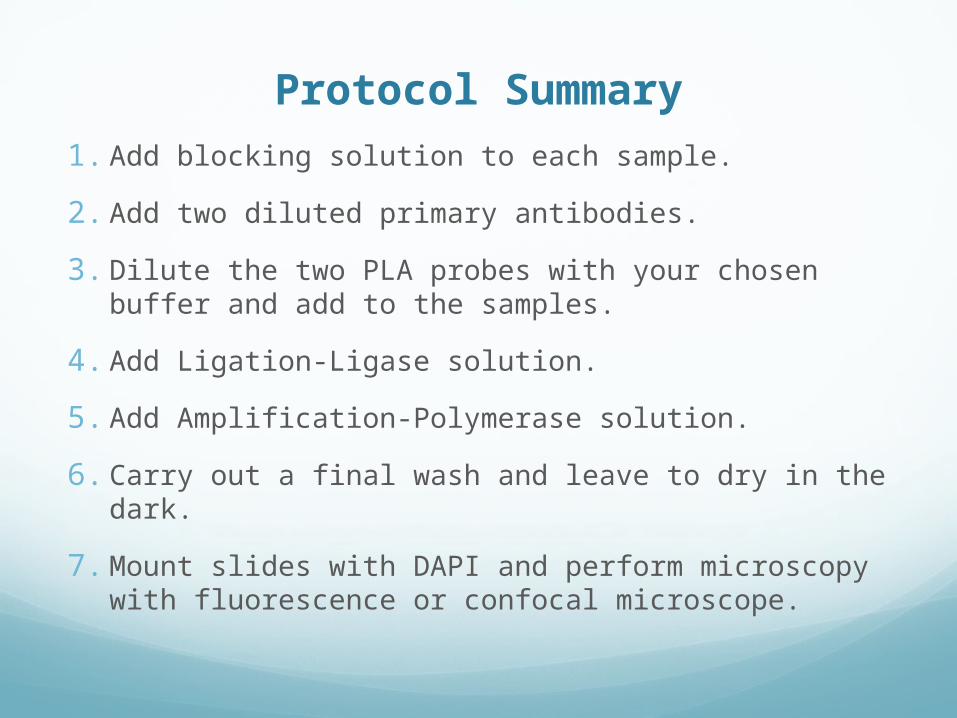
Protocol Summary
1. Add blocking solution to each sample.
2. Add two diluted primary antibodies.
3. Dilute the two PLA probes with your chosen buffer and add to the samples.
4. Add Ligation-Ligase solution.
5. Add Amplification-Polymerase solution.
6. Carry out a final wash and leave to dry in the dark.
7. Mount slides with DAPI and perform microscopy with fluorescence or confocal microscope.

Typical Results
One antibody, positive and negative control
Two antibodies, positive and negative control

In our assay we used:
Cells: MNS-70 cells developed by Masato Nakafuku
These cells have high Ascl1 and GSX2 levels.
Antibodies: 1- Goat Anti ASH1 (Mash1/Ascl1)
2- Rabbit Anti GSX1/2
PLA Probes: anti goat (plus) ; anti rabbit (minus)
Block: 10% Normal Donkey Serum in Tris Buffered Saline with 0.1% Tween 20
Detection reagent: Orange- (Cy3 spectra)
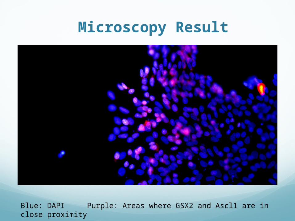
Microscopy Result
Blue: DAPI Purple: Areas where GSX2 and Ascl1 are in close proximity
Results
There were many cells that did not have any of the desired proteins in close proximity (25 nm) to one another, but there were some that did show to be in close proximity.
All of the regions in which Ascl1 and GSX2 were shown to be within close proximity to each other were found in the nuclear regions of the cells.