Embed Size (px)
Citation preview

Study on MRI Diagnosis i. (. rerebraR diseases in Dogs
犬の脳疾患のMRI診防1'関する研究
Shinji Tamura
田村慎司
2007
一1一

CONTENTS
0riginal Articles 40ral Announcement 5Chapter 1
Sequential Magnetic Resonance lmaging of an lntracranial Hematoma in a dog
Case History Report 13
Chapter 2
Multiple metastases of thyroid cancer in the cranium and pituitary gland in two dogs
1ntroduction 23 Case Histories 24
-2一

Chapter 3
Magnetic Resonance lmaging Findings ofNeuroaxonal Dystrophy
in a Papillon Puppy
Abstract
Introduction
Case History
Discussion
References
Figures
Chapter 4
A case of a dog with intracranial meningioma treated with hydroxyurea
Abstract
Introduction
Case History
Discussion
References
Figures
General Discussion
Acknowledgments
一3一
34
35
36
39
42
45
48
49
50
53
56
58
61
69

Original Articles
1. Shinj i Tamura, Yumiko Tamura, Take shi Tsuka, Kazuyuki Uchida
Sequential Magnetic Resonance lmaging of an inttracranial hematoma in a dog
Veterina7 y Radiolog v and Ultrasound Volume 47 I ssue 2 Page 142-146 (2006)
2. Shinj i Tamura, Yumiko Tamura, Nobutaka Suzuoka, Ay a Ohoka, Takahisa
Hasegawa, Kazuyuki Uchida
Multiple metastases of thyroid cancer in the cranium and pituitary gland in two dogs
Journal ofSmall A nimal Practice Volume 48 I ssue 4 Page 237-239, April (2007)
3. Shinj i Tamura, Yumiko Tamura, Kazuyuki Uchida
Magnetic Resonance lmaging Findings of Neuroaxonal Dystrophy in a Papillon Puppy
Journal ofSmall A nimal Practice Volume 48 I ssue 8 Page 458-461, August (2007)
4. Shinj i Tamura, Yumiko Tamura, Ay a Ohoka, Takahisa Hasegawa, Kazuyuki Uchida
A canine case of skull base meningioma treated with hydroxyurea
The Journal of Veterinary Medical Science Volume 69 Issue 12(2007) in press
一4一
Oral Announcement
O3/10/13 平成15年度日本小動物獣医学会(中国)
犬の外傷性脳実質内血腫のMRIの経時的変化
田村慎司、田村由美子
03/12/14 第21回獣医神経病研究会
犬の外傷性脳実質内血腫のMRIの経時的変化
田村慎司、田村由美子
05/12/11第25回獣医神経病研究会
甲状腺癌が下垂体を含む頭蓋内に多発性転移した犬の2例
田村慎司、田村由美子、山本正義、鈴岡宣孝、大岡恵、長谷川孝寿、
内田和幸
06/06/11第26回獣医神経病研究会
神経軸索性ジストロフィーのパピヨンの1例
田村慎司、田村由美子、梶谷幸子、梶谷剛、来田千晶、内田和幸
06/12/10 第27回獣医神経病研究会
ハイドロキシウレアにより治療した頭蓋内髄膜腫の犬の1例
田村慎司、田村由美子、大岡恵、長谷川孝寿、内田和幸
一5一
Introduction
Diseases of the cerebral diseases in dogs fall into various categories, including
degenerative, metabolic, neoplastic, inflammatory, infectious, idiopathic, iatrogenic,
traumatic and vascular disease, and also malformation. They range from diseases such
as inter vertebral disk disease, a disease that has been widely recognized since old times
and that has relatively well-established pathology and diagnostic and treatment
approaches, to tho se such as necrotizing meningoencephalitis, for which neither the
cause nor treatment has been identified. Due to the aging dog population, practicing
veterinarians are likely to be faced with cerebral disease s in dogs more frequently than
ever before. At the same time, as dog owners become more aware of the quality of
veterinary care, they are expecting better treatment of these diseases.
Veterinary magnetic resonance imaging (MRI) was introduced partially in
response to such demand in the practice, and since then, antemortem diagnosis of
central nervous system (CNS) diseases has improved dramatically. CNS diseases,
especially those of the brain, used to be diagnosed using a black box model, based only
on the localization of lesions by neurological examinations, and speculation regarding
the characteristics of the disease based on factors including signalment, onset pattern
and cerebrospinal fluid (CSF) analysis. More detailed diagnosis used to be possible only
after necropsy. lntroduction of computed tomography (CT) into veterinary medicine
permitted the visualization (albeit partial) of intracranial diseases, and introduction of
MRI permitted clearer imaging. Because of these advances in diagno stic technology,
一6一

along with the aforementioned aging dog population, diseases that were previously
thought not to affect so many dogs (e. g. cerebral infarction) actually turned out to be
relatively common. Brain tumor also turned out to be a disease that practitioners are
frequently faced with. Advances in diagnostic technology also paved the way for causal
treatment (e. g. surgical removal of a brain tumor) for CN S diseases that used to be, with
some exceptions, treated mainly using symptomatic therapies. ln veterinary diagnostic
imaging, however, diagnostic criteria for humans are often used, and for diseases such
as necrotizing meningoencephalitis and granulomatous meningoencephalitis, for which
there are no human counterparts, no diagnostic criteria or other standards exist for
humans that can be used for dogs. Thus, there is an urgent need for MRI findings in
dogs to be verified via histopathological diagnosis on an ongoing basis. Therefore, for
disease s with no previously reported cases of spontaneous onset in dogs, 1 assessed the
clinical symptoms together with the findings from MRI, histopathological studies and
various examinations. 1 present the results of my asse ssments here, so that they can be
used as base s for antemortem diagnosis in the future.
In Chapter 1,1 describe the changes that occurred over time in a dog with
spontaneously occurring traumatic cerebral intraparenchymal hematoma, at a total of
seven observation points from one hour to 14 days after the injury. Histological
examination was conducted at autopsy after the dog died on the 14th day of injury. The
changes that were seen in MR images were similar to those reported in humans with the
same condition. Thus, 1 obtained data that could be a robust basis for future antemortem
MRI diagnosis of intracranial hemorrhage in dogs, including information on the onset
-7一

pattern and ti me elapsing between onset and MRI examination.
In Chapter 2, 1 describe the MRI characteri stics of two canine cases of
multiple intracranial (including the pituitary gland) metastases of thyroid carcinoma,
and the results of histopathological examination. 1 also discuss the reasons why there is
no previous report of tumor metastases in the canine pituitary gland in the literature
based on the clinical symptoms observed in these two cases.
In Chapter 3, 1 describe a case of neuroaxonal dystrophy in a papillon puppy,
which was examined using MRI twice, at three and a half and six months of age. I
discuss the changes in the MR images relative to the changes in clinical symptoms.
After the puppy was euthanized at six months of age, histopathological examination was
also conducted, and herein 1 also discuss these findings relative to the MRI features.
In Chapter 4, 1 describe the case of a miniature schnauzer with meningioma in
the cranial base, which was treated with hydroxyurea chemotherapy. The efficacy of
this treatment regimen was monitored over seven months using MRJ.
In all cases summarized in Chapters 1 to 3, the identity of the disease in
question could be speculated on based on the signalment, history, and the results of
physical, neurological and general laboratory examinations. However, MRI
examinations provided information on the localization and/or characteristics of the
lesions, and permitted more detailed diagnoses than those possible on the basis of other
data. The MR[1 findings could be verified by a definitive diagnosis made on the basis of
histopathological examination. Moreover, for the case described in Chapter 4, MRI was
used to monitor the efficacy of chemotherapy, and changes in tumor size over time
-8一
during therapy were confirmed. The information on various cerebral diseases in dogs
presented here will be usefu1 for the antemortem diagnosis of clinical cases of these
diseases and for the monitoring oftherapeutic response.
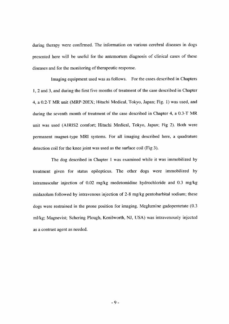
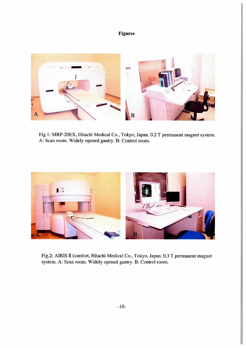
Imaging equipment used was as follows. For the cases described in Chapters
1, 2 and 3, and during the first five months of treatment of the case described in Chapter
4, a O. 2-T MR unit (MRP-20EX; Hitachi Medical, Tokyo, Japan; Fig. 1) was used, and
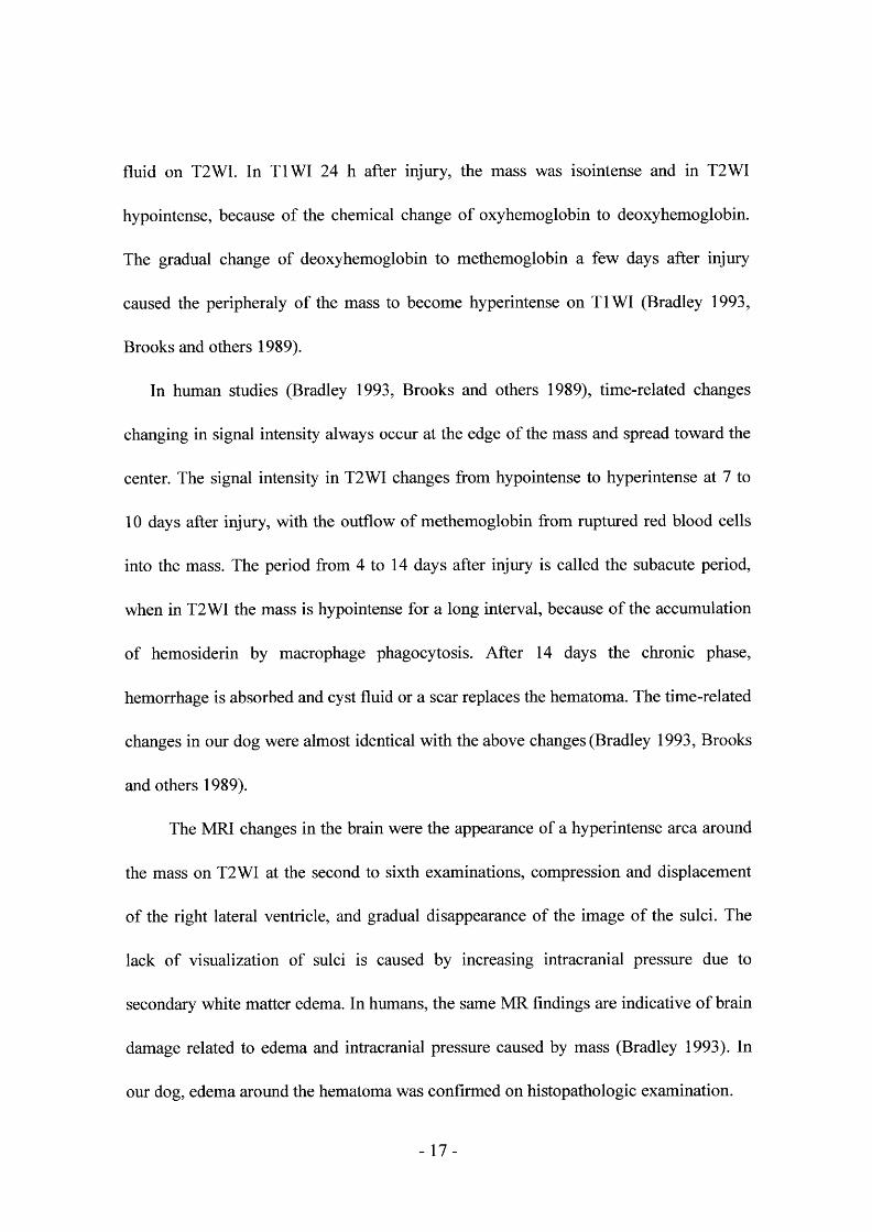
during the seventh month of treatment of the case described in Chapter 4, a O. 3-T MR
unit was used (AIRIS2 comiort; Hitachi Medical, Tokyo, Japan; Fig 2). Both were
permanent magnet-type MRI systems. For all imaging described here, a quadrature
detection coil for the knee joint was used as the surface coil (Fig 3).
The dog described in Chapter 1 was examined while it was immobilized by
treatment given for status epilepticus. The other dogs were immobilized by
intramuscular injection of O. 02 mg/kg medetomidine hydrochloride and O. 3 mg/kg
midazolam followed by intravenous injection of 2-8 mg/kg pentobarbital sodium; these
dogs were restrained in the prone position for imaging. Meglumine gadopentetate (O. 3
ml/kg; Magnevist; Schering Plough, Kenilworth, NJ, USA) was intravenously inj ected
as a contrast agent as needed.
一9一
Figures
r.
MA 'L一,
iiiii,. i
L. ・一 一
t
. ,
@. er”;. ”rl '
N nii一
門
x
・一一一 ザ
麟レ耀要
B
Fig. 1: MRP-20EX, Hitachi Medical Co. , Tokyo, Japan. O. 2 T permanent magnet system.
A: Scan room. Widely opened gantry. B: Control room.
r
重述 i'1
下
写
1・:
L
ミ
'1'
吐.
xxx
)
r J=':一:L=7. [・
11
Fig. 2: AIRIS l l comfort, Hitachi Medical Co. , Tokyo, Japan. O. 3 T permanent magnet
system. A: Scan room. Wi dely opened gantry. B: Control room.
一一 P0一

Figures
,,mi一
い
σ
Fig. 3: Quadrature detection (QD) coil for human stifle joint of AIRIS 11 comfort,
Hitachi Medical Co. ,, Tokyo, Japan.
一11一

Chapterl
Sequential Magnetic Resonance lmaging
Hematoma in a dog
of an lntracranial
Abstract
An 8-year-old Yorkshire terrier developed acute onset coma and seizure after cranial
trauma. Intracranial hemorrhage was suspected from the clinical signs and history. Low
field MR imaging revealed a round mass within the right cerebral hemisphere,
compressing the right lateral ventricle and displacing the longitudinal fissure to the left.
The lesion was hypointense on T l-weighted images and hyperintense on T2-weighted
images, consistent with an acute hemorrhage. MR imaging was performed every 24 h
for 6 day s from 1 h after the inj ury, and then on day 14 of ho spitalization. With time,
the signal intensity changed to hyperintense on T 1-weighted images. On T2-weighted
images the center of the mass changed to hypointense, and then to hyperintense with a
hypointense rim. These changes of signal intensity were related to hemogrobin
oxidation.
一12一

Case History Report
An 8-year一一〇ld female Yorkshire tenier developed continuous circling to the right
and excitement soon after falling from a height of l m. There was right-sided
rhinorrhagia and right conj unctival bleeding, as well as changing of consciousne ss
status and absent pupillary light response. l ntracranial hemorrhage secondary to cranial
trauma was suspected (Braund and others 1994). lmmediate intravenous inj ection of 0. 2
mg/kg midazolam hydrochloride succeeded in halting the circling and excitement. 2. O
mg/kg phenobarbital, 6. 0 mgtkg pentobarbital, and 2. 0 mg/kg dexamethasone were
administered to immobilize the dog and prevent of the clinical sign.
Thirty minutes after the inj ury, MR imaging was performed. lmages were obtained
with a O. 2 Te sla permanent magnet (MRP-20EX, Hitachi Medical Co. , Japan). T l
weighted images (T I WI) were acquired using a repetition time (TR) of 500 ms and an
echo delay time (TE) of 20 ms. T2 weighted images (T2wr) were acquired using a TR
of 4000 ms and TE of 120 ms. Transverse and dorsal plane images were acquired. The
slice thickness was 5 mm, without an interslice gap. There was a mass approximately 20
mm in diameter that formed a well defined globe and was located in the rostroventral
part of the right frontal lobe (Figure. 1 A, B). The volume of the mass corresponded to
200/o of the cross-sectional area. The midline of the brain hemispheres was displaced
toward the left. On T l wr the mass was isointense and on T2WI slightly hyperintense to
gray matter. The mass did not enhance on T I WI after IV inj ection of meglumine
gadopentetate (O. 3mYkg, Magnevist, Shering Tokyo). The mass was interpreted as a
hyperacute intracranial hematoma. Continuous light sedation with antiepileptic drugs
-13一

was needed the dog to prevent excitement. To monitor changes in the mass, MR
imaging was performed daily while antiepileptic therapy was administered.
On day 2, the mass on T l wr had no change in signal intensity, and on T2WI had
become hypointense, centrally. Bleeding was assumed to have stopped because the
mass had not enlarged. Glycerin was administered to decrease the intracranial pressure.
On day 3, in T I WI the mass was heterogeneously hyperintense(F igure. 1 C). ln T2WI,
there was an increase in size of the hypointense center of the mass(Figure. 1 D). On day
5, there was peripheral hyperinteisity in T I WI (Figure. 1 E, F). On T2WI, a
hyperintense area of white matter that was thought to be edematous on day 2, worsened
from days 3 to 6. On T2WI the cerebral gyri and sulci gradually became unclear from
day s 2 to 6. The T I WI and T2WI signal intensities of the mass were unchanged on day
6. Treatment with antiepileptics for 6 days and with glycerin for 5 days succeeded in
discontinuing sedation without excitement, producing the appearance of reaction to
sound and touch on 7 day, and a short period of standing on day 9. However, the dog
died suddenly on day 14 because of status epilepticus. ln MR examination soon after
death, the size of the mass was unchanged, and the mass was now hyperintense at the
center and hypointense at the rim on T2WI, but had almost no change on T l WI (Figure.
1 G, H). The edema around the mass was reduced on T2WI. On T2wr the cerebral gyri
and sulci were clearly seen. Grossly, the mass extended through most of the right
cranial cavity and displaced the midline markedly to the left.
Necropsy was performed on day 14. There was a large hematoma occupying the
right hemisphere, from the lateral ventricle to the zona striata. Around the hematoma
-14一

there was malacia as well as spongy changes of the neuropil, consisting of Wallerian
degeneration of neuronal axons, accumulation of fat-granular cells, and
neovascularization. Large numbers of swollen oligodendroglia and atypical astrocytes
were distributed diffusely throughout the cerebral parenchyma on both sides of the
frontal lobe, the right occipital lobe, and the thalamencephalon. There were ischemic
changes in the pyramidal cells scattered widely in the cerebral cortex and hippocampus.
In the medulla oblongata, there were a number of swollen oligodendroglia and atypical
astrocytes, and the neurons exhibited ischemic necrosis. ln the leptomeninges, small
numbers of neutrophils and macrophages had accumulated around the blood vessels,
and the endothelium of the vessels was hyperplastic. These histologic findings indicated
a primary hematoma (Summers and others 1995), with sub sequent brain edema and
menlngltls.
一15一

Discussion
Time-related changes on MR images relative to the stage of advancement of
hematomas from hyperacute to chronic have been used as criteria for selecting treatment
(Bradley 1993, Brooks and others 1989). There are two reports of the MR imaging
features of intracranial hemorrhage in animal s (Thomas and others 1997, Vernau and
others 2002). They describe subacute hematoma associated with cerebral vascular
malformation and hemorrhage within an araclmo id cy st. However, time-dependent
evaluation of MR imaging patterns has not yet been defined in dogs.
Our applications of sequential MR imaging in a dog with suddenronset neurologic
problems after cranial trauma resulted in the diagnosis of intracranial hematoma and
successfu1 recording of the evolving signal intensity of the lesion (Table 1). At the first
examination we recognized the intracranial mass to be clearly separate from the brain,
both morphologically and in terms of signal intensity. Reports in humans of time-related
MR imaging changes of hematomas have shown a progressive chemical reaction of
iron ions as components of hemoglobin (Bradley 1993, Brooks and others 1989). lt is
also known that the features of MR images of hematomas differ according to the
strength of the magnetic field (Brooks and others 1989). With a low-field device,
increasing of signal intensity on T l WI due to T l reduction starts earlier than high-field
MRI (Brooks and others 1989). The hematoma in our dog was observed with a
low-field permanent magnet.
The chemical form of hemoglobin in hyperacute lesions is oxyhemoglobin, which
influences the signal intensity similar to that of white matter on T I WI and similar to
-16一
fiuid on T2WI. I n T l wr 24 h after inj ury, the mass was isointense and in T2WI
hypointense, because of the chemical change of oxyhemoglobin to deoxyhemoglobin.
The gradual change of deoxyhemoglobin to methemoglobin a few days after inj ury
caused the peripheraly of the mass to become hyperintense on T l wr (Bradley 1993,
Brooks and others 1989).
In human studies (Bradley 1993, Brooks and others 1989), time-related changes
changing in signal intensity always occur at the edge of the mass and spread toward the
center. The signal intensity in T2wr changes from hypointense to hyperintense at 7 to
10 days after inj ury, with the outfiow of methemoglobin from ruptured red blood cells
into the mass. The period from 4 to 14 days after inj ury is called the subacute period,
when in T2wr the mass i s hypointense for a long interval, because of the accumulation
of hemosiderin by macrophage phagocytosis. After 14 days the chronic phase,
hemorrhage is absorbed and cyst fluid or a scar replaces the hematoma. The time-related
changes in our dog were almost identical with the above changes (Bradley 1993, Brooks
and others 1989).
The MRI changes in the brain were the appearance of a hyperintense area around
the mass on T2WI at the second to sixth examinations, compression and displacement
of the right lateral ventricle, and gradual disappearance of the image of the sulci. The
lack of visualization of sulci is caused by increasing intracranial pressure due to
secondary white matter edema. ln humans, the same MR findings are indicative of brain
damage related to edema and intracranial pressure caused by mass (Bradley 1993). ln
our dog, edema around the hematoma was confirmed on histopathologic examination.
一17一
It was interesting to observe the recovery process synchronously with changes in the
clinical signs and the MR findings. However, the dog died suddenly on day 14 after
inj ury because of status epilepticus. ln response to a request by the owner, the dog was
treated at home from day 9 onward. However, the dog had laryngeal paralysis making
administration of oral antiepileptic drugs impossible; this is likely why the dog entered
status epilepticus.
一18一

References
Bradley W. G. Jr. (1993) MR appearance of hemorrhage in the brain, Radiology 189,
15-26.
Braund K. G. (1994) Localization using neurological syndromes. ln: Clinical
Syndromes in Veterinary Neurology 2nd ed London: Mosby,.
Brooks R. A. , Di Chiro G. , Patronas N. (1989) MR imaging of cerebral hematomas at
different field strengths: theory and applications. 1 Comput Assist Tomogr 13, 194一
一206.
Summers B. A. , Cummings J. F. , de Lahunta A. (1995) Inj uries to the central nervous
system. ln: Veterinary Neuropathology, London Mo sby.
Thomas W. B. , Adams W. H. , McGavin M. D. , Gompf R. E. (1997) Magnetic
resonance imaging appearance of intracranial hemorrhage secondary to cerebral
vascular malformation in a dog. Vet Radiol Ultrasoun 38 , 371-375.
Vernau KM, LeCouteur RA, Sturges BK, et al. (2002) lntracranial intra-arachnoid cyst
with intracy stic hemorrhage in two dogs. Vet Radiol Ultrasoun. 43 , 449454.
一19一

Figures
Figurel. Transverse MR images of the brain. There is a 1arge mass in the right frontal
lobe. lhe mass is isointense on T l-weighted images (A) and slightly hyperintense on
T2-weighted images (B) 1 h after injury. The signal intensity increases gradually at the
edge of the mass on T l-weighted images (C: day 3, E: day 5, G: day 14). T2-weighted
images of the mass (D: day 3, F: day 5, H: day 14) show a hypointense area in the
center of the mass; on day 14 the mass is hyperintense at the center and hypointense at
the rim. The amount of edema around the mass (small arrow) had increased by day 2
and decreased by day 14 on T2-weighted images. The cerebral gyri nad sulci
(arrowhead)were indistinct丘om days 2 to 6 but visible again by day 14.
一20一

引智羅;瀦ll塁麹1
旨 : 1 ヨ : ! : : 1 : i : i : i i ヨ : i ! :9)iO l
Q l自i ロ. ヨ1
劃 ロ21ぐレ
Q I当i
器陰 コレロく「づ 1
暫 I
I I
l l
I l
l l
I l
I l
ロ ロ
ロ コ
bl l●i∋ l l. $l l l紆l l8; lQ l l到§壁暮景茎諺i ■
I
l ■
曜 耀
膠 i
I ■
■ 1
ロ
コ ロ
劃舅1雛鷲勇1 l I
■ 耀
l I
■ 腫
コ ロ
コ ロ
コ ロ
1 ⇒b l
§iあ℃ 1口 1ハ
ロ
81お コ
:Σil
Ig l
引:劉磐i I扁1Φi ロ島1 の コこつ
コ
:● 1
港塁§隠 二二 冒1鎚肇総髭§
'li
琵1←l i ■
ゆ ロ
Ol 旨 i器 』邑 i。日おお 資]℃しぎ自有有嚢. Ol℃馨 i●妻k
i霞§ lpt vl…蒔vvv℃等i蕪
i-No「寸woり一 1/ i i弩身i碁拳
禽
巴妻
lB器 脇畜 墜婁劃1易Q IE一一 ee

Chapter2
Mukiple metastases of thyroid cancer in the crani”m and
pituitary gland in two dogs
Abstract
Two dogs, a 14-year-old female, American Eskimo dog and a 14-year-old male,
Maltese dog, were presented with thalamic syndromes, including lowered levels of
consciousness, poor postural responses and masses in the neck region. ln both dogs,
magnetic resonance imaging (MRI) revealed multiple masses inside the cranium,
including the pituitary gland. One dog died丘om status epilepticus two days after MRI
and the other died two months after MRI from respiratory failure. These dogs were
histopathologically diagnosed with multiple metastase s of thyroid cancer occuning
inside the cranium, including the pituitary gland. To the authors' knowledge, this is the
first time this tumour pattern has been reported in dogs, but it is possible that it is not
uncommon.
. 22一

Introduction
In humans, intracranial metastasis of thyroid cancer is a rare condition
representing only 1 per cent of thyroid cancer cases (McWilliams and others 2003). To
the authors' knowledge, no canine cases have previously been reported. Metastatic
tumours in the pituitary area are also uncommon in humans, accounting for 1 to 26 per
cent of pituitary tumours (Bell and others 2001, Simon and others 2004), and in dogs,
there is only one previously report of metastatic tumours in the pituitary (a transmissible
venereal tumour [Spence and others 1978]). This report describes two canine cases of
multiple metastases of thyroid cancer in the cranium, including the pituitary gland.
一23一

Case Histories
Case 1
A 14-year-old female, American E skimo dog was presented with reduced appetite and
energy of three weeks' duration, and cluster seizures that had been occurring since the
day before.
During a physical examination, masses in the neck and mamrnary glands, as well
as swelling of the mandibular lymph nodes, were found. Neurological examination
revealed a lowered level of consciousne ss and a poor postural response in both
hindlimbs and the left forelimb. Serum chemistry findings were normal. Chest
radiographs showed a round mass in the cranial lobe of the left lung.
On magnetic resonance imaging (MRI), on both T l一 and T2-weighted images, an
extraparenchymal mass with the same signal intensity as the surrounding brain
parenchyma was seen in the area extending from inside the sella turcica towards the
upper part. The image of the mass was enhanced by intravenous administration of O. 3
ml/kg meglumine gadopentetate (Magnevist; Schering Plough). Oedema was noted in
the thalamus along the edge of the mass. Another mass was found on the border
between the gray and white matter in the left temporal lobe, which was thought to be a
metastatic tumour on the basis of its location, its roughly round shape and the severe
oedema in the surrounding white matter (Fig 1).
On the basis of these findings, tumours in the pituitary, thyroid and mammary
glands, and metastatic tumours in the temporaHobe and lung were suspected. The
cluster seizures were treated with phenobarbital and dexamethasone, but the dog died
-24一
from status epilepticus two day s after MRI examination.
On postmortem examination, a red mass in the temporal lobe and a milky-white
mass in the pimitary area were found. Swo llen mandibular lymph nodes and many
nodules in the lung and spleen were also observed. Histopathologically, the neck mass
comprised solid growths of tumour cells with round nuclei varying in size, undergoing
active mitosis. The tumours were positive for thyroglobulin and negative for calcitonin
and adrenocorticotropic hormone(ACTH)oll imnユunostai血g(Fig 2). Similar cells
were observed in the other masses. On the basis of these results, the dog was diagnosed
with follicular thyroid cancer and systemic metastasis. The difference in the colours of
the temporal lobe and pituitary masses was attributed to the differences in blood vessel
distribution in the surrounding tissue. The mass in the manrmary gland was diagnosed
as a benign mixed tumour.
Case 2
A 14-year-old male, Maltese dog was presented with claudication of the left hindlimb
and anisocoria. lt had had reduced appetite and energy for the past three months, and
had been diagno sed with a thyroid tumour and hypothyroidism two months previously,
after which the dog had been treated with thyroid hormone.
On physical examination, masses in the neck and swelling of the mandibular
lymph nodes were found. Neurologically, lowered levels of consciousness and postural
responses in all limbs, as well as anisocoria, were observed. In serum chemistry, an
elevated cholesterol level (390. 7 mg/dl) and lowered values of T4 and FT4 (6. e5 nmol/1
-25一
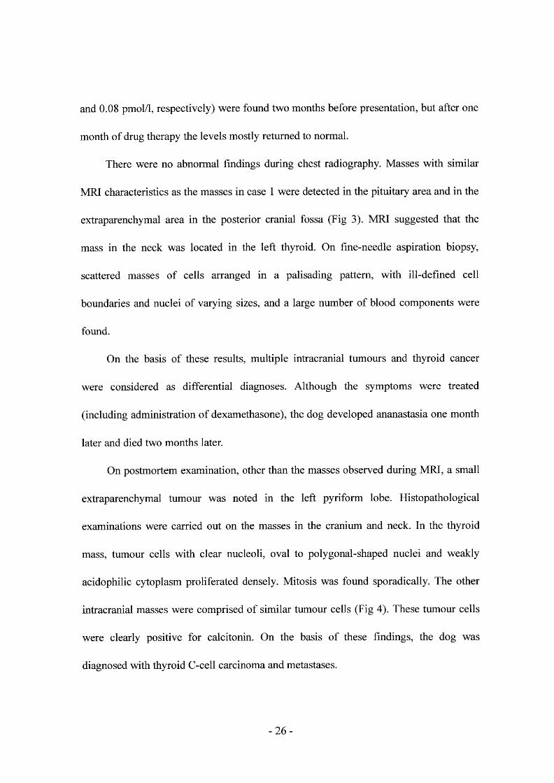
and O. 08 pmol/1, respectively) were found two months before presentation, but after one
month o f drug therapy the levels mostly returned to normal.
There were no abnormal findings during chest radiography. Masses with similar
MRJ characteristics as the masses in case 1 were detected in the pituitary area and in the
extraparenchymal area in the posterior cranial fossa (Fig 3). MRI suggested that the
mass in the neck was located in the left thyroid. On fine-needle aspiration biopsy,
scattered masses of cells arranged in a palisading pattern, with ill-defined cell
boundaries and nuclei of varying sizes, and a large number of blood components were
found.
On the basis of these results, multiple intracranial tumours and thyroid cancer
were considered as differential diagnoses. Although the symptoms were treated
(including administration of dexamethasone), the dog developed ananastasia one month
later and died two months later.
On postmortem examination, other than the masse s observed during MRI, a small
extraparenchymal tumour was noted in the left pyriform lobe. Histopathological
examinations were carried out on the masses in the cranium and neck. In the thyroid
mass, tumour cells with clear nucleoli, oval to polygonal-shaped nuclei and weakly
acidophilic cytoplasm proliferated densely. Mitosis was found sporadically. The other
intracranial masses were comprised of similar tumour cells (Fig 4). These tumour cells
were clearly positive for calcitonin. On the basis of these findings, the dog was
diagnosed with thyroid C-cell carcinoma and metastases.
一26一

Discussion
It i s likely that the number of reported cases of intracranial metastasis in cats and
dogs i s less than those in humans because, in many cases, animals with tumours die or
are euthanased before metastasis occurs. The incidence of intracranial metastatic
tumours might also have been underestimated because craniotomy is not routinely
carried out at postmortem examinatioh in cats and dogs (Bagley 2005). However, the
structure of the aortic arch branch in cats and dogs, which i s different from that in
humans, may prevent intracranial metastasis from occurring readily (Summers and
others 1995). Although metastases of thyroid cancer into the cranium or pituitary gland
have not been previously reported in dogs, the two cases reported here were
encountered within a short period of time. So such metastases might not necessarily be
rare conditions.
In case 1, intracranial lesions were strongly indicated by the epileptic seizures
caused by the temporal lobe lesion. The lowered levels of consciousness and poor
postural responses observed in both cases are symptoms of thalamic syndromes caused
by pituitary metastasis and are characteristic of such disorders. However, if a thorough
examination is not carried out at the initial presentation, these symptoms may not be
linked to intracranial diseases and may be attributed to debility instead. Furthermore, in
neither case did any clinical finding suggest an abnormal secretory function of the
pituitary gland.
Metastatic tumours in the pituitary area rarely produce clinical symptoms in
humans (Ruelle and others 1992). Therefore, it i s likely that metastatic tumours in the
-27一

pituitary area in dogs have been under-reported because these tumours produce no clear
clinical symptoms and tend to be overlooked. ln fact, it could be difficult to detect
primary pituitary tumours other than those producing ACTH. If lowered levels of
consciousne ss and poor postural response are erroneously interpreted as lethargy and
peripheral neuropathy, respectively, they could be confused with the clinical symptoms
of hypothyroidism. Therefore, in cases with hypothyroidism caused by thyroid cancer,
thyroid preparations alone could be administered wi thout intracrdliial diseases ever
being suspected.
In humans, it i s difficult to diagnose metastatic tumours in the pituitary area before
surgery (Komninos and others 2004). Endocrine function tests (for example,
ACTH-stimulation tests) are usefu1 for differential diagnosis, especially for
ACTH-producing tumours. The presence of central diabetes insipidus is the most
crucial criterion for the differential diagnosis of pituitary metastatic tumours in humans,
because it is found in 1 per cent of cases of pituitary adenoma and in 45. 2 per cent of
cases of metastatic tumour (Schubiger and Haller 1992). In dogs, however, 10 to 20 per
cent o f pituitary macroadenoma cases develop pituitary macroadenoma syndrome at, or
immediately after, diagnosi s (Nelson 1998). As central diabetes insipidus is a
component of pituitary macroadenoma syndrome, it is unlikely to be a usefu1 criterion
for differential diagnosis in dogs. Dumbbell-shaped tumours extending from the
intrasellar to the suprasellar area are likely to be metastatic tumours (Komninos and
others 2004). Moreover, the presence of multiple tumours suggests that they are
metastatlc.
一28一

It will be a challenge in the future to develop an approach for the differential
diagno sis of canine metastatic tumours in the pituhary area.
一29一
References
Bagley, R. S. (2005) Clinical clues to brain tumor. ln: Fundamentals of Veterinary
Clinical Neurology. Eds R. S. Bagley. Blackwell, Ames. pp 133
Bell, C. D. , Kovacs, K. , Horvath, E. , Smythe, H. & Asa, S. (2001) Papillary carcinoma
of thyroid metastatic to the pituitary gland. Archives(ゾ、Pathologソa加1 Laboratoり2
ルledicine l 25 935-938 ,
Komninos, J. , Vlassopoulou, V. , Protopapa, D. , Korfias, S. , Kontogeorgos, G. , Sakas, D.
E. & Thalassino s, N. C. (2004) Tumors metastatic to the pituitary gland: case report
and literature review. 」∂urnal qプC伽'cal Endocrinolog:ソand、Metabolis〃289,
574-580
McWilliams, R. R. , Giarrnini, C. , Hay, 1. D. , Atkinson, J. L. , Stafford, S. L. & Buckner,
J. C. (2003) Management of brain metastases from thyroid carcinoma: a study o f 16
pathologically confirmed cases over 25 years. Cancer 98, 356-362
Nelson, R. W. (1998) Endocrine di sorders. In: Small Animal Internal Medicine. Eds R.
W. Nelson and C. G. Couto. Mosby, St Louis, MO, USA. pp 672-807
Ruelle, A. , Palladino, M. & Andrioli, G. C. (1992) Pituitary metastases as presenting
lesions of malignancy. Journal ofNeurosurgical Sciences 36, 5 1-54
Schubiger, O. & Haller, D. (1992) Metastases to the pituitary一一hypothalamic axis. An
MR study of 7 symptomatic patients. Ne uroradiology 34, 131-134
Simon, N. , Quyyumi, S. A. & Rothman, J. G. (2004) Follicular thyroid cancer
presenting as a sellar mass: case report and review of the literature. Endocrine
一30一

Practice 10. 62-66 ,
Spence, J. A. , Holt, P. E. , Sayer, P. D. , Rottcher, D. & Cooper, J. E. (1978) Metastasis
of a transmissible venereal tumor to the pituitary. Jo urnal of Small A n imal Practi'ce
19, 175-184
Summers, B. A. , Cummings, J. F. & de L ahunta A. (1995) Metastatic central nervous
system tumors. ln: Veterinary Neuropathology. Eds B. A. Surmners, J. F. Cumming s
and A. de Lahunta. Mosby, St Louis, MO, USA. pp 391-395
一31一

Figures
t'
FIG 1. Axial magnetic resonance image of case 1 at the level of the pituitary gland. (A)
Post-contrast T 1-weighted(T l W)image. Contrast-enhanced加unours can be seen in the
pituitary area and in the left temporal lobe. (B) T2-weighted (T2W) image. Oedema can
be seen around the tumour (arrow). Cerebrospinal fluid can be seen between the tumour
and thalamus, suggesting that it is located outside the brain parenchyma (arrowhead)
難ず ド
総・. ,
ら
ia,ptiseyAsst:・sg-ggi}1t
lts
鍼.
. 讐i黙 濡.
”: j W. . . . ?)N
. 難li誕蕨'
r,夢
解.
'一
.
褻
v 一
応、e Aノ
翼唖.
一;・. y
刀
自る凄「
:・ . 農
「'ぐ ● .
e l
・縫d
A一
聾ご. 聾罰‘
FIG 2. Photomicrograph of止e tUmour in the pitUitary area in case 1. Tumour cells
derived丘om thyroid follicUlar cells form a solid grow血, which is split into honeycomb-
like components by fme-grained connective tissue, and appears to replace the normal
cells. Capillaries are growing in the stroma. Haematoxylin and eosin (H&E). ×400
一32一

Figures
・購.
1ン委「i・・いJ
FIG 3. MRI post-contrast TIW paramedian sagittal image of case 2. Contrast-enhanced
tumours can be seen in the pituitary area and in the extraparenchymal area of the
posterior cranial fossa
ゴだロ ヨ ロ の ゆ ロ
e一,一・k ,1'ぺ勲
∴触『 . . 、巳
ゴ き ロ
し ゆ
,、こ鮮註
㌧. 、ぞ1 ・P . “
ロ ぼロ ね
庭瓢ダ》辱畢.
ぜ. 、. ,も、夕
曳く集;か
艦誤避. ,. i. fL ・・V一
. ナ. . ヒ∴←「臨強聴一. tl
FIG 4. Photomicrographs of the tumour in the pituitary area in case 2. (A) Tumour cells
similar to those in the thyroid show irregular glandular-cavity-like proliferation. ln the
stroma, infiiltration of inflammatory cells consisting mainly of neu廿ophils is obvious, and
a slight increase in microglial cell number and vascular endothelial proliferation can be
seen in the surrounding area. H&E. ×400. (B) Calcitonin-stained section. Cells are clearly
positive for calcitonin. ×400
一33一
Chapter3
Magnetic Resonance lmaging Findings of
Neuroaxonal Dystrophy in a Papillon Puppy
Abstract
A3. 5-month-old papillon puppy was brought to our clinic with chief
complaints of progressive quadriparesis, ataxia and head tremor. Lesions in the
cerebellum, brain stem and spinal cord were suspected on the basis of a neurological
examination. No abnormality was found in a clinicopathological examination or on
MRI. Among differential diagnosis, an inflammatory disease, a degenerative condition
or a storage disorder were considered on the basis of these results. After this time, the
initial symptoms progressed, and glossoplegia and dysphagia developed at 6 months of
age. At a second MRI, severe atrophy of the entire brain was found. After these
examinations, the puppy was euthanized and histopathologically diagnosed with
neuroaxonal dystrophy. B ecause MR[( detected abnormal features that were
characteristic of neuroaxonal dystrophy in thi s case, we speculate that MRI can assist in
the premortem diagnosis of this disease.
一34b

Introduction
Neuroaxonal dystrophy is a degenerative disease of the central nervous
sy stem that is characterized by the degeneration of neurons and axons. Cases in dogs,
cats, rats, sheep, horses and humans have been described in the literature (Franklin and
others 1995). Canine cases have been reported, though sporadically, in several breeds
(Cork and others 1983), including rottweilers (Chri sman and others 1984, Bennett and
others 1997, Evans and others 1988, S iso and others 2001), Chihuahuas (Blakemore and
others 1985), collie sheep dogs (Clark and others 1982), Jack Russell terriers (Sacre and
others 1993) and English cocker spaniels (McLellan and others 2003). There is also a
report of neuroaxonal dystrophy in all members of a litter of five papillon puppies, in
which the clinical course and histopathological features of the disease are described in
detail (Franklin and others 1995). According to the report of Franklin and colleagues,
progressive ataxia, hypermetria and reduced po stural reaction of the limbs were
observed, and axonal degeneration was found histopathologically in both the white and
gray matter. However, at present, there is no known method for premortem diagnosis of
neuroaxonal dy strophy. We report here another case of neuroaxonal dystrophy in a
papillon puppy, for which we describe MRI changes over time, in addition to describing
a clinical course and histopathological features that are similar to those of the cases
reported by Franklin and colleagues.
一35一

Case History
A 3. 5-month-old female papillon, weighing 1. 1 kg, was referred to our clinic
for the evaluation of progressive neurologic abnormalities including te廿aparesis, ataxia
and head tremor, which were frrst observed at 2 months of age. The puppy had been
born a singleton. The puppy could not stand, but adopt a prone position. Vo luntary
movements were observed in the upper body, and hypermetria was observed in the
forelimbs. Both hind limbs tended to be extended. There was no impairment of
consciousness. A neurological examination at the initial presentation showed reduced
postural reaction of the limbs, lack of blink response to menace, and head tremor, which
was aggravated by excitation and disappeared when at rest. Urination was involuntary
and could be easily induced by applying pressure manually. At that time lesions in the
cerebellum, brain stem and spinal cord were suspected. No abnormality was found in a
complete blood count, or a biochemical examination. Among differential diagnosis in a
young animal with progressive multifocal signs, an inflammatory disease, a
degenerative condition or a storage disorder were considered on the basis of these
findings, but leukocyte deformation, which indicates a storage disease, was not
observed. Head MRI was performed using a O. 2-T system (MRP-20EX, Hitachi
Medical Corporation, Tokyo). T l-weighted images were acquired using a repetition
time of 500 ms and an echo delay time of 20 ms. T2-weighted images were acquired
using a repetition time of 4000 ms and an echo delay time of 120 ms. Transverse, dorsal
and sagittal plane images were acquired. The slice thickness was 5 mm, and there was
no i nterslice gap. No contrast medium was used. No clear abnormality was found in
-36一

either T2一 or T l-weighed images (Fig. 1).
The puppy was not treated for the condition, and head tremor, hypermetria,
and extension of the hind limbs became gradually aggravated, and generalized
amyotrophy progressed. At 6 months of age, the puppゾs level of consciousness
decreased, it was tetraplegic with increased tone in all four limbs and even the tail, and
the hind limbs could not be bent manually; therefore, the patellar reflex could not be
examined. Dysphagia prevented the puppy from taking any food. Algesthesia of the
limbs was normal (Fig. 2). Complete blood count and biochemical examination results at
this time were normal. A second head MRI showed clearly delineated cerebral sulci and
enlarged lateral ventricles, indicating severe atrophy of the cerebrum. In a sagittal image,
atrophy was also observed in the interthalamic adhesion, cerebellum, mesencephalon,
pons and medulla oblongata, and the fourth ventricle was enlarged. The arbor vitae of
the cerebellum was less clear relative to its appearance at initial presentation (Fig. 3). In
a cerebrospinal fluid sample obtained via a cisternal tap, the cell count was normal
(〈2/pl) and protein levels were slightly lowered (6. 0 mg/dl; reference value 8. 0-30. O
mg/dl). The level of lg-G antibody against canine distemper virus measured by the
immunoperoxidase method was 5120-fold for serum and less than 5-fold for
cerebrospinal fluid, indicating no infection with this virus. After these examinations, the
puppy was euthanized for humane reasons.
In a pathological autopsy, mild atrophy of the cerebellum was observed
(Fig. 4). A histopathological examination detected small intracellular vacuoles diffusely
present and reactive astrocytes diffusely proliferating in the white matter of the whole
-37一

central nervous sy stem. There was no substantial change in the cortex. In the cerebellum,
degeneration and enlargement of many axons was observed, especially in the fastigial
nucleus, interpositus nucleus and dentate nucleus. ln the cortex, a small number of
Purkinj e cells were lost or had degenerated, and a moderate number of torpedoes were
sporadically found in the granular layer. ln the medulla oblongata, loss and
degeneration of neurons in the olivary nuclei and a large number of small spheroids
were observed. ln the spinal cord, vacuoles were present diffusely, mainly in the white
matter. In the dorsal horn, neurons were decreased in number and many axons had
degenerated and become enlarged. ln the ventral horn, motor neurons were mostly
normal. Normal structures were maintained in the femoral and sciatic nerves (Fig. 5). On
the basis of these findings, the puppy was diagnosed with neuroaxonal dystrophy.
一38一

Discussion
Based on the histopathological examination, we speculated that the loss of
blink response to menace and head tremor observed in this puppy were clinical
symptoms caused by a disorder of the cerebellum. We considered that the impairment
of consciousness that appeared concurrently with the progression of the other symptoms
was a clinical manifestation of an impaired reticular activating system, that the
glossoplegia and dysphagia were manifestations of a functional disorder of the medulla
oblongata, and that the spastic quadriparesis was a manifestation of disordered upper
motor neurons due to impaired white matter tracts in brain stem and spinal cord. As was
also reported by Franklin and others (1995), in the present case the patellar refiex was
normal at the age of 19 weeks, but we could not examine the reflex at the age of 6
months because of the rigidity of the puppy's knee j oints.
In our case, the neurological findings at first presentation mainly suggested
dysfunction of the cerebellum, brain stem and spinal cord. In addition, a degenerative
disease was suspected because of the chronic progressive cerebellar manifestation that
developed from 2 months of age. The clinical symptoms and histopathological changes
were very similar to those reported by Franklin and colleagues (1995). ln our case, the
puppy did not have any littermates, so we camiot comment on whether entire litters tend
to have the same disease, as was observed in a previous instance. In our case, as for the
puppies described by Franklin and colleagues, the cause of the disease was not known.
Ahyperintense cerebellum on T2-weighted MR and fluid-attenuated
inversion recovery (FLAIR) images, and an elevated cerebellar diffUsion pattern on
-39一

MRI are characteri stic of neuroaxonal dystrophy in humans (Sener 2004). In human
cases, the most characteristic microscopic feature is cerebellar atrophy. ln the present
case, a hyperintense cerebellum was not observed on T2-weighted MR images, and
FLAIR and diffusion-weighted images could not be acquired with our MRI system.
There have been several reports describing the MRI findings for canine cases of
degenerative diseases of the central nervous system, including storage diseases. These
have tended to comprise atrophy of the central nervous tissue and bilateral high signal
lesions in T2-weighted MR images (Kaye and others 1992, Cozzi and others 1998,
Mariani and others 2001, Merwe and others 2001, Koie and others 2004, Garosi and
others 2005, Matsuki and others 2005). ln these degenerative diseases, the degree of
atrophy observed in the MR images and the MR signal intensity in the lesions would be
expected to change depending on factors including the progression of degeneration in
the nervous tissue, the substances accumulated in the lysosomes and the degree of
calcification. Lesions for which degeneration has progressed tend to appear with high
signal intensity on T2-weighted MR images and low signal intensity on T l-weighted
MR images, whereas when calcification occurs, low signal intensity is seen in both
sequences. In the present case, progressive atrophy in the cerebrum, cerebellum and
brain stem was found on MRI. MR images obtained at the age of 3. 5 months were
normal, suggesting that unable to detect MR changes in an early stage. Appearance of
major lesions in the second study, suggesting reinforcing the hypothesis of a progressive
disorder, and observation of diffuse atrophy, again reinforcing the hypothesis of a
degenerative disease and evident in a late stage of this animal's disease. Post-contrast
-40一

images were not acquired in the present study, but would be necessary in order to
distinguish neuroaxonal dystrophy from other conditions, including inflammatory
lesions.
In the present case, the imaging findings were consistent with the lesions found
histopathologically. Spinal cord MRI was not performed in the present case, but we
speculate that abnormality would have been detected given the observed
histopathological changes. Repeated MRI studies of brain and spinal cord in these
animal s, corresponding to different stages of the disease would be usefu1 for the
antemortem diagnosis of neuroaxonal dystrophy in papillons, in association with other
factors including breed, age of onset and clinical symptoms.
一41一

References
Bennett, P. F. & Clarke, R. E. (1997) Laryngeal paralysis in a rottweiler with
neuroaxonal dystrophy. A ustralian Veterinary Journal 75, 784-786
Blakemore, W. F. & Palmer, A. C. (1985) Nervous disease in the chihuahua
characterised by axonal swellings. Veterinary Record 117, 498-499
Chrisman, C. L. , Cork, L. C. & Gamble, D. A. (1984) Neuroaxonal dystrophy of
Rottweiler dogs. Journal of the A merican Veterinary Medical A ssociation 184,
464-467
Clark, R. G. , Hartley, W. J. , Burgess, G. S. , Cameron, J. S. & Mitchell G. (1982)
Suspected inherited cerebellar neuroaxonal dystrophy in collie sheep dogs. New
Zealandレieterinaリヘノburnal 30,102-103
Cork, L. C. , Troncoso, J. C. , Price, D. L. , Stanley, E. F. & Griffin, J. W. (1983) Canine
neuroaxonal dystrophy. Journal ofNeuropathology and Experimental Neurology 42,
286-296
Cozzi, F. , Vite, C. H. , Wenger, D. A. , Victoria, T, & Haskins, M. E. (1998) MRI and
electrophysiological abnormalities in a case of canine globoid cell leucodystrophy.
/ournal ofSmall A nimal Practice 39, 401-405
Evans, M. G. , Mullaney, T. P. & Lowrie, C. T. (1988) Neuroaxonal dystrophy in a
rottweiler pup. Journal of the. 4merican Ve terina1ッMedical. 4∬ociation 192,
1560-1562
Franklin, R. J. , Jeffery, N. D. & Ramsey, 1. K. (1995) Neuroaxonal dystrophy in a litter
of papillon pups. Journal ofSmall Animal Practice 36, 441-444
-42一

Garosi, L. S. , Penderis, J. , McConnell, J. F. &Jakobs, C. (2005)L-2-hydroxyglutaric
aciduria in a West Highland white tenier. Ve terinary Record 156, 145-147
Kaye, E. M. , Alroy, J. , Raghavan, S. S. , Schwarting, G. A. , Adelman, L. S. , Runge, V. ,
Gelblum, D. , Thalhammer, J. G. & Zuniga, G. (1992) Dysmyelinogenesis in animal
model of GM I gangliosidosis. Pediatric IVeurology 8, 255-261
Koie, H. , Shibuya H. , Sato, T. , Sato, A. , Nawa, K. , Nawa, Y. , Kitagawa, M. , Sakai, M. ,
Takahashi, T. , Yamaya, Y. , Yamato, O. , Watari, T. & Tokuriki, M. (2004) Magnetic
resonance imaging of neuronal ceroid lipofuscinosis in a border collie. Journal of
Veterinary Medical Science 66, 1453-1456
Mariani, C. L. , Clemmons, R. M. , Graham, J. P. , Phillips, L. A. & Chrisman, C. L.
(2001) Magnetic resonance imaging of spongy degeneration of the central nervous
system in a Labrador Retriever. Veterinary Radiology and UltTasound 42, 285-290
Matsuki, N. , Yamato, O. , Kusuda, M. , Maede, Y. , Tsuj imoto, H. & Ono, K. (2005)
Magnetic resonance imaging of GM2-gangliosidosis in a golden retriever. Canadian
Veterinary Journal 46, 275-278
McLellan, G. J. , Cappello, R. , Mayhew, 1. G. , Elks, R. , Lybaert, P. , Watte, C. &
Bedford, P. G. (2003) Clinical and pathological observations in English cocker
spaniels with primary metabolic vitamin E deficiency and retinal pigment epithelial
dystrophy. Ve terinai y Record 153, 287-292
Sacre, B. J. , Cummings, J. E & De Lahunta, A. (1993) Neuroaxonal dy strophy in a Jack
Russell terrier pup resembling human infantile neuroaxonal dy strophy. The Cornell
Ve terinarian 83, 133-142
-43一

Sener, R. N. (2004)Di伽sion-weighted and conventional MR imaging findings of
neuroaxonal dystrophy. A merican lournal of7Veuroradiology 25, 1269-1273
Siso, S. , Ferrer, 1. & Pumarola M. (2001) Juvenile neuroaxonal dystrophy in a
Rottweiler: accumulation of synaptic proteins in dy strophic axons. A cta
Neuropathologica 102, 501-504
van der Merwe, L. L. & Lane, E. (2001) Diagnosis of cerebellar cortical degeneration in
a Scottish terrier using magnetic resonance imaging. Journal of Small A nimal
Practice 42. 409-412 '
一44一

Figures
ム
D
tf'
Fig. 1. MR images at initial presentation(at 3. 5 months of age). (A)T2-weighted and
(B)Tl-weighted transverse images at the level of the thalamus. (C)T2-weighted
median sagi賃al image. There was no clear abnormality in either the T2-or T 1-weighted
の
lmages・
Fig. 2. Appearance of the puppy at 6 months of age. Spastic quadriparesis was observed.
一45一

Figures
イ
x_:・ン,
“
Fig. 3. MR images obtained at 6 months of age. (A) T2-weighted and (B) T l-weighted
transverse images at the level of the thalamus. Cerebral atrophy was observed. (C) T2-
weighted median sagittal image. The cerebellum and brain stem were atrophied, and the
arbor vitae of the cerebellum was less clear relative to its appearance at initial
presentatlon.
Fig. 4. Sagittal cut section of the fixed cerebellum. Mild atrophy of the cerebellum was
observed.
一46一

g
Fignres
ぐ 9 . . ● コ
の ド』 、'ノ ㌃ 、'も. ロ コ な 鍵鱈・♂ :・
き.
'
v 1智■
, ~ ・見 層' ・ド 隔
ゲ
つ ノ. . 一,‘'〃!喚・‘
. 樽.
B,:
g
ぞジ”
冒 」
'. Z・ 一 :・. . 1'. . ,,. . ・ ・一. t': lii ,,
. 、き・. ∵∵,
. . ‘冤,誉
コ コ し ご コ し
∫篤憾 . . '☆、. メ豪㌻
Fig. 5. Photomicrographs of histopathological specimens. (A) Cerebellum. ln the white
matter, axonal vacuolar degeneration was observed to a 1arge extent and reactive astrocytes
were proliferating diffusely. In the cortex, a sma11 number ofPurkinj e cells had been lost or
had degenerated, and torpedoes were sporadically found in the granular layer. Hematoxylin
and eosin, ×200. (B) Cross-section of spinal cord vvhite matter. Vacuolation of axons
ranging in size from small to 1arge was observed diffusely throughout the white matter.
Hematoxylin and eosin, × 40.
一47一

Chapter4
A Canine Case of Skull Base Meningioma Treated with
Hydroxyu rea
Abstract
An 11-year-old female miniature schnauzer was tentatively diagnosed with the skull
base meningioma, based on several examinations. Because surgical treatment was
difficult, and outpatient radiation therapy was not available in the local area,
chemotherapy with hydroxyurea combined with dexamethasone was selected. The
patient's clinical symptoms improved after one week of treatment, and the tumor size
was obviously reduced on MRI perfomied 37 days after treatment began. The patient
received hydroxyurea for 7 months, with symptoms remaining stable, and the tumor
re-increased to almo st the same size at 7 months as that at the initial examination. At
that time, hydroxyurea was discontinued. The patient died from pulmonary edema 14
months after treatment began. Pathologically, the tumor was diagnosed as a
memngloma.
一48一

Introduction
In dogs, unlike in humans and cats, intracranial meningioma is likely to infiltrate
the surrounding ti ssue s, making radical resection of the tumor difficult [10]. Although a
combination of surgery and radiation therapy has been shown most effective for
treatment of the disease, surgical intervention is sometimes more difficult in dogs than
in humans, depending on the affected site, because the temporal muscles of dogs are
much thicker than those of humans [1, 2]. Few veterinary hospitals provide radiation
therapy for tumors, which requires anesthesia to be administered multiple times during
the course of treatment. Therefore, veterinarians treating dogs with intracranial
meningioma need other treatment options. We report a canine case of inoperable
meningioma in the skull base that was treated with hydroxyurea, which has been used to
treat human meningiomas [7, 8]. Hydroxyurea chemotherapy was combined with
steroid treatment, resulting in reduced tumor size and improved clinical symptoms. .
一49一

Case History
An 11-year-old female miniature schnauzer presented with a four-to-five-month
hi story of progressive impairment of vision and hearing. On physical examination,
bradycardia was observed. A neurological examination revealed a decreased level of
consciousne ss, loss of pupillary light reflex, absence of menace response, and slightly
reduced postural reactions in all 1imbs. No abnormalities were found with respect to
hematology or blood chemistry, or in thoracic and abdominal X-ray examinations.
Magnetic resonance imaging (MRI) of the brain (O. 2T MRP20-EX; Hitachi Medico,
Tokyo, Japan)revealed a mass in the extraparenchymal area spreading丘om the sella
turcica to the right olfactory bulb, which was hypointense on T1-weighted images
(Tlwrs), hyperintense on T2-weighted images (T2wr s), uniformly strongly enhancing
upon intravenous administration of O. 3 ml/kg meglumine gadopentetate (Magnevist;
Schering Plough), and had a dural tail sign. Mid-sagittal plane images showed that the
thalamus and midbrain were dislocated to the dorsocaudal side by the mass effect.
Edema was also observed around the mas s (Fig. 1). We compared changes in tumor size
over time, using the maximum width and height (excluding the dural tail), on transverse
MRI images at the pituitary level. lnitially, the tumor was 14. 2 mm × 10. 3 mm in size
(width × height). Meningioma in the skull base was suspected, but surgical excision was
considered difficult because of the location and size of the tumor. We offered the
patient's owner several treatment options, including radiation therapy, chemotherapy,
symptomatic treatment with steroids, and any combination of these options. After
discussion, we decided to administer hydroxyurea chemotherapy combined with
-50一

symptomatic treatment with a steroid.
Initially, 30 mg/kg hydroxyurea was administered orally three times a week, plus
O. 5 mg/day oral dexamethasone to treat the edema around the tumor. A complete blood
count (CBC) was obtained every two weeks to detect any myelosuppression in response
to hydroxyurea treatment. One week after treatment began, the dog's vision and hearing
recovered, and the level of consciousness returned to normal. On MRI performed at 37
day s after treatment began, the tumor was smaller and the surrounding edema had
disappeared. At that time, the tumor was 9. 5 mm × 9. 1 mm in size (Fig. 2).
Dexamethasone was tapered, and mild worsening of the neurological symptoms,
particularly the visual and auditory ones, was observed at times. The symptoms were
controlled by an increase in the dexamethasone dose. Five months after treatment began,
loss of vision and depression developed. At that time, the tumor was 9. 8 mm × 9. 5 mm
in size, with no edema (Fig. 2), and the dexamethasone dose was O. 5 mg/day. lncreasing
the hydroxyurea dose to 45 mg/kg three times a week with no change to the
dexamethasone dose relieved these symptoms. Packed cell volume (PCV) gradually
decreased during the course of treatment, which was possibly caused by the
hydroxyurea. At 7 months after the initial presentation, when the PCV had decreased to
360/o (from 480/o at the initial presentation), hydroxyurea was discontinued. At that time,
the dexamethasone dose was O. 25 mg/day. On MRI carried out at the same time as the
PCV measurement (O. 3T AIRI S2 Comfort; Hitachi Medico, Tokyo), the tumor was 14. O
mm × 11. 4 mm in size, without edema (Fig. 2). One month after discontinuation of
hydroxyurea treatment, the PCV fluctuated around 400/o. Neutropenia was not observed
-51一
at any time during treatment. Two months after treatment began, hepatomegaly and
elevated hepatic enzyme levels were noted. Although these symptoms were considered
to be related to dexamethasone, it was difficult to determine the appropriate time at
which to discontinue the drug. At around 1 1 months after the first presentation, signs o f
Cushing's syndrome began to appear, and at 14 months, the patient died at her owner's
home from pulmonary edema of unknown cause. On post-mortem MRI, no definitive
evaluation was possible because contrast media could not be used, although the tumor
was seen to be enlarged on several T2wr s relative to images obtained at 7 months.
At necropsy, a large extramedullary mass, 15. 2mm x 12. 2mm in diameter, white to red
in color, was found on the ventral surface of the brain. Histopathological examinations
revealed that the mass consisited of solid proliferation of ovoid to spindle-shaped tumor
cells, sometimes forming whorl structures and a few psammoma bodies. In the stroma
there were mild neutrophilic infriltration and cholesterol deposits. Based on the se
findings, the tumor was diagnosed as meningioma, meningothelial type (Fig. 3). Based
on findings including systemic calcinosi s (in the lungs, kidneys, spleen, liver, and
cerebrovascular vessels), severe vacuolar degeneration of hepatic cells, and atrophy of
the adrenal cortex, we determined that the patient had eventually developed iatrogenic
Cushing's syndrome. Apart from these changes, pulmonary congestive edema
was also obserbed.
一52一

Discussion
Hydroxyurea is an antimetabolite that specifically affects the S stage of the cell
cycle [4]. Hydroxyurea can be used for years with acceptable and reversible toxicity in
humans, and for this reason it may be the optimal drug for treating slow-growing tumors
with low mitotic indices, such as unresectable and recurrent meningioma, despite the
controversy over its efficacy [4]. In dogs, hydroxyurea is used for the treatment of
chronic lymphocytic leukemia (CLL) [6], polycythemia [9], and essential
thrombocythemia [3]. Lomustine and carmustine, which pass through the blood-brain
barrier and are used for certain human brain tumors, are not effective treatments for
canine meningioma [1]. A previous case report described a dog with meningioma that
survived for 13 months on lomustine and prednisolone [5], although no data on tumor
size after treatment were provided. For these reasons we selected hydroxyurea to treat
the present case.
In the present case, the cytostatic and tumor-shrinking effects of hydroxyurea in
combination with the antiedema effect of dexamethasone appeared to result in symptom
relie£ Specifically, when symptoms worsened after 5 months of treatment, the
hydroxyurea dose was increased, resulting in suppression of tumor growth and
reduction in tumor size. After a discussion about treatment with the owner, hydroxyurea
chemotherapy was started at a low dose o f 30 mg/kg, based on the therapeutic do se
range used for dogs with CLL. One of the side effects of hydroxyurea is progressive
myelosuppression, which can be attenuated before it becomes severe by temporarily
decreasing the dose or discontinuing the medication when neutropenia is detected
-53一

during routine CBC monitoring [6]. We discontinued hydroxyurea treatment because of
a persistent decrease in PCV, although neutropenia did not occur in this case. It is
unclear whether the gradual decrease in P CV was due to hydroxyurea-induced
myelosuppression, or in fact whether discontinuation of treatment was appropriate.
Although hydroxyurea is used at a dose of 30-50 mg/kg in dogs with CLL, fUrther
investigation of the optimal dose range for treating canine meningiomas is necessary.
In the present case, symptomatic treatment with dexamethasone eventually
induced iatrogenic Cushing's syndrome. wnen using steroids, matters warranting
consideration are selection of steroids, temporary use of steroids only when clinical
symptoms worsen, and combined use with other antiedema agents such as
acetazolamide.
In a review of the treatment outcomes for intracranial meningioma in dogs, it was
found that the median survival time was approximately 3. 9 months with symptomatic
treatment with steroids alone, 7 months with surgery alone, and 16. 5 months with
surgery and radiation therapy [1]. In that analysis, however, the location of the tumor,
the tumor size at diagnosis, the histological type of the tumor, and the patient's age were
not taken into consideration. Therefore, although our patient survived for 14 months
with chemotherapy combined with a steroid, care should be taken when comparing her
survival time with those of other reported cases. ln the present case, more important is
the fact that the combination of steroid and hydroxyurea chemotherapy reduced the
tumor size, which suggests that this combination therapy may be an effective approach
for treating meningiomas in dogs. Multimodal therapies combining chemotherapy with
-54一

surgery or radiation therapy can also be considered. There is little information available
in the l iterature about chemotherapy for canine brain tumors, including meningiomas,
and the matter has not yet been extensively investigated [1]. More hydroxyurea-treated
cases of canine meningioma should be studied in order to further investigate the optimal
dosage and efficacy of thi s chemotherapeutic agent.
一55一

References
1. Adamo, P. F. , Forrest, L. and Dubielzig, R. 2004. Canine and feline meningiomas:
Diagnosis, treatment, and Prognosis, Compend. Contin. Educat. Pract. Vet. 26:
951-960.
2. Bagley, R. S. 2005. pp. 303-322. Treatment of important and common diseases
involving the intracranial nervous system of dogs and cats. ln: Fundamentals of
Veterinary Clinical Neurology. (Bagley R. S. ed), Blackwell Publishing Ltd, Ames,
IA. U. S. A. '
3. Bass, M. C. and Schultze, A. E. 1998. Essential thrombocythemia in a dog: case
report and literature review. 」. A m. A nim. Hosp. Assoc. 34: 197-203.
4. Hoshino, T. , Nagashima, T. , Murovic, J. A. , Wilson, C. B. , Davis, R. L. 1986
Proliferative potential of human meningiomas of the brain. A cell kinetics study with
bromodeoxyuridine. Cancer 58:1466-1472.
5. Jung, D. 1. , Kim, H. J. , Park, C. , Kim, J. W. , Kang, B. T. , L im, C. Y. , Park, E. H. , Sur,
J. H. , Seo, M. H. , Hahm, D. H. and Park, H. M. 2006. Long-term chemotherapy with
lomustine of intracranial meningioma occurring in a miniature schnauzer. Z Ve t. Med.
Sci. 68: 383-386.
6. Leifer, C. E. , Matus, R. E. , Patnaik, A. K. and MacEwen, E. G. 1983. Chronic
myelogenous leukemia in the dog. X A m. Vet. Med. Assoc. 183: 686-689.
7. Chamberlain, M. C. 2004. lntracranial meningiomas, Curr. Treat. Opin. Neurol. 6:
297-305.
8. Mason, W. P. , Gentili, F. , Macdonald, D. R. , Hariharan, S. , Cruz, C. R. and Abrey,
一56一

L. E. 2002. Stabilization of disease progression by hydroxyurea in patients with
recurrent or unresectable meningioma. 」. Neurosurg. 97: 341-346.
9. Moore, K. W. and Stepien, R. L. 2001. Hydroxyurea for treatment of polycythemia
secondary to right-to-left shunting patent ductus arteriosus in 4 dogs. 1. Ve t. lntern.
Med. 15: 418-421.
10. Sumniers B. A. , Cummings J. F. , deLahunta A. 1995. pp. 351-401. Tumors of the
central nervous sy stem, ln: Veterinary Neuropathology (Summers B. A. , Cummings
J. F. , deLahunta A eds), Mosby, St Louis USA.
一57一

Figures
Fig. 1. Brain MRI scans obtained at presentation. A: Transverse T2WI at the pituitary level.
B: TIWI at the same level. C: Postcontrast T I WI at the same level. D: Dorsal postcontrast
TlWI. E: Sagittal postcontrast T 1 WI. A tumor mass with marked enhancement can be seen
in the extraparenchymal area from the sella turcica to the right olfactory bulb. Edema in the
adj acent thalamus can be seen on T2wr. Mass effect can be seen in the sagittal image.
一58一
Figures
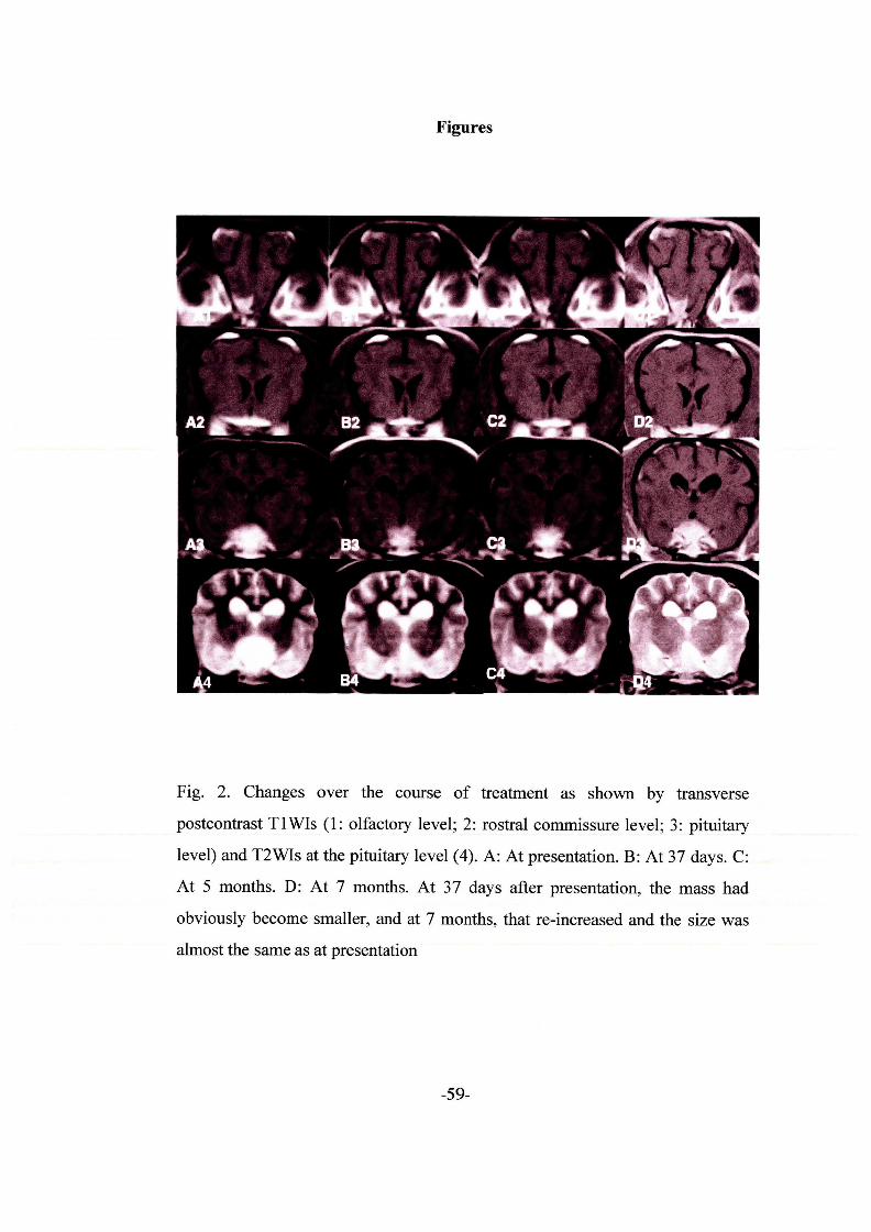
Fig. 2. Changes over the course of treatment as shown by transverse
postcontrast T I WIs (1: olfactory level; 2: rostral commissure level; 3: pituitary
level) and T2WIs at the pituitary level (4). A: At presentation. B: At 37 days. C:
At 5 months. D: At 7 months. At 37 days after presentatioq the mass had
obviously become smaller, and at 7 months, that re-increased and the size was
almost the same as at presentation
一59一

Figures
芦
汽.
:k'
蓬1;
:・
:lgs :
難
'殴. . t 騰 へ 1、、
墜. . 1. ・1旗
螺蓋・
',. . . E・' 轤凵E』玄
;i'
?t
感
! Y. .
v 二.
1
' Ni
℃・・舵IN:
}・
li'tt
!畠1
' 'L
纏i鞍'
Fig. 3. A: Gross fmdings at autopsy. The tmor had spread extensively in the
sku11 base.
Fig. 3. B: Histopathological features of the neoplastic mass. Solid
proliferation of sma11 ovoid to spindle-shaped cells mimicking
meningothelial cells with occasional psammoma body form. ation
(arrow). H&E. original magnification x 40Q.
一60一
General Discussion
MRI is considered the most usefu1 diagnostic imaging method in human
medicine, and its utility for small animals is being gradually acknowledged in clinical
veterinary practice. Here 1 evaluate the usefulness of MR[1 for diagnostic imaging
cerebral diseases in dogs. In this section, 1 will summarize each chapter and
comprehensively discuss the MRI diagnosis of cerebral diseases in dogs.
In Chapter 1, 1 describe the case of a dog with intracranial hematoma, and
discuss the changes in MR images over time. ln this case of spontaneous intracranial
hematoma, similar changes were observed in MR images over time as those that are
seen in human hematomas and secondary lesions in the surrounding brain parenchyma.
This finding will provide a robust basis for future differential diagnosis of intracranial
mass lesions in dogs. lfrapid antemortem diagnosis becomes possible, more appropriate
treatment can be implemented. For example, lesions could be surgically removed, or, as
in the case described in Chapter 1, if the symptoms are relatively mild and the
hemorrhage does not persist (as seen in longitudinal observations), supportive treatment
could be used.
In Chapter 2, 1 describe two clinical cases of multiple intracranial metastases
of thyroid carcinoma in dogs. MRJ revealed multiple tumors in the intracranial area,
including the pituitary gland. 1 expect that intracranial or pituitary gland metastases of
tumors will be encountered in clinical practice more often in the future, so their
differential diagnosis will hopefully be elucidated at a later date. ln addition, it is
-61一

possible that there exist a large number of nonhormone-producing tumors of the
pituitary gland that have been difficult to detect.
In Chapter 3, a case of neuroaxonal dystrophy in a papillon puppy is
described in detail. This case had almost the same clinical symptoms and
histopathological findings as cases reported by Franklin et al. in 1995. ln addition to
clinical symptoms and histopathological findings, 1 also discuss the changes in MRI
findings over time, which showed progressive atrophy of the cerebrum, cerebellum and
brain stem. These findings confirmed the progressive and degenerative nature of this
disease and provide usefu1 information for future antemortem diagnosis when
considered in association with information such as the breed of the dog, age (in months)
at onset, and clinical symptoms. A number of similar degenerative diseases of the CNS
are known in dogs and, therefore, a task for the future is to establish an approach for
differential diagnosis using MRI.
In Chapter 4, 1 describe a canine case of meningioma in the cranial base,
which was treated using hydroxyurea chemotherapy. The change in tumor size over
time was monitored using MRI, and the reduction in tumor size observed showed that
the chemotherapy regimen was effective. Monitoring intracranial tumors using MRI is
usefu1 for confirming therapeutic efficacy and modifying the course of treatment,
because it can be used to obj ectively vi sual ize the therapeutic response. In the future, I
plan to use MRI to monitor the process of treating brain tumors, encephalitis and other
diseases.
For cerebral diseases, the diagnostic procedure starts with estimating the age
-62一

of onset, and identifying the pattern of symptoms and other characteristics of the case
based on their signalment and information obtained from the owner. Next, neurological
and other symptoms at the time of presentation to the practitioner are identified via
physical and neurological examinations. wren neurological disease is suspected, at this
point the location of the lesion(s) is speculated on based on the results of the
neurological examination. If the lesion(s) is thought to be located in the CNS, after
systemic diseases that influence the nervous system are ruled out via biochemical
examinations of blood and urine, specific examinations including MRI, C SF tests,
electrophysiological tests and biopsies are performed. By comprehensively considering
all the findings from these examinations, a list of differential diagnoses i s produced and
a course of treatment is determined.
In the cases described in Chapters 1, 2 and 4, MRJ examination revealed
intracranial mass lesions. lntracranial mass lesions can be tumors, inflamed tissue,
abscesses, hemorrhages or a combination of these. ln the cases described in Chapter 3,
progressive atrophy was observed in the CNS and, therefore, degenerative disease was
suspected. Relative to the MRI characteristics of the lesions in other cases, the mass
lesion in the case described in Chapter 1 was characterized by having high signal
intensity in T l-weighted images from acute to chronic stage, and undergoing changes in
signal intensity over time. These characteri stics are al so observed in intracranial
hematoma in humans. ln the cases described in Chapter 2, an extraparenchymal mass
located in the pituitary gland and a spherical mass on the border between the gray and
white matter were observed. The locations of the cerebral masses and their multiplicity
-63一

suggested metastatic tumor, but a combination of cerebral intraparenchymal tumor and
primary pituitary tumor could not be ruled out. ln the case described in Chapter 4, a
widespread extraparenchymal mass was observed in the cranial base, which was
isointense relative to the surrounding brain parenchyma in T l一 and T2-weighted images,
had uniform strong enhancement, and showed the dural tail sign. On the basis of these
MRI findings, the extraparenchymal large mass lesion in the brain was strongly
suspected to be meningioma. However, other diseases, including malignant histiocytosis
and lymphoma, are known to produce similar lesions, although their incidences are
much lower than that of meningioma. For this reason, a definitive diagnosis of
meningioma was not made before the results of the histological examination were
obtained. Thus, when carrying out an MR examination of an intracranial mass lesion,
several characteristic features were noted, including the location, morphology, and
signal intensity of the mass, and the presence or absence of and degree of enhancement.
Although the MRI findings did not necessarily permit a definitive diagnosis, they are
likely to play an important role in the differential diagnosis of mass lesions. As for
conditions other than mass lesions, in the case described in Chapter 3, progressive
atrophy was found and degenerative disease was suspected. However, at this point too
few cases of this disease this disease have been described to permit identification of the
disease-specific patterns of atrophy or changes in signal intensity in MR images;
therefore, detection of such patterns is a task for the future.
As mentioned above, a diagnosis (of intracranial hematoma) could be made
solely on the basis of MR[1 findings for the case described in Chapter 1, but MRI
-64一
findings alone did not permit a conclusive diagnosis for the cases described in Chapters
2, 3 and 4. ln the cases described in Chapter 2, metastatic tumor was thought to be
possible because a mass in the thyroid gland area was detected during a physical
examination of the whole body, and because a blood test did not indicate Cushing
syndrome. ln the case described in Chapter 3, neuroaxonal dy strophy was suspected
based on signalment and clinical symptoms, and MRI confirmed the presence of an
abnormality that was consistent with the clinical features of this disease.
As we have seen, MRI enables CNS lesions to be localized and the type of
lesion, for example, mass lesions as seen in the cases described in Chapters 1, 2 and 4,
atrophic lesions as seen in the case described in Chapter 3, as well as diffuse
inflammatory lesions, to be more accurately determined. In addition, by combining MRI
data with signalment, information from the owner, and the results of other examinations
in accordance with the procedure for diagnosing neurological diseases that was outlined
earlier, diagnosis can be further narrowed down. Thus, MRI data is likely to play an
extremely important role in the process of diagnosing these CNS diseases and deciding
on a course of treatment. ln particular, visualizing lesions using MRI makes it possible
to progress to specific treatment such as surgical treatment or radiation therapy as
needed.
Over the past few years, while 1 have been conducting the research presented
here, workers both in Japan and abroad have been gathering MRI data from dogs with
CNS diseases, and the amount of data available is increasing. MRI is being widely
adopted by veterinary care facilities in Japan at a rate that was unthinkable in 2001,
一65一

when it was first introduced in my clinic. Case reports including MRI findings are now
not infrequently presented at academic meetings. As is the case in medical practice, in
small animals practitioners started to use MRI not only for diagnostic purposes at frrst
presentation, but also for monitoring the results of treatment for some diseases,
including brain tumors and encephalitis. One example of such use was outlined in
Chapter 4, highlighting the utility of using MRI in this way. However, the potential of
MRI is far from being fully harnessed in veterinary practice. lnterpreting each image
thoroughly and obtaining data from more cases would permit image-based diagnoses to
become more detailed and accurate in the future. Furthermore, the imaging sequences
used in this research were only spin echo T l-weighted images, fast spin echo
T2-weighted images and T l-weighted images using contrast medium, but proton
density-weighted, gradient echo T2“一weighted and fluid attenuated inversion recovery
images are also often used for imaging small animals. There are a variety of other
methods using different imaging sequences, including a method that distinguishes fat
and water, diffUsion-weighted images and MR angiography, but there is virtually no
report on the application of these methods in animals. More detailed differential
diagnosis will become possible in the future when these and other newly developed
sequences are used.
Diagnostic imaging for CNS diseases advanced considerably with the
introduction of MRI. Relative to humans, in small animals markedly fewer CNS
diseases have been named, and there is a larger number of idiopathic diseases,
indicating that these diseases have been insufficiently classified according to their
-66一

causes. Therefore, establishing a more detailed classification of these diseases and their
diagnostic methods would be clinically important. To achieve this, it is crucial to
continue keeping detailed treatment records for each clinical case, as these records
represent valuable data. ln addition, it is necessary to fUrther improve the accuracy of
diagnosis of CNS diseases by considering other diagnostic methods, such as
electrophysiological tests, molecular biological tests and tests for potential
disease-specific markers in the C S F, including anti-astrocyte autoantibody, which has
been tested for in very few cases reported in the literature. Further improvements will be
achieved also by accumulating reports in which the findings of various examinations
including MRI and the results of histopathological diagnosis are comprehensively
analyzed. Furthermore, there are diseases in dogs (e. g. Borna disease) for which only a
tiny number of cases have been reported so far, and the number of such diseases can be
expected to increase. The epidemiological and pathological features of these newly
encountered diseases should also be studied in the future. Additionally, 1 speculate that
because two Japanese people died from rabies in November 2006, measures against
rabies, including education campaigns encouraging people to be immunized against
rabies, and inclusion of rabies in the differential diagnosis for dogs with CNS symptoms,
will be considered more important than before.
The number of diseases that can be diagnosed before an animal's death needs
to be increased by collecting information regarding diagnosis via the actions 1 described
above relating to the observation of clinical cases. At the same time, attention should be
directed also to elucidating the pathology of CNS diseases in cats and dogs, and
-67一

focusing surgical and medical treatment on the causes of these diseases. 1 consider that
these are relatively unexplored themes.
So far, I have discussed the MRI diagnosis of CNS diseases in dogs丘om the
academic perspective of veterinary medicine. From a slightly different standpoint, that
of a small animal practitioner, 1 believe that specialized examination, diagnosis and
treatment of animals at secondary care facilities after referral by general practitioners'
facilities should benefit not only the animals and their owners but also the general
practitioners who refer these cases. General practitioners would benefit in two ways.
First, a benefit that general practitioners are already receiving is the feeling of security
of being unburdened of a difficult case, knowing that the animal and its owner wili
return once the disease resolves. Second is another benefu that 1 think general
practitioners should receive, which is academic feedback with respect to the outcomes
of treatment and new information generated by the referred cases. S econdary care
facilities should directly and indirectly feed back the information collected from the
accrued clinical cases that they have treated, for exaMple regarding what type o f
disorder should be suspected on the basis of which symptoms, or in what situations
cases need to be urgently referred to secondary care facilities, so that the cases can be
referred as smoothly as possible.
一68一
Acknowledgments
1 would like to thank those people who assisted and cooperated with me in
various ways during this research. Without these people, 1 would not have been able to
complete this work.
Sincere thanks to: Dr. Kazuyuki Uchida and hi s colleague s at the Department
of Veterinary Pathology, Faculty of Agriculture, Miyazaki University, who have
infallibly agreed to conduct histopathological examinations for me on a tight schedule,
provided excellent histological photos, and given me valuable assistance with writing
this thesis; Dr. Takato shi Hasegawa, Dr. Aya Ohoka and Dr. Noritaka Suzuoka at
Misasa Animal Hospital, Dr. Masayoshi Yamamoto at Hatsukaichi Animal Hospital,
and Dr. Takeshi Kaj iya and Dr. S achiko Kaj iya at Kaj iya Animal Hospital, who referred
the clinical cases that 1 described here; Dr. Take shi Tsuka, Research Associate in the
Laboratory for Veterinary Diagnostic lmaging, Faculty of Agriculture, Tottori
University, who translated my case report on intracranial hematoma and provided
advice on the report and the format of this thesis; my fellow practitioners including Dr.
Akira S akurada at Sakurada Animal Hospital, Dr. Kazuhiro Itamoto at Itamoto Animal
Hospital, Dr. Naotami Ueoka at Ueoka Animal Hospital, Dr. Etsuo Kawano at Kawano
Veterinary Clinic, Dr. Yasuhiro Sonoda at Sonoda Animal Hospital, Dr. Yasuhiro Wada
at Wada Animal Hospital, Dr. Hitoshi Ohnura and Dr. Kotoe Ohmura at Pal Animal
Hospital, Dr. Yuko Hori at Tao Animal Hospital and other doctors, who were kind
enough to attend rehearsals of my presentations for academic meetings in their precious
-69一
free time, and helped me to find and correct mistakes in the abstract, presentation draft
and slides; the staff members of Tamura Animal Clinic, Mari Matsumura, lzumi
Yamamoto and Miyuki Ono, who helped me with the examination, treatment, and
pathological autopsy ofthe dogs.
1 am very gratefu1 to Prof. Mikihiko Tokuriki at Nihon University for
triggering my interest in these esoteric neurological diseases when 1 was a student, and
to Prof. Emeritus S anenori Nakama, Prof. Yasuho Taura at the Department of
Veterinary Surgery, Faculty of agriculture and Prof. Munekazu Nakaichi at the
Department of Veterinary Radiology, F aculty of Agriculture, all at Yamaguchi
University, who allowed me to experience many actual cases of neurological diseases
and introduced me to MRI. Clandestine thanks also to my wayward college friends, who
trained and strengthened me up via our all-night mah-j ong games, when 1 did not have
enough physical strength to tolerate the long hours of research work. Furthermore, I
sincerely thank Dr. Keij i Kamakura at K Animal Hospital, who educated me as a small
animal practitioner during the two years after 1 graduated from college, who strongly
recommended that 1 introduce MRI into my practice after opening a clinic, and who
taught me about the idea of secondary veterinary care.
1 would also like to thank my parents who raised me to be a diligent worker
with the mottos “there i s no talent that can exceed exertion” and “if you don't work you
won't eat”. In addition, 1 deeply thank my wife, Dr. Yumiko Tamura, who i s al so a staff
member of Tamura Animal Clinic, who supported me in every way possible, with
housework, day-to-day practice, night surgery, treatment during holidays, and, above all,
一70一

waking up thi s l ate-sleeper at the same time every morning. 1 thank and pray for my late
dog, Moj o, who faithfully welcomed me at the entrance to my home, however late I
returned, exhausted after a day's work.
1 sincerely thank the owners of the animals studied in this research, who were
kind enough to let me examine and treat their pets, even though they were deeply
concerned about their suffering from mysterious neurological diseases, and moreover,
after their deaths, agreed to a pathological autopsy despite their sorrow.
Lastly, 1 dedicate this thesis to the five dogs, Sara, Mari, Takl〈un, Eve and
Chris, who provided valuable data that will help dogs suffering from similar diseases in
the future;Ipray for the souls of these dogs.
一71一