Embed Size (px)
Citation preview
HAL Id: insu-00852201https://hal-insu.archives-ouvertes.fr/insu-00852201
Submitted on 20 Aug 2013
HAL is a multi-disciplinary open accessarchive for the deposit and dissemination of sci-entific research documents, whether they are pub-lished or not. The documents may come fromteaching and research institutions in France orabroad, or from public or private research centers.
L’archive ouverte pluridisciplinaire HAL, estdestinée au dépôt et à la diffusion de documentsscientifiques de niveau recherche, publiés ou non,émanant des établissements d’enseignement et derecherche français ou étrangers, des laboratoirespublics ou privés.
Study on Europium Doped HydroxyapatiteNanoparticles by Fourier Transform Infrared
Spectroscopy and Their Antimicrobial PropertiesSimona Liliana Iconaru, Mikael Motelica-Heino, Daniel Predoi
To cite this version:Simona Liliana Iconaru, Mikael Motelica-Heino, Daniel Predoi. Study on Europium Doped Hydroxya-patite Nanoparticles by Fourier Transform Infrared Spectroscopy and Their Antimicrobial Properties.Journal of Spectroscopy, 2013, Article ID 284285, 10 p. �10.1155/2013/284285�. �insu-00852201�

Study on europium doped hydroxyapatite nanoparticles by Fourier transform infrared
spectroscopy and their antimicrobial properties
Simona Liliana Iconaru1,2, Mikael Motelica-Heino2, Daniela Predoi1*
1Department of Multifunctional Materials and Structures Laboratory, National Institute of
Materials Physics, 105 BisAtomistilor, P.O. Box MG 07, 077125 Magurele,
Romania,1*[email protected]
2ISTO, Université d’Orléans, Orléans cedex 02, 45067, France
Abstract
In this work, Fourier Transform Infrared spectroscopy (FT-IR) analysis were
conducted on europium doped hydroxyapatite, Ca10-xEux(PO4)6(OH)2 nanocrystalline
powders (Eu:HAp) with 0 ≤ xEu ≤ 0.2. Antimicrobial studies were also performed for the first
time on Eu:Hap. The antimicrobial properties of Eu:HAp nanoparticles with 0 ≤ xEu ≤ 0.2 on
Gram-negative (E coli ATCC 25922, Pseudomonas aeruginosa 1397) and Gram-positive
(Staphylococcus aureus 0364, Enterococcus faecalis ATCC 29212) bacteria systems and a
species of fungus (Candida albicans ATCC 10231) were reported. Our study demonstrates
that the antimicrobial activity of Eu:HAp nanoparticles is dependent on the europium
concentration.
Keywords: nanocrystalline, hydroxyapatite, europium, antimicrobial activity.
1. Introduction
Hydroxyapatite ( Ca10(PO4)6(OH)2 ) is the main inorganic component of human bones
and teeth, showing a very good biocompatibility, bioactivity and osteoconductivity due to its
nontoxic, and noninflammatory properties [1-9]. Therefore, it has been widely used in many
fields, like bio-medical applications, as a bioactive coating material for metallic implants,
clinical bone augmentation, dental implantology, tumors treatment and cell activation, or it
could be also used as a carrier in drug delivery systems [10]. Moreover, trivalent rare-earth-

ion-doped hydroxyapatite can also be used as biological fluorescent probes due to their
excelent luminescent properties.
In the last years, luminescence imagining, especially in the region of near-infrared
spectrum showed a great interest for biomedical applications, focusing on the development
and characterization of rare-earth based inorganic luminescent nanoparticules [11-14]. The
most important applications could be found on pharmaceutical industry or biological and
medical diagnostics [15-17].
When doped with rare-earth ions, hydroxyapatite is being considered a fluorescent
probe, the luminescence intensity depending on the concentration of rare-earth’s doping ions,
the degree of crystallinity and the crystal structure of the host material [18-20]. Over the past
decade, several methods for obtaining luminescent inorganic nanoparticles have been
developed, including coprecipitation and sol-gel synthesis which allow to adjust particles
morphology, size, structure and composition in order to adapt their physical and chemical
properties [14, 21-23].
Fluorescent labeling is an indispensable technique which is widely used for
performing nondestructive observations both in vivo and in vitro by replacing the calcium
ions in hydroxyapatite crystal lattice using rare earth luminescent ions [24-26]. The
hexagonal hydroxyapatite allows the substitutions of many rare earth ions without any
changes in the crystal structure [27-30]. Nowadays, the rare earths ions doped hydroxyapatite
nanoparticles are being studied very intently as cell labeling materials, as a result of their
strong luminescence under visible light spectrum. [31-36]
Compared to other rare earth elements, trivalent Europium Eu3+ ions have a simple
electronic energy level scheme and hypersensitive transitions. The Eu3+ doped calcium
apatites represent a good biological probe candidate due to their low toxicity and stable
luminescence over time and it has been proven that a small amount of europium in the
bioactivity behaviour has no harmful effects [37-41]. The ionic radius similarity between
Ca2+ and Eu3+ in the apatite lattice makes them a good host for Eu3+ doping [4].
Recently, great attention has been paid to europium doped hydroxyapatite (Eu:HAp)
potential use as biological probe [30]. Previously, the europium doped hydroxyapatite was
studied applying PL [32-33]. On the other hand, IR spectroscopy is a powerful method in
ionic investigation and has been used extensively in phosphate minerals research [42–44].
Concerning the phase composition of the as-prepared Eu:HAp various studies have
been reported [31-33, 45]. In our recent studies no other additional phases were observed [45]
for the synthesized Eu:HAp samples and the diffraction patterns were identical to that
obtained for stoichiometric apatite. In this paper, the obtained Eu:Hap samples were
systematically characterized by Fourier transform infrared (FTIR) spectroscopy. The aim of
this work is to contribute to the study of the influence of europium in the structure of
hydroxyapatite. On the other hand, cell viability from various doses of the Eu:HAp samples
at two different time points were compared with corresponding culture media values.After 24
h and 48h exposure to all types of Eu:HAp at 10 μg /ml concentration, the morphology of the
cells was preserved. Moreover, we report in this work for the first time the bacterial studies
on Eu:HAp. Furthermore, in this article, original stidies on antimicrobial activities of Eu:HAp
against Gram-positive (Staphylococcus aureus 0364, Enterococcus faecalis ATCC 29212),
Gram-negative (E coli ATCC 25922, Pseudomonas aeruginosa 1397) and fungal strains
(Candida albicans ATCC 10231) are presented.
2. Materials and methods
2.1. Samples
All the reagents for synthesis, including ammonium dihydrogen phosphate
[(NH4)2HPO4], calcium nitrate [Ca (NO3)24H2O], and europium nitrate [Eu(NO3)36H2O]
(Alpha Aesare) were used as purchased, without purification. Europium doped
hydroxyapatite (Eu:Hap, Ca10-xEux(PO4)6(OH)2) nanoparticles were performed by setting xEu
= 0.01, xEu = 0.02,xEu = 0.05, xEu = 0.1, xEu = 0.2 and [Ca+Eu]/P as 1.67 in accord with [45].
2.2. Fouriertransforms infrared (FTIR) spectroscopy.
The functional groups present in the prepared nanoparticles and thin films were
identified by FTIR using a Perkin Elmer, Spectrum BX spectrometer. In order to obtain the
nanoparticles spectra, 1% of the nanopowder was mixed and ground with 99% KBr. Tablets
of 10 mm diameter were prepared by pressing the powder mixture with a pressure of not
more than 10 psi. The spectrum was recorded in the range of 400 to 4000 cm-1 with 4 cm-1
resolution. The first FTIR spectra were obtained after 256 scans at room temperature(25 ±
0.50C). The second derivative IR spectra were acquired after 5-point smoothing of the
original IR spectra.For selected spectral ranges, the peak fitting analyses were performed
using procedures of J. Kolmas et al. [46]: (i) baseline correction, (ii) second derivative

calculation and self deconvolution assessment in order to determine the number and positions
of the bands and (iii) curve fittings with fixed peak positions using Lorentzienne lines. In the
previous studies Matsuhiro et al. [47] and Gómez-Ordóñezet al. [48] showed thatsecond-
derivatives of FTIR spectra are generally used as an aid for wave number determination of
weak absorption bands onto improve resolution of overlapped bands in the originalspectra.To
that end, in our studies derivation including Savitzky-Golay algorithm with nine smoothing
points was performed.
2.3. Antimicrobial studies.
The microbial strains identification was confirmed by aid of VITEK II automatic
system. VITEK cards for identification and susceptibility testing were inoculated and
incubated according to the manufacturer's recommendations. Microbial suspensions of
1.5x10⁸ CFU/mL corresponding to 0.5 McFarland density obtained from 15-18 h bacterial
cultures developed on solid media were used in our experiments. The tested substances were
solubilised in DMSO and the starting stock solution was of 5000 μg/mL concentration. The
qualitative screening was performed by an adapted disk diffusion method [49-53].
3. Results and discussions
3.1. IR Absorbance Spectra
The FTIR spectra of Eu:HAp were recorded in the typical, absorption mode from KBr
pellets.IR absorbance spectra of EuHAp samples with various europium concentrations (0 ≤
xEu ≤ 0.2) are shown in the 4000 cm-1 to 400 cm-1 range in Figure 1. Inourpreviousstudies
[45] we haveshown that for all the samples the presence of strong [OH]- vibration peak (632
cm−1) could be noticed. The broad bands in the regions 1600–1700 cm−1 and 3200–3600
cm−1 correspond to H–O– H bands of lattice water. Fowler B.O. in his infrared studies on
apatites [54] showed that the band at 3570 cm−1 is characterized by [OH]- stretching mode
and the band at 632 cm−1 characterized by [OH]- arises from stretching librational mode. The
bands at around 1090 cm−1 and about1040 cm−1 can be attributed to the 3 [PO4]3- while the
band at 962 arises from 1 [PO4]3-. The 602 cm−1 and 564 cm−1 bands appear from 4 [PO4]
3-.
Markovik et al. [55], presented that the sharpness of bands, especially sharpness of the 632
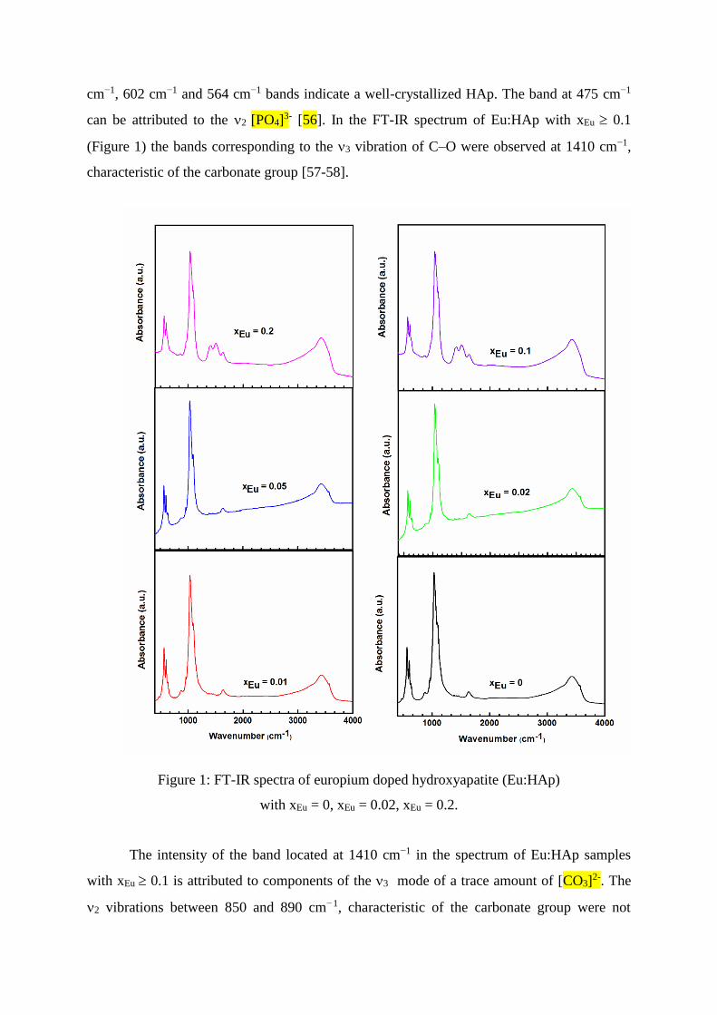
cm−1, 602 cm−1 and 564 cm−1 bands indicate a well-crystallized HAp. The band at 475 cm−1
can be attributed to the 2 [PO4]3- [56]. In the FT-IR spectrum of Eu:HAp with xEu 0.1
(Figure 1) the bands corresponding to the 3 vibration of C–O were observed at 1410 cm−1,
characteristic of the carbonate group [57-58].
Figure 1: FT-IR spectra of europium doped hydroxyapatite (Eu:HAp)
with xEu = 0, xEu = 0.02, xEu = 0.2.
The intensity of the band located at 1410 cm−1 in the spectrum of Eu:HAp samples
with xEu 0.1 is attributed to components of the 3 mode of a trace amount of [CO3]2-. The
2 vibrations between 850 and 890 cm−1, characteristic of the carbonate group were not

detected because 2 [CO3]2- band at 872 cm−1 is hidden by [HPO4]2- band at 875 cm-1.
Similar comportment was observed by Markovik et al. [55] in his studies on preparation and
comprehensive characterization of calcium hydroxyapatite. Markovik et al. and Holcomb
D.W. et al. [59] showed that the [CO3]2- band at 1410 cm−1 derives from [CO3]
2- (designated
by “B-type” carbonate that replace PO43- ions in the hydroxyapatite lattice). The band at 1510
cm−1was also detected in the FT-IR spectrum of Eu:HAp with xEu 0.1. The band at 1510
cm−1 derives from [CO3]2- (designated by “A-type” carbonate) that replacesOH- ions in the
hydroxyapatite lattice [55, 60]. In all the spectra of Eu:HAp samples, the band at 875 cm-1
was detected. This band is supposed to arise due to [HPO4]2- ions from several reasons [55].
In Figure 1 we observed that the contribution of the area that corresponds to the
phosphate bands decreases when the europium concentration in the samples increases. The
bands at 475 and 962 cm−1 progressively disappear with the increase of europium
concentration. When the xEu = 0.2 the bands at 475 and 962 cm−1 are almost absent. We can
also observe in the Eu:HAp spectra a broadening of peak vibration with the decrease of the
europium concentration. This behaviour was observed by Owada et al. [61] in sintered Y-
doped hydroxyapatite.
3.2. IR Second Derivative Spectra
In order to complete structural information on the analyzed Eu:HAp samples with 0 ≤
xEu ≤ 0.2 we agreed to perform derivative analysis and peak fitting of selected 4, 3, 2 and
1phosphate bands. The second derivative of the spectrum of Eu:HAp samples (xEu = 0, xEu =
0.02, xEu = 0.2) in the 4, 3 and 1 [PO4]3- bands are shown in Figure 2.Only the results
obtained for pure HAp (xEu = 0) and Eu:HAp (xEu = 0.02, xEu = 0.2) samples are shown,
which clearly assigned the strong changes that occur in the FT-IR spectra of HAp in the
presence of Eu. 1 [PO4]3- band was observed at around 962 cm-1 in the second derivative
spectra. On the other hand, in the second derivative spectra was identified a 2 [PO4]3- band at
475 cm-1. In concordance with precedent studies, the bands assigned in the second derivative
spectra of Eu:HAp (0 ≤ xEu ≤ 0.2) can beattributed to molecular vibrations of the phosphate
[PO4]3- in a apatitic stoichiometric environment of hydroxyapatite [62].
To evaluate the subtle spectral changes occurring as a consequence of the europium
doped hydroxyapatite, the spectra in the spectral regions of 450–700 cm-1(2and 4 [PO4]3-

domain) and 900–1200 cm-1(1 and 3 [PO4]3- domains) were analyzed by means of second
derivative (Figure 2) and curve fitting analysis (Figure 3).
In Figure 2 we also observethe second derivative band at 633 cm-1 that derives from
the OH- librational mode. IR wavenumber position (cm-1) of the 4, 3, 2and 1 [PO4]3- bands
of Eu:HAp spectrum from second derivative is presented in Table 1. Ten bands were detected
for phosphate bands of hexagonal Eu:HAp samples.
Table 1. IR wavenumber position (cm-1) of 4, 3, 2 and 1 [PO4]3- bands of Eu:HAp
spectrum from second derivative.
Figure 3 shows the FT-IR spectrum of hydroxyapatite (xEu = 0), the phosphate 4, 3,
2 and 1 regionswith experimental and calculated contours overlaid along with the individual
subbands (blue) as determined by a curve fitting analysis. Five components were needed for a
satisfactory fit in the spectral region of 450–700 cm-1 (2 and 4 [PO4]3- domain) and eight in
the spectral region of 900–1200 cm-1(1 and 3 [PO4]3- domains). FT-IR spectrum of Eu:Hap
with xEu = 0.02 is also presented and five components were needed for a satisfactory fit in the
spectral region of 450–700 cm-1(2 and 4 [PO4]3- domain) and six in the spectral region of
900 –1200 cm-1(1 and 3 [PO4]3- domains). For Eu:Hap samples withxEu =0.2 four
components were needed for a satisfactory fit in the spectral region of 450–700 cm-1 (2 and
4 [PO4]3- domain) and five important components in the spectral region of 900–1200 cm-1 (1
and 3 [PO4]3- domains).
Assignments Position (cm-1) References
1 [PO4]3- 962 62
2 [PO4]3- 475 62
3 [PO4]3- 1033; 1045; 1073;
1090
62
4 [PO4]3- 569; 575; 588;
602
62
Present in newly precipitated
apatite.
1111 62-63
[HPO4]2-- containing apatites 1144 63
[OH]- group ( librational mode) 633 63-64

Figure 2: Second derivative of Eu:HAp (xEu = 0, xEu = 0.02, xEu = 0.2) spectrum
of the 4, 3, 2 and 1domains.

Figure 3: FT-IR deconvoluted spectra of the 4, 3, 2 and 1 domain
for Eu:HAp (xEu = 0, xEu = 0.02, xEu = 0.2).
The individual component for phosphate 2 region (featured near 475 cm-1) decreases
for the samples with xEu = 0.02 and disappears when xEu increases to 0.2. The high frequency
shoulder is more clearly defined for pure HAp than in the case of the Eu:HAp with xEu = 0.02

and xEu = 0.2 in the 1 and 3 phosphate regions. The band requires at least eight components
for an adequate fit to the spectrum, compared with only six in the case of Eu:HAp with xEu =
0.02 or only five in the Eu:HAp with xEu = 0.2. In this series of materials it was not realistic
to attempt to identify particular components characteristic the particle size. However, a
correlation was found between the percentage area of the 1, band (near 960 cm-1) and the
crystal size. The individual component for the phosphate 1 region (band near 960 cm-1)
disappears when xEu increases to 0.2. On the other hand, the individual component for
phosphate 3 region (band near 1040 cm-1) decreases for the samples with xEu = 0.02.
However, in the Figure 2 and 3, we observe the disappearance of the band at 1144 cm-1,
which is associated with [HPO4]2- ions [65].We note that the main molecular species that
gave rise to the Eu:Hap (xEu = 0, xEu = 0.02, xEu = 0.2) absorbance in the 900-1200 cm-1
region was assigned to the phosphate ion, [PO4]3-.
3.3. Antimicrobial studies
A study on enhancement of osteoblast proliferation on europium doped
hydroxyapatite has rarely been reported. Anselme K. [66] in his study concerning osteoblast
adhesion on biomaterials and Garcia AJ, et al. [67] in the study of bio-adhesive surfaces to
promote osteoblast differentiation and bone formation showed that often synthetic materials
do not support osteoblast adhesion and this may result in poor cell differentiation and limited
bone formation. Keselowsky BG et al. [68] in the study on surface chemistry and Mc Farland
CD et al.[69] in his study on protein adsorption and cell attachment to patterned surfaces
demonstrated that the effect of surface properties on cellular response depends on differences
in species, concentration, and biological activity of adsorbed proteins, which may be obtained
from different sources, i.e., biological fluids and cell-mediated synthesis and deposition.
The antimicrobial activity of Eu:HAp (0 ≤ xEu ≤ 0.2) nanoparticles was tested using
the most common bacterial pathogens and fungus: E coli ATCC 25922 (Gram-negative),
Pseudomonas aeruginosa 1397 (Gram-negative), Staphylococcus aureus 0364 (Gram-
positive), Enterococcus faecalis ATCC 29212 (Gram-positive) and Candida albicans ATCC
10231 (fungus).
The antimicrobial studies on Eu:HAp (0 ≤ xEu ≤ 0.02) nanoparticles indicated that
antimicrobial activity is present.The results of antimicrobial activity of Eu:HAp (0.05 ≤ xEu ≤
0.2) nanoparticles are shown in Figures 4-6. For the as-prepared Eu:HAp samples an

antibacterial activity was not observed on E coli ATCC 25922 (Figures 4-6). The Eu:HAp
nanoparticles with xEu = 0.05 showed a good antibacterial activity on Enterococcus faecalis
ATCC 29212) (Figure 6) for all the concentrations studied (from 0.031 mg/ml to 1 mg/ml).
For the samples of Eu:HAp with xEu = 0.1 and xEu = 0.2 (Figures 5-6) we have observed that
the inhibition of Enterococcus faecalis ATCC 29212) was more evident. In the samples with
xEu = 0.1, the inhibition was observed for concentrations higher than 0.062 mg/ml. For the as-
prepared Eu:Hap samples with xEu = 0.2 the inhibition was observed for all concentrations.
For Candida albicans ATCC 10231 a good inhibition was observed for samples with xEu =
0.2 (Figure 6).
Figure 4:Antimicrobial activity of as-prepared Eu:HAp samples (xEu = 0.05) on E coli ATCC
25922, Pseudomonas aeruginosa 1397, Staphylococcus aureus 0364, Enterococcus
faecalisATCC 29212 and Candida albicans ATCC 10231.

Figure 5: Antimicrobial activity of as-prepared Eu:HAp samples (xEu = 0.1) on E coli ATCC
25922, Pseudomonas aeruginosa 1397, Staphylococcus aureus 0364, Enterococcus faecalis
ATCC 29212 and Candida albicans ATCC 10231.
Figure 6: Antimicrobial activity of as-prepared Eu:HAp samples (xEu = 0.2) on E coli ATCC
25922, Pseudomonas aeruginosa 1397, Staphylococcus aureus 0364, Enterococcus faecalis
ATCC 29212 and Candida albicans ATCC 10231.
For samples with xEu = 0.05 and xEu = 0.1 the inhibition was observed at high
concentrations (Figures 4-6). Additionally, a very good inhibition of Pseudomonas
aeruginosa 1397 has been noticed when the concentration of Eu:Hap (0.05 ≤ xEu ≤ 0.2)
increased from 0.125 mg/ml to 1 mg/ml. Raimondi et al. [70] and Morones et al.[71],
studying the inhibition of bacterial growth by differentially shaped nanoparticles, showed that
the antimicrobial efficacy of the nanoparticles depends on the shape of the nanoparticles.
One of the aims of this study was to obtain an Eu:Hap stoichiometric apatite and to
contribute to the study of the influence of europium in the structure of hydroxyapatite. We
note that it is possible to determine the type of apatite, nonstoichiometric or stoichiometric,
using Fourier deconvolution techniques. This study allowed highlighting the stoichiometry of
Eu:HAp biomaterials, based on changes in the phosphate 1 and 3 absorbances in the 900-
1200 cm-1 spectral region. The Sauer GR et al. [72] in their studies claimed that the presence
of the [PO4]3- doublet at 602 and 567 cm-1 in all the composites suggest that the precursor
phase of the HAp was octacalcium phosphate, OCP, (Ca8H2[PO4]6). On the other hand, they

showed that the OCP precursor ensures a more crystalline and ordered HAp phase. Granja PL
et al. [73] in their previous studies have affirmed that if the precursor had been amorphous
calcium phosphate (ACP) (Ca2[PO4]3), these [PO4]3- bands should be a broad singlet instead
of a doublet. Moreover, Hutchens SA et al. in 2006 [74] evidenced that ACP precipitation
requires the rapid interaction between Ca2+ and [PO4]3-at high supersaturation instead of
precursor complexation with other species. A previous study of apatite minerals realized by
Rey et al. in 1991 [75], using Fourier deconvolution analysis, has attributed a 1020 cm-1 band
to nonstoichiometric apatite containing [HPO4]2- and [CO3]
2-, and the band at 1125cm-1in
FT-IR deconvoluted spectra to stoichiometric apatite. Due to the presence of the band at
around 1127 cm-1 in all the prepared samples, our present studies have shown that the
Eu:HAp is a stoichiometric apatite.
Moreover, our present study demonstrates that the antibacterial activity of Eu:HAp
nanoparticles is dependent on the europium concentration. Furthermore, the inhibitory effect
was found to be dependent to the increase of concentration from 0.031 mg/ml to 1 mg/ml.
Conclusions
In the present work, we contributed to the study of the influence of europium in
structure of hydroxyapatite. Using Fourier deconvolution techniques we showed that it is
possible to determine the type of Eu:HAp apatite, nonstoichiometric or stoichiometric. The
spectra of Eu:Hap samples in the spectral regions of 450–700 cm-1 and 900–1200 cm-1 were
analyzed by means of second derivative and using Fourier deconvolution analysis. This study
allowed highlighting the stoichiometry of Eu:HAp biomaterials, based on changes in the
phosphate 1 and 3 absorbances in the 900-1200 cm-1 spectral region.
The antimicrobial activity of Eu:HAp (0 ≤ xEu ≤ 0.2) nanoparticles was tested using
the most common bacterial pathogens and fungus: E coli ATCC 25922 (Gram-negative),
Pseudomonas aeruginosa (Gram-negative), Staphylococcus aureus 0364 (Gram-positive),
Enterococcus faecalis ATCC 29212 (Gram-positive) and Candida albicans ATCC
10231(fungus).
In summary, this study on the antimicrobial activity of Eu:HAp (0 ≤ xEu ≤ 0.2)
nanoparticlesdescribes a nanotechnology-based strategy luminescent Eu3+ doped
hydroxyapatite represents a potential application for drug release and targeting based on their
luminescent properties. These results and methods could be interesting for academic and

industrial researchers in biomaterials, potential orthopedic medical materials and drug
carriers.
Competing interests
The authors declare that they have no competing interests.
Acknowledgments
The financial and encouragement support provided by the Ministery of Educations of
Romaniaunder the project IFA-CEANo. C2-06. The authors gratefully acknowledgments the
support given to this work by Dr. F. Massuyeau from Institut des Matériaux-Jean Rouxel,
Nantes.
References
1. LeGerosR Z, LeGeros JP:Calcium phospate bioceramics:past, present, future. Key Eng
Mater 2003, 3:240–242.
2. Hench LL,Wilson J: Suface Active Biomaterials. Science1984, 226:630-636.
3. Suchanek W, Yoshimura M: Processing and properties of hydroxyapatite based
biomaterials for use as hard tissue replacement implants. J Mater Res 1998,13:94-117.
4.Srinivasa Rao Ch, Upendra Kumar K, Jayasankar C K: Luminescence properties of Eu3+
ions in phosphate-based bioactive glasses. Solid State Sci 2011,13(6):1309-1314.
5. Hench LL, Splinter RJ, Allen WC, Greenlee TK: Bonding mechanism at the interface of
ceramic prosthetic materails. J Biomed Mater Res 1971, 2:117-141.
6.Izquierdo-Barba I, Vallet Regi M: Fascinating properties of bioactive templated glasses: A
new generation of nanostructured bioceramics.Solid State Sci2011,13:773-783.
7.Vallet-Regí M, Balas F, Arcos D: Mesoporous materials for drug delivery. Angew Chem Int
Ed 2007,46:7548–58.
8.Hench LL, Polak JM: Third Generation Biomaterials. Science 2002, 295(5557):1014-1017.
9.Arcos D, Del Real RP, Vallet-Regí M: Biphasic materials for bone grafting and
hyperthermia treatment of cancer. J Biomed Mater Res A 2003,65:71–78.
10.Hench LL: Bioceramics: from concept to clinic. J Am Ceram Soc1991,74:1487–510,.
11. Rao JH, Dragulescu-Andrasi A, YaoH Q: Fluorescence imaging in vivo: recent
advances.Curr Opin Biotechnol 2007,18:17-25.
12. Zhou J, Sun Y, Du XX, Xiong LQ, Hu H, Li FY: Dual-modality in vivo imaging using
rare-earth nanocrystals with near-infrared to near-infrared (nir-to-nir) upconversion
luminescence and magnetic resonance properties. Biomaterials 2010, 31:3287-95.
13. Chen F, Huang P, Zhu Y-J, Wu J, Cui D-X: Multifunctional Eu3+/Gd3+ dual-doped
calcium phosphate vesicle-like nanospheres for sustained drug release and imaging.
Biomaterials 2012, 33:6447-6455.
14. Dembski S, Rupp S, Milde M, Gellermann C, Dyrba M, Schweizer S, Batentschuk M,
Osvet A,Winnacker A: Synthesis and optical properties of luminescent core–shell structured
silicate and phosphate nanoparticles. Opt Mater 2011, 33(7):1106-1110.
15. Chander H: Development of nanophosphors - A review. Mater Sci Eng R
2005,49(5):113–155.
16.Höppe HA: Recent developments in the field of inorganic phosphors. Angew Chem Int
Ed 2009,48(20):3572–3582.
17. Shen J, Sun L-D, Yan C-H: Luminescent rare earth nanomaterials for bioprobe
applications. Dalton Transactions 2008,42:5687–5697.
18. De Araujo TS, Macedo ZS, De Oliveira P, Valerio M: Production and characterization of
pure and Cr3+-doped hydroxyapatite for biomedical applications as fluorescent probes. J
MaterSci 2007,42:2236–43.
19.Jagannathan R, Kottaisamy M: Eu3+ luminescence: A spectral probe in M5(PO4)3X
apatites (M=Ca or Sr; X=F-, Cl-, Br- or OH-). J Phys Condens Matter, 1995,7 (44):8453.
20. Graeve OA, Kanakala R, Madadi A, Williams BC, Glass KC: Luminescence variations in
hydroxyapatites doped with Eu2+ and Eu3+ ions. Biomaterials 2010,31:4259–4267.
21. Dittmeyer R, Keim RW, Reysa G, Oberholz A. in Chemische Technik: Prozesse und
Produkte. Band 2: Neue Technologie. Wiley-VCH, Weinheim 2004.
22.Tissue BM: Synthesis and luminescence of lanthanide ions in nanoscale insulating hosts.
Chem Mater 1998,10:2837-2845.
23. Groza A: Review of the processes identified during the polymerization of organic and
organosilicon liquid films in atmospheric pressure air corona discharges. Rom Rep Phys 64,
2012:1227–1242.
24.Wang F, Tan WB, Zhang Y, Fan X, Wang M: Luminescent nanomaterials for biological
labelling. Nanotechnology 2006,17(1): R1–R13.

25. Meiser F, Cortez C, Caruso F: Biofunctionalization of Fluorescent Rare-Earth-Doped
Lanthanum Phosphate Colloidal Nanoparticles. Angew Chem Int Ed 2004,43:5954-5957.
26. Han Y, Wang X, Dai H, Li S: Synthesis and luminescence of Eu3+ doped hydroxyapatite
nanocrystallines: Effects of calcinations and Eu3+ content. J Lumin 2012, 135:281-287.
27. Rakovan J, Reeder RJ: Intracrystalline Rare Earth Element distributions in apatite:
Surface structural influences on zoning during Growth. Geochim Cosmochim Acta 1996,
60:4435-4445.
28.Reisfeld R, Gaft M, Boulon G, Panczer G, Jorgensen CK: Laser-induced luminescence of
rare-earth elements in natural fluor-apatites. J Lumin 1996, 69:343-353.
29.Mayer I, Layani JD, Givan A, Gaft M, Blanc P: La ions in precipitated hydroxyapatites. J
Inorg Biochem 1999,73(4):221-226.
30. Martin P, Carlot G, Chevarier A, Den-Auwer C, Panczer G: Mechanisms involved in
thermal diffusion of rare earth elements in apatite. J Nucl Mater 1999, 275:268-276.
31. Doat A, Fanjul M, Pelle F, Hollande E, Lebugle A: Europium-doped bioapatite: a new
photostable biological probe, internalizable by human cells. Biomaterials 2003,24:3365-
3371.
32. Doat A, Pelle F, Gardant N, Lebugle A: Synthesis of luminescent bioapatite nanoparticles
for utilization as a biological probe. J Solid State Chem 2004, 177:1179-1187.
33.Doat A, Pellé F, Lebugle A: Europium-doped calcium pyrophosphates : allotropic forms
and photoluminescent properties. J Solid State Chem 2005, 1(7):2354-2362.
34.Mondejar SP, Kovtun A, Epple M: Lanthanide-doped calcium phosphate nanoparticles
with high internal crystallinity and with a shell of DNA as fluorescent probes in cell
experiments. J Mater Chem 2007,17:4153–4159.
35.Ciobanu CS, Iconaru SL, Le Coustumer P, et al.: Antibacterial activity of silver-doped
hydroxyapatite nanoparticles against gram-positive and gram-negative bacteria. Nanoscale
Res Lett 2012,7:324.
36. Ciobanu CS, Massuyeau F, Andronescu E, Stan MS, Dinischiotu A, Predoi D:
Biocompatibility Study Of Europium Doped Crystalline Hydroxyapatite Bioceramics. Dig J
Nanomater Bios 2011,6(4):1639-1647.
37. Capobianco JA, Proulx PP, Raspa N: Laser-excited fluorescence spectroscopy and crystal
field analysis of europium (III)-doped cordierite glass. Chem Phys Lett 1989,160:591-596.
38.Lavin V, Babu P, Jayasankar CK, Martin IR, Rodriguez VD: On the local structure of
Eu3+ ions in oxifluoride glasses. Comparison with fluoride and oxide glasses. J Chem Phys
2001, 115(23):10935-10944.
39. Zambelli M, Abril M, Lavin V, Speghini A, Bettinelli M: Fluorescence line narrowing
spectroscopy of Eu3+ in a niobium tellurite glass. Phys Non-Crystalline Solids 2004, 345–
346(15):386–390.
40. KushidaT: Site-selective fluorescence spectroscopy of Eu3+ and Sm2+ ions in glass. J
Lumin 2002,100(1–4):73-88.
41. Doweidar H: Density–structure correlations in Na2O–CaO–P2O5–SiO2 bioactive glasses.
J Non-Cryst Solids 2009, 355(9):577-580.
42. Frost RL, Mills SJ, Weier ML: Peisleyite an unusual mixed anion mineral—a vibrational
spectroscopic study. Spectrochim Acta Part A 2005,61(1–2):177-184.
43. Fleet ME, Liu X: Coupled substitution of type A and B carbonate in sodium-bearing
apatite. Biomaterials 2007, 28(6):916-926.
44. Sivakumar M, Sampath Kumart TS, Shantha KL, Panduranga Rao K: Development of
hydroxyapatite derived from Indian coral. Biomaterials 1996,17(17):1709-1714.
45.Ciobanu CS, Iconaru SL, Massuyeau F, Constantin LV, Costescu A, Predoi D: Synthesis,
Structure, and Luminescent Properties of Europium-Doped Hydroxyapatite Nanocrystalline
Powders. J Nanomater 2012, doi:10.1155/2012/942801.
46. Kolmas J, Jaklewicz A, Zima A, Bućko M, Paszkiewicz Z, Lis J, Ślosarczyk A,
Kolodziejski W: Incorporation of carbonate and magnesium ions into synthetic
hydroxyapatite: The effect on physicochemical properties. J Mol Struct 2011,987:40–50.
47. Matsuhiro B, Rivas P: Second-derivative Fourier transforms infrared spectra of seaweed
galactans. J Appl Phycol 1993,5(1):45-51.
48. Gómez-Ordóñez E, Rupérez P: FTIR-ATR spectroscopy as a tool for polysaccharide
identification in edible brown and red seaweeds. Food Hydrocolloids 2011,25:1514-1520.
49. Uggeri J, Guizzardi S, Scandroglio R, Gatti R: Adhesion of human osteoblasts to
titanium: A morpho-functional analysis with confocal microscopy”, Micron 2010,41:210–
219.
50. Buttke TM, McCubrey JA, Owen TC: Use of an aqueous soluble tetrazolium/formazan
assay to measure viability and proliferation of lymphokine-dependent cell lines. J Immunol
Methods1993,157(1–2):233–240.

51. Limban C, Chifiriuc MC: Antibacterial Activity of New Dibenzoxepinone Oximes with
Fluorine and Trifluoromethyl Group Substituents. Int Journal Mol Sci 2011,12(10):6432-
6444.
52. Limban C, Marutescu L, Chifiriuc MC: Synthesis, Spectroscopic Properties and
Antipathogenic Activity of New Thiourea. Derivatives Molecules 2011, 16(9):7593-7607.
53. Marutescu L, Limban C, Chifiriuc MC, Missir A-V, Chirita IC, Caproiu MT: Studies on
the antimicrobial activity of new compounds containing thiourea function. Biointerface
Research in Applied Chemistry 2011,1(6):236-241.
54. Fowler BO: Infrared studies on apatites. I. Vibrational assignment for calcium, strontium
and barium hydroxyapatites utilizing isotopic Subtitutions. Inorg Chem 1974,13:194-207.
55.Markovik M, Fowler BO, Tung MS: Preparation and comprehensive characterization of a
calcium hydroxyapatite reference material. J Res Natl Inst Stan 2004,109:553-568.
56. Jastrzebski W, Sitarz M, Rokita M, Bułat K: Infrared spectroscopy of different
phosphates structures, Spectrochimica Acta Part A, 2011,79:722-727.
57. Nakamoto K In: Nakamoto K: Infrared and Raman spectra of inorganic and coordination
compounds. New York: Wiley 1978.
58. LeGeros RZ, Bonel G, Legros R: Types of ‘‘H2O’’ in human enamel and in precipitated
apatites. Calcif Tissue Res 1978, 26:111–118.
59. Holcomb DW, Yung RA: Thermal decomposition of human tooth enamel, Calcified
Tissue International1980,31:189-20.
60. Elliott JC: The crystallographic structure of dental enamel and related apatites. PhD
Thesis, University of London1974.
61. Owada H, Yamashita K, Umegaki T, Kanazawa T, Nagai M: Humidity-sensitivity of
yttrium substituted apatite ceramics. Solid State Ionics 1989,35(3-4):401–404.
62. Castro Ribeiro C, Gibson I, Barbosa MA: The uptake of titanium ions by hydroxyapatite
particles-structural changes and possible mechanisms. Biomaterials 2006, 27:1749–1761.
63. Rey C, Shimizu M, Collins B, Glimcher MJ: Resolution enhanced Fourier transform
infrared spectroscopy study of the environment of phosphate ion in the early deposits of a
solid phase calcium phosphate in bone and enamel and their evolution with age:
investigations in the n3 PO43domain. Calcif Tissue Int 1991,49:383–388.
64. Kay MI, Young RA, Posner AS: Crystal structure of hydroxyapatite. Nature 1964,
204:1050–2.

65. Paluszkiewicz C, Slosarczyk A, Pijocha D, Sitarz M, Bucko M ,Zima A, Chroscicka A,
Lewandowska-Szumieł M: Synthesis, structural properties and thermal stability of Mndoped
hydroxyapatite, Journal of Molecular Structure, 2010, 976:301-309.
66. Anselme K: Osteoblast adhesion on biomaterials. Biomaterials 2000, 21:667–81.
67. Garcia AJ, Reyes CD: Bio-adhesive surfaces to promote osteoblast differentiation and
bone formation. Journal of Dental Research 2005, 84:407–13.
68. Keselowsky BG, Collard DM, Garcia AJ: Surface chemistry modulates focal adhesion
composition and signaling through changes in integrin binding. Biomaterials 2004, 25:5947–
54.
69. McFarland CD, Thomas CH, De Filippis C, Steele JC, Healy KE: Protein adsorption and
cell attachment to patterned surfaces. J Biomed Mater Res 2000, 49:200–10.
70. Raimondi F, Scherer GG, Kotz R, Wokaun A: Nanoparticles in energy technology:
examples from electochemistry and catalysis. Angew Chem Int Ed 2005,44:2190-2209.
71. Morones JR, Elechiguerra JL, Camacho A, Ramirez JT: The bactericidal effect of silver
nanoparticles. Nanotechnology 2005,16:2346-2353.
72. Sauer GR, Wuthier R E: Fourier transform infrared characterization of mineral phases
formed during induction of mineralization bycollagenase-released matrix vesicles in vitro.
The Journal of Biological Chemistry 1988, 27:13718–24.
73. Granja PL, Barbosa MA: Cellulose phosphates as biomaterials. Mineralization of
chemically modified regenerated cellulose hydrogels. J Mater Sci2001,36:2163–72.
74. Hutchens SA, Benson RS, Evans BR, O’NeillH M, Rawn CJ: Biomimetic synthesis of
calcium-deficient hydroxyapatite in a natural hydrogel. Biomaterials 2006,27:4661–4670.
75. Rey C, Shimuzu M, Collins B, Glimcher MJ: Resolution enhanced fourier transform
infrared spectroscopy study of the environment of phosphate ions in the early deposits of a
solid phase of calcium-phosphate in bone and enamel, and their evolution with age. I.
Investigations in the v4 P04 domain.Calcif Tissue Int 1990, 46:384-394.