Embed Size (px)
Citation preview

See discussions, stats, and author profiles for this publication at: https://www.researchgate.net/publication/322123890
Macroscopic & Microscopic Molecular-associated Treatments of Monosodium
Glutamate-induced Uterine Fibroid via Aqueous Extract of Ginger Rhizomes: A
Study on Adult Female Wistar Rat...
Article · December 2017
DOI: 10.9734/JCTI/2017/35217
CITATIONS
0READS
90
6 authors, including:
Some of the authors of this publication are also working on these related projects:
Investigation of remedial properties of Cannabidiol on hippocampal structures and functions in Kainic acid-induced epilepsy in Wistar rats model View project
Farnesyl Diphosphate Synthase (FDS) of Azadirachta indica View project
John AFEES Olanrewaju
Babcock University
33 PUBLICATIONS 14 CITATIONS
SEE PROFILE
Owolabi Joshua
Babcock University
59 PUBLICATIONS 45 CITATIONS
SEE PROFILE
Olatunji Sunday yinka
Babcock University
37 PUBLICATIONS 13 CITATIONS
SEE PROFILE
All content following this page was uploaded by John AFEES Olanrewaju on 25 January 2018.
The user has requested enhancement of the downloaded file.

_____________________________________________________________________________________________________ *Corresponding author: Email: [email protected], [email protected];
Journal of Cancer and Tumor International 6(4): 1-21, 2017; Article no.JCTI.35217 ISSN: 2454-7360
Macroscopic & Microscopic Molecular-associated Treatments of Monosodium Glutamate-induced
Uterine Fibroid via Aqueous Extract of Ginger Rhizomes: A Study on Adult Female Wistar Rats
A. J. Olanrewaju 1,2*, J. O. Owolabi 1, S. Y. Olatunji 1, E. I. Oribamise 1,
O. I. Omotuyi 3 and A. B. O. Desalu 1
1Department of Anatomy, Benjamin S. Carson School of Medicine, Babcock University, Ilishan-Remo,
Ogun State, Nigeria. 2Department of Anatomy, College of Health Sciences, University of Ilorin, Ilorin, Kwara State, Nigeria.
3Centre for Bio-computing and Drug Development, Adekunle Ajasin University, Akungba-Akoko, Ondo State, Nigeria.
Authors’ contributions
This work was carried out in collaboration between all authors. Authors EIO and AJO designed, wrote
the protocol, supervised the study, performed the statistical analysis and wrote the first draft of the manuscript. Authors ABOD, JOO and EIO managed the analyses of the study. Authors OIO, EIO and
AJO managed data collection, the literature searches and laboratory work. All authors read and approved the final manuscript.
Article Information
DOI: 10.9734/JCTI/2017/35217
Editor(s): (1) Saritha Sandra D’souza, University of Wisconsin, Madison, USA.
(2) Bing Yan, Department of Oncology, Hainan Branch of PLA General Hospital, China. Reviewers:
(1) A. Ukwubile, Cletus, Federal Polytechnic Bali, Nigeria. (2) Marie Alfrede Mvondo, University of Dschang, Cameroon.
(3) V. Harshini, India. Complete Peer review History: http://www.sciencedomain.org/review-history/22481
Received 1 st July 2017
Accepted 28 th November 2017 Published 27 th December 2017
ABSTRACT
Background: Uterine fibroids are benign (non-cancerous) tumours that develop in the uterus [1,2]. They have been and still are a major reproductive threat to women of child-bearing age especially to those of the African race [3,4]. Ginger has been reported to have anti-tumor, anti-cancerous, anti-oxidant and anti-inflammatory effects [5].
Original Research Article

Olanrewaju et al.; JCTI, 6(4): 1-21, 2017; Article no.JCTI.35217
2
Aim: To determine the effects of aqueous ginger extract on Monosodium Glutamate (MSG)-induced uterine fibroid in adult female wistar rats. Study Design: The present study involved the administration of Monosodium Glutamate (MSG) to induce uterine fibroid and the preventive and curative effects of aqueous ginger extract on this as seen physically, gross morphologically and histologically. While hormonal measures were carried out to assess the effects of MSG in inducing uterine fibroid in the adult female wistar rats; molecular, macroscopic and microscopic parameters, amongst others, were assessed to evaluate the effects of ginger extract as a preventive and/or curative agent. Place and Duration of Study: Department of Anatomy, Benjamin S. Carson (Snr.) School of Medicine, Babcock University, Ilishan-Remo, Ogun State, Nigeria between January 2017 and May 2017. Methodology: Acclimatization lasted 10 days, after which oral administration of Monosodium Glutamate (MSG) and Aqueous ginger extract ensued to determine the prophylactic (Ginger extract for 25 days, followed by MSG for 25 days) and curative (MSG for 25 days, followed by ginger extract for 25 days) effects of ginger on MSG-induced uterine fibroid in adult female wistar rats. Administration lasted 50 days, after which the experimental animals were sacrificed via cervical dislocation, gross anatomical images were captured, uteri specimens were weighed, fixed in 10% formal saline for histological analysis, and molecular studies were also carried out on the uterus to assay Matrix Metalloproteinase-2 (MMP-2). Results: Uterus of induced animals showed abnormal gross morphology (i.e., uterine fibroid), histology, abnormally high MMP2 levels, increased weight; however the prophylactic and high-dosed curative treatment with ginger extracts mitigated these effects, i.e., ginger extract both prevented the onset of fibroid and ameliorated the effects of MSG thereby reducing fibroid volume. Conclusion: The results of this study suggests that ginger can be used as a preventive and curative agent against uterine fibroid and may offer a non-surgical therapy of treating women with fibroids, whilst contributing greatly to knowledge.
Keywords: Matrix Metalloproteinase-2 [MMP2]; Monosodium Glutamate (MSG); uterus; fibroid; ginger;
rat.
1. INTRODUCTION Uterine fibroids are non-malignant tumors that develop in the uterus. They grow in various locations on and within the uterine walls or in the uterine cavity (Figure 1) and are therefore described as subserosal, submucosal or intramural fibroids [6,11]. With an estimated incidence of 20%–40% in women during their reproductive years, uterine fibroids are the commonest benign uterine tumors, [7,8,9], and may also be referred to as myoma, leiomyoma, leiomyomata, and fibromyoma, or simply fibroid. It is note-worthy to state that the growth and location are the main factors that determine if a fibroid leads to symptoms and problems. In fact, a small lesion can be symptomatic if located within the uterine cavity while a large one on the outside of the uterus may go unnoticed [8]. Histologically, they consist of large amounts of extracellular matrix that contain collagen, fibronectin, and proteoglycan [10]. Leiomyoma in the uterus consists of whorled/fascicular patterns of bland smooth muscle fibres that are separated by well-vascularized connective tissue [12]. These smooth muscle fibers resemble the
muscle cells of the normal myometrium and are embedded in a fibrous stroma. Also, there is abundance of well vascularized Extracellular Matrix (ECM) in uterine fibroids [13,14]. The smooth muscle cells in uterine fibroids are elongated with eosinophilic or occasional fibrillar cytoplasm and distinct cell membranes, also they contain fairly large and conspicuous nuclei. [15].
Figure 1. Source: Human Anatomy Charts (November 30, 2015)
Olanrewaju et al.; JCTI, 6(4): 1-21, 2017; Article no.JCTI.35217
3
Uterine fibroids contain abnormal deposition of ECM components that play important role in the pathogenesis [16,17]. The growth of uterine fibroids takes place due to an increase in cell proliferation and deposition of the ECM [18]. Also, ECM stiffness has been reported to contribute to fibroid growth [19], the stiffness of ECM that surrounds the cells depends on the amount of cross-linking of the newly secreted altered collagen. Amyloid deposition is usually also characterized in uterine fibroids conditions [20]. Although various types of proteinases are involved in ECM degradation, the major enzymes are considered to be matrix metalloproteinases (MMPs) [21], they are designated matrixins that hyrdole components of the Extracellular Matrix. They play central roles in many biological processes including embryogenesis and morphogenesis, angiogenesis, wound healing, tissue repair and remodelling in response to injury and in the progression of diseases such as atheroma, arthritis, cancer, tissue ulceration and cardiovascular disease [21]. Gelatinases (MMP-2 and MMP-9) digest gelatin via their fibronectin type II repeats that binds to gelatin/collagen, and they can also digest a number of ECM molecules including type IV, V and XI collagens, laminin and others. MMP-2, but not MMP-9 can also digest collagens I, II and III in a similar manner to the collagenases [22]. One study has shown that fibroid growth is associated with increased activity of MMP-2 [23]. Also, according to Wolanska et al. in 2004, the expression of MMP-1, MMP-2 and MMP-3 are elevated in larger uterine fibroids [23]. MMP-2 and MMP-9 are the key enzymes that degrade major collagen in ECM component [24]. MSG: Monosodium Glutamate (MSG) is a salt of glutamate, synthesized from L-glutamic acids and used as a flavour enhancer in foods [25]. Various studies have reported the toxicity of MSG in humans and experimental animals [26-29]. Monosodium glutamate has been used to induce uterine fibroids in experimental animals [30,31]. Ginger is the common name for Zingiber officinale Roscoe, reported to be one of the more commonly used spices in the world [32]. The most commonly consumed part of ginger is the rhizome, or the vertical portion of the root and studies have shown that ginger extracts had different pharmacological effects such as anti-
inflammatory [33], anti-oxidant [34], and anti-tumor effects [35]. Some active compounds of ginger include:
• Gingerols, which are the major constituents of fresh ginger and are found slightly reduced in dry ginger has been reported to have anti-oxidant [36], antitumor [37,38], anti-inflammatory activities, amongst others.
• Paradol also has anti-oxidant and anti-cancerous effects [39,40].
• Shogoal, according to researches done by Park et al. 2006 [5] has Anti-oxidant and anti-inflammatory activities. 6-shogaol showed anticancer activities through the inhibition of cell invasion reduction of matrix metalloproteinase-9 expression, anti-proliferation activity and anti-invasion [41,42]. The medicinal, chemical, and pharmacological properties of ginger have been extensively reviewed [35,43-45].
These reports, active ingredients and the various non-empirical data pertaining to the ginger and uterine fibroid, amongst others, may serve as basis to access the potential preventive and curative effects of ginger on uterine fibroids. 2. MATERIALS AND METHODS
2.1 Study Area The study was carried out at the department of Anatomy, Benjamin S. Carson School of Medicine, Babcock University, Ilishan-Remo, Ogun State. Molecular studies were carried out at CBDD, Adekunle Ajasin University, Akungba-Akoko, Ondo State, Nigeria. 2.2 Procurement and Administration of
Monosodium Glutamate (MSG) MSG was used to induce uterine fibroid in adult female wistar rats as it mimics many aspects of the disease in the experimental animal. The solid form of MSG in form of vedan was obtained from a major seasoning shop at Agric Bus-stop, Ikorodu, Lagos State, Nigeria. The solid MSG was dissolved in distilled water to produce the desired concentration of 900 mg/kg body weight and stored in the refrigerator below (-4°C). On administration, the dissolved MSG was ‘fed’ orally to the experimental animals through orogastric intubation as instructed by Table 1.

Olanrewaju et al.; JCTI, 6(4): 1-21, 2017; Article no.JCTI.35217
4
The median lethal dose of MSG for rats is 15,000 mg/kg [46]. Also, Koffuor et al. in their article, “Effect of Ethanolic Stem Bark Extract of Blighia unijugata (Sapindaceae) on Monosodium Glutamate-Induced Uterine Leiomyoma in Sprague Dawley Rats”, administered a dosage of 600 and 800 mg kg MSG to induce uterine fibroid in the experimental animals [31,47]. 2.3 Procurement and Administration of
Ginger Extract Ginger extract was used as a prophylactic and curative agent in the present study. Fresh ginger rhizomes were purchased from Kaduna State Ginger Processing Company. They were washed in distilled water and ground in distilled water using a kitchen blender (Gray NutriBullet 12-Piece High-Speed Blender, with a High-torque power base and 600-watt motor). Procurement and administration of Ginger extract: The dried ginger rhizome was procured in ground powdered form and mixed with distilled water and extracted from the cold maceration of the mixture. The ground mixture was filtered through a fine cotton cloth and the aqueous extracts was administered orally to the animals in various grades (i.e., 500 mg/kg, 900 mg/kg, 1,700 mg/kg) as instructed by the Table 1. LD50 of Aqueous ginger extract=33,500 mg/kg (Shoyakugaku, 1983) In a study by Rong et al. titled, “A 35-day gavage safety assessment of ginger in rats”, 500, 1000 and 2000 mg/kg body weight of ginger were administered to Sprague-Dawley rats to test the safety and efficacy of ginger on the uterus amongst other organs of the experimental animals [48]. 2.4 Experimental Animals’ Husbandry
and Design After receiving ethical approval from Babcock University Health Research Ethics Committee (BUHREC) of reference number NHREC/17/12/2013, Forty two (42) healthy, non-pregnant adult female wistar rats (Rattus novergicus) weighing between 190-230g were obtained from, housed, and cared for at the Babcock University animal house, Babcock University, Ilishan-Remo. The animals were housed in suitable, clean and dry animal holding facility in well-ventilated plastic cages at room temperature under natural light and dark cycles
while unnecessary restraint and overcrowding was avoided as rats were split into random groups of six (6). Beddings were ensured to be non-allergenic, dust-free, inedible, absorbent, non-toxic and free of pathogenic organisms, and were changed regularly to prevent the build-up of pathologic ammonia. Rats were properly fed to ensure adequate growth and development and prevent malnutrition-induced disease conditions. Drinking water was made available and accessible. A good housing measure was observed as recommended by the Animal Research Review Panel [49]. Also, the body weight of the animals were taken on the first day of administration, on the 25th day and on the 50th day using a sensitive Camry weighing balance so as to access weight gain and/or loss in each group. The animals were allowed to acclimatize for a period of 10 days with free access to feed and water, and although not reported, just prior to the commencement of administration during the acclimatization period, the estrous cycle of each animal was monitored for five days so as to confirm non-gravid state in the adult female wistar rats as pregnancy would have affected results. Hence, it was ensured that no animal was in the diestrous phase of estral cycle at the commencement of the administration. In the present study, the estrous cycle was not monitored during the period of administration, hence there was no record on the effect of the fibroid growth or ginger action on estrous cycle of the experimental animals; although fibroid growth has been reported to be affected by hormonal changes during menstrual cycle [50] With seven (7) animals in six (6) group, the following treatment regimen was followed to achieve the aim of the present study: 2.5 Confirmation of the Presence of
Uterine Fibroid For each of the animals in groups A, E and F, blood tests were carried out on the 25th day to confirm the presence of fibroids before administration of ginger extract. These were done using the Accubind ELISA kit (2013) to test for the levels of estradiol and progesterone in the serum of the respective animals. The animals in Groups E and F showed abnormally high levels of serum estradiol and progesterone in contrast to those of the control group. It is also note-worthy to state that Obochi et al. in 2009 [30], Zia et al. in 2012 [52] and Koffour et al. in 2013 [31],

Olanrewaju et al.; JCTI, 6(4): 1-21, 2017; Article no.JCTI.35217
5
amongst other authors have carried out biochemical assays on serum estradiol, serum progesterone as these hormones have reported to be notable markers for uterine fibroid. 2.6 Sacrifice of Experimental Animals and
Organ Harvest One day after the last administration, the experimental animals were weighed and then sacrificed by cervical dislocation after blood collection through the orbits. The skin and peritoneum of the abdomen were opened after cleansing with 70% ethanol, the uterus was excised carefully using a pair of dissecting scissors and forceps, weighed and fixed in 10% formosaline after a little part has been sectioned for molecular assays. Proper disposal of carcasses was ensured.
2.7 Relative Uterus Weight (RUW) and Gross Organ Morphology
Just before sacrifice, the animals were weighed and after the sacrifice, following tissue excision, organ weight of the uterus was also measured using an air-tight AWS (American Weigh Scale)- 1kg digital scale so as to obtain RUW which was calculated as the whole organ (uterus) weight divided by the whole body weight as a means of evaluating the development and size of a
particular organ relative to the whole body; this was employed to evaluate organ mass relative to the whole body mass across animal groups [Relative Uterus Weight= Uterus weight/ Body weight]. Also pictures of the uterus were taken in JPEG format to depict gross morphological images of the fibroid and non-fibroid uteri. 2.8 Methods: Tissue Processing for
Histological Procedure 2.8.1 Haematoxylin and Eosin for general
histoarchitecture of uterus The harvested tissue samples were immersion-fixed in 10% formo-saline at room temperature after which they were dehydrated in ascending grades of alcohol (70%, 95%, 100%, and 100%) and cleared with two changes of xylene for one hour, 30 minutes each. Thereafter, the cleared tissues were then transferred into two changes of molten paraffin wax I and II for one and half hour each for infiltration process; the tissue blocks were then serially sectioned at 6µm thickness using a microtome. Strips of sections were gently lowered into the surface of a warm water bath at 40°C. The floated sections were mounted on egg albumin-coated microscopic slides, and put in an oven maintained at 60°C for 30 minutes to fix the tissue firmly on the slide.
Table 1. Table showing treatment regimen design for control and experimental groups
GROUP (No of animals)
Treatment schedule Rationale
Group A (7) A placebo of water Control group Group B (7) Animals will be treated orally with 900 mg/kg MSG
only for 25 consecutive days (Negative control group) To induce Uterine fibroid
Group C (7) Animals receiving 900 mg/kg Ginger extract only for 25 consecutive days
(Positive control group) A placebo for ginger extract
Group D (7) Animals receiving 900 mg/kg Ginger extract orally for 25 days, one day later, followed , two days later, by oral administration 900 mg/kg MSG for 25 consecutive supplemental days
(Prophylactic group) To prevent the onset of Uterine fibroid
Group E (7) Animals receiving oral administration of 900 mg/kg MSG for 25 consecutive days, followed , two days later, by oral administration of low dose (500 mg/kg) Ginger extract for 25 consecutive supplemental days
(Curative group 1) To reverse/reduce uterine fibroid induced by MSG
Group F (7) Animals receiving oral administration of 900 mg/kg MSG for 25 consecutive days, followed , two days later, by oral administration of high dose (1,700 mg/kg) Ginger extract for 25 consecutive supplemental days
(Curative group 2) To reverse/reduce uterine fibroid induced by MSG
N.B.: MSG and ginger extract have antagonist actions, as reported by Waggas in 2009 [51].

Olanrewaju et al.; JCTI, 6(4): 1-21, 2017; Article no.JCTI.35217
6
Thereafter, the slides were dewaxed with two changes of xylene and hydrated with decreasing alcohol concentration and then immersed in water for 5 minutes. Regressive staining and counter staining of the sectioned tissues then followed, with Ehrlich's hematoxylin and with Eosin respectively. Tissues were washed in tap water and dehydrated by rinsing in increasing concentration of alcohol and then xylene-I. They were then placed in xylene-II until mounting done by placing a drop of mountant DPX (A mixture of Distyrene, a Plasticizer, and Xylene) on top of the sections and the cover slip was applied. 2.8.2 Periodic Acid Schiff (PAS) for
mucopolysaccharides in the uterus The PAS reaction in tissue sections is useful for the demonstration of mucopolysaccharides. Reagents: The Sections were deparaffinized and hydrated to water to deionized water, the slides were then immersed in 0.5% periodic acid solution for 5 minutes at room temperature and rinsed in several changes of distilled water. Then the slides were then immersed in Schiff’s Reagent for 15 minutes at room temperature and+ washed in running tap water for 5 minutes. Also, the slides were counterstained in Haematoxylin Solution for 90 seconds, after which they were rinsed in running tap water and then dehydrated in graded alcohols, cleared in xylene and mounted in xylene based mounting media and cover-slipped. 2.8.3 Masson trichome for collagen detection 2.8.3.1 Staining procedure After mordanting the tissue sections in Bouin's solution at a temperature of 60°C for 1 hour, they were then microwaved 1 minute, and allowed to stand 15 minutes. They were washed in running tap water for 5 minutes to remove the picric acid and then immersed in Weigert's working hematoxylin for 10 minutes, blued in running tap water for 5 minutes, and rinsed in distilled water. The tissue sections were immersed in Biebrich scarlet for 5 minutes, and thereafter rinsed in distilled water. They were immersed in Phosphotungstic acid for 10 minutes after which the solution was discarded. The tissue sections were transferred directly into Aniline blue for 5
minutes and rinsed in distilled water and immersed in 1% Acetic acid for 1 minute after which the solution was discarded and the sections were rinsed in distilled water. Dehydration, clearing, and coverslip then ensued respectively. 2.8.4 Molecular assay: Gene profilling and
expression of MMP-2 2.8.4.1 The procedure include RNA Isolation using the RNA Snap method with the implementation of the RNA Snap kit whose components include: 18 mM EDTA (Ethylenediaminetetraacetic acid), 0.025% SDS (Sodium Dodecyl Sulfate), 95% Formamide, 1% 2-mercapto-ethanol. RNA Quantification via the use of a spectrophotometer after the machine has been calibrated by means of distilled deionized water (ddH2O). Reverse transcription of RNA to cDNA with the use of Reverse transcriptase enzyme. Gene amplification was done using Polymerase Chain Reaction after specific cocktail containing 2.5UL of master mix, 0.5UL of forward primer and 0.5UL of reverse primer of the MMP-2 was added. (FORWARD 5'-3': CCACGTGACAAGCCCATGGGGCCCC REVERSE 5'-3': GCAGCCTAGCCAGTCGGATTTGATG) Gel electrophoresis: Following gene amplification, tracking dye was added to each amplicon (the resultant amplified genes), each of which was then placed in solidified agarose gel to be used for electrophoresis on a fixed electrophoresis tank and moved to the UV transilluminator for gene expression to be observed (The amount of DNA that moves towards the positive pole is the expressed gene) 2.9 Statistical Analysis Bar graphs were obtained by the software Graph Pad Prism for Windows version 5 (GraphPad Software, San Diego, CA, USA) and subjected to One-Way Analysis of Variance (ANOVA) with Newman-Keuls's post hoc test. P ≤ 0.05 was considered statistically significant in all analysis. The results are presented as mean S.E.M.
Olanrewaju et al.; JCTI, 6(4): 1-21, 2017; Article no.JCTI.35217
7
3. RESULTS
3.1 Results from Fibroid Confirmation
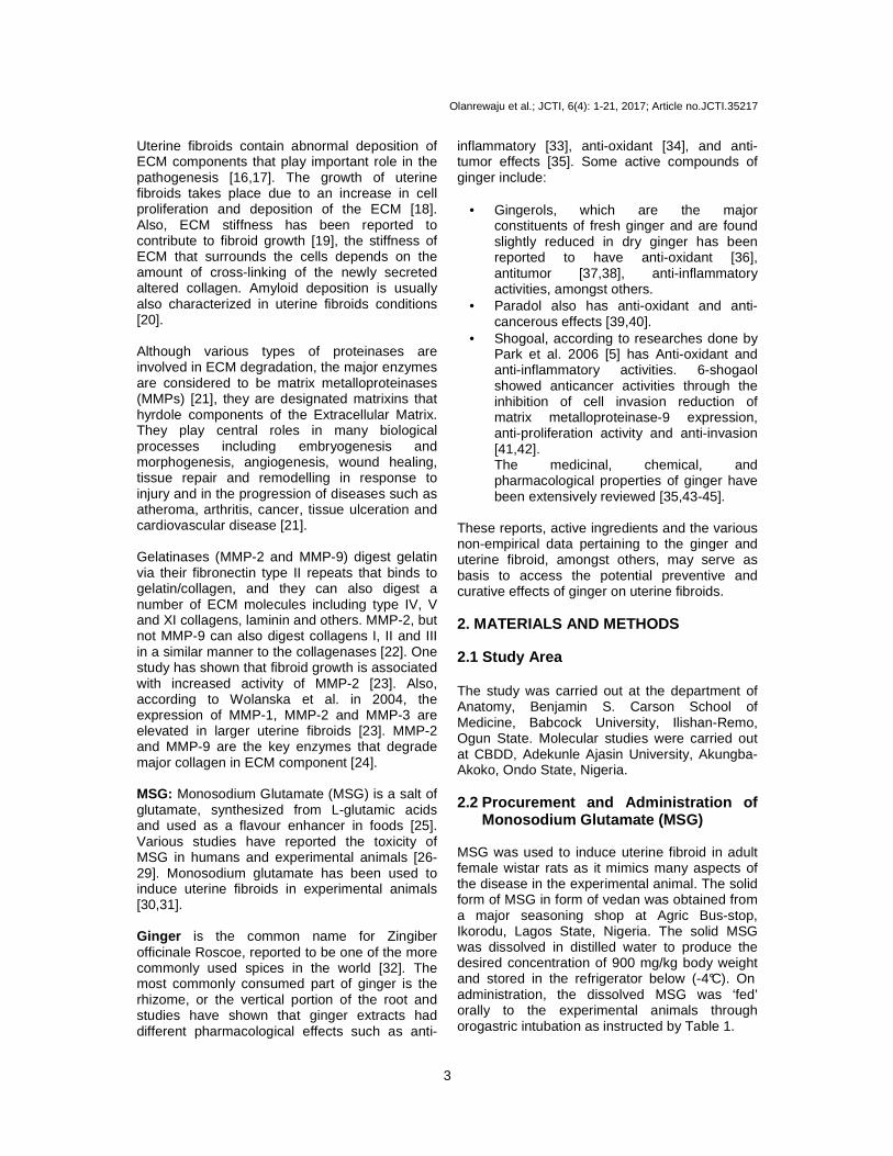
3.1.1 Serum estradiol level Group A: Control group Group E: MSG � Ginger low dose Group F: MSG � Ginger high dose Results from biochemical confirmation of uterine fibroid before commencement of ginger extract showed in Figure 1. that estradiol levels of groups E (25.28+1.414) and F (26.23±1.403)
were significantly higher than that of the group A (7.677±0.4707). 3.1.2 Serum progesterone level Group A: Control group Group E: MSG � Ginger low dose Group F: MSG � Ginger high dose Results from biochemical confirmation of uterine fibroid before commencement of ginger extract showed in Figure 2 that estradiol levels of groups E (12.45±0.6867) and F (12.71±0.7123) were significantly higher than that of the group A (6.390±0.3635).
A E F0
10
20
30
εµ
Groups
Est
radi
ol l
evel
(pg
/ml)
Figure 2. Bar graph showing estradiol levels (pg/ml ) across groups A, E, F ε= P<0.05 when compared with Group E, µ=P<0.05 when compared with Group F
A E F0
5
10
15
εµ
Groups
Pro
gest
eron
e le
vels
(ng
/ml)
Figure 3. Bar graph showing progesterone levels (ng /ml) across groups A, E, F
ε= P<0.05 when compared with Group E, µ=P<0.05 when compared with Group F 3.2 Mean Body Weight (MBW) The mean body weights of the animals were calculated as final body weight + initial body weight/2. Group A: Control Group B: MSG only Group C: Ginger only Group D: Ginger � MSG Group E: Ginger + MSG Group F: MSG � Ginger (Low dose) Group G: MSG � Ginger (High dose)

Olanrewaju et al.; JCTI, 6(4): 1-21, 2017; Article no.JCTI.35217
8
Groups A B C D E F Mean±SEM (g) 209.2±0.9972 216.3±1.216 203.5±0.4183 207.7±1.848 210.7±1.056 202.4±0.8308
3.3 Relative Uterus Weight (RUW) RUW (g) = Uterus weight/Body weight Group A: Control Group B: MSG only Group C: Ginger only Group D: Ginger � MSG Group E: Ginger + MSG Group F: MSG � Ginger (Low dose) Group G: MSG � Ginger (High dose)
Groups A B C D E F Mean± SEM (g)
0.00215± 0.0001147
0.004117± 0.0001759
0.0020± 0.0001581
0.003683± 0.0001990
0.00225± 0.0001118
0.00184± 0.0001806
Figure 4. Bar graph showing Mean Body Weight of ani mals across groups α= P<0.05 when compared with Group A, *=P<0.05 when compared with Group B, σ=P<0.05 when compared with Group C, λ=P<0.05 when compared with Group D, ε= P<0.05 when compared with Group E, µ=P<0.05
when compared with Group F
A B C D E F0.000
0.001
0.002
0.003
0.004
0.005
Groups
Rel
ativ
e U
teru
s W
eigh
t (g) αµσε αµσε
Figure 5. Bar graph showing Relative Organ Weight o f animals across groups α= P<0.05 when compared with Group A, σ=P<0.05 when compared with Group C ε= P<0.05 when compared with Group E, µ=P<0.05 when compared with Group F
A B C D E F
0
50
100
150
200
250 µσ
Groups
Mea
n bo
dy w
eigh
t (g)
µσλαε µσ µσ

Olanrewaju et al.; JCTI, 6(4): 1-21, 2017; Article no.JCTI.35217
9
3.4 Gross Organ Morphology of Uterus Specimens
Figure 6. Gross morphological images showing the ut erus of groups (a) Control (b) MSG only (c) Ginger only (d) Ginger ���� MSG (e) MSG ���� Ginger (low dose) (f) MSG ���� Ginger (high dose).
Images are in JPG format (black arrows: suspected i ntramural and pedunculated fibroids) 3.5 Histological Analysis
Olanrewaju et al.; JCTI, 6(4): 1-21, 2017; Article no.JCTI.35217
10
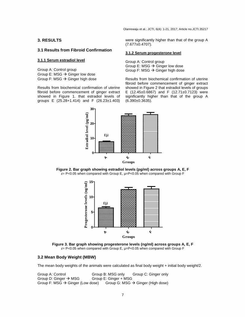
Plate 1. Photomicrographs showing panoramic views o f Uterus general morphological presentations in Wistar rats across groups A-F. Hem atoxylin and Eosin stain (X100). The
Perimetrium (P), Myometrium (M), The Endometrium (E ), uterine glands (G), uterine lumen (L), Highly vascularized extracellular matrix (yellow arr ows), Whorls of bland smooth muscle cells (black arrow). Group A: Control group Group B: Uter ine fibroid Group C: Ginger only Group D: Ginger only ���� Uterine fibroid Group E: MSG ���� Ginger (Low dose) Group F:
MSG ���� Ginger high dose
Plate 2. Photomicrographs showing panoramic views u terus general morphological presentations in Wistar rats across groups A-F. Per iodic Acid Schiff ‘PAS’ (X100). The
Myometrium (M), Endometrium (E), Amyloid deposition in pseudo-capsule vessel (G), uterine Lumen (L), severe endometriosis (yellow arrows), Ne crotic bodies (N) are well demonstrated.
Nuclei: Blue colouration Group A: Control group Group B: MSG only Group C: Ginger only
Group D: Ginger ���� MSG Group E: MSG ���� Ginger (Low dose) Group F: MSG ���� Ginger high dose

Olanrewaju et al.; JCTI, 6(4): 1-21, 2017; Article no.JCTI.35217
11
Plate 3. Photomicrographs showing panoramic views u terus general morphological presentations in Wistar rats across groups A-F. Mas son Trichrome stain (X100). The
perimetrium (P), Myometrium (M), Endometrium (E), u terine glands (G), uterine Lumen (L), severe endometriosis (yellow arrows), Apoptotic bod ies (B), Necrotic bodies (N) and uterine
blood vessels are well demonstrated. Group A: Control group Group B: MSG only Group C: Ginger only
Group D: Ginger ���� MSG Group E: MSG ���� Ginger (Low dose) Group F: MSG ���� Ginger high dose
Summary report: B, E - severe endometriosis, extracellular matrix accumulation with red blood cells was observed, excessive amyloid and collagen deposition. A, C, F- presents normal histology. D- mild histopathological changes were observed 3.6 MMP-2 Results
Groups
Relat
ive E
xpre
ssion
MMP-
2
A B C D E F
0
50
100
150
Figure 7. Bar graph showing relative expression of MMP-2 across groups through RNA isolation of tissue samples using RNA Snap kit

Olanrewaju et al.; JCTI, 6(4): 1-21, 2017; Article no.JCTI.35217
12
4. DISCUSSION
• Results from an attempt to confirm the presence of uterine fibroid in this study showed that the groups induced with uterine fibroid showed increased levels of estradiol - E2 (the form of estrogen found in non-pregnant conditions) and progesterone when tested. There was a remarkable rise in the estradiol level of groups E and F when compared to the control group. This can be attributed to the fact that estrogen promotes the growth of uterine fibroids, as these benign tumors have been described as estrogen-feeding tumors [53,54]. MSG which was used to induce the uterine fibroid has been reported to increase estrogen levels in experimental animals thus causing fibroid growth. The mechanism behind the MSG-induced fibroid formation and growth doesn’t just involve the hormones estrogen and progesterone but also circulating enzymes and hormone receptors. Aromatase and 17beta-hydroxysteroid dehydrogenase enzymes are aberrantly expressed in fibroids. Some of the known factors that increase aromatase activity are age, insulin, obesity, gonadotropins, and alcohol [67]. Aromatase and 17beta-hydroxysteroid dehydrogenase convert circulating androstenedione (a pro-hormone released by the adrenal cortex & ovaries) into estradiol [68]. Estradiol action is then mediated by its nuclear receptors ERα and ERβ to promote other uterine-fibroid-inducing conditions, amongst which is to induce the production of Progesterone Receptor (PR) which is then responsible for the response of uterine tissues to progesterone secreted by the ovaries. Upon ligand binding, these receptors act as transcriptional factors that up- or down-regulate gene expression by interacting with the regulatory regions of target genes. Estrogen also promotes fibroid growth by up-regulating Transforming Growth Factor-β3 which leads to increased cell proliferation, Insulin Growth Factor-1, Epidermal Growth Factor Receptor, and Platelet-derived Growth Factor (PDGF), and also promotes the abnormal survival of leiomyoma cells by reducing apoptosis through down-regulation of p53, increasing expression of the anti-apoptotic factor PCP4 and antagonizing PPAR-gamma
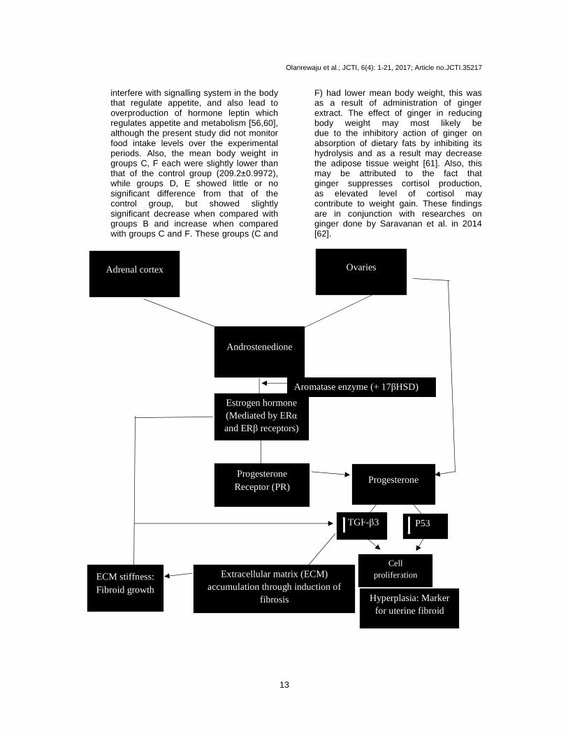
signaling [69]. Progesterone also promotes growth of leiomyoma through up-regulating EGF, TGF-beta1 and TGF-beta3, and promotes survival through up-regulating Bcl-2 expression and down-regulating TNF-alpha [70,71]. Estrogen and Progesterone, through their up-regulation of TGF-β3 and down-regulation of p53, increases cell proliferation and survival and enhances extracellular matrix formation through induction of fibrosis which is characterized by resistance to apoptosis leading to the persistence of cells, and secretion of collagen and other ECM components by those cells leading to abundant disposition of highly cross-linked, disoriented, and often widely dispersed collagen fibrils. Also, the stiffness of ECM that surrounds the cells depends on the amount of cross-linking of the newly secreted altered collagen. ECM stiffness have been reported to contribute to fibroid growth. [19]. The following sketch gives a concise explanation to this mechanism and will help in understanding how the other parameters evaluated in this study affects or are affected by uterine fibroid:(Figure 8)
• The results obtained from the analysis of mean body weight across the groups revealed that the group administered with MSG only had the highest mean body weight (216.3±1.216) when compared with the other groups (Figure 4). This can be attributed to the fact that with fibroid growth, there is increase in weight of the ‘host’ [55], and may also imply that MSG increases body weight over time [56] although as seen in the graph the risk of weight gain attributed to MSG intake was modest when compared with the control group. This weight gain could also have been as a result of evidence that shows that MSG stimulates the pancreas to produce about three times the normal amount of insulin it produces [57]. This unnatural amount of insulin then converts the sugar in the blood into fat very efficiently. Dr. Russsell Blaylock in his book Excitotoxins..The Taste that Kills, reports that in animal studies MSG creates a lesion in the hypothalamus that correlates with abnormal development, including obesity [58,59]. Studies have also suggested that increased food intake induced by MSG through leptin resistance and insulin action may also lead to an increase in weight gain as MSG might
Olanrewaju et al.; JCTI, 6(4): 1-21, 2017; Article no.JCTI.35217
13
interfere with signalling system in the body that regulate appetite, and also lead to overproduction of hormone leptin which regulates appetite and metabolism [56,60], although the present study did not monitor food intake levels over the experimental periods. Also, the mean body weight in groups C, F each were slightly lower than that of the control group (209.2±0.9972), while groups D, E showed little or no significant difference from that of the control group, but showed slightly significant decrease when compared with groups B and increase when compared with groups C and F. These groups (C and
F) had lower mean body weight, this was as a result of administration of ginger extract. The effect of ginger in reducing body weight may most likely be due to the inhibitory action of ginger on absorption of dietary fats by inhibiting its hydrolysis and as a result may decrease the adipose tissue weight [61]. Also, this may be attributed to the fact that ginger suppresses cortisol production, as elevated level of cortisol may contribute to weight gain. These findings are in conjunction with researches on ginger done by Saravanan et al. in 2014 [62].
P53 TGF-β3
Androstenedione
Aromatase enzyme (+ 17βHSD)
Estrogen hormone (Mediated by ERα and ERβ receptors)
Progesterone Receptor (PR)
Progesterone
Extracellular matrix (ECM) accumulation through induction of
fibrosis
ECM stiffness: Fibroid growth
Cell proliferation
Hyperplasia: Marker for uterine fibroid
Ovaries Adrenal cortex

Olanrewaju et al.; JCTI, 6(4): 1-21, 2017; Article no.JCTI.35217
14
• The uterus of animals with uterine fibroids were photographed and weighed to compare it with the uterus of control animals. There was observed change in the size of the uterus and some fibrous extensions suggested to be uterine fibroid (pedunculated), there was also statistical significant increase in the relative uterus weight of the animals in group B in comparison with group A (Figure 5). These could be attributed to fibroid growth in that group, induced by MSG administration. Fibroids do not only grow outside the uterus (as seen in pedunculated leiomyomas), but also grow cause the uterus to increase while growing from the inside [63]. This abnormal growth of the uterus and outgrowth on the uterus hence caused the organ weight of the uterus to be increased in the group induced with uterine fibroid. It is also important to note that an increase in weight and size of the uterus as seen in Figures 5 and 6usually indicates hyperplasia (cell proliferation), an early neoplastic process and a marker for uterine fibroid. This hyperplastic effect suggests that increased MSG produces a corresponding increase in synthesize of these biochemical markers that enhance proliferation process of the uterus. Gross morphologically, there was observable growth of pedunculated and submucous (black arrows) uterine fibroids in the specimen representative of group B, confirming the induction of tumour growth by Monosodium Glutamate (MSG). Generally, circulating steroid hormones modulates the growth of fibroid arising from uterine smooth muscle cells, which has been associated with periods of increased estrogen secretion. Therefore, as previously stated, the ability of estrogen to modulate the growth dynamics of uterine fibroid cells occurs by mechanisms involving modulation of cells proliferation. The growth of fibroid during periods of increased estrogen secretion is primarily due to cellular hypertrophy resulting in increase in intracellular volume [72]. In subsequent groups where ginger was administered, there was significant statistical decrease in the uterus weight and structural size in comparison to that of the negatively-induced group therefore implying that ginger may have played a pivotal role in reducing organ weight and/or shrinking the growing tumor. Gross
morphologically, there was presence of what can be referred to as ‘shrinking-in-progress’ of suspected fibroid in the prophylactic and curative I groups (i.e., groups D, E); while groups C, F showed little or no observable difference in comparison with group A, with no fibroid growth or complete shrinkage of benign tumour in corresponding cases. This could be attributed to the anti-tumor effects accorded to ginger and its related compounds e.g., Gingerol [64,65], Zerumbone [66]. Also, to take into notice is the little effect ginger had as a prophylactic agent with regards the evaluation of uterus weight. There was only a slight decrease in group D as compared to group B, and an abnormal increase in uterus weight as compared with the normal (control group).
• Histologically, the photomicrographs of the uterus were captured at X100magnification for appreciation of cellular components. Plate 1A showed the control group at a magnification of X100 stained with H & E dye. The muscular wall and the endometrial lining of the uterus are clearly seen. The glands embedded in stroma are also depicted, the layers of the uterine wall: perimetrium, myometrium and endometrium are clearly seen. The uterine lumen is also depicted in the photomicrograph. A normal uterine histology is clearly portrayed. Plate 1B showed bland/whorled/fascicular patterns of smooth muscle fibres, embedded in a fibrous stroma and separated by well-vascularized connective tissue. Deposition of abundant extracellular matrix was also depicted. Extracellular disorganized matrix is a peculiar characteristic of fibroid growth, mainly consisting of collagen subtypes, fibronectin, and proteoglycans. TGF-β3 plays an essential role in ECM overproduction in uterine fibroids by inducing the expression of collagen type 1, fibronectin, laminin and proteoglycans [73]. These ECM-related genes such as collagen type 1 and fibronectin are overexpressed in uterine fibroids. Norian et al. reported that mechanical signals are transmitted from the ECM scaffold via transmembrane receptors to the internal cytoskeleton in order to maintain an isometric state and demonstrated that the ECM microenvironment of leiomyoma cells is characterized by increased mechanical stress and that the viscoelastic properties

Olanrewaju et al.; JCTI, 6(4): 1-21, 2017; Article no.JCTI.35217
15
of the ECM contribute substantially to the increased tissue stiffness of leiomyoma [74,75]. Myometrial cells react to, and may be protected from, external loads by the mechanical properties of the surrounding matrix through secretion of ECM. Hence, the mechanical properties of leiomyoma are a key feature of these tumors and may contribute to their growth [75]. ECM of fibroids is not only excessive in amount, but it is abnormally formed, and the components of such critical structural elements, such as the orientation of the fibrils and their length are altered in fibroids [76]. Plate 1C showed a normal histo-achitecture similar to that of the control group. This could be attributed to the fact that ginger has no adverse effects on the uterus. This is in conjunction with work done by Xianglu et al. in 2009 [48]. Plates 1D, 1E, 1F showed reversal of the histopathology observed in plate 1B, with some bland smooth muscle fibers depicted, vascularized extracellular matrix seen in plates 1D and 1E, but plate 1F showed similar histology to the control group It could be said that ginger extract, just like Vitamin D, induces a remarkable reduction in the expression of collagen and other TGF-β3-dependent key profibrotic factors in human fibroid cells in a dose-dependent fashion [77].
PAS was used to investigate the pathogenesis of amyloid presented in uterine leiomyoma. Amyloidosis represents a spectrum of diseases that result from deposition of amyloid in extracellular matrix, leading to disruption of normal function and a broad but nonspecific clinical manifestations [79]. Plate 2A showed normal histology with little or no amyloid deposition observed. Plate 2B showed that all the muscle fiber interstitiums within fibroid entities were negative for amyloid deposition while fibroid capsule and vascular amyloid deposition positive which suggest that blood-derived ingredients and/or interstitium affected by metabolism of leiomyoma, may lead to conformational change of amyloid precursor protein and gradual amyloid deposition. Also, amyloid deposited in tissue looked as block mass. Inference from these results and our findings support that metabolic changes in the setting of functional alterations in local microenvironment with uterine leiomyoma, may be related to the amyloid deposition. Also concluded was that local tissue metabolism
changes resulted from fibroid cell functional alterations, may be related to the amyloid deposition [78]. Results are in conjunction studies done by Wu lin who, in 2000, found amyloid in uterine leiomyoma [80]. As seen in the plates, ginger extract in the subsequent groups seemed to reduce amyloid deposits especially in groups D and F. Although little or no research has been carried out on the activity of ginger extract in reducing amyloidosis in uterine leiomyoma cases, there has been few findings that portray ginger as an inhibitor of beta-amyloid peptide-induced cytokine and chemokine expression in cultured THP-1 monocytes [81]. Also, curcumin, a ginger family member, has been reported to reduce beta-amyloid plaques in Alzheimer’s disease condition [82]. Plate 3A showed normal histology with minimal collagen deposition. Plate 3B showed collagen accumulation. This was due to the accumulation of Extracellular matrix of which collagen is a major component of. The collagen production also occurs by tumor myocytes, the interstitium of the tumor fascicle is expanded by abundant blue collagen that separates the red staining fibroid myocytes. No interspersed fibroblasts are noted in the blue collagenous stroma, indicating that the stromal matrix has been produced by the tumor myocytes themselves. Also many of the myocytes of the central portion of the stained section in plate 3B are hypertrophied in size as cellular hypertrophy is a marker for uterine fibroid. Most leyomyomata demonstrate much more extracellular matrix deposition than normal myometrium does, and present as encapsulated collagen-rich masses of smooth muscle cells. LM typically have 50% more collagen than normal myometrium and an increased proportion of type I collagen [83]. Another sources revealed that collagen of type III in LM tissue is highly deposed too [84]. Ginger extracts in groups D and G seemed to nearly-abrogate these effects. Ginger extract, owing to some of its active compounds, down-regulated TGF–beta 3 which in turn reduces cell proliferation, hence reducing fibrosis that causes accumulation of collagen. Ginger extract reduced collagen deposits just like in previous studies [84,85].
• In conjunction with work done by Bogusiewicz et al. in 2007 [88], the present study showed an increase in MMP-2 level in group B when compared to group A. This can be attributed to the fact that uterine fibroids express higher levels of MMP activity than adjacent normal myometrium [86] to degrade the excess
Olanrewaju et al.; JCTI, 6(4): 1-21, 2017; Article no.JCTI.35217
16
ECM in cases of uterine fibroid. The dysfunction and disorganization of ECM homeostasis causes a paradoxical increase in MMPs in uterine fibroids. As stated earlier, uterine fibroids grow slowly by the deposition of a wide array of extracellular matrix (ECM) components which are under continuous physiological degradation process important for development, tissue repair and remodelling by MMPs [21]. There is also greater remodeling of the ECM in UL as they express higher levels of specific metalloproteinases (MMPs) including MMP2 and MMP11 [87]. The MMP-2 level in the ginger group C was less than that of the control group. Ginger administration in subsequent groups led to a decrease in expression of this proteinase enzyme, down to the level noticed in the normal myometrium. The other ginger-administered groups also showed decreased levels of MMP-2. Ginger played a major role in reducing this enzyme that is usually aberrantly expressed in uterine fibroid conditions. This finding is in conjunction with previous findings also [85,89,90,91].
4. CONCLUSION In conclusion, the results from this study have shown that MSG caused uterine fibroid as seen morphologically, increased the body weight of induced animals, increased their uterus weight, caused increased ECM, amyloid and collagen deposits histomorphologically and increased Matrix Metalloproteinase-2 (MMP-2) levels. However, high-dose curative treatments of aqueous extract of ginger rhizomes near-completely to completely mitigated the induced effects. Although, the prophylactic effect of ginger wasn’t as prominent as that of the high-dosed curative, there was a remarkable difference in the negatively-induced and prophylactic when observed in some parameters. The low-dose curative treatment was not as prominent as the other ginger-based groups, there was little or no therapeutic effect at that dose. These findings indicate that dose-dependent ginger extract may therefore offer the possibility of preventing and treating women with fibroids especially at higher doses for extended period without the need for surgery.
CONSENT It is not applicable.
ETHICAL APPROVAL As per international standard or university standard, written approval of Ethics committee has been collected and preserved by the authors. ACKNOWLEDGEMENT The authors acknowledge God Almighty for the success of this study. All references cited in this study are greatly acknowledged. The Centre for Bio-computing & Drug Development, Adekunle Ajasin University, Akungba-Akoko, Ondo State is acknowledged. Also, we acknowledge every and all individual that participated including the Oribamise family that contributed financially to this study, and the faculty members of the anatomy department of Benjamin S. Carson (Snr.) School of Medicine, Babcock Univeristy for all their support. COMPETING INTERESTS Authors have declared that no competing interests exist. REFERENCES 1. Levy G, Hill M, Beall S, Zarek S, Segars J,
Catherino W. Leiomyoma: Genetics, assisted reproduction, pregnancy and therapeutic advances. Journal of Assisted Reproduction and Genetics. 2012;29(8): 703–712.
2. Tavassoli F, Schnitt S, Hoefler H, Boecker W, Rosai J, Heywang-Kobrunner SH. Intraductal proliferative lesions. World Health Organization Classification of Tumours. Pathology and Genetics of Tumours of the Breast and Female Genital Organs. 2003;4.
3. Ishikawa H, Reierstad S, Demura M, Rademaker AW, Kasai T, Inoue M, Usui H, Shozu M, Bulun SE. High aromatase expression in uterine leiomyoma tissues of African-American women. Journal of Clinical Endocrinology and Metabolism. 2009;94(5):1752–1756.
4. Baird DD, Dunson DB, Hill MC, Cousins D, Schectman JM. High cumulative incidence of uterine leiomyoma in black and white women: Ultrasound evidence. American Journal of Obstetrics and Gynecology. 2003;188(1):100–107.

Olanrewaju et al.; JCTI, 6(4): 1-21, 2017; Article no.JCTI.35217
17
5. Park YJ, Wen J, Bang S, Park SW, Song SY. [6]-Gingerol induces cell cycle arrest and cell death of mutant p53-expressing pancreatic cancer cells. Yonsei Med J. 2006;47:688-697.
6. Vitiello D, McCarthy S. Diagnostic imaging of myomas. Obstetrics and Gynecology Clinics of North America. 2006;33(1):85–95.
7. Crum CP, Lester SC, Cotran RS. Female genital system and breast. In: Kummar V, Cotran RS, Robbins S, editors. Patología Humana. Madrid: Elsevier. 2004;679-717.
8. Wallach EE, Vlahos NF. Uterine myomas: An overview of development, clinical features, and management. Obstet Gynecol. 2004;104(2):393-406.
9. Martínez CM, Ibáñez C, Corpa JM. Simultaneous uterine leiomyoma and endometrial hyperplasia in a white-nosed monkey (Cercopithecus nictitans). First case report. An. Vet. (Murcia). 2010;26: 61-68.
10. Sankaran S, Manyonda IT. Medical management of fibroids (PDF). Best Pract Res Clin Obstet Gynaecol. 2008;22(4): 655–676.
11. Brosens I. Uterine leiomyomata: Pathogenesis and management. Abingdon, England, Informa Healthcare/ Taylor & Francis; 2006.
12. Veronika Aleksandrovych, Tomasz Bereza, Marek Sajewicz, Jerzy A. Walocha, Krzysztof Gil. Uterine fibroid: Common features of widespread tumor. Folia Medica Cracoviensia. 2015;LV,1:61–75.
13. Bereza T, Lis G, Mitus J, Sporek M, Chmielewski P, Kolber W, Mazur M, Goncerz G, Kuniewicz M. Blood vessels of the intratumoral septa in uterine leiomyomata. Folia Med Cracov. 2013; 53(2):99–106.
14. Walocha JA, Litwin JA, Miodoński AJ. Vascular system of intramural leiomyomata revealed by corrosion casting and scanning electron microscopy. Hum Reprod. 2003;18(5):1088–1093.
15. Bulun SE. Uterine fibroids. New England Journal of Medicine. 2013;369:1344-1355.
16. Stewart EA, Friedman AJ, Peck K, Nowak RA. Relative overexpression of collagen type I and collagen type III messenger ribonucleic acids by uterine leiomyomas during the proliferative phase of the menstrual cycle. Journal of Clinical Endocrinology and Metabolism. 1994; 79(3):900–906.
17. Malik J. Segars, Catherino WH. Integrin �1 regulates leiomyoma cytoskeletal integrity and growth. Matrix Biology. 2012;31(7-8): 389–397.
18. Walker R, Lupien JR. School of Biological Sciences, University of Surrey, UK, and Food and Nutrition Division, FAO of the United Nations, Italy. The safety evaluation of monosodium glutamate. Journal of Nutrition. 2000;130(4S Supp):1049S-1052S.
19. Leppert PC, Baginski T, Prupas C, Catherino WH, Pletcher S, Segars JH. Comparative ultrastructure of collagen fibrils in uterine leiomyomas and normal myometrium. Fertility and Sterility. 2004; 82(supplement 3):1182–1187.
20. Liu Y, Whelan RJ, Pattnaik BR, Ludwig K, Subudhi E, Rowland H, Claussen N, Zucker N, Uppal S, Kushner DM, Felder M, Patankar MS, Kapur A. Terpenoids from Zingiber officinale (Ginger) induce apoptosis in endometrial cancer cells through the activation of p53. PLoS One. 2012;7:e53178.
21. Visse R, Nagase H. Matrix metalloproteinases and tissue inhibitors of metalloproteinases. Circ. Res. 2003; 92(8):827-39.
22. Patterson ML, Atkinson SJ, Knauper V, Murphy G. Specific collagenolysis by gelatinase A, MMP‐2, is determined by the hemopexin domain and not the fibronectin-like domain. FEBS Letters. 2001;503(2-3):158-62.
23. Wolanska M, Sobolewski K, Bankowski E, Kaworski S. Matrix metalloproteinases of human leiomyoma in various stages of tumor growth. Gynecol Obstet Invest. 2004;58:14-18.
24. Zucker S, Lysik RM, Zarrabi MH, Moll U. M(r) 92,000 type IV collagenase is increased in plasma of patients with colon cancer and breast cancer. Cancer Res. 1993;53:140-146.
25. Ikonomidou C, Turski L. Glutamate in neurodegenerative disorders. In: Stone TW Ed. CNS neurotransmitters and neuromodulators: Glutamate. Boca Raton, Florida: CRC Press. 1995;253-272.
26. Eweka AO, Om’Iniabohs FAE. Histological studies of the effects of monosodium gluatamate on the ovaries of adult wistar rats. Annals of Medical and Health Sciences Research. 2011;1(1):37-43.
27. Dixit SG, Rani P, Anand A, Khatri K, Chauhan R, Bharihoke V. To study the

Olanrewaju et al.; JCTI, 6(4): 1-21, 2017; Article no.JCTI.35217
18
effects of monosodium gluatame on histomorphorlogy of cortex of kidney in adult albino rats. 2014;36(2):266-270.
28. Edward Group. The harmful effects of monosodium glutamate (MSG). Global Healing Center; 2014.
29. Veronika Husarova, Daniela Ostatnikova. Monosodium glutamate toxic effects and their impliations for human intake: A review. JMED Research; 2013.
30. Obochi GO, Malu SP, Obi-Abang M, Alozie Y, Iyam MA. Effect of garlic extracts on monosodium glutamate (MSG) induced fibroid in Wistar Rats. Pakistan Journal of Nutrition. 2009;8(7):970-76. DOI: 10.3923/pjn.2009.970.976
31. Koffuor AG, Annan K, Kyekyeku JO, Fiadjoe HK, Enyan E. Effect of ethanolic stem bark extract of Blighia unijugata (Sapindaceae) on Monosodium Glutamate-Induced Uterine Leiomyoma in Sprague-Dawley Rats. British Journal of Pharmaceutical Research. 2013;3(4):880-896.
32. Koh EM, Kim HJ, Kim S, Choi WH, Choi YH, Ryu SY, Kim SY, et al. Modulation of macrophage functions by compounds isolated from Zinigiber officinale. Planta Medica. 2009;75(2):148-151.
33. Young HY, Luo YL, Cheng HY, Hsieh WC, Liao JC, Peng WH. Analgesic and anti-inflammatory activities of [6]-gingerol. J Ethnopharmacol. 2005;96:207–210.
34. Stoilova I, Krastanov A, Stoyanova A, Denev P, Gargova S. Antioxidant activity of a ginger extract (Zingiber officinale). Food Chem. 2007;102:764-770.
35. Shukla Y, Singh M. Cancer protective properties of ginger: A brief review. Food Chem Toxicol. 2007;45(5):683-690.
36. Masuda Y, Kikuzaki H, Hisamoto M, Nakatani N. Antioxidant properties of gingerol related compounds from ginger. Biofactors. 2004;21(1-4):293-296.
37. Hu R, Zhou P, Peng Y-B, Xu X, Ma J, Liu Q, et al. 6-Shogaol induces apoptosis in human hepatocellular carcinoma cells and exhibits anti-tumor activity in vivo through endoplasmic reticulum stress. Plos ONE. 2012;7(6):e39664. DOI: 10.1371/journal.pone.0039664
38. Nafiseh Shokri Masshadi, Reza Ghiasvand, […], Mohammad Reza Mofid. Anti-oxidative and anti-inflammatory effects of finger in health and physical Activity: review of current evidence. International
Journal of Preventive Medicine. 2013; 4(Suppl 1):S36-S42.
39. Chung WY, Jung YJ, Surh YJ, Lee SS, Park KK. Antioxidtaive and tumor promoting effects of [6]-paradol and its homologs. Mutation Res. 2001;496:199-206.
40. Keum YS, Kim J, Lee KH, editors et al. Induction of apoptosis and caspase-3 activation by chemopreventive [6]-paradol and structurally related compounds in KB cels. Cancer Lett. 2002;177(1):41-47.
41. Ling H, Yang H, Tan SH, Chui WK, Chew EH. 6-Shogaol, an active constituent of ginger, inhibits breast cancer cell invasion by reducing matrix metalloproteinase-9 expression via blockade of nuclear factor-KB cativation. Br J Pharmacol. 2010; 161(8):1763-77.
42. Choudhury D, Das A, Bhattacharya A, Chakrabarti G. Aqeous extract of ginger shows antiproliferative activity through disruption of microtubule netwroek cancer cells. Food Chem Toxicol. 2010;48(1): 871-880.
43. Ali BH, Blunden G, Tanira MO, Nemmar A. Some phytochemical, pharamacological and toxicological properties of ginger (Zinigiber officinale Roscoe): A review of recent research. Food Chem Toxicol. 2008;46(2):409-420.
44. White B. Ginger: An overview. Am Fam Physician. 2007;75(11):1689-91.
45. Nicoll R, Henein MY. Ginger (Zinigiber officnale Roscoe): A hot remedy for cardiovascular disease? Int J Cardiol. 2009;131(3):408-9.
46. Walker R, Lupien JR. School of Biological Sciences, Univeristy of Surrey, UK, and Food and Nutrition Division, FAO of the United Nations, Italy. “The Safety evaluation of monosodium glutamate”. Journal of Nutrition. 2000;130(4S Suppl): 1049S-52S.
47. Shoyakugaku Zasshi. 1983;37(1):37-38. 48. Xianglu Rong, Gang Peng, […], Yuhao Li.
A 35-day gavage safety assessment of ginger in rats. Regul Toxicol Pharamacol. 2009;54(2):118-123.
49. Animal Research Review Panel. Guidelines for the Housing of Rats in Scientific Institutions. Guideline. 2002;20: 1-74.
50. Friedman AJ, SM Lobel, MS Rein, RL Barbieri. Efficacy and safety consideration in women with uterine leiomyomas treated with Gonadotropin Releasing Hormone
Olanrewaju et al.; JCTI, 6(4): 1-21, 2017; Article no.JCTI.35217
19
agonist. The Estroen Threshhold Hypothesis. Am J. Obstet Gyneacol. 1990;163:114-119.
51. Abeer M. Waggas. Neuroprotective evaluation of extract of giner (Zinigiber officinale) root in monosodium glutamate-induced toxicity in different brain areas male albino rats. Pakistan Journal of Biological Sciences. 2009;12:201-212.
52. Muhammad Sarwar Zia, Khadija Qamar, Ruhila Hanif, Moazzam Khalil. Effect of monosodium glutamate on the serum estrogen and progesterone levels in female rat and prevention of this effect with diltiazem. J Ayub Med Coll Abbottabad. 2014;26(1), 18-20
53. Center for Uterine Fibroids. What are fibroids? Boston: Brigham and Women's Hospital. 2006;2011. Sept 19 [cited 2011 Mar 13]; [about 3 screens].
54. Veronica Medikare, Lakshmi Rao Kandukuri, Venkateshwari Ananthapur, Mamata Deenadayal, Pratibha Nallari. The genetic bases of uterine fibroids; A review. J Reprod Infertil. 2011;12(3):181–191.
55. Leah Johnson. Can fibroids cause weight gain?. Fibroid Treatment Collective; 2016.
56. Adam Marcus. MSG linked to weight gain. American Journal of Clinical Nutrition; 2011. bit.ly/kv9cvF
57. Hugues Chevassus, Eric Renard, Gyslaine Bertrand, Isabelle Mourand, Raymond Puech, Nathalie Molinier, Joël Bockaert, Pierre Petit, Jacques Bringer. Effects of oral monosodium (l)-glutamate on insulin secretion and glucose tolerance in healthy volunteers. Br J Clin Pharmacol. 2002;
53(6):641–643. DOI: 10.1046/j.1365-2125.2002.01596.x
58. Russell L. Blaylock MD. Excitotoxins: The Taste That Kills. 1994;81.
59. Dani Veracity. The link between monosodium glutamate (MSG) and obesity. National Health News and Scientific Discoveries; 2005. Available:http://www.naturalnews.com/009379_monosodium_glutamate_MSG.html#ixzz4qrTUhWeI
60. Ka He, Liancheng Zhao, Martha L Daviglus, Alan R Dyer, Linda Van Horn, Daniel Garside, Liguang Zhu, Dongshuang Guo, Yangfeng Wu, Beifan Zhou, Jeremiah Stamler. Association of monosodium glutamate intake with overweight in Chinese adults: The INTERMAP study. Obesity (Silver Spring). 2008;16(8):1875–1880.
61. Badreldin H. Ali, Gerald Blunden, Musbah O. Tanira, Abderrahim Nemmar. The phytochemical, pharmacological and toxicological properties of ginger (Zingiber officinale Roscoe): A review of recent research. Food and Chemical Toxicology. 2008;46 409–420.
62. Ganapathy Saravanan, Ponnusamy Ponmurugan, Machampalayam, Arumugam Deepa, Balasubramanian Senthilkumar. Anti-obesity action of gingerol: Effect on lipid profile, insulin, leptin, amylase and lipase in male obese rats induced by a high-fat diet; 2014. Available:wileyonlinelibrary.com DOI: 10.1002/jsfa.6642
63. Rachel Ross, Live Science Contributor. Uterine Fibroids: Symptoms, Diagnosis and Treatment. Live Science; 2017. Available:https://www.livescience.com/34804-uterine-fibroids.html
64. Shafina Hanim Mohd Habib, Suzana Makpol, Noor Aini Abdul Hamid, Srijit Das, Wan Zurinah Wan Ngah, Yasmin Anum Mohd Yusof. Ginger extract (Zingiber officinale) has anti-cancer and anti-inflammatory effects on ethionine-induced hepatoma rats. Clinics. 2008;63(6):807–813.
65. EK Radhakrishnan, Smitha V. Bava, Sai Shyam Narayanan, Lekshmi R. Nath, Arun Kumar T. Thulasidasan, Eppurathu Vasudevan Soniya, Ruby John Anto. [6]-gingerol induces caspase-dependent apoptosis and prevents pma-induced proliferation in colon cancer cells by inhibiting MAPK/AP-1 signaling. PLoS One. 2014;9(8):e104401.
66. Kirana C, McIntosh GH, Record IR, Jones GP. Antitumor activity of extract of zingiber aromaticum and its bioactive sesquiterpenoid zerumbone. Nutr. Cancer. 2003;45:218-225.
67. Purohit V. Can alcohol promote aromatization of androgens to estrogens? A review. Alcohol. 2000;22(3):123-7.
68. Yanyan Hong, Shiuan Chen. Aromatase, estrone sulfatase, and 17β-Hydroxysteroid dehydrogenase: Structure-FUNCTION STUDIES AND INHIBITOR Development. Mol Cell Endocrinol. 2011;340(2): 120–126.
69. Davis BJ, Risinger JI, Chandramouli GVR, Bushel PR, Baird DD, Peddada SD. Gene expression in uterine leiomyoma from tumors likely to be growing (from Black Women over 35) and Tumors Likely to Be

Olanrewaju et al.; JCTI, 6(4): 1-21, 2017; Article no.JCTI.35217
20
Non-Growing (from White Women over 35). PLoS ONE. 2013;8(6):e63909. Available:https://doi.org/10.1371/journal.pone.0063909
70. Julie Kim J, Elizabeth C. Sefton. The role of progesterone signalling in the pathogenesis of uterine leiomyoma. Mol Cell Endocrinol. 2012;358(2):223–231.
71. Cermik D, Arici A, Taylor HS. Coordinated regulation of HOX gene expression in myometrium and uterine leiomyoma. Fertil Steril. 2002;78:979–84.
72. Fisher B, Costantino JP, Redmond CK, Fisher ER, Wickerham DL, Cronin WM. Endometrial cancer in tamoxifen-treated breast cancer patients: Findings from the National Surgical Adjuvant Breast and Bowel Project (NSABP) B-14. J. Natl. Cancer Inst. 1994;86:527-537.
73. Ding L, Xu J, Luo X, Chegini N. Gonadotropin releasing hormone and transforming growth factor β activate mitogen-activated protein kinase/extracellularly regulated kinase and differentially regulate fibronectin, type I collagen, and plasminogen activator inhibitor-1 expression in leiomyoma and myometrial smooth muscle cells. J Clin Endocrinol Metab. 2004;89:5549–5557.
74. Rogers R, Norian J, Malik M, et al. Mechanical homeostasis is altered in uterine leiomyoma. American Journal of Obstetrics and Gynecology. 2008;198(4): 474.e1–474.e11.
75. Norian JM, Owen CM, Taboas J, et al. Characterization of tissue biomechanics and mechanical signaling in uterine leiomyoma. Matrix Biology. 2012;31(1): 57–65.
76. Phyllis C. Leppert, William H. Catherino, James H. Segars. A new hypothesis about the origin of uterine fibroids based on gene expression profiling with microarrays. Am J Obstet Gynecol. 2006;195(2):415–420.
77. Ayman Al-Hendy, Marwa Badr. Can vitamin D reduce the risk of uterine fibroids? Womens Health (Lond Engl). 2014;10(4):353–358.
78. Jinping Liu, Fei Zhai, Peng Ge, Jinhai Lu, Yi Qin, Xuguo Sun. Investigation of amyloid deposition in uterine leiomyoma patients. Health. 2012;4(8):522-525.
79. Sun XG, Hu KS, Song L, Feng Q. Diag- nosis of amyloidosis. Journal of Chinese Modern Medi- cine. 2009;6:292-295.
80. Wu L, Yi GW, Lei JX. 22 cases of pregnancy with uterine fibroids. Journal of
Fourth Military Medical University. 2000; 21:310.
81. Grzanna R, Phan P, Polotsky A, Lindmark L, Frondoza CG. Ginger extract inhibits beta-amyloid peptide-induced cytokine and chemokine expression in cultured THP-1 monocytes. J Altern Complement Med. 2004;10(6):1009-13.
82. Shrikant Mishra, Kalpana Palanivelu. The effect of curcumin (turmeric) on Alzheimer's disease: An overview. Ann Indian Acad Neurol. 2008;11(1):13–19.
83. Brosens I. Uterine leiomyomata: Pathogenesis and management. Abingdon, England, Informa Healthcare/Taylor & Francis; 2006.
84. Tarek K Motawi, Manal A Hamed, Manal H Shabana, Reem M. Hashem, Asmaa F Aboul Naser. Zingiber officinale acts as a nutraceutical agent against liver fibrosis. Nutr Metab (Lond). 2011;8:40.
85. Narasimharao Bhagavathula, Roscoe L. Warner, Marissa DaSilva, Shannon D. McClintock, Adam Barron, Muhammad N. Aslam, Kent J. Johnson, James Varani. A combination of curcumin and ginger extract improves abrasion wound healing in Corticosteroid-Damaged Hairless Rat Skin. 2009;17(3):360.
86. Sunil K. Halder, Kevin G. Osteen, Ayman Al-Hendy. (Vitamin D3 inhibits expression and activities of matrix metalloproteinase-2 and -9 in human uterine fibroid cells. Hum Reprod. 2013;28(9):2407–2416.
87. Faezeh Koohestani, Andrea G. Braundmeier, Arash Mahdian, Jane Seo, JiaJia Bi, Romana A. Nowak. Extracellular matrix collagen alters cell proliferation and cell cycle progression of human uterine leiomyoma smooth muscle cells. PLoS One. 2013;8(9):e75844.
88. Bogusiewicz M, Stryjecka-Zimmer M, Postawski K, Jakimiuk AJ, Rechberger T. Activity of matrix metalloproteinase- 2 and -9 and contents of their tissue inhibitors in uterine leiomyoma and corresponding myometrium. Gynecological Endocrinology. 2007;23(9):541–546.
89. Ann M. Bode, Zigang Dong. The amazing and mighty ginger. Herbal Medicine: Biomolecular and Clinical Aspects. 2nd edition Chapter 7.
90. Binita Koirala Sharma, David Charles Klinzing, John Donnie Ramos. Zingiber officinale roscoe aqueous extract

Olanrewaju et al.; JCTI, 6(4): 1-21, 2017; Article no.JCTI.35217
21
modulates matrixmetalloproteinases and tissue inhibitors of metalloproteinases expressions in dengue virus-infected cells: Implications for prevention of vascular permeability. Tropical Journal of Pharmaceutical Research. 2015;14(8): 1371-1381.
91. Sahdeo Prasad, Amit K. Tyagi. Ginger and Its constituents: Role in prevention and treatment of gastrointestinal cancer. Gastroenterology Research and Practice. 2015;Article ID 142979:11. Available:http://dx.doi.org/10.1155/2015/142979
© 2017 Olanrewaju et al.; This is an Open Access article distributed under the terms of the Creative Commons Attribution License (http://creativecommons.org/licenses/by/4.0), which permits unrestricted use, distribution, and reproduction in any medium, provided the original work is properly cited.
Peer-review history: The peer review history for this paper can be accessed here:
http://sciencedomain.org/review-history/22481
View publication statsView publication stats