Embed Size (px)
Citation preview

S T U D I E S ON ISOLATED N U C L E I
1. Isolation and Chemical Characterization of a
Nuclear Fraction from Guinea Pig Liver
R A C H E L E M A G G I O , Ph.D., P H I L I P S I E K E V I T Z , Ph.D., and
G E O R G E E. P A L A D E , M.D.
From The Rockefeller Institute
ABSTRACT
This article describes a method for the isolation of nuclei from guinea pig liver. It involves the homogenization of the tissue in 0.88 M sucrose-l.5 mM CaC12 followed by centrifugation in a discontinuous density gradient in which the upper phase is the homogenate and the lower phase is 2.2 M sucrose-0.5 mM CaC12. Based on DNA recovery, the isolated fraction contains 25 to 30 per cent of the nuclei of the original homogenate. Electron microscopical observations showed that ~88 per cent of the isolated nuclei come from liver cells (the rest from von Kupffer cells and leucocytes) and that ~90 per cent of the nuclei appear intact, with well preserved nucleoli, nucleoplasm, nuclear envelope, and pores. Cyto- plasmic contamination is minimal and consists primarily of the nuclear envelope and its attached ribosomes. The nuclear fraction consists of ~22.3 per cent DNA, ~4.7 per cent RNA, and ~73 per cent protein, the DNA/RNA ratio being 4.7. Data on RNA extracti- bility by phosphate and salt and on the base composition of total nuclear RNA are included.
I N T R O D U C T I O N
Numerous methods based on a variety of ap- proaches have been used for the mass separation of nuclei from various tissues (cf. references 1, 2). In some of these methods (3-5) which follow the original procedure of Behrens (6), the tissue is frozen, ground, and lyophilized; the ensuing powder is suspended in an organic solvent, and the nuclei isolated by the differential or isopycnic centrifugation of this suspension. These procedures have the advantage of minimizing losses of water- soluble substances from the nucleus, and the disadvantage of poor preservation of nuclear morphology and extensive contamination by cyto- plasmic elements.
Procedures using aqueous media are more expeditive and in some cases yield better preserved nuclei. Citric acid solutions introduced by Stone-
burg (7) and subsequently used with various modifications by Marshak (8), Dounce (9-11), Mirsky and Pollister (12), and Dounce et al. (13) facilitate separation of the nuclei from the cyto- plasm, but precipitate the nucleoplasm. Under light optics, the appearance of nuclei isolated in sucrose solutions approaches that of nuclei in vivo (14). When packed during centrifugation, how- ever, such nuclei clump (14), presumably because of DNA 1 leakage (15). Clumping can be pre- vented by adding, to the sucrose solution, Ca ~- (16, 17) or Mg ~- (15) in low concentrations, or
x The abbreviations used in this paper are: DNA, deoxyribonucleic acid; RNA, ribonucleic acid; PCA, perchloric acid; TCA, trichloroacetic acid; AMP, adenylie acid; CMP, cytidylic acid; GMP, guanylic acid; UMP uridylic acid.
267
on May 19, 2018jcb.rupress.org Downloaded from http://doi.org/10.1083/jcb.18.2.267Published Online: 1 August, 1963 | Supp Info:

NaC1 and phosphate buffer (18). 2 Nuclei pre-
pared in aqueous media lose protein (20, 21) and probably RNA (21); moreover, if nuclear clump-
ing is extensive, the cytoplasmic contamination of the nuclear fraction becomes a problem difficult
or impossible to control by washing. Another non-
electrolyte repeatedly used in the past for the iso- lation of nuclear fractions is glycerol (22-25, @
reference 15). More recently good results were reported with a highly concentrated sucrose solu- tion by Chauveau and Chauveau et al. (26-29)
and, after some modifications, by Zbarskii and
Georgiev (30), Wilczok and Chorazy (31), and Sporn et al. (32). Finally, preliminary reports indicate that satisfactory results can be obtained by the use of detergents (33, 34). In this paper we describe a method for the isolation of a reasonably
pure nuclear fraction from guinea pig liver homogenates, and present data about the gross
chemistry of this fraction and about the properties of the RNA it contains.
M A T E R I A L S AND M E T H O D S
M a t e r i a l s
The animals used were young (albino) guinea pigs weighing 250 to 300 gm, fasted for 1 day before each experiment. The livers were removed under ether anesthesia.
Cell fractionation was carried through with Spinco model L ultracentrifuges, using either angle head rotors (No. 40) for some preliminary experiments, or swinging bucket rotors (SW 25, SW 39) for pre- liminary and final experiments. Centrifugal fields are given for the middle of the tubes.
All chemicals were of reagent grade.
M e t h o d s
C E L L F R A C T I O N A T I O N : Wheproceduresarede- scribed in detail in the experimental part since their development is the main object of this paper. As a starting point for our experiments we used the pro- cedure of Chauveau et M. (27), which seemed to us to be the best available at the time.
E L E C T R O N M I C R O S C O P Y : Liver tissue was fixed for 2 hours at 0 ° in 0.039 M ( 1 per cent) OsO4 in either acetate-Veronal buffer, pH 7.6 (35), or phos- phate buffer, 0.1 M, pH 7.7, (36); dehydrated in ethanol; and embedded in methacrylate or Epon according to standard procedures.
2 For more detailed information on the influence of pH, ionic strength, dielectric constant, and divalent cation concentration of the medium upon nucleo- plasm precipitation and nucleohistone extraction, see references 15, 18, and 19.
Pellets were fixed in situ, i.e. at the bottom of the tube, in either: (a) 0.039 M OsO4 in 0.88 M sucrose for ~15 hours at 0°; (b) 3.3 M formaldehyde (HCHO) (10 per cent) in acetate-Veronal buffer s for 3 hours at 0 °, followed by 2 hours in 0.039 M OsO4 in acetate- Veronal buffer (pH 7.6) at 0°; (c) 3.3 M HCHO in 0.1 M phosphate buffer (pH 7.6) followed by 0.039 M OsO4 in the same buffer; or (d) 0.24 M glutaraldehyde (37) in 0.1 M phosphate buffer (pH 7.6) followed by 0.039 M OsO4 in the same buffer, other conditions being the same as for (b). 4
All pellets were dehydrated in ethanol; cut into orientable strips when in 95 per cent ethanol; and finally embedded either at 47 ° in a 2:8 mixture of methyl methacrylate:n-butyl methaerylate, with 2 per cent benzoyl peroxide as a catalyst, or at 60 ° in Epon. Polymethacrylate or Epon cradles were used to embed the strips in the desired position. Sections were cut parallel to the direction of the sedimenta- tion, taking care to include the whole thickness of the pellet in the slice. They were subsequently stained with lead hydroxide (39), uranyl acetate (40), or both, and examined systematically from the top to the bottom of the pellet, to detect any layering or contamination. The microscope used was an RCA model EMU~2b or a Siemens Elmiskop I.
CHEMICAL DETERMINATIONS : The cell fractions were resuspended in H20 and treated with TCA to a final concentration of 10 per cent to precipi- tate proteins and nucleoproteins. After 15 hours at 0 °, the precipitate was separated by centrifugation; washed twice with cold 5 per cent TCA; extracted twice with either 95 per cent ethanol or ethanol: ether, 3: 1, at 60°; extracted twice with ether; and finally air dried. The dry powder was digested for 20 hours at 37 ° in 0.5 N K O H to convert the RNA into a mixture of 2', 3' mononucleotides (41). Total N was determined by nesslerization in small samples of this alkaline digest after H2SO4 combustion.
The digest was subsequently acidified at 4 ° with 70 per cent PCA (final PCA concentration 2 per cent) to precipitate unhydrolyzed proteins and DNA. The precipitate was separated by centrifugation and, after decanting the supernate, was washed with a small volume of 2 per cent PCA. The combined acid super- hate and washings were neutralized with K O H and
3 The final pH of this fixative mixture is 8,0 to 8.3 when the initial pH of the buffer is 7.6 and the formaldehyde is previously neutralized. As recently shown, acetate-Veronal is not an effective buffer in the presence of HCHO (37). 4 In the double fixation procedures (b) and (c), the first fixative mixture was carefully decanted and the pellet covered in situ, without washing, with the second fixative. In procedure (d) the pellet was washed in situ for 2 hours with phosphate buffer (pH 7.6) after decanting the glutaraldehyde and be- fore adding the OsO4.
268 T h E JOURNAL OF CELL BIOLOGY " VOLUME 18, 1963

the ensuing KCIO4-precipitate removed by centrifu- gation. RNA was determined in the neutralized supernatant by the orcinol reaction (42), using a yeast RNA as standard. ~ DNA was determined in the hot TCA extract (43) of the PCA residue by the diphenylamine reaction (44). 6 Since some protein was digested by KOH, protein N was obtained by subtracting the sum of RNA-N and DNA-N from total N. Nucleic acid N was calculated as 15.5 per cent of RNA and 16.1 per cent of DNA. Protein was estimated as 6.25 X protein N.
The alkaline digestion was preferred to the Schneider procedure (43) for it gave a good separa- tion of DNA from RNA (negligible amounts of DNA were found in the PCA extract) and permitted the subsequent study of the nucleotide composition of the RNA. The PCA residue was not checked for unextracted RNA, for the orcinol reaction is of little value in the presence of large amounts of DNA.
E X T R A C T I O N P R O C E D U R E S : The methods developed by Allfrey et al. (45) and subsequently mod- ified by Osawa et al. (46) were used to extract various RNA's from nuclear fractions isolated from 30 gm liver by our procedure and from 14 gm liver by the method of Chauveau et al. (27). Each fraction was homogenized in 60 ml 0.1 M K2HPO4-KH2PO4 buffer, pH 7.1, and subsequently centrifuged for 20 minutes at 20,000 g to obtain a residue and a super- nate which represents the pH 7.1 extract (46). The residue was reextracted as above with 100 ml of the same buffer. The second residue was further extracted by resuspending it in 60 ml 1 M NaC1. After 2 to 3 hours of continuous stirring at ~ 4 °, the preparation was centrifuged for 60 minutes at 30,000 g to yield two fractions: the pellet, or salt residue, and the supernate, or salt extract.
All RNA fractions obtained by phosphate and NaC1 extractions were hydrolyzed in 0.5 N K O H and the hydrolysate analyzed for RNA, DNA, and N. No recovery data were obtained in experiments involving salt extraction of nuclear fractions because of the impossibility of dispensing uniformly the nuclei in 1 M NaCI.
D E T E R M I N A T I O N O F N U C L E O T I D E S I N
RNA HYDROLYSATES: The four major nucleo- tide components of RNA were isolated from the alka- line hydrolysates by paper chromatography carried out as in Davis et al. (47) with the following modifica- tions: the paper used was Whatman No. 1 filter paper washed as recommended by Crestfield and Allen (48) ; the chromatograms were developed in the first direction by a descending flow (7 hours) of the isobutyrate buffer of Magasanik et al. (49) (10 parts
5 The sample obtained from Dr. J. F. Kitsch consisted of 91 per cent RNA and 9 per cent protein. 6 Absorbency was converted into milligrams, using a standard curve worked out for calf thymus DNA by Dr. V. Allfrey and Dr. A. E. Mirsky.
isobutyri~ acid, 6 parts 0.5 N NH4OH, V/V, pH 3.6 to 3.7); after drying in air, the chromatograms were developed in the second direction, at constant tem- perature (20 ° to 21 o), by a descending flow (17 hours) of the tertiary amyl alcohol-formic acid-water (14 : 6: 3; v : v : v) solvent of Hanes and Isherwood (50), prepared as recommended by Crestfield and Allen (48). Areas containing the nucleotides were detected under ultraviolet light and cut from the papers. The nucleotides were extracted by shaking the cut paper with 1 to 2 ml of 0.01 N HC1 for 30 minutes, and their amounts determined by using a Beckman UV spec- trophotometer and the spectral data of Wyatt (51).
From the 0.05 to 0.2 mg of hydrolyzed RNA pres- ent in each sample of original concentrate, 60 to 70 per cent was recovered after chromatography, as calculated from the sum of the amount§ of the four nueleotides.
R E S U L T S
1. P r e l i m i n a r y E x p e r i m e n t s
In the procedure of Chauveau et al. (27), the liver is homogenized in a 2.2 M sucrose solution (density 1.28 gm/cc) and the nuclei isolated by differential centrifugafion at 40,000 g for 60
minutes. The procedure has the inherent ad- vantage of preventing contaminat ion of the nu-
clear pellet by cytoplasmic components lighter than 1.28 gm/cc. It yields a preparat ion which in the light and electron microscopes was reported to appear homogeneous and to consist of well pre- served nuclei (27, 28).
In our hands a 1:20 (w :v) liver homogenate in 2.2 M sucrose yielded upon centrifugation at 40,000 g for 60 minutes (rotor No. 40) a gelatinous, slightly pink pellet which in the electron micro-
scope appeared to consist predominantly of nuclei slightly, but noticeably contaminated by bundles of collagen fibrils, debris which looked like twisted and extensively dehydrated cells, and fragments of cytoplasm still at tached to nuclei (Figs. l and 2).
In pellets fixed in 0.039 M O s O 4 in 0.88 M sucrose, the nuclei appeared extensively swollen and frequently ruptured, and showed little or no differentiation between nucleoli and nucleo- plasm. The appearance was noticeably better in pellets fixed in H C H O or glutaraldehyde followed
by OsO4 (procedures b to d) : recognizable nucleoli were present, and swelling and disruption were less pronounced, but many nuclei still appeared ruptured.
We concluded that both the homogeneity of the nuclear fraction and the preservation of the nuclei
RACHELE MA~OIO, PHILIP SIEKEVITZ, AND GEORGE E. PALADE I.~olated Nuclei. I 269

270 THE JOURNAL OF CELL BIOLOGY • VOLUME ]8, 1963

FmuR~.s 1 and 2 Nuclear fraction isolated in 2.2 M sucrose (Chauveau-Moul6 procedure, reference 27).
The preparation consists of intact (N1), partially damaged (N2), and extensively disrupted (N3) nuclei; it also contains cytoplasmic contaminants mostly in the form of compressed, twisted masses of mixed cytoplasmic and nuclear components (Cy). In Fig. 2, such masses are shown at a higher magnifi- cation to illustrate their composition which is exclusively cytoplasmic (in the plane of this section) for Cyl, mixed nuclear (left half) and cytoplasmic (right half) for Cy~, and predominantly nuclear, with some ribosome-containing cytoplasm (right lower corner) for Cy3.
Nucleoli (n), chromatin masses (ch), and coarsely granular nuc]eoplasmic regions (gn) can be recog- nized in some nuclei. The nuclear envelopes show numerous blebs (arrows).
Pellet fixed in glutaraldehyde followed by OsO4 (procedure d) and embedded in Epon. Sections stained in uranyl acetate followed by Pb(OH)2. Fig. 1, >( 3,200; Fig. 2, X 14,000.
needed improvement, and we also noted that double fixation of the pellets in H C H O - O s O 4 was decidedly better than fixation in OsO4 alone.
Further experiments showed that contamina- tion by collagen fibrils and tissue debris could be eliminated by removing the bulk of the connective tissue and vasculature of the liver before homog- enization (see below), and by filtering the homogenate through cheese cloth (cf. reference 17).
Nuclear damage was reduced by carrying the
homogenization in 0.88 M sucrose and subse- quently increasing the concentration of sucrose to 2.2 M in the final homogenate before centrifuga- tion. Since the improvement was only partial, we carried out a series of experiments to test the effects of Ca 2+ added to the sucrose solution. In t h e past, Ca ~- was used to reduce or prevent clumping of nuclei which we assume to be due to small nuclear ruptures, difficult to detect by light microscopy, rather than to leakage through an intact envelope (cf. reference 15). The addition of
RACHELE MAGGIO, PHILIP SIEKEVITZ, AND GEORGE E. PA~AD~ !solated Nuclei. I 271

CaC12 (final concentration 3.0 mM) to 2.2 M sucrose in a Chauveau et al. (27) type of separation led to the formation of a deposit on the centrifugal wall of the tube (rather than a conventional pellet) and caused coarse and general precipitation of the nucleoplasm (Fig. 3). It is noteworthy that a similar CaCIz concentration (3.0 mM) has been used in the past to isolate nuclei from many tissues (c]. reference 1). It is not known whether the mor- phology of these nuclei is similarly affected, but it has been reported that calf thymus nuclei, isolated in 0.25 M sucrose-3.0 mM CaCI~, do not lose the ability to incorporate amino acids into proteins (52). In a subsequent experiment we tested, following the example of Hogeboom et al. (17), the effect of Ca 2+ addition to sucrose, the homogenate being centrifuged in a 2 layer dis- continuous density gradient. The upper layer con- sisted of liver homogenate in 0.88 ra sucrose con- taining 1.0 mM CaCI~, the lower layer of 2.2 M sucrose containing either 0.5 or 1.0 or 1.5 mM CaC12. In the electron microscope all the pellets obtained showed minimal damage of the nuclear envelope and no precipitation of the nucleoplasm. A 2 layer system consisting of liver homogenate in 0.88 M sucrose with 1.5 m i CaC12 over 2.2 M sucrose with 0.5 mM CaC12 was also tested, found successful, and adopted for the final procedure.
~. F i n a l Procedure
a. HOMOGENIZATION: The livers were cut into relatively large pieces, cleaned of visible vessels and connective tissue, washed in a small volume of homogenization medium, gently blotted with cheese cloth, and subsequently passed through a tissue masher provided with a stainless steel plate with perforations of ~1 mm diameter. This operation removed most of the connective tissue framework and coarse vasculature of the organ. The ensuing tissue pulp was collected in small batches and gently homogenized in 0.88 M sucrose containing 1.5 mM CaCI~, using a glass homog- enizer provided with a loosely fitting Teflon pestle (0.009 inch clearance), moderate speed
(500 RI, M), and 4 to 6 up and down movements of the pestle. The volume of the pooled homogenized batches was adjusted to 1:20, liver weight:final volume, and the homogenate filtered through four layers of cheese cloth which retained a small but visible residue of tissue debris. All operations sub- sequent to the removal of the livers were carried out in a cold room at ~ 4 °.
The 9 to 30 gm fresh liver tissue needed for an experiment were processed in 9 gm batches, using three SW 25 rotors each with a capacity of 60 ml homogenate; i.e., 3 gm fresh tissue. The prepara- tion of these batches was so staggered as to pre- vent the "aging" of the homogenates, for storing the latter at 0 ° for 1 hour or more prior to cen- trifugation resulted in slightly reddish-colored pellets. Color was found to be a reliable indication of contamination.
b. SEPARATION: 20 ml of filtered homog- enate corresponding to 1 gm tissue were layered over 10 ml 2.2 M sucrose containing 0.5 mM CaC12 and centrifuged for 90 minutes at 53,000 g in a SW 25 rotor.
The pellet obtained at the bottom of the tube had the shape of a thin ring, and was colorless and slightly opalescent. In the phase contrast micro- scope, it appeared to consist of a homogeneous population of well preserved nuclei. A detailed description of the preparation at the electron microscope level is given in a following section.
A relatively thick reddish-tan layer formed at the interface between the two sucrose solutions. In the phase contrast microscope it was found to consist mainly of mitochondria and intact or partially broken liver cells, among which red blood cells and free nuclei were encountered.
Lipid droplets formed a white pellicle on top of the 0.88 M sucrose layer. The latter appeared cloudy at the end of centrifugation, presumably on account of still unsedimented microsomes.
c. WASHING: I n s o m e cases, the nuclear pel- lets were resuspended in a small volume of 0.88 M sucrose (usually 12 ml per 9 gm original liver tissue). Aliquots of 1.5 to 2.0 ml were layered on top of a discontinuous density gradient (1.5 ml of
FIGURE 3 Nuclei isolated in ~.~ M sucrose-3.0 mM CaCI2. At this Ca 2+ concentration, the nuclear content shows complete and severe precipitation in relatively large irregular masses (Pl, p2). Some of these masses (p2) may be or contain nucleoli. The nuclear en- velope is still recognizable in many places (arrows). Vesicles (v) of unknown origin are regularly encountered in these precipitated nuclei.
Pellet fixed in 0.039 M OsOa in 0.88 M sucrose and embedded in methacrylate. Section stained with Pb(OH)~. X ~7,0OO.
272 THI~ JOURNAL OF CELL BIOLOGY " VOLUME 18, 1963

RACHELE MAGGm, PHILIP SIEKEVITZ, AND GEORGE E. PALADE Isolated Nuclei. 1 273

1.5 M sucrose over 2 ml of 2.2 M sucrose) in a 5 ml tube of the SW 39 rotor and centrifuged for 45 minutes at 30,000 g. In terms of DNA amounts, 85 per cent of the original nuclear fraction was recovered in the washed preparations.
3. Electron Mic ro sco p y
a. COMPOSITION OF THE NUCLEAR FRAC- TION: The pellets obtained as described above consist almost exclusively of isolated nuclei (Figs. 4 a and b). Contamination by tissue debris or cell fragments was generally absent, and contamination by recognizable cytoplasmic components, i,e., mitochondria, centrioles, and elements of the endoplasmic reticulum, was only occasionally encountered. Counts in a representative fraction gave less than one mitochondrion or mitochondrial fragment for 10 nuclei (actually 172 mitochondria for 1830 nuclear profiles). The fractional volume of the mitochondrial contamination is therefore <~ 0.09 per cent. In their purified nuclear frac- tions, Hogeboom et al. (17) counted 1.5 mito- chondria per nucleus under light optics. Micro- somes and mitochondria are generally lighter than 1.28 gm/cc , hence we assume that they were brought down by nuclei incompletely freed of their surrounding cytoplasm. Since some cytoplasmic components appeared free in the pellet, we must further assume that their point of attachment to a nucleus was missed by the section.
The large majority of the sedimented nuclei could be identified as liver cell nuclei on account
of their size and structure. As in intact liver cells, they were large (3 to 5/z diameter), spherical, and provided with a variable number of nucleoli and a rather homogeneous, finely textured nucleo- plasm. A small number of nuclei could be recog- nized as coming from yon Kupffer cells or from leucocytes for, like the nuclei of such cells in situ, they were small (2 to 3/~ diameter), either oval or irregular in shape, and usually showed a charac- teristic peripheral concentration of dense chroma- tin material (Figs. 5 and 6).
The frequency of damaged nuclei was relatively small: about 10 per cent of the profiles showed completely ruptured nuclear envelopes and hernia- tion of nucleoplasm. Only occasionally did broken nuclei or recognizable nuclear fragments, mainly nucleoli, occur in the preparation. Table I gives in percentages the frequency of the various types mentioned among a total of 1217 nuclei counted in a representative preparation.
b. FINE STRUCTURE OF ISOLATED NUCLEI: The isolated nuclei appeared remarkably well preserved after fixation in H C H O - O s O 4 (Figs. 4 and 7). In mest of them the nucleoplasm showed, as in situ, a fine even texture comprised of fila- mentous or "granular" elements 5 to 10 m# in diameter, more or less evenly distributed between the nuclear envelope and the nucleoli. Further nucleoplasmic differentiations were visible in the form of coarsely granular fields interrupting the continuity of the finely textured material (Fig. 7). Evidence already obtained by electron micros-
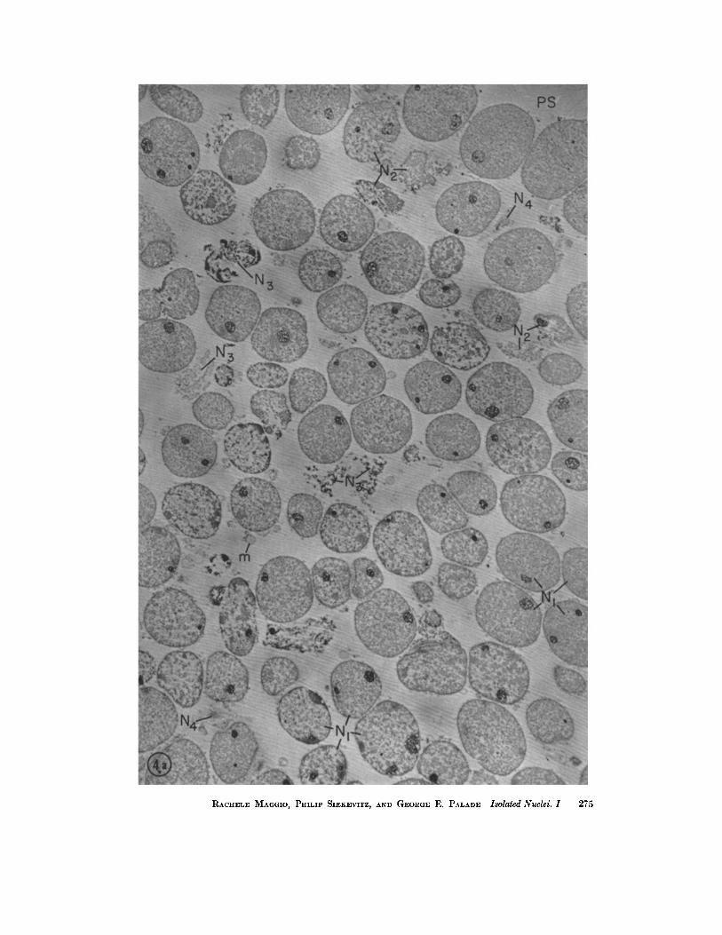
FIGURES 4 a and 4 b Nuclear fraction isolated from a liver homogenate (prepared in 0.88 M sucrosc-l.5 mM CaCl2) by centrifugation in a discontinuous density gradient (homogenate/~.2 M sucrose-0.5 mM CaCl~).
The preparation consists mainly of apparently intact nuclei (N1), slightly deformed by packing. Partially damaged (N2), or completely disrupted (N3) nuclei are relatively rare. Most nuclei appear in medial sections; less numerous lateral or grazing sections are marked N4.
Cytoplasmic contamination can be recognized more clearly in Fig. 4 b. I t consists of few mitochondria (m) and elements of the cndoplasmic reticulum ("microsomes") still attached to the nuclear envelopes (arrows).
Many nuclear profiles contain one or more nucleoli (n). The nucleoplasm shows a certain amount of differentiation between finely textured chromatin (eh) and coarsely granular, non-chromatin regions (gn). This detail is illustrated more clearly in Fig. 7.
Fig. 4 a, which shows a layer ~100 g from the surface of a nuclear pellet marked PS towards its bottom, demonstrates the relative homogeneity of the preparation, as well as the variation in nucleolar structure and nucleoplasmie texture between the top and the deeper layers of the pellet.
Pellet fixed in 3.3 M HCHO in 0.1 M phosphate buffer (pH 7.6) followed by 0.089 M OsO4 in the same buffer; embedded in methacrylate. Sections stained with 1%(0H)2 and covered with a carbon film. The fine striations in the eml:edding matrix represent wrinkles developed under the electron l:eam. Fig. 4 a, X ~,800; Fig. 4 b, X 5,200. (Fig. 4 b on following page)
274 THE JOURNAL OF CELL BIOLOGY - VOLUME 18, 1968
RACHEI~E MAGGIO, PHILIP SIEKEVITZ, AND GEORGE E. PALADE Isolated Nuclei. 1 275

276 THE JOURNEL OF CEL~ .BIOLOGY • VOLUME 18, 1963

copy, combined with either cytochemical spot tests (Feulgen (53, 54), indium (55)), enzymatic digestion (56), or autoradiography (57, 58), indicates that the deoxyribonucleoprotein (chro- matin, chromosomes) occupies the finely textured fields.
The nucleoli appear as more or less spherical
63) is usually present around each nucleus (Figs. 5 to 7) but in some cases its outer membrane is ruptured or discontinuous (Fig. 8 a). Blebs and points of continuity with the rest of the endo- plasmic reticulum are sometimes encountered (Fig. 8 a). Attached ribosomes are clearly recogniz- able on this outer membrane whenever present
FmVRE 5 Limited field in a nuclear pellet isolated by our procedure (see text and legend for Fig. 4). It shows nuclei derived from hepatic (N1) and yon Kupffer (N2) cells, and demonstrates clearly the
difference between these two nuclear types. Note the characteristic, high peripheral condensation of ehromatin in N2.
Pellet fixed in 3.3 M HCHO in 0.1 M phosphate buffer (pH 7.6) followed by 0.039 M OsO4 in the same buffer; embedded in methacrylate. Section stained with Pb(OH)2. )< 14,000.
masses (diam = 1.0 to 1.5 /z) of tightly packed small granules (diam = 10 to 15 m/z) and fine dense fibrils (cf. references 59-62). In sections these masses show many discontinuities or lacunae (diam ~ 100 m#) which appear either "empty" or occupied by a fibrillar material comparable in texture to chromatin. At their periphery, the nucleoli are in broad and intimate contact with the chromatin masses of the nucleus (Fig. 7).
A distinct, well preserved envelope (cf. reference
(Figs. 8 a to c, 9). Nuclear pores (diam ~-~ 90 m/z) are also recognizable (Figs. 8 and 9); most of them appear plugged by a moderately dense material (Figs. 8 a to c, 9), whereas a few seem to be free of such obstruction (Fig. 8 b). No evidence of nucleoplasm outflow or escape is found at the pores level.
The nucleoplasm texture was usually coarser in the deep layers of the pellets, presumably re- fleeting changes taking place during fixation
RACHELE MAGGIO, PHILIP SIEKEVITZ, AND GEonaE E. PALADE Isolated Nuclei. 1 277

in situ rather than differences established during fractionation. Swelling and extraction of the nucleoli occurred sometimes but were restricted to a thin upper layer in the pellet and affected primarily the material occupying the nucleolar lacunae.
The preceding description applies to nuclei
4. Nuclear Recovery
It follows that the fraction obtained is reasonably homogeneous and that the nuclei of which it is comprised show little structural damage. When the recovery is checked, however, it turns out that the fraction is incomplete in the sense that it does not
F m v l ~ 6 Limited field in a nuclear fraction prepared by our procedure. The nuclei marked N1 are derived from parenchymatous hepatic cells; and that marked N~ from a yon Kupffer cell. The group of nuclear profiles designated N3 represent the lobated nucleus of a leucocyte. Note the compact character and peripheral location of chromatin in N2 and especially in Na. Note also differences in the appearance of their nucleoli.
Preparation for electron microscopy as for Fig. 5. X 17,000.
fixed in H C H O OsO4 either in acetate-Veronal or in phosphate buffer. The general over-all preservation was slightly better in the last case but the fine structural features of the nuclei appeared unaffected by the variables involved in the two formulae. Glutaraldehyde followed by OsO4 (procedure d) gave less satisfactory results: the nucleoplasm was coarsely precipitated and the nuclear envelope less well preserved; the nucleoli, however, showed fewer "empty" lacunae.
contain all the DNA, presumably all the nuclei, of the original homogenate. In fact, only 25 to 35 per cent of the DNA of the filtered homogenate was recovered in the nuclear fraction, the rest remaining above the 0.88 M/2.2 ~ sucrose inter- face. Moreover, phase contrast microscopy showed that the dense band formed at the inter- face contained many nuclei. Since only 13 per cent of the latter were within apparently intact cells and 11 per cent in broken cells, whereas 76 per
278 ThE JOURNAL OF CELL BIOLOGY • VOLUME 18, 1963

cent appeared free, we assumed tha t the sedi- men ta t ion of the lat ter was prevented by excessive crowding at the interface. Indeed, increasing the concent ra t ion of the homogena te from 1:20 to 1:10 and 1:5 (w:v) led to a decrease in recovery from 30 to 12 and 8 per cent, respectively. Length- ening the centr i fugat ion t ime of a 1:20 homog-
T A B L E I
Type and Preservation of Nuclei in a Representative Nuclear Fraction
Nuclear profiles were counted in 33 different fields taken at r andom from the top to the bot- tom of a nuclear fraction pellet and micro- graphed at an original magnificat ion of 3,000. A few sections from a pellet, fixed in situ in 3.3
H C H O in 0.1 u phosphate buffer followed by 0.039 M OsO4 in the same buffer, were used for the counting.
The number of cytoplasmic tabs of any con- sequence found in all the fields counted was 14. Since they were generally smaller than a nucleus, the cytoplasmic contamina t ion is es- t imated at less than 0.5 per cent of the mass of the nuclear fraction. The obligatory cyto- plasmic contamina t ion represented by the nu- clear envelope is not included in this estimate.
Preservation Per
Type of nucleus Intact Ruptured Total cent
Liver cell nu- 968 108" 1,076 88.5 clei
yon Kupffer 93 18 111 9.1 cell nuclei
Leucocyte nu- 8 - - 8 0.6 elei
Unidentified~ 22 - - 22 1.8 Fotal 1,091 126 1,217
* This figure (10 per cent of total liver cell nuclei) comprises par t ia l ly ruptured as well as completely f ragmented nuclei; i.e., " f reed" nucleoli.
Mostly profiles too small for rel iable identifica- t ion (grazing sections).
enate did not not iceably improve the situation, whereas e l iminat ion of the interface proved more effective. Centr i fugat ion of a homogena te pre- pared directly in 2.2 M sucrose- l .5 mM CaCI~, or brought up to this concentra t ion after initial homogenizat ion in 0.88 M sucrose- l .5 mM CaC12, yielded nuclear pellets in which 50 to 55 per cent of the DNA of the initial, filtered prepara t ion was
recovered. These figures agree with the recovery reported by Chauveau et al. (27, 28), but the frequency of damaged nuclei and the extent of cytoplasmic contamina t ion also increased in these pellets (Fig. 10). Since for our purpose homo- geneity was more impor tan t than yield, we adopted the procedure described.
5. Gross Chemistry of Isolated Nuclei
The amounts and relative concentra t ions of RNA, DNA, and protein found in seven different nuclear fractions isolated by our procedure are given in Table II . The relatively large var iat ions in amounts per g ram fresh tissue can be explained by variat ions in yield from one exper iment to another . The smaller spread among percentages and ratios could reflect inheren t biological var ia- tions, differences in the extent of con tamina t ion or in the extract ion of macromolecular com- ponents. O n the average, R N A makes up 4.7, D N A 22.3, and protein 73 per cent of the mass of the nuclear fraction, when the sum of these com- pounds is taken as 100. Lipids and carbohydra tes were not determined.
The rat io of D N A to R N A varies from 3.8 to 5.6. If we assume tha t all the D N A in the cell is in the nucleus, tha t ~,25 per cent of the D N A in the filtered homogenate is recovered in our nuclear fraction, and tha t no R N A is extracted from the nuclei dur ing their isolation, it can be calculated tha t 8 to 12 per cent of the cellular R N A is found associated with the nucleus isolated by our procedure.
At tempts to improve the fraction by washing met with l imited success. As shown in Table I I I , there was a slight decrease in R N A and an equally small increase in DNA content in washed nuclear fractions. The effect on protein content was even less conclusive. The ratios of D N A and R N A to protein showed little or no change, when compared to those of unwashed fractions, whereas the D N A to R N A ratio was slightly increased.
6. Phosphate Extractible Nuclear R N A
Only 3 per cent of the original R N A was extracted by phosphate buffer from nuclear fractions isolated by our procedure, whereas the corresponding figure was ~ 1 5 per cent in the case of nuclear fractions isolated according to Chau- veau et al. (27). The la t ter value is considerably lower than the ~ 4 3 per cent reported by Hot t a and Osawa (64) for ra t hepat ic nuclear fractions
]~ACHELE MAGGIO, PHILIP SIEKEVITZ, AND GEORGE ]~. PALADE Isolated Nuclei. I 279

280 THE JOURNAL OF CELL BIOLOGY • VOLUME 18, 1963

obtained by the same procedure (27), and the ~ 3 0 per cent reported by Osawa et al. for calf thymus nuclei (46). Since Ca ~+ appeared notice- ably to affect the results, the effect of the cation on the composition of the nuclear fractions and on the extractibility of their R N A was investigated in more detail. Nuclear fractions were isolated in parallel from the same pool of liver tissue either in 2.2 M sucrose or in 2.2 r~ sucrose-l .5 mM CaCl2 and their gross chemistry determined. The D N A / R N A ratio was 5.3 and the percentage of phosphate extractible R N A was 6 per cent in the presence of Ca ~-, the corresponding figures being 4.8 and 15 per cent in the absence of the cation. Since Ca ~- addition permits the isolation of fractions in which nuclei are better preserved and less contaminated by cytoplasmic components, we assumed that the increase in D N A / R N A ratio and the decrease in the amount of phosphate extractible R N A reflect a decrease in cytoplasmic contamination. This assumption is supported by the following findings: (a) The R N A of cyto- plasmic fractions, especially that of microsomes, is easily extracted with phosphate buffer. Indeed we found that 60 to 80 per cent of the original R N A and 50 to 70 per cent of the original protein were extracted by 0.1 ~t potassium phosphate buffer from microsomal fractions isolated from guinea pig livers. 7 The phosphate extract of the microsomal fractions yielded, upon centrifugation for 2 hours at 105,000g, a clear supernate and a pellet which accounted for 85 per cent of the R N A of the original extract, and which in the
7 It should be noted that the amount of RNA present in microsomal fractions isolated by current pro- cedures (65, 66) was the same whether 0.88 M sucrose or 0.88 M sucrose-l.5 mM CaC12 was used for prepar- ing the original homogenate. The finding indicated that CaCI2 did not bring down with the microsomes any supernatant RNA which might subsequently be extracted by phosphate buffer.
electron microscope was found to consist of isolated vesicles and free particles, in all probability, detached ribosomes. (b) The nucleotide com- position of phosphate extractible nuclear R N A was found to be generally similar to that of cytoplasmic RNA's (see Table IV). (c) In appendix and thymus (64), the turnover of phosphate extractible, nuclear R N A is similar to that of the corresponding microsomal R N A (see, however, reference 68). (d) The phosphate extract of the nuclear fraction yielded, upon centrifugation for 60 minutes at 105,000 g, a pellet which accounted for 70 per cent of the R N A of the original phosphate extract. In the electron microscope, this pellet was found to consist of vesicles and free particles, presumably ribosomes (Fig. 11). This RNA-containing pellet is highly reminiscent in its morphology of the pellet obtained from the phosphate extracts of microsomal fractions. Its elements could represent contaminating microsomes or fragments of nuclear envelopes. (e) The residue obtained after the phosphate extraction of the nuclear fractions consists of still recognizable nuclei on account of their size and shape (Figs. 12 and 13). The nucleoplasm texture is, however, coarsened and the nuclear envelope extensively lost. Recognizable cytoplasmic contamination is drastically reduced.
7. Sal t Inso lub le Nuc l ear R N A
Extraction with 1 M NaC1 for 2 to 3 hours removed on ly~10 per cent, leaving behind 90 per cent of the R N A of our nuclear fractions. Table IV gives the nucleotide composition of this major, salt-insoluble fraction of nuclear R N A and compares it with that of a series of cytoplasmic RNA's. ~[he data show that the salt insoluble nuclear R N A is different from all the cytoplasmic RNA's examined: it has a lower G M P q- CMP, and a higher U M P q- A M P content than the latter, a feature already noted by Osawa et al. (46) for liver and other tissues.
FIGURE 7 Parts of 4 profiles of liver cell nuclei in a nuclear fraction isolated by our procedure. In the central profile, three nucleoli (nl, n~, na) show the dense granular tex- ture that characterizes these bodies in ~'tu. Some of their lacunae arc, however, larger and of lighter content (x) than in intact cells. Fine textured chromatin regions appear: l) around the nucleoli (chl) ("nucleolus-associatcd ehromatin"), 2) at the periphery of the nucleus, immediately below the nuclear envelope (ch2), and 3) scattered throughout the rest of the profile (ch3). The rest of the nucleus is occupied by a heterogeneous, coarsely granular material (gn). Nuclear envelopes can be seen at arrows. Contaminating micro- somes are marked m.
Preparation for electron microscopy as for Fig. 5. X ~0,000.
RACHELE Mxooto, PHILIP SIEKEVITZ, AND GEORGE E. PALADE Isolated Nuclei. 1 281

282 TH~ JOURNAL OF CELL BIOLOGY • VOLUME 18, 1963

D I S C U S S I O N
1. M e t h o d
Our procedure for the isolation of liver cell nuclei includes a number of features taken from already available methods. It is based primarily on the procedure of Chauveau et al. (27, 28) from which it retains the centrifugation in a viscous, highly concentrated sucrose solution (2.2 M) as an efficient means of separating nuclei from cyto- plasmic components. Nuclear damage is reduced by homogenization in 0.88 M sucrose. Nuclear aggregation is prevented by the addition of Ca ~- as already done by Schneider and Petermann (16) and by Hogeboom et al. (17). Finally, non- nuclear contamination is almost entirely elimi- nated by the preliminary filtration of the homog- enate and its centrifugation in a discontinuous density gradient.
2. N u c l e a r F r a c t i o n
a. M o R P H O L O G Y : The nuclear fraction so prepared has a number of favorable features. I t is homogeneous in the sense that it consists almost entirely of nuclei of which more than 88 per cent come from parenchymatous liver cells. It shows little preparative damage at the electron micro- scopical level, for ,-~90 per cent of the nuclear profiles seen in sectioned pellets appear intact. Finally, the fraction has been systematically and repeatedly examined and its composition in terms of subeellular components is well documented.
The preparation has also certain disadvantages of which the most obvious at present is low re- covery; the fraction accounts only for 25 to 30 per cent of the DNA of the parent homogenate. This shortcoming, common to nuclear fractions isolated by many other procedures (cf. references
1 and 2), is not particularly disturbing in our case in which homogeneity is more desirable than complete recovery.
The systematic electron microscopical examina- tion of our preparations revealed that the nuclear envelope is generally preserved, that there is no evidence of nucleoplasm loss through the nuclear pores, and that the structural organization of the nuclei does not visibly change upon isolation. I t follows that our nuclear preparation is mor- phologically satisfactory and that it contains, in addition to a negligible number of varied cyto- plasmic components, an "obligatory" or " ir- reducible" cytoplasmic contaminant, namely the nuclear envelope with its attached ribosomes. As originally established by Watson (63) and now generally accepted, the nuclear envelope is part of the general system of cytoplasmic membranes known as the endoplasmic reticulum. This oblig- atory contaminant is probably present in all nuclear fractions isolated in aqueous media (see, however, reference 68) although in many cases it may appear negligible when compared to the gross contamination introduced by other cyto- plasmic elements. The nuclear envelope appears to be partially removed from nuclei isolated in detergent solutions (34, 69).
b. c H e ~t I S T R Y : It is difficult to compare our fraction with nuclear preparations obtained by other procedures because the pertinent data are incomplete and frequently expressed in different ways that do not permit direct or meaningful comparison. If DNA/pro te in and RNA/pro te in ratios are used as criteria, our preparation con- tains a higher concentration of DNA and R N A than the nuclear fractions isolated by Chauveau et al. (27, 28) or by Hogeboom et al. (17) from rat liver (Table V). The difference can be explained only in part by a decrease in cytoplasmic con-
FIGURE 8 Normal sections through nuclear envelopes in nuclear fractions isolated by our procedure. The outer and inner nuclear membrane are marked om and ira, respec- tively. Ribosomes attached to the outer membrane are indicated by arrows. FIGURE 8 a The outer membrane shows a bleb at b (former point of continuity with the rest of the endoplasmic reticulum?) and is missing to the left of d. A number of nuclear pores are visible at pp and op; some of them appear "plugged" (pp), one is "open" (op). X 5~,000. FmURE 8 b "Plugged" (pp) and "unplugged" (op) pores are shown at a higher magnifi- cation than in Fig. 8 a. The body marked x might be a displaced plug (annulus). X 65,000. FIGnRE 8 C The micrograph shows clearly the nuclear envelope, with a series of plugged pores (pp) and a tenuously attached microsomal vesicle (m). Part of the nucleolus is visible in the lower right quarter. Chromatin masses are marked ch.
Preparation for electron microscopy as for Fig. 5. X 75,000.
•ACHELE MAGGIO, PHILIP SIEKEVITZ, AND GEORGE E. PALADE Isolated Nueld. I 283

FIGURE 9 Grazing section showing structural details at the periphery of a hepatic cell nucleus. Nuclear fraction isolated by our procedure.
The outer membrane of the nuclear envelope appears at om; the cavity of the perinuclear cisterna can be seen only at c, probably due to a local blebbing. Around the rest of the profile, the inner and outer membranes overlap to give an irregular corona of moderate density (ram). The outer membrane hears numerous attached ribosomes (arrows), some of them disposed in rows or spirals. The section cuts through nuclear plugs (annuli) at successive levels from the outside (pl) to the inside (p2) of the nucleus. Note that the plugs (diam __~ 800 A) seem to have a central orifice (diam ~ 100 A) surrounded by a corona of smaller (diam ~_ 60 A) openings. Channels of circular profile (o) cut through the peripheral chroma- tin (ch) under the pores.
Preparation for electron microscopy as for Fig. 5. X 77,000.
FIGURE 10 Nuclear fraction isolated by differential centrifugation from a liver homoge- nate prepared in ~.~ M sucrose-l.5 mM CaCI2.
The preparation consists of nuclei which show a coarser texture than those in Fig. 4. The number of damaged and disrupted nuclei is also higher and as a result "free" nucleoli (fn) are more frequently encountered. The masses marked Cy represent aggregates of disrupted nucleoplasm and cytoplasmic contaminants.
Preparation for electron microscopy as for Fig. 5. X 1~,000.
284 THE JOURNAL OF CELL BIOLOGY • VOLUME 18, 1963

RACHELE MAGGIO, PHILIP SIEKEVITZ, AND GEORGE E. PALADB Isolated Nuclei. I 285

T A B L E II
Chemical Composition of Hepatic Nuclear Fractions (Guinea Pig)
The nuclear fraction was isolated by the procedure given on page 272. RNA, DNA and total N were determined in the alkaline hydrolysate of the fraction as indicated in the text. Protein and Protein N were calculated by subtracting from total N,RNA-N, and DNA-N. Protein was estimated as 6.25 X protein N.
Wet weight RNA/ D N A / DNA/ Exp. processed RNA DNA Protein protein protein RNA
mg/gm* per ~nt~ mg~m* per ~nt~ mg~m* percent~ 1 30 0.089 4.6 0.484 25.4 1.34 70.0 0.066 0.34 5.4 2 30 0.124 4.6 0.498 18.4 2.10 77.0 0.059 0.24 4.0 3 30 0.117 4.3 0.531 19.7 2.03 76.0 0.058 0.26 4.5 4 30 0.089 4.9 0.500 27.8 1.21 67.3 0.073 0.41 5.6 5 15 0.099 4.4 0.400 18.6 1.70 77.0 0.058 0.24 4.0 6 15 0.094 4.7 0.525 26.3 1.36 69.0 0.068 0.38 5.6 7 9 0.091 5.4 0.342 21.0 1.22 73.6 0.074 0.28 3.8
Averages: 0.100 4.7 0.468 22.3 1.57 73.0 0.065 0.31 4.7
* per gram original tissue. t the sum of RNA q- DNA -1- protein being considered 100.
T A B L E I I I
Effect of Washing upon Hepatic Nuclear Fractions (Guinea Pig)
The fraction was isolated as described on page 272 and washed by recentrifugation in a discontinuous density gradient. RNA, DNA, and protein determined or calculated as for Table II. DNA recovery reached 85 per cent of the DNA of the original unwashed fraction in Exp. 1, and 75 per cent in Exp. 2.
RNA/ D N A ] DNA/ Exp. Nuclei RNA DNA Protein Protein Protein RNA
mg/gm* per ¢ent~ mg/gm* per ¢ent~ mg/gm* per cent~ 1 unwashed 0.099 4.5 0.40 18.5 1.70 77.0 0.058 0.24 4.0
washed 0.064 3.4 0.36 19.2 1.45 77.4 0.044 0.25 5.6
2 unwashed 0.073 4.0 0.40 20.0 1.49 76.0 0.049 0.27 5.5 washed 0.055 3.6 0.31 20.8 1.11 75.6 0.048 0.28 5.7
* per gram original tissue. the sum of RNA -b DNA -}- protein being considered 100.
tamination, for the latter is known to be of limited extent in the fractions mentioned. A different species (guinea pig), differences in losses of nuclear protein and RNA, variable extent of nuclear rupture and possibly selection of heavy nuclei (cf. reference 70) are other variables that may be involved. In this respect it is noteworthy that, in the examples mentioned, the concentrat ion of nucleic acids increases with the decrease in recovery. With the data at hand it is impossible
to decide, however, what is the relative importance of these factors.
3. PO4-Extractible Nuc lear R N A
On the strength of the results presented we conclude that the phosphate extractible RNA of the nuclear fraction isolated by the Chauveau-
Moul~ procedure represents cytoplasmic con- taminat ion; and that the smaller amounts of such R N A in our fraction may reflect, to a large extent,
286 THE JOURNAL OF CELL BIOLOGY • VOLUME 18, 1963

T A B L E IV
Nucltotide Composition of the RNA' s Obtained from Various Nuclear Fractions and from Some Cytoplasmic Fractions of Guinea Pig Liver
The RNA's were hydrolyzed and the nucleotides separated by chromatography and determined by spectrophotometry as given in the text. Each figure represents a separate experiment on a different RNA preparat ion.
Moles/100 moles of total nucleotides RNA source CMP GMP UMP AMP
A + c Gd-U
Salt residue of nu- 23.3, 20.5, 26.3, 31.0, 29.5, 27.5, 20.9, 21.0, 0.79, 0.71, clear fraction:~ 22.8 30.3 28.3 18.6 0.71
Phosphate extract of 26.8, 24.0 31.9, 38.0 25.0, 22.7 16.3, 15.3 0.76, 0.65 nuclear fraction*
Soluble RNA 28.0, 27.8 33.5, 34.6 20.0, 20.0 18.5, 17.6 0.87, 0.83 Microsomal fraction 27.8 35.2 20.6 16.4 0.79 Microsomal RNP 31.4, 28.0 30.0, 37.0 20.5, 17.5 18.1, 17.5 0.98, 0.84
particles First postmicrosomal 29.7 31.1 21.8 17.4 0.89
fract ion
* Prepared from nuclei isolated in 2.2 M sucrose. This phosphate extract contained ~ 15 per cent Of the tntal RNA of the nuclei.
Prepared from nuclei isolated according to our method. The nuclei were t reated for 2 hours with 1 M NaCI; the salt residue obtained after this t rea tment contained over 90 per cent of the RNA of the whole nuclei. Phosphate extracted only ~ 3 per cent of the total RNA of these nuclei.
the existence of an irreducible cytoplasmic con-
taminat ion , namely the presence of a t tached
ribosomes on the outer m e m b r a n e of the nuclear
envelope. O u r results and conclusions must be
contrasted with those reported and arr ived at by
other workers (45, 46, 64, 68) who found tha t a
large propor t ion of the R N A of nuclear fractions,
i.e. up to ~ 4 0 per cent of the total, could be
extracted with 0.i ~a phosphate buffer, pH 7.1.
Such results were obta ined with nuclear fractions
isolated from liver, thymus, and appendix in a
0.25 M sucrose conta ining 3 to 4 mM CaCI~.
Fur the r studies could show to what extent our
R E F E R E N C E S
conclusions apply to the h igh percentage of PO4-extractible R N A reported in these fractions.
Part of this work was made possible by a grant (A- 1635) to Dr. Siekevitz from the National Institute of Arthritis and Metabolic Diseases, National Insti- tutes of Health, United States Public Health Service.
Dr. Maggio is a Fellow of the Population Council, Rockefeller Foundation. Her present address is the Department of Comparative Anatomy, University of Palermo, Palermo, Italy.
A preliminary report of these findings was made at the meeting of the American Society for Cell Biology in San Francisco, November, 1962.
Received for publication, January 10, 1963.
1. ALLFREY, V., in The Cell, (J. Brachet and A. E. Mirsky, editors), New York, Academic Press, Inc., 1959, 1, 193.
2. ROODYN, D. B., Internat. Rev. Cytol., 1959, 8, 279. 3. BEHRENS, M., in Handbuch der Biologischen Ar-
beitsmethoden, (E. Abderhalden, editor), Berlin and Vienna, Urban and Schwarzen- berg, 1938, 5/10, 1363.
R., and FREER, R. M., J. Gen. Physiol., 1950, 33, 629:
5. ALLFREY, V., San~RN, H., MmSKV, A. E., and SAETR~N, H., J. Gen. Physiol., 1952, 35, 529.
6. BEHRENS, M., Z. Physiol. Chem., 1932, 209, 59. 7. STONEBURG, C. A., J. Biol. Chem., 1939, 129, 189. 8. MARSH~K, A., J. Gen. Physiol., 1941, 25, 275. 9. DOUNCE, A. L., J. Biol. Chem., 1943, 147,685.
4. DOUNCEj A. L., TISHKOFF, G. H., BARRNETT, S. _ 10. DOUNCE, A. L., J. Biol. Chem., 1943, 151, 221.
RACHELE MAOGIO, PmLIP SIEKEVITZ, AND GEORGE E, PALADE Isolated Nuclei. I 287

FIGURE 11 Section of a pellet obtained by centrifuging at 105,000 g for ~ hours a phosphate extract of a nuclear fraction obtained by our procedure.
The pellet accounts for 70 per cent of the extracted RNA. As illustrated by this figure, the bottom layer of the pellet consists of isolated vesicles (v), attached and free ribosomes (r), and larger particles, possibly glycogen (g). The top layer (not illustrated) is comprised mainly of free ribosomes.
Pellet fixed in 3.8 M HCHO in acetate-Veronal buffer followed by 0.039 M OsO4 in the same buffer (pH 7.7); embedded in methacrylatc. Section stained with Pb(OH)2. X 66,000.
11. DOUNCE, A. L., Ann. New York Acad. Sc., 1948, 50, 982.
12. MIRSKy, A. E., and POLLISTER, A. W., J. Gen. Physiol., 1946, 30, 101, 117.
13. DOUNCE, A. L., WITTER, R. F., MONa'Z, K. J., PATE, S., and COTTONE, M. A., J. Biophysic. and Biochem. Cytol., 1955, 1, 139.
14. HOGEBOOM, G. H., SCHNEIDER, W. C., and Pa- LADE, G. E., J. Biol. Chem., 1948, 172,619.
15. PHILPOT, J. ST. L., and STANIER, J. E., Biochem. J. , 1956, 63,214.
16. SCHNEIDER, R. M., and PETERMANN, ~VI. L., Cancer Research, 1950, 10, 751.
17. HOGEBOOM, G. H., SCHNEIDER, W. C., and
STRIEBICH, J., J. Biol. Chem., 1952, 196, 111.
18. WILBUR, K. M., and ANDERSON, N. G., Exp.
Cell Research, 1951, 11, 47.
FIGURES 12 and 18 The preparation consists of shrunken and altered, but still recog- nizable, nuclei interconnected by many fine strands of moderate density (J). Fig. 18 shows that the extraction has not caused whole scale disruption of nuclei. Fig. 12 demon- strates that the nuclcoplasm has a tighter and coarser texture than before extraction. Nucleoli (n) are still recognizable. The nuclear envelope has been removed from most of the perimeter of the profiles. A few remnants are marked by arrows. The one in the upper left corner still bears a few attached ribosomes.
Pellet fixed and embedded as for Fig. 11. Sections stained with uranyl acetate fol- lowed by Pb(OH)2. Fig. 12, )< 18,000; Fig. 18, )< 3,600.
288 THE JOUI~NAI~ OF CI$1~L BIOLOGY • VOLUME 18, 1963

RACHELE MAGGIO, PHILII" SIEKEVITZ, AND GEOI:iGE E. PALADE Isolated Nuclei. I 289

T A B L E V
Comparison of the Gross Chemistry of Hepatic Nuclear Fractions Isolated in Sucrose Solutions
Medium DNA per cent DNA RNA RNA/
Material Procedure Sucrose Ca ~-+ recovery protein protein DNA
Rat liver Hogeboom 0.25 M 1.8 mM 92* 0.14 0.054 0.38
et al. (17) 0.34 M 0.18 mM 78~ 0.20 0.049 0.24 Rat liver Chauveau 2.2 M 45--55 0.21 0.034 0.16
et al. (27) Guinea pig liver Maggio 0.88 M 1,5 mM 25--30§ 0.31 0.065 0.21
et al. 2.2 M 0.5 mM
* recovery and ratios calculated from data for nuclear fractions isolated from unfiltered homogenates. (Table I, reference 17.)
recovery and ratios calculated from data for nuclear fractions isolated from filtered homogenates pre- pared from twice perfused livers. (Table I I I , reference 17.) The recovery figure represents 90 per cent of the DNA in the filtered homogenate. § per cent DNA recovered from filtered homogenates.
19. ANDERSON, N. G., and WILBUR, K. M., J. Gen. Physiol., 1952, 35, 781.
20. STERN, H., and MIRSKY, A. E., J. Gen. Physiol., 1953, 37, 177.
21. KAY, R. M., SMELLIE, R. M. S., HUMPHREY, G. F., and DAVlDSON, J. N., Biochem. J., 1956, 62, 160.
22. SCHNEIDER, R. M., Fed. Proc., 1952, 11, 194. 23. DALLAM, R. D., and THOMAS, L., Fed. Proc.,
1953, 12, 193. 24. SCHNEIDER, R. M., Exp. Cell Research, 1955, 8, 24. 25. POORT, C., Biochim. et Biophysica Acta, 1957, 25,
34. 26. CHAUVEAU, J., Compt. rend. Acad. so., 1952, 235,
902. 27. CHAUVEAU, J., MouL~, Y., and ROUILLER, C.,
Exp. Cell Research, 1956, 11, 317. 28. CHAUVEAU, J., Bull. Soc. Chim. Biol., 1957, 39,
1521. 29. MouLl~, Y., and CHAUVEAU, J. Exp. Cell Re-
search, 1959, suppl. 7, 156. 30. ZBARSKn, J. B., and GEORGIEV, G. P., Biokhimiya,
1959, 24, 192. 31. WILCZOK, T., and CHORAZY, K., Nature, 1960,
188, 516. 32. SPORN, M. B., WANKO, T., and DINGMAN, W.,
J. Cell Biol., 1962, 15, 109. 33. ZALTA, J-P., ROZENCWAjG, R., CARASSO, N.,
and FAVARD, P., Compt. rend. Acad. sc., 1962, 255 ,412 .
34. FISHER, H. W., and HARRIS, H., Proc. Roy. Soc. London, Series B, 1962, 156, 521.
35. PALADE, G. E., J. Exp. Med., 1952, 95,285. 36. MILLONIG, G., J. Appl. Physics, 1961, 32, 1637. 37. HOLT, S. J., and HicKs, R. M., Nature, 1961,
191,832.
38. SABATINI, D. D., BENSCH, K., and BARRNETT, R.J . , J. CellBiol., 1963, 17, 19.
39. KARNOVSKY, M. J., J. Biophysic. and Biochem. Cytol., 1961, 11, 729.
40. GIBBONS, I. R., and GRIMSTONE, A. V., o r. Bio- physic, and Biochem. Cytol., 1960, 7,697.
41. COHN, W., J. Biol. Chem., 1960, 235, 1488. 42. MEJBAUM, W., I . Physiol. Chem., 1939, 258, 117. 43. SCHNEIDER, W. C., J. Biol. Chem., 1945, 161,293. 44. DISCHE, Z., Mikrochemie, 1930, 8, 4. 45. ALLFREY, V. G., MIRSKY, A. E., and OSAWA, S.,
J. Gen. Physiol., 1957, 40, 451. 46. OSAWA, S., TAKATA, K., and HOTTA, Y., Bio-
chim. et Biophysica Acta, 1958, 28,271. 47. DAVIS, F. F., CARLUCCI, A. F., and ROUBEIN,
I. F., J. Biol. Chem., 1959, 234, 1525. 48. CRESTFIELD, A. M., and ALLEN, F. W.,
Chromatographic Methods, 1957, 2, 9. 49. MAOASANIK, B., WIMBLER, E., DUNIOER, R.,
ELSON, D., and CHARGAFF, E., 3". Biol. Chem., 1950, 136, 37.
50. HANES, C. S., and ISHERWOOD, F. A., Nature, 1949, 164, 1107.
51. WYATT, G. R., in Nucleic Acids (E. Chargaff and J. N. Davidson, editors), New York, Academic Press Inc., 1955, 1, 523.
52. ALLFREY, V. G., and MIRSKY, A. E., Proc. Nat. Acad. Sc., 1957, 43, 589.
53. MosEs, M. J., J. Biophysic. and Biochem. Cytol., 1956, 2 suppl. 4, 397.
54. HUXLEY, H. E., and ZUBAY, G., J. Biophysic. and Biochem. Cytol., 1961, 11, 273.
55. WATSON, M. L., and ALDRIDGE, W. G., J. Cell Biol., 1961, 11,257.
56. LEDUC, E. H., and BERNHARD, W., J. Biophysic. and Biochem. Cytol., 1960, 10, 437.
290 THE JOURNAL OF CELL BIOLOGY • VOLUME 18, 1963

57. REVEL, J. P., and HAY, E. D., Exp. Cell Research, 1961, 25, 476.
58. HAY, E. D., and REVEL, J. P., Anat. Rec., 1962, 142, 339.
59. BERNHARD, W., BAUER, A., GRoPP, A., HAGU- ENAU, F., and OBERLING, C. H., Exp. Cell Re- search, 1955, 9, 88.
60. BERNHARD, W., Exp. Cell Research, 1959, suppl. 6, 17.
61. I-IORSTMANN, E., and KNOOP, A., Z. Zellforsch., u. Mikr. Anat., 1957, 46, 100.
62. LAFONTAINE, J. G., and CHOUINARD, L. A., J. Cell Biol., 1963, 17, 167.
63. WATSON, M. L., J. Biophysic. and Biochem. Cytol., 1955, 1,257.
64. HOTTA, Y., and OSAWA, S., Biochim. et Biophysica Acta, 1958, 28, 642.
65. SIEKEVlTZ, P., and PALADE, G. E., J. Biophysic. and Biochem. Cytol., 1958, 4, 309.
66. SIEKEVITZ, P., and PALADE, G. E., J. Biophysic. and Biochem. Cytol., 1958, 4, 557.
67. KIT, S., Arch. Biochem. and Biophysic., 1960, 88, 1. 68. DAVISON, P. F., and MERCER, E. H., Exp. Cell
Research, 1956, 11, 237. 69. HUBERT, M.-T., FAVARD, P., CAEASSO, N.,
ROZENCWAJG, R., and ZALTA, J-P., J. Micr., 1962, 1,435.
70. FALZONE, J. A., BARROWS, C. H., and YIENOST, M. J., Exp. Cell Research, 1962, 26, 552.
RACHELE MAGOlO, IOHILIP SIEKEVITZ, AND GEORGE E. PALADE Isolated Nuclei. I 291


![The Behavior of Multi-Story Buildings Seismically Isolated ...file.scirp.org/pdf/OJER_2014121817203398.pdf · Isolated System Hybrid Isolation ... [11]. The importance of ... -This](https://img.dokumen.tips/doc/110x75/5b348ac87f8b9a8b4b8c2651/the-behavior-of-multi-story-buildings-seismically-isolated-filescirporgpdfojer.jpg)


