Embed Size (px)
Citation preview

Proc. Nati. Acad. Sci. USAVol. 78, No. 4, pp. 2179-2183, April 1981Biochemistry
Structure of a B-DNA dodecamer: Conformation and dynamics*(DNA structure/sugar puckering in DNA/principle of anticorrelation/torsion angles in DNA/thermal vibration in DNA)
HORACE R. DREWt, RICHARD M. WINGtt, TSUNEHIRO TAKANOt, CHRISTOPHER BROKAt§, SHOJI TANAKAt¶,KEIICHI ITAKURAII, AND RICHARD E. DICKERSONttNorman W. Church Laboratory of Chemical Biology, California Institute of Technology, Pasadena, California 91125; and IlCity of Hope National Medical Center,Duarte, California 91010
Communicated by P. D. Boyer, December 31, 1980
ABSTRACT The crystal structure of the synthetic DNA do-decamer d(CpGpCpGpApApTpTpCpGpCpG) has been refined toa residual error ofR = 17.8% at 1.9-A resolution (two-o'data). Themolecule forms slightly more than one complete turn of right-handed double-stranded B helix. The two ends of the helix overlapand interlock minor grooves with neighboring molecules up anddown a 21 screw axis, producing a 190 bend in helix axis over the11-base-pair steps ofthe dodecamer. In the center ofthe molecule,where perturbation is least, the helix has a mean rotation of 36.9°per step, or 9.8 base pairs per turn. The mean propeller twist (totaldihedral angle between base planes) between APT base pairs in thecenter of the molecule is 17.30, and that between COG pairs on thetwo ends averages 11.50. Individual deoxyribose ring conforma-tions as measured by the C5'-C4'-C3'-03' torsion angle 8, exhibitan approximately Gaussian distribution centered around the Cl'-exo position with 8., = 1230 and a range of 790 to 157. Purinesugars cluster at hig; 8 values, and pyrimidine sugars cluster atlower 8. A tendency toward 2-fold symmetry in sugar conforma-tion about the center of the molecule is detectable in spite of thedestruction of ideal 2-fold symmetry by the molecular bending.More strikingly, sugar conformations of paired bases appear tofollow a "principle of anticorrelation," with 8 values lying ap-proximately the same distance to either side of the center value,6 = 1230. This same anticorrelation is also observed in other DNAand DNARNA structures.
In the 28 years since a double helix model for B-DNA was pro-posed by Watson and Crick (2), direct evidence for its structurehas been based on refinement of models having standard bondparameters against x-ray diffraction data from oriented fibers(3-6). This has had two disadvantages: loss of information be-cause of rotational disorder about the fiber axis, and lack of in-formation about the effect ofspecific base sequences, aside froma limited number of experiments on homopolymers and alter-nating copolymers. Recent advances in triester methods ofDNA synthesis have made possible the preparation of mol-ecules ofpredetermined base sequence, in quantities and puri-ties suitable for single-crystal x-ray analysis. This paper pre-sents the results ofa structure analysis and refinement ofa com-plete turn of right-handed B-DNA, with the sequenced(CpGpCpGpApApTpTpCpGpCpG). A preliminary report ofthe partially refined structure has appeared (7).Our structure analysis of the dodecamer C-G-C-G-A-A-T-T-
C-G-C-G is a logical extension of the earlier analyses ofC-G-C-G-C-G (8) and C-G-C-G (9). Both of the latter adopted a left-handed zigzag or Z helix, a totally unexpected conformationwhose relevance to biological DNA of more varied sequencebecame particularly interesting. In December 1979 we werefortunate in growing large single crystals of dodecamer, syn-thesized at Pasadena and Duarte. This sequence is ofparticular
significance because it contains an EcoRI restriction site, G-A-A-T-T-C, and because it brackets a Z-incompatible A-A-T-T seg-ment with two Z-compatible C-G-C-G ends, offering a test ofthe tendency of mixed-sequence DNA to adopt the Z confor-mation. In spite of favorable salt conditions, the central A-A-T-T segment apparently is sufficient to counteract the Z-formingtendencies of the tetrameric C-G-C-G ends, resulting in a clas-sical Watson-Crick B helix throughout.Structure analysis and refinementThe dodecamer crystallizes in space group P212121 with a =24.87 A, b = 40.39 A, c = 66.20 A, and two single strands orone double helix per asymmetric unit. Two isomorphous heavyatom derivatives were obtained by de novo triester synthesiswith 5-bromodeoxycytidine at the third position along eachstrand and by diffusion of the anticancer agent cisplatin [cis-dichlorodiamminoplatinum(II)] into pregrown crystals. Phasesfrom this analysis were used to obtain a starting model for re-strained least-squares refinement (10).
Initial energy parameters for DNA as obtained from MichaelLevitt were modified so that conformational energy would re-strain the molecule to a sterically acceptable structure butwould allow the x-ray data to determine this structure. Internalsugar ring bond angles were set to the average oftheir C2'-endoand C3'-endo values but were left flexible enough that otherconformations could be obtained (11). The residual error or Rfactor for 2725. two-oa data between 8.0 and 1.9 A was reducedfrom 42% to 18.1% by 50 cycles of position and temperaturefactor refinement. During this process, 10 superimposed Four-ier/difference Fourier maps were inspected in order to makemanual corrections and introduce solvent molecules. At the endofthe 50 cycles, all 5534 zero-o-data between 8.0 and 1.9 A wereincluded in refinement. (Data out to 2.2-A resolution weremeasured at least twice on different crystals.) After 62 cycles,the final R factor for 486 DNA atoms and 80 ordered solventmolecules is 23.9% for the complete zero-a data or 17.8% forthe two-oa data (calculated with the same parameter set). Theworst bond length in the structure deviates by 0.03 A from itsideal value, and the worst bond angle deviates by 4.40. Bothintensity data and final coordinates have been deposited withthe Brookhaven Protein Data Bank.Nonuniform motion in the helixThe refined dodecamer structure is shown in Fig. 1. It is a right-handed, Watson-Crick B double helix with an average of 10.1base pairs per turn over the entire helix. Two striking depar-
* This is paper no. 1 of a series; paper no. 2 is ref. 1.t Present address: Department of Chemistry, University of California
at Riverside, Riverside, CA 92521.§ Present address: Department of Chemistry, University of Californiaat San Diego, La Jolla, CA 92093.
¶ Present address: Suntory Institute for Biomedical Research, Osaka,Japan.
2179
The publication costs ofthis article were defrayed in part by page chargepayment. This article must therefore be hereby marked "advertise-ment" in accordance with 18 U. S. C. §1734 solely to indicate this fact.

Proc. Nati. Acad. Sci. USA 78 (1981)
FIG. 1. "Vibration diagram" representation of the C-G-C-G-A-A-T-T-C-G-C-G double helix viewed into the wide groove (Upper) and 900 to theright (Lower). Chains are identified in text and tables by consecutive base numbering C1, . . ., G12 for one chain and C13,. . ., G24 for the other.C1 is paired with G24, G2 with C23,. . ., A5 with T20, etc. Base pair Cl G24 is at the top ofeach helix, and G12 C13 is at the bottom. The radiusof each atom has been set to u/3; u is the rms displacement as obtained from its individual isotropic temperature factor parameter, B = 8irfu2.
2180 Biochemistry: Drew et al.
~~~~~Proc. Nati. Acad. Sci. USA 78 (1981) 2181
tures from the simplest classical B helix, discussed in the pre-
liminary report (7), are the propeller twist of each individual
base pair (total dihedral angle between base planes) and the 190
bend in helix axis over 11 base pair steps. This bending is to the
right in Fig. 1 Upper and concave toward the viewer in Fig. 1
Lower. Although probably induced by hydrogen bonds be-
tween molecules in the crystal, this bending requires less than
0.5 kcal/mol relative to a straight helix and illustrates the in-
herent flexibility of the DNA double helix.
The double helix structure is depicted in Fig. 1 in a manner
that illustrates the relative displacement of atoms as calculated
from individual isotropic temperature factors, B. The larger the
atom, the greater is its mean displacement in the crystal struc-
ture, whether this is dynamic (thermal vibration) or static (po-sitional variation between crystallographically equivalent mol-
ecules). Deoxyribose and phosphate group atoms on the outside
of the helix are less restricted to a fixed position (B,,vg = 42 and
51, respectively) than are atoms within base pairs nearer the
helix center (Bavg = 28). Part of this effect may arise from the
inherent flexibility of a deoxyribose ring. Lacking a hydroxylgroup on the 2' carbon atom, it does not experience the steric
clash that a ribose ring does in shifting between C3'-endo and
C2'-endo conformations (11). For comparison purposes, in the
only available example of a RNA polymer, tRNA (12), the tem-
perature factors are more similar between phosphate groups
(Bavg = 48) and base pairs (BayI= 39).Th~e motion that we see in Aie crystal is entirely consistent
with, but not necessarily identical to, the coupled sugar-
phosphate motion proposed to explain the nanosecond NMR
relaxation times of DNA in solution (13). If the displacementseen in Fig. 1 is interpreted as molecular vibration, then the
sugar-phosphate backbone on the outside vibrates with a
greater amplitude than does the core of the helix, and base pairsat the upper and lower ends of the helix move more than those
at the center. Atoms involved in the outer hydrogen bonds of
any one base pair frequently vibrate more than those in the
central hydrogen bond, consistent with a propeller twist mo-
tion. The reasonableness of the vibrational interpretation of the
displacements in Fig. 1 is sufficient cause for favoring this ex-
planation over the alternative one of static disorder within the
crystal.In many cases the deoxyribose ring appears to be rocking
about the C1'-N bond to the base, as suggested by the average
B values of34 for Cl', 40 for C2', 42 for C3' and 01', and 44
for C4'. Such a Cl '-N rocking motion could lead to a continuum
of sugar conformations between C2'-endo and O1'-endo and,as will be seen in the following section, this is indeed what is
observed in the dodecamer. However, a more detailed analysis(1) suggest that these represent genuine structural differences
in conformation, rather than "frozen vibrations."
Variations in sugar conformation
Glycosyl and main-chain torsion angles for the refined dodeca-
mer are listed in Table 1, along with ideal values that have been
proposed for A- and B-DNA (6, 14). The main chain torsion
angles a through ~ are close to the (gauche-, trans, gauche',trans, trans, gauche-) values expected for an ideal B helix (5),with the greatest variation in the C5'-C4'-C3'-03' torsion angle
The conformation of a deoxyribose ring is closely related to
this angle in the manner shown along the top of Fig. 2 (15). For
the dodecamer, the clarity of definition of the C5'-C4'-C3'-03'
backbone chain in the electron density map permits the estab-
lishment of to within roughly 100. The shape of each deoxy-ribose ring in the electron density map is compatible with the
ring conformation predicted by torsion angle 6, but the preci-sion with which can be determined makes it the better guideto sugar conformation at less than atomic resolution (9).
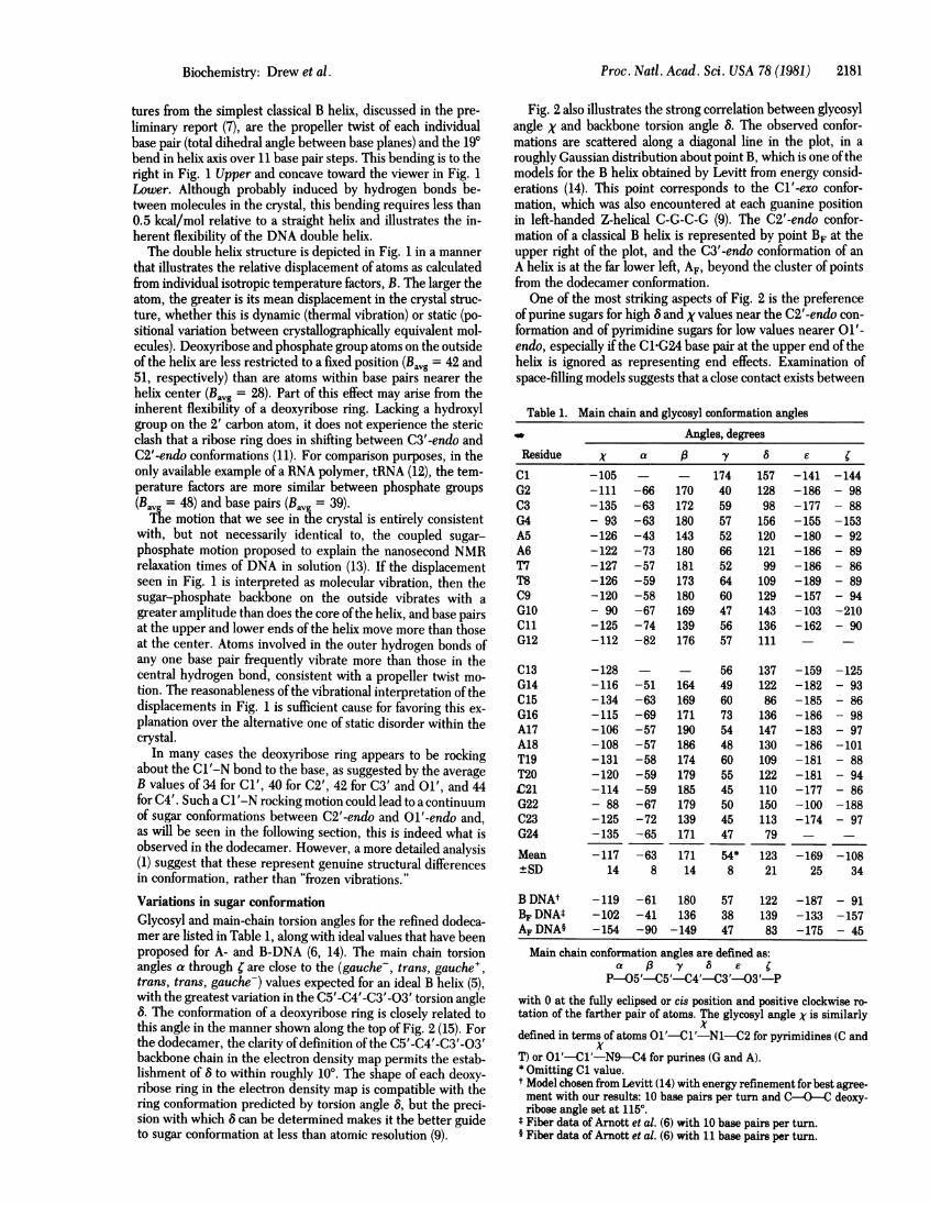
Fig. 2 also illustrates the strong correlation between glycosyl
angle X and backbone torsion angle 6. The observed confor-
mations are scattered along a diagonal line in the plot, in a
roughly Gaussian distribution about point B, which is one of the
models for the B helix obtained by Levitt from energy consid-
erations (14). This point corresponds to the C1'-exo confor-
mation, which was also encountered at each guanine position
in left-handed Z-helical C-G-C-G (9). The C2'-endo confor-
mation of a classical B helix is represented by point BF at the
upper right of the plot, and the C3'-endo conformation of an
A helix is at the far lower left, AF, beyond the cluster of points
from the dodecamer conformation.
One of the most striking aspects of Fig. 2 is the preferenceof purine sugars for high and X values near the C2'-endo con-
formation and of pyrimidine sugars for low values nearer 01'-
endo, especially if the Cl-G24 base pair at the upper end of the
helix is ignored as representing end effects. Examination of
space-filling models suggests that a close contact exists between
Table 1. Main chain and glycosyl conformation angles4W ~~~~~~~Angles, degreesResidue x a (3 y 8 eCl -105 - - 174 157 -141 -144G2 -111 -66 170 40 128 -186 - 98C3 -135 -63 172 59 98 -177 - 88G4 - 93 -63 180 57 156 -155 -153AS -126 -43 143 52 120 -180 - 92A6 -122 -73 180 66 121 -186 - 89T7 -127 -57 181 52 99 -186 - 86T8 -126 -59 173 64 109 -189 - 8909 -120 -58 180 60 129 -157 - 94G10 - 90 -67 169 47 143 -103 -210ClH -125 -74 139 56 136 -162 - 90G12 -112 -82 176 57 111 - -
013 -128 - - 56 137 -159 -125G14 -116 -51 164 49 122 -182 - 93C15 -134 -63 169 60 86 -185 - 86G16 -115 -69 171 73 136 -186 - 98A17 -106 -57 190 54 147 -183 - 97A18 -108 -57 186 48 130 -186 -101T19 -131 -58 174 60 109 -181 - 88T20 -120 -59 179 55 122 -181 - 94.C21 -114 -59 185 45 110 -177 - 86G22 - 88 -67 179 50 iSO -100 -188C23 -125 -72 139 45 113 -174 - 97G24 -135 -65 171 47 79 - -
Mean -117 -63 171 54* 123 -169 -108±SD 14 8 14 8 21 25 34
BDNAt -119 -61 180 57 122 -187 - 91BFDNAM -102 -41 136 38 139 -133 -157AFDNA§ -154 -90 -149 47 83 -175 - 45
Main chain conformation angles are defined as:a j3 y 8 e C
P--05'-C5'-C4'-C3'--03'-Pwith 0 at the fully eclipsed or cis position and positive clockwise ro-tation of the farther pair of atoms. The glycosyl angle X is similarly
xdefined in terms of atoms O1'-Cl'-N1--C2 for pyrimidines (C and
xT) or Ol'-Cl'-N9--C4 for purines (G and A).* Omitting Cl value.tModel chosen from Levitt (14) with energy refinement for best agree-ment with our results: 10 base pairs per turn and C-O---C deoxy-ribose angle set at 1150.
* Fiber data of Arnott et al. (6) with 10 base pairs per turn.§ Fiber data of Arnott et al. (6) with 11 base pairs per turn.
Biochemistry: Drew et al.

Proc. Natl. Acad. Sci. USA 78 (1981)
-80r
3'en(O.O) 2'ex(O.6)4'ex(O.I) O'en(O.6)T;$ $
-90k
-100K
-110k
x-120k
-130k
-150k
-160k
-170170 80 90 100 110
FIG. 2. Correlation plot betweenC5'-C4'-C4'-03' torsion angle 8 (ora numbered circle, with angles takenideal A and B helices as derived fr(refinement (14). The distribution ofccentered at point B. Purines are shcdines, by light ones. Notice the correbases related by a 2-fold axis normalthe anticorrelation (constant 8 sum)gether on one step of the helix (1/2410 standard endo and exo conformamol relative to C3'-endo are given a
the H atom of a sugar C1' and t1200. This contact cannot be ru
ative without introducing otherciH6 and the hydrogens attached t,to more negativeX eliminates thiangle involved in the connectionbered ring of a purine keeps theaway from the sugar ring that no
the purine sugars are free to adoiC2' -endo conformation.
In view of the gentle slope ofvicinity of point B (figure 2 of refconformations seen in Fig. 2 is I
B helix in DNA is as much an ove
a helix in proteins. The DNA isformational states listed at the t(and rather shallow potential wepoint B. Visual inspection of theconformations can best be descrito the right of8 = 1290 in Fig.1050 and 1290, and Ol'-endo forlone C3'-endo or A-like sugar pi
the second strand. (This assignmsubjective at intermediate angleTwo other features of the disti
cial mention. Sugars related bthrough the center of the molecaxis(1 and 13, 2 and 14, 3 and 1andX values, indicating local pr
in spite of the overall 190 bendi1 Upper (and discussed in ref.differ in 8 by a mean (± SD) ofc
i'en(2) 2'ex(O.2) 3'en(2.0) expected, and the only unusual feature is that this conforma-tional symmetry is preserved in spite ofthe bending ofthe helixaxis.
o 0 The second correlation is more surprising and potentially ofmore fundamental significance. The conformations of sugars inpaired bases (1/24, 2/23, 3/22, etc.) tend to be anticorrelated:
BF(D
if one conformation lies to the left of center in Fig. 2, the other0ed 0 conformation lies a similar distance to the right of center. This
14
Ois a stronger statement than the previous observation that pu-0 rines generally prefer higher 8 and X values than do pyrimi-
dines. The midpoint in 8 for each base pair have a mean of© < I)(H)122.80, near the Cl'-exo conformation, with a mean deviation
of only 5.60. Some base pairs such as 5/20, 6/19, and 2/23 havesimilar sugar conformations centered closely at Cl'-exo. Otherssuch as 1/24, 10/15, and 3/22 have quite disparate conforma-tions, one of them nearer C2'-endo or C3'-exo and the othernear Ol'-endo or even C3'-endo. If the difference between 8values for two paired bases is defined as the conformationalspread, then the CCG base pairs have a larger mean spread(36.90) than do the A-T base pairs (20.80), although the statistical
I20 0 0I50l osignificance of this is not clear with only 12 base pairs to120 130 140 150 160 compare.This behavior of sugar conformations in paired bases is suf-
glycosyl torsion angle X and the ficiently striking to be formalized by defining it as the "principledY). Each sugar is represented by of anticorrelation": deoxyribose sugars attached to paired basesIfrom Table 1. AF, BF, and B locate in B-DNA tend to adopt 8 values that are equidistant to eitherom fiber diffraction (6) or energy side of a central 8 = 1230 (or Cl'-exo) value. It probably is a-onformations is close to Gaussian consequence of wrapping sugar-phosphate chains having flex-wn by heavy circles, and pyrimi- iblequenceo f rings around base pairs of fixed dimensions.ilation (similar 8 values) between.to the helix (1/13,2/14, etc.), and One would not necessarily expect to find anticorrelation in sugarbetween bases that are paired to- conformations ofpaired bases in RNA, with its more constrainedt, 2/23, etc.). The 8 values for the ribose ring, and, indeed, such anticorrelation is not present inLtions and their energies in kcal/ the one available example, tRNA (16). When the midpoints inlong the top of the plot (15). 8 values for paired bases in tRNA are calculated, omitting the
first base pair at either end of a double-helical stack to eliminatehe 02 of a pyridimine at X = end effects, the mean of these values is 82.80 and the meanelieved by making X less neg- deviation is 2.40. But the mean spread or difference in 8 valueslashes between the pyrimidine between paired bases is only twice this, 5.4°. The half-spreado either C2' or C3', but a shift from the midpoint is no larger than the uncertainty in midpointe clash. In contrast, the larger position, indicating no statistically significant anticorrelation in* of a sugar to the five-mem- sugar conformation. By comparison, in the DNA dodecamer C-N3 and H8 atoms far enough G-C-G-A-A-T-T-C-G-C-G the mean is 122.8°, the mean devia-close contacts result. Hence, tion is 5.60, and the mean spread over all base pairs is 31.50,
pt higherX values and an ideal 6 times as great.The anticorrelation principle also is observed in the nonin-
'the total energy curve in the tercalated T-A base pair step of the 2:1 intercalation complexf. 15), the scatter in individual of daunomycin with C-G-T-A-C-G (17). Sugar pucker infor-hardly surprising. The "ideal" mation provided in ref. 17 indicates that the Cs and Gs brack-rsimplification as is the "ideal" eting the intercalator molecules would be scattered at the uppernot aware of the discrete con- right of Fig. 2 with the exception of an anomalous C1 confor-op of Fig. 2, only of a smooth mation at the beginning of the chain. But the unintercalated11 centered in the vicinity of T3-A4 is a beautiful example of anticorrelation. (Because thestereo drawings indicates that complex has an internal 2-fold axis of crystallographic symme-ibed as C2'-endo for all sugars try, the two strands are identical. T3 on one strand is followed2, CL'-exo for sugars between by A4 on the same strand and hydrogen bonded to A4 on thesugars 15, 3, and 7, with one opposite strand.) T3 has the (X, 8) conformation (-131°, 96°),uckering at the 3' terminus of and A4 has (- 107°, -- 144°). (The X values quoted in ref. 17ent is necessarily arbitrary and should have 1800 subtracted from each to bring them into accordS.) with IUPAC-IUB conventions.) In addition, the tendency,ribution in Fig. 2 deserve spe- noted by both Sobell and colleagues (18) and Rich et al. (19, 20),My an approximate 2-fold axis for many intercalating groups (but not daunomycin) to induceule perpendicular to the helix a (C3'-endo) -3', 5'-(C2'-endo) conformation at the bases brack-L5, etc.) tend to have similar 8 eting the intercalator, whether in DNA or RNA helices, can beeservation of 2-fold symmetry regarded as an extreme limit of the principle of anticorrelation.in the helix axis visible in Fig. Zimmerman and Pheiffer (21) have recently found a RNA-DNA7). Such 2-fold-related sugars hybrid, poly(rA)-poly(dT), that can be induced to adopt a B helix)nly 16.5° ± 8.30. This is to be under conditions ofhigh humidity. Its 8 values as deduced from
03
2182 Biochemistry: Drew et al.
-r--
-130~
9

Proc. Natl. Acad. Sci. USA 78 (1981) 2183
Table 2. Local helix parameters
Base Propeller Helix twist Base pairs per Rise per basepairs twist,0 (qi) angle,*'(0) turnt (n) pair, A (h)
Cl/G24 13.2 ±2.0 38.3 ± 1.1 9.40 ± 0.27 3.36 ± 0.01G2/C23 11.7±2.1 39661 9.09± 1.40 3.38±0.08C3/G22 7.2 ± 2.1 33521 10.75 ± 0.67 3.26 ± 0.05G4/C21 13.2 1.9 37.4 ± 1.7 9.63 ± 0.44 3.30 ± 0.10A5/T21 178 ±2+ 1 375 ± 0.9 9.60 ± 0.23 3.27 ± 0.02A6/T9 1.8 2- 32.2 ± 2.1 11.18 ± 0.73 3.31 ± 0.03
T8/A18 17.1 .9 36.0 ± 2.8 10.00 ± 0.78 3.29 ± 0.01T8/A7 1.1 2- 41.4 ± 2.1 8.70 ± 0.42 3.14 ± 0.02
(G/C 8.9 ± 1.9 32.3 ± 1.3 11.11 ± 0.45 3.56 ± 0.07C11/C14 4172±1.9 44.7 ± 5.4 8.05 ± 0.97 3.21 ± 0.18C11G14 172 ±1- 37.0 ± 1.9 9.73 ± 0.50 3.54 ± 0.19G12/C13 6.2 ± 2.3
Mean 13.4 ± 4.9 37.3 ± 3.8 9.75 ± 0.98 3.33 ± 0.13
A DNA(5, 22) 32.7 11.0 2.56
B DNA(5, 22, 23) 36.0 10.0 3.38
C DNA(23) 38.6 9.33 3.31
D DNA(23) 45.0 8.0 3.03
Helical parameters were found by using vectors between atoms Cl'and the attached N of one base and the equivalent atoms of the nextbase up the same chain, with a program kindly provided by John Ro-senberg. Standard deviations are shown at individual steps and sta-tistical variation over the entire helix is shown with the means. Pro-peller twist is the dihedral angle between base planes in the same basepair and is twice the fiber twist in 22 and 23.* Rotation per base pair.t n = 360/0.
fiber diffraction patterns are 970 and 1520 for rA and dT, re-spectively. The midpoint of these values, 124.50, is only 1.70away from the mean DNA dodecamer value.
Local helix parametersA helix-generating program was used to determine the rotationangle, orientation of the local helix rotation vector, and rise perbase pair along the local axis for each of the 11 base pair stepsalong the dodecamer. The individual rotation angles and riseper base pair are listed in Table 2, along with the measuredpropeller twist at each base pair. These numbers probably aremore accurate than any other results of the analysis, becausebase planes are well defined in the map and are established bya large number of atomic positions. The propeller twist of AFTbase pairs in the center of the molecule is a remarkably uniform17.30 ± 0.40 (mean ± SD), and much ofthe variation at the twoends probably arises from overlapping of helices up the c axis(figure 3 of ref. 7). The smaller mean, 11.50 ± 5.10 for the twoC-G-C-G ends, could also reflect the flattening influence of thethird hydrogen bond in each base pair.
The individual helical twist or rotation values for the five cen-tral base pair steps that are unperturbed by overlapping ends(from G4 to C9) give a mean of 36.90 ± 3.3°, corresponding to9.8 base pairs per turn. The three base pair steps at either endare wound somewhat more tightly, 37.60 ± 4.5° or 9.6 base pairsper turn. The overall value of 35.80 or 10.1 base pairs per turn
that was reported in ref. 7 arises because several of the localtwist vectors in the overlapping ends ofthe molecule are sharplytilted from the mean helix axis, altering their twist angles inprojection. Space does not permit a detailed discussion of localand global helix twist vectors here, but this is discussed in detailelsewhere (1). It can only be mentioned here that these dis-placements in local helix rotation vectors show a 2-fold sym-metry about an axis through the center of the molecule, per-pendicular to the page in Fig. 1 Upper or horizontal in Fig. 1Lower. This ideal symmetry axis is expected from the chemicalidentity of the two strands but is destroyed by the 190 bend inthe helix axis. Hence, these local variations in helix twist vectorare observed in spite of the bend in the helix, not because ofit.
We thank Peter Dembek for his help in synthesis of the dodecamerand Lillian Casler for her assistance in preparing the figures. This workwas carried out with the support of National Institutes of Health GrantsGM-12121 and GM-24393 and National Science Foundation GrantPCM79-13959. H.R.D. was the recipient of a National Institutes ofHealth Predoctoral Traineeship. This is contribution no. 6317 from theDivision of Chemistry and Chemical Engineering.
1. Dickerson, R. E. & Drew, H. R. (1981) J. Mol. Biol., in press.2. Watson, J. D. & Crick, F. H. C. (1953) Nature (London) 171,
737-738.3. Langridge, R., Wilson, H. R., Hooper, C. W., Wilkins, M. H.
F. & Hamilton, L. D. (1960)J. Mol. Biol. 2, 19-37.4. Fuller, W., Wilkins, M. H. F., Wilson, H. R., Hamilton, L. D.
& Arnott, S. (1965) J. Mol. Biol. 12, 60-80.5. Arnott, S. & Hukins, D. W. L. (1972) Biochem. Biophys. Res.
Commun. 47, 1504-1509.6. Arnott, S., Chandrasekaran, R., Birdsall, D. L., Leslie, A. G. W.
& Ratliff, R. L. (1980) Nature (London) 283, 743-745.7. Wing, R. M., Drew, H. R., Takano, T., Broka, C., Tanaka, S.,
Itakura, K. & Dickerson, R. E. (1980) Nature (London) 287,755-758.
8. Wang, A. H.-J., Quigley, G. J., Kolpak, F. J., Crawford, J. L.,Van Boom, J. H., Van der Marel, G. & Rich, A. (1979) Nature(London) 282, 680-686.
9. Drew, H. R., Takano, T., Tanaka, S., Itakura, K. & Dickerson,R. E. (1980) Nature (London) 286, 567-573.
10. Jack, A. & Levitt, M. (1978) Acta Crystallogr. A34, 931-935.11. Westhof, E. & Sundaralingam, M. (1980)J. Am. Chem. Soc. 102,
1493-1500.12. Sussman, J. L., Holbrook, S. R., Warrant, R. W., Church, G. M.
& Kim, S.-H. (1978) J. Mol. Biol. 123, 607-630.13. Hogan, M. E. & Jardetzky, 0. (1980) Biochemistry 19, 3460-3468.14. Levitt, M. (1978) Proc. Natl. Acad. Sci. USA 75, 640-644.15. Levitt, M. & Warshel, A. (1978) J. Am. Chem. Soc. 100,
2607-2613.16. Holbrook, S. R., Sussman, J. L., Warrant, R. W. & Kim, S.-H.
(1978)J. Mol. Biol. 123, 631-660.17. Quigley, G. J., Wang, A. H.-J., Ughetto, G., van der Marel, G.,
van Boom, J. H. & Rich, A. (1980) Proc. Natl. Acad . Sci. USA 77,7204-7208.
18. Jain, S. C., Tsai, C.-C. & Sobell, H. M. (1977)J. Mol. Biol. 114,317-331.
19. Rich, A., Quigley, G. J. & Wang, A. H.-J. (1979) in Stereody-namics ofMolecular Systems, ed. Sarma, R. H. (Pergamon, NewYork), pp. 3154330.
20. Rich, A., Wang, H.-J. & Quigley, G. J. (1980) in Frontiers ofBioorganic Chemistry and Molecular Biology, ed. Ananchenko,S. N. (Pergamon, New York), pp. 327-337.
21. Zimmerman, S. B. & Pheiffer, B. H. (1980) Proc. Nat. Acad. Sci.USA 78, 78-82.
22. Arnott, J., Dover, S. D. & Wonacott, A. J. (1969) Acta Crystal-logr. B25, 2192-2206.
23. Arnott, J. & Selsing, E. (1975) J. Mol. Biol. 98, 265-269.
Biochemistry: Drew et al -