Embed Size (px)
Citation preview

The Plant Cell, Vol. 11, 1081–1092, June 1999, www.plantcell.org © 1999 American Society of Plant Physiologists
Structure of a Plant Cell Wall Fragment Complexed to Pectate Lyase C
Robert D. Scavetta,
a,1
Steven R. Herron,
a
Arland T. Hotchkiss,
b
Nobuhiro Kita,
a,2
Noel T. Keen,
c
Jacques A. E. Benen,
d
Harry C. M. Kester,
d
Jaap Visser,
d
and Frances Jurnak
a,3
a
Department of Physiology and Biophysics, 346-D Med Sci I, University of California, Irvine, California 92697-4560
b
U.S. Department of Agriculture–Agricultural Research Service, Eastern Regional Research Center, 600 E. Mermaid Lane, Wyndmoor, Pennsylvania 19038-8598
c
Department of Plant Pathology, University of California, Riverside, California 92521
d
Department of Molecular Genetics of Industrial Microorganisms, Wageningen Agricultural University, Dreijenlaan 2, 6703 HA Wageningen, The Netherlands
The three-dimensional structure of a complex between the pectate lyase C (PelC) R218K mutant and a plant cell wallfragment has been determined by x-ray diffraction techniques to a resolution of 2.2 Å and refined to a crystallographic
R
factor of 18.6%. The oligosaccharide substrate,
a
-
D
-Gal
p
A-([1
→
4]-
a
-
D
-Gal
p
A)
3
-(1
→
4)-
D
-Gal
p
A, is composed of fivegalacturonopyranose units (
D
-Gal
p
A) linked by
a
-(1
→
4) glycosidic bonds. PelC is secreted by the plant pathogen
Er-winia chrysanthemi
and degrades the pectate component of plant cell walls in soft rot diseases. The substrate hasbeen trapped in crystals by using the inactive R218K mutant. Four of the five saccharide units of the substrate are wellordered and represent an atomic view of the pectate component in plant cell walls. The conformation of the pectatefragment is a mix of 2
1
and 3
1
right-handed helices. The substrate binds in a cleft, interacting primarily with positively
charged groups: either lysine or arginine amino acids on PelC or the four Ca
2
1
ions found in the complex. The observedprotein–oligosaccharide interactions provide a functional explanation for many of the invariant and conserved aminoacids in the pectate lyase family of proteins. Because the R218K PelC–galacturonopentaose complex represents an in-termediate in the reaction pathway, the structure also reveals important details regarding the enzymatic mechanism.Notably, the results suggest that an arginine, which is invariant in the pectate lyase superfamily, is the amino acid thatinitiates proton abstraction during the
b
elimination cleavage of polygalacturonic acid.
INTRODUCTION
Pectate lyases are depolymerizing enzymes that degradeplant cell walls, causing tissue maceration and death. Theenzymes normally are secreted by phytopathogenic organ-isms and are known to be the primary virulence agents insoft rot diseases caused by
Erwinia
spp (Collmer and Keen,1986; Kotoujansky, 1987; Barras et al., 1994). In the latterorganisms, the enzymes exist as multiple, independentlyregulated isozymes that share amino acid sequence identityranging from 27 to 80%.
Pectate lyases share sequence similarities with fungalpectin lyases, plant pollen proteins, and plant style proteins(Henrissat et al., 1996). The three-dimensional structures of
five members of the superfamily have been determined andinclude
Erwinia chrysanthemi
pectate lyase C (PelC) (Yoderet al., 1993; Yoder and Jurnak, 1995),
E. chrysanthemi
pec-tate lyase E (PelE) (Lietzke et al., 1994),
Bacillus subtilis
pec-tate lyase (
B. subtilis
Pel) (Pickersgill et al., 1994),
Aspergillusniger
pectin lyase A (PLA) (Mayans et al., 1997), and
A. niger
pectin lyase B (PLB) (Vitali et al., 1998). All share a similarbut an unusual structural motif, termed the parallel
b
helix, inwhich the
b
strands are folded into a large, right-handedcoil. The enzyme structures differ in the size and conforma-tion of the loops that protrude from the parallel
b
helix core.As deduced from sequence similarity and site-directed mu-tagenesis studies, the protruding loops on one side of theparallel
b
helix form the pectolytic active site (Kita et al.,1996). The structural differences of the loops are believed tobe related to subtle differences in the enzymatic and macer-ation properties of the proteins.
Pectate lyases catalyze the cleavage of pectate, the de-esterified product of pectin, which is the major component thatmaintains the structural integrity of cell walls in higher plants
1
Current address: 6803 South Ivy Way, Inglewood, CO 80112.
2
Current address: Kanagawa Institute of Agricultural Science, 1617Kamikisawa, Hiratsuka, Kanagawa 259-12, Japan.
3
To whom correspondence should be addressed. E-mail [email protected]; fax 949-824-8540.

1082 The Plant Cell
(Carpita and Gibeaut, 1993). The pectate backbone is com-posed of blocks of polygalacturonic acid (PGA), which is ahelical homopolymer of
D
-galacturonic acid (Gal
p
A) unitslinked by
a
-(1
→
4) glycosidic bonds. The blocks of PGA areseparated by stretches in which (1
→
2)-
a
-
L
-rhamnose resi-dues alternate with Gal
p
A (McNaught, 1997). Blocks of PGAmay contain as many as 200 Gal
p
A units and span 100 nm(Thibault et al., 1993). Cations are necessary to neutralize PGAin solution and, as a consequence, influence its structure.
In the presence of Ca
2
1
, PGA assumes a 2
1
helical confor-mation in dilute polymer concentrations (Morris et al., 1982;Powell et al., 1982) and a 3
1
helix at high concentrations ineither a gel or solid form (Walkinshaw and Arnott, 1981a,1981b). Because the PGA concentration in the plant cell wallupon demethylation of pectin lies near the critical conforma-tional transition point, considerable speculation exists as tothe in situ structure of PGA. A popular view is the “eggboxmodel” in which Ca
2
1
ions cross-link the uronic acid moi-eties of neighboring antiparallel chains of PGA together. Al-though the eggbox model generally is depicted with PGA ina right-handed 2
1
helical conformation, the original literaturesuggests that cross-linking between Ca
2
1
and PGA in aright- or left-handed 3
1
helical conformation is feasible aswell (Grant et al., 1973; Kohn, 1975).
The results of a recent nuclear magnetic resonance studysuggest that the Ca
2
1
–PGA complex in the plant cell wall ismuch more complex than the simple eggbox model. Thiscomplex contains both 2
1
and 3
1
helices of PGA as well asintermediate conformational states (Jarvis and Apperley,1995). Nuclear magnetic resonance, molecular modeling,and molecular dynamic analyses of pectic disaccharidesand trisaccharides also have reported that PGA has both 3
1
and 2
1
helical conformations (Hricovini et al., 1991; DiNola etal., 1994; Gouvion et al., 1994). Disaccharide hydration andsodium salt formation may shift the predicted PGA helicalconformation from 3
1
to 2
1
(DiNola et al., 1994; Gouvion etal., 1994; Catoire et al., 1997).
All proteins in the pectate lyase superfamily are believedto share a similar enzymatic mechanism, but the catalyticroles of the amino acids in the active site region have notbeen identified. For reasons of technical convenience, re-cent studies have focused on PelC. The enzyme randomlycleaves PGA by a
b
elimination mechanism, generating pri-marily a trimer end product with a 4,5-unsaturated bond inthe galacturonosyl residue (
a
-
L
-4-eno-threohexosylpyrano-syluronic acid [
a
-
L
-4-en-thrHex
p
A]) at the nonreducing end(Preston et al., 1992). PelC has an in vitro pH optimum of 9.5and requires Ca
2
1
for pectolytic activity. Structural studies
Figure 1. Stereoview of the Ca21 Ions and TetraGalpA Superimposed upon the Simulated Annealed OMIT Electron Density Map of the PelCR218K–Substrate Complex Contoured at 1.0s.
The Ca21 ions are represented by yellow spheres. TetraGalpA as well as the interacting amino acids are represented by rods by using the Inter-national Union of Pure and Applied Chemistry coloring code: carbon atoms are gray; oxygen atoms, red; and nitrogen atoms, blue. The R218Kbackbone is represented by green ribbons. Individual amino acids that are shown are labeled at the a-carbon.
PelC Complexed to Plant Cell Wall Fragment 1083
have shown that Ca
2
1
is bound to the enzyme at a locationthat was first suggested in a PelC–Lu
3
1
complex (Yoder etal., 1993) and later confirmed by structural studies of a
B.subtilis
Pel–Ca
2
1
complex (Pickersgill et al., 1994). The roleof Ca
2
1
has not been established. In the
b
elimination reac-tion, the reaction is initiated by proton abstraction from C-5of the galacturonosyl residue on the reducing side of theglycosidic scissile bond. The group or groups that initiateproton abstraction and transfer the proton to the glycosidicoxygen have not been identified. Potential candidates in-clude two invariant amino acids in the superfamily, Asp-131and Arg-218 in PelC nomenclature, as well as four aminoacids, Glu-166, Asp-170, Lys-190, and Arg-223, which areinvariant within the pectate lyase subfamily (Henrissat et al.,1996). Site-specific mutations at the latter PelC positionsabolish pectolytic as well as maceration activity (Kita et al.,1996). Notably, the pectolytic region is devoid of conservedhistidine, serine, or tyrosine residues, which frequently areimplicated in
b
elimination enzymatic mechanisms with alower pH optimum. In this study, we have taken advantageof the impaired catalytic properties of one PelC mutant,R218K, to form a stable substrate–enzyme complex thatcould be studied by x-ray diffraction techniques. The resultsprovide an atomic view of a pectate fragment,
a
-
D
-Gal
p
A-([1
→
4]-
a
-
D
-Gal
p
A)
3
-(1
→
4)-
D
-Gal
p
A (pentaGal
p
A), and the iden-tification of the key amino acids involved in oligosaccharide
binding. In addition, the results provide tentative identifica-tion of the amino acid that initiates proton abstraction.
RESULTS
Conformation of the PentaGal
p
A Substrate
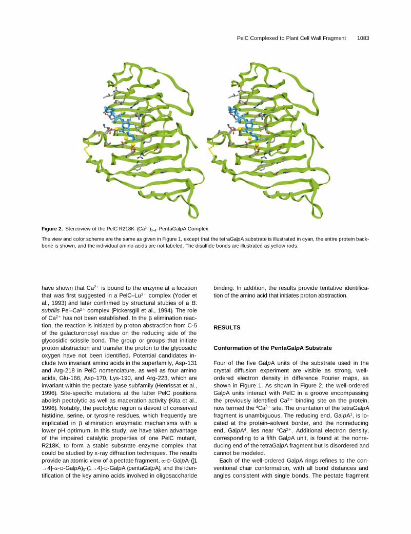
Four of the five Gal
p
A units of the substrate used in thecrystal diffusion experiment are visible as strong, well-ordered electron density in difference Fourier maps, asshown in Figure 1. As shown in Figure 2, the well-orderedGal
p
A units interact with PelC in a groove encompassingthe previously identified Ca
2
1
binding site on the protein,now termed the
4
Ca
2
1
site. The orientation of the tetraGal
p
Afragment is unambiguous. The reducing end, Gal
p
A
1
, is lo-cated at the protein–solvent border, and the nonreducingend, Gal
p
A
4
, lies near
4
Ca
2
1
. Additional electron density,corresponding to a fifth Gal
p
A unit, is found at the nonre-ducing end of the tetraGal
p
A fragment but is disordered andcannot be modeled.
Each of the well-ordered Gal
p
A rings refines to the con-ventional chair conformation, with all bond distances andangles consistent with single bonds. The pectate fragment
Figure 2. Stereoview of the PelC R218K–(Ca21)3–4–PentaGalpA Complex.
The view and color scheme are the same as given in Figure 1, except that the tetraGalpA substrate is illustrated in cyan, the entire protein back-bone is shown, and the individual amino acids are not labeled. The disulfide bonds are illustrated as yellow rods.

1084 The Plant Cell
folds into an unbent right-handed helical conformation, withthe observed helical angles compared with the idealized 21
and 31 helices in Table 1. Two of the three glycosidic bondshave c torsional angles that approximate the 31 helix observedin fiber diffraction studies of PGA–Ca21 gels (Walkinshawand Arnott, 1981a, 1981b). One of the glycosidic bonds, be-tween GalpA2 and GalpA3, has a c torsional angle similar toa 21 helix. Consequently, the overall appearance of the pec-tate fragment conformation is of a 31 helix, but with the mid-dle segment distorted into 21 helix, as illustrated in Figures3A and 3B. Given the greater number of GalpA3 contacts, aslisted in Table 2, the deviation from the 31 helical conforma-tion is probably a result of specific interactions with the en-zyme. The deviation is not likely due to other effects, suchas pectate concentration, hydration, or cation type, whichare postulated to cause the transition between 21 and 31 he-lical conformations of pectate. If the R218K–(Ca 21)3–4–GalpA5 structures are representative of interactions that oc-cur within the plant cell wall, then endogenous proteins alsoare likely to distort the PGA conformation from any helicalstates observed under in vitro conditions.
Coordination of Ca21 Ions
In wild-type PelC, a Ca21 ion coordinates to seven ligands, in-cluding two water molecules, both carboxyl oxygens of Asp-
131, and one carboxyl oxygen from each of Asp-129, Glu-166, and Asp-170. In the R218K complex with pentaGalpA,the equivalent 4Ca21 coordinates to the same groups, with asingle exception—a carboxyl oxygen from GalpA4 replacesone of the water molecules. In addition to 4Ca21, three addi-tional Ca21 ions have been identified. Two of the additionalCa21 ions, 2Ca21 and 3Ca21, are fully occupied, and thethird, 1Ca21, has a partial occupancy of z50%. Each Ca21
ion bridges the carboxyl group of each GalpA unit to theprotein. In addition, 2Ca21 and 3Ca21 link the uronic acidmoieties of GalpA2, GalpA3, and GalpA4. The coordinationaround each Ca21 ion is listed in Table 3. The observed Ca21
positions are very different from the interstrand Ca21 ions
Table 1. Bond Angles (t) and Torsional Rotations (f and c)about Glycosidic Bonds in the Refined Structures of thePelC–(Ca21)3–4–PentaGalpA Complexesa
Glycosidic Bond tb fc cd
31 Helixe 117.08 80.08 89.08
21 Helixf 117.08 80.08 161.08
GalpA1–GalpA2 117.08 54.08 90.08
GalpA2–GalpA3 116.68 117.08 157.08
GalpA3–GalpA4 120.48 73.08 51.08
a Torsional angles were determined by looking from the nonreducingend side (with prime) down the bond of interest to the reducing endside and determining the angle of rotation created from the planes ofO-59–C-19–O-4 and C-19–O-4–C-4 for f and C-19–O-4–C-4 andO-4–C-4–C-5 for c. A cis configuration is taken to be 08, and a transconfiguration is taken to be 1808. A negative sign is a rotation fromcis in a counterclockwise direction, and a positive sign is a rotationfrom cis in a clockwise direction.b t, the C1–O–C49 bond angle.c f, the torsional rotation about the C1–O bond.d c, the torsional rotation about the O–C49 bond.e The values for the 31 helix are those determined by the program O(Jones et al., 1991) from the model published by Walkinshaw andArnott (1981b).f The values for the 21 helix are those determined by the program Ofor a galacturonic acid model constructed using the parameters foralginic acid by Atkins et al. (1973).
Table 2. Atomic Distances of 3.0 Å or Less between the Oxygen Atoms of TetraGalpA and Amino Acids, Ca21 Ions, orWater Molecules
GalpA Atomsa Interacting Atomsb Distance (Å)
GalpA1
Ring interaction Tyr-268O-6A GA1Wat1 2.9O-6B 1Ca21 2.9
GalpA2
O-2 Asp-162 O-d2 2.7O-2 GA2Wat1 2.7O-3 2Ca21 2.6O-5 1Ca21 2.9O-6A 1Ca21 2.6O-6A Arg-245 NH-1 2.8O-6B Arg-245 NH-2 3.0O-6B GA2Wat2 2.8
GalpA3
O-2 Arg-223 NH-2 2.9O-3 Ser-196 O 2.8O-3 Arg-223 NH-1 2.9O-5 2Ca21 2.5O-5 2CaWat2 3.0O-6A Lys-190 N-z 2.8O-6A 2Ca21 2.3O-6A 2CaWat2 2.9O-6B 3Ca21 2.4O-6B 3CaWat3 2.8
GalpA4
O-2 2CaWat2 2.9O-3 GA4Wat1 2.8O-4 GA4Wat2 2.7O-4 Ser-308 O 2.8O-5 3Ca21 2.4O-6A 4Ca21 2.5O-6A 3Ca21 2.4O-6A GA4Wat2 2.9O-6B 4CaWat1 2.6
a The positions of the atoms are indicated in Figure 4.b xWaty refers to the Y water molecule associated with the X GalpAunit. zCa21 refers to the Z position of the Ca21 ion as defined in Fig-ure 4.

PelC Complexed to Plant Cell Wall Fragment 1085
postulated to link PGA helices together (Walkinshaw andArnott, 1981a, 1981b; Morris et al., 1982; Powell et al., 1982).In the present structure, the Ca21 ions link not only the oli-gosaccharide to the protein but also adjacent uronic acidmoieties within a single pectate strand.
Protein–Ca21–TetraGalpA Interactions
The protein–Ca21–tetraGalpA interactions are represented inFigure 4, and all relevant interatomic distances are summa-rized in Table 2. Electrostatic interactions dominate, with thenegatively charged uronic acid moieties primarily interactingwith positively charged groups: either lysine or arginine onPelC or the four Ca21 ions found in the complex. The car-boxyl oxygens of GalpA2 and GalpA3 interact strongly withArg-245 and Lys-190, respectively, whereas a carboxyl oxy-gen of GalpA4, at a distance of 3.2 Å from Lys-172, forms aweaker interaction. Lys-172 is highly conserved, and Lys-190 is invariant in the pectate lyases, but neither amino acidis found among the pectin lyases that bind a neutral methy-lated form of pectate. Arg-245 is conserved only amongPelC subfamily members but not in the PelE subfamilywhose members rapidly cleave the substrate to an unsatur-ated dimer. Several additional interactions between tet-raGalpA and the protein were observed, but notably, themost specific ones involve GalpA3. Arg-223, another invari-ant amino acid in the pectate lyase subfamily, forms hydro-
gen bonds with the C-2 and C-3 hydroxyl groups of GalpA3,the orientation of which partially defines the galactoseepimer. The C-3 hydroxyl group also forms a hydrogen bondwith a nonconserved Ser-196. In GalpA2, the C-2 hydroxylinteracts with Asp-162, and in GalpA1, the ring forms astacking interaction with Tyr-268. Both amino acids are con-served but only in the PelC subfamily. In addition to interac-tions with the protein and Ca 21 ions, the tetraGalpAsegment is highly solvated, forming many hydrogen bondswith water molecules that increase in frequency from GalpA1
to GalpA4. Collectively, the observed protein–tetraGalpA in-teractions provide a functional role for all invariant and con-served amino acids in the pectolytic region of the pectatelyases, except one, Arg-218.
Position of Scissile Bond
Crystals of wild-type PelC, which are isomorphous withR218K crystals, cleave pentaGalpA when diffused into crys-tals. Because the R218K mutant is catalytically inactive anda saturated tetraGalpA has been observed, the R218K–(Ca21)3–4–GalpA5 complexes represent a Michaelis complexin the reaction pathway. Can the scissile glycosidic bond beidentified with certainty? PelC and subfamily members havebeen reported to cleave a pectate substrate, yielding a tri-mer as the primary unsaturated end product (68 to 72%;Preston et al., 1992). In the crystal structure, an unsaturatedtrimeric end product would result only if the scissile bond
Figure 3. Stereoview of the TetraGalpA Structure Superimposed upon Modeled Right-Hand 21 and 31 OligoGalpA Helices.
The tetraGalpA structure determined at 2.2 Å is shown in both (A) and (B) as cyan rods.(A) The modeled 31 oligoGalpA helix (Walkinshaw and Arnott, 1981a) is shown in red.(B) The modeled 21 oligoGalpA helix (Atkins et al., 1973) is shown in yellow.

1086 The Plant Cell
occurred between GalpA3 and GalpA4. Moreover, only inter-actions with GalpA3 and GalpA4 involve highly conservedand invariant amino acids within the pectate lyase family.GalpA3 forms the most protein interactions, which appear tocause the greatest distortion from the 31 helical conforma-tion of the tetraGalpA. In contrast, there are fewer interac-tions with GalpA1 and GalpA2, and all involve amino acidsthat are conserved only within the PelC subfamily.
To confirm the position of the scissile glycosidic bond, weinvestigated the enzymatic cleavage patterns of oligogalac-turonates with different degrees of polymerization under op-timized assay conditions for PelC. The composition of boththe saturated and unsaturated end products was analyzed,
and the results, in Table 4, demonstrate that a pentaGalpAsubstrate has two observed modes of binding on PelC. Theprimary mode yields, as products, an unsaturated trimer (4-en-thrHexpA-[GalpA]2) and a saturated dimer at a frequencyof 71%. A secondary binding mode occurs at a 29% fre-quency, yielding an unsaturated dimer (4-en-thrHexpA-GalpA)and a saturated trimer. When reduced pentaGalpA, contain-ing a galactonic acid at the reducing end, is used as the sub-strate, the cleavage pattern produces a reduced, unsaturatedtetramer ([4-en-thrHexpA-GalpA]2-L-Gal-onic, where L-Gal-onic refers to L-galactonic acid) and a saturated monomer ata 68% frequency. In addition, the cleavage pattern producesa reduced, unsaturated trimer (4-en-thrHexpA-GalpA-L-Gal-onic) and a saturated dimer at a 32% frequency. The newpattern is indicative of a shift toward the nonreducing end inthe position of the scissile bond, because the galactonicacid unit now lies outside the enzyme in the primary bindingmode. Because the galactonic acid unit is open rather thanin a ring structure, the reduced saccharide cannot partici-pate in the same interactions and occupy the GalpA1 site onthe protein–substrate complex. The only bond position thatis consistent with the observed primary-mode cleavage pat-terns for the reduced and unreduced pentaGalpA substrateis that between GalpA3 and GalpA4 in the crystals of the pro-tein–substrate complex.
DISCUSSION
The b elimination reaction in pectolytic cleavage is believedto involve three processes: neutralization of the carboxylgroup adjacent to the scissile glycosidic bond, abstractionof the C-5 proton, and transfer of the proton to the glyco-sidic oxygen. In the R218K–(Ca21)3–4–pentaGalpA structures,the carboxyl group of GalpA3 is neutralized by interactionswith 3Ca21 and 2Ca21 as well as by Lys-190, an invariantamino acid in the pectate lyase subfamily. Lys-190 also mayserve an additional role, which is to partially protonate thecarboxylic acid group, stabilizing an enolic intermediate aspostulated and defined by Gerlt and colleagues (Gerlt et al.,1991; Gerlt and Gassman, 1992, 1993). Either or both ef-fects serve to decrease the pKa (the negative log of the dis-sociation constant) of the a proton at C-5, making it moresusceptible to an attack by a base.
It is more difficult to definitively identify the group(s) re-sponsible for proton abstraction and transfer. In our struc-ture, there are no amino acids, water molecules, or Ca21
ions within 3 to 4 Å of any C-5 atom or glycosidic oxygen forany GalpA unit. If the wild-type PelC structure is superim-posed upon the R218K–substrate structure, as shown inFigure 5, there are minimal changes in the conformation ofany side chain. However, one guanidinium nitrogen of thewild-type amino acid Arg-218 is positioned within 2.6 Å ofC-5 of GalpA3, and the other nitrogen, at a distance of 2.7 Å,interacts with an oxygen of the carboxyl group. The latter in-
Table 3. Ca21-Coordinating Ligandsa in thePelC R218K–(Ca21)3–4–PentaGalpA Complexes
Ca21 Ligand Distance (Å)
1Ca21b Lys-218 N-z 2.6GalpA1 O-6B 2.9GalpA2 O-5 2.9GalpA2 O-6A 2.61CaWat1 2.8
2Ca21 Asp-160 O-d2 2.3Asp-162 O-d2 2.4GalpA2 O-3 2.6GalpA3 O-5 2.5GalpA3 O-6A 2.22CaWat1 2.62CaWat2 2.2
3Ca21 Glu-166 O-d1 2.5Glu-166 O-d2 2.5GalpA3 O-6B 2.4GalpA4 O-5 2.4GalpA4 O-6A 2.43CaWat1 2.53CaWat2 2.43CaWat3 2.4
4Ca21 Asp-129 O-d1 2.3Asp-131 O-d1 2.5Asp-131 O-d2 2.4Glu-166 O-d1 2.4Asp-170 O-d2 2.4GalpA4 O-6A 2.54CaWat1 2.2
a zCa21 refers to the Z position of the Ca21 ion as defined in Figure 4.xWaty refers to the Y water molecule associated with either the XGalpA unit or the X Ca21 ion. The labels for the oxygen atoms aredefined in Figure 4.b The observed electron density is best suited to a Ca21 with a 50%occupancy rather than to a water molecule. The 1Ca21 ion is in thesame location, relative to the uronic acid of GalpA1, as are the otherCa21 ions that coordinate GalpA units. However, unfavorable con-tact with the well-ordered and fully occupied lysine, Lys-218, wasobserved. It is not possible to determine whether 1Ca21 interactswith an unprotonated lysine at pH 9.5 in the crystals in 50% of themolecules.

PelC Complexed to Plant Cell Wall Fragment 1087
teraction is likely to be responsible for the lowered pKa calcu-lated for Arg-218. By using the MEAD program (Bashfordand Gerwert, 1992), the calculated pKa values for all PelCarginine groups, except for Arg-218, fell within the range of12.0 to 12.5. In contrast, the calculated pKa value for Arg-218 is 9.5, approximately the same as the pH optimum ofthe reaction. It is highly unusual for an arginine to act as ageneral base during catalysis. However, as the H285R mu-tant of the acyl–acyl carrier protein thioesterase illustrates(Yuan et al., 1995), it is not impossible. The site-specific mu-tation of the catalytic histidine to an arginine shifts the enzy-matic pH optimum from 8.5 to 12. In PelC, the orientation ofArg-218 suggests a catalytic role, which is consistent withother known data, including the high pH optima for allpectate lyase–catalyzed reactions in vitro, the catalytic im-pairment of the R218K mutation, and the invariance of acomparable arginine in the pectate lyase superfamily.
In the structures presented in this study, no alternative at-oms are close enough to the glycosidic oxygen betweenGalpA3 and GalpA4 to serve as a proton donor. When a par-tially flattened GalpA3 ring, expected during a b elimination
reaction, is modeled, a water molecule lies within 3 to 4 Å ofthe glycosidic oxygen. The same water molecule, desig-nated as 3CaWat2, coordinates strongly to 3Ca21 and possi-bly is activated by the Ca21 ion. Additional experiments areunderway to test the novel enzymatic mechanism implied bythe structural results.
In summary, the structures of the R218K–(Ca21)3–4–penta-GalpA complexes provide an atomic view of a pectate com-ponent of the plant cell wall, revealing a right-handed, mixed21 and 31 helical conformation for the observed tetraGalpAfragment and unanticipated Ca21 positions. The complexrepresents a Michaelis complex in the reaction pathway,and the details have led to a possible catalytic mechanism,involving a novel role for an arginine as a C-5 proton ab-stractor. Moreover, the structure provides a functional ex-planation for all of the invariant and conserved residues inthe pectolytic active site region of the pectate lyases. Unfor-tunately, the structure does not provide an explanation foranother set of invariant residues, the vWiDH amino acid se-quence, located in a second putative active site (Henrissatet al., 1996), but one that is too small to accommodate long
Figure 4. Schematic Representation of R218K and Ca21 Ion Interactions with TetraGalpA at a Distance of <3.0 Å.
GalpA1 is the reducing saccharide, and GalpA4 is the nonreducing terminus. The interactions are designated with dotted lines, and the distancesare given in Table 4. Oxygen atoms are represented by circles, with the corresponding number, and the carbon atoms are assumed at the inter-section of bonds designated in boldface lines. Water molecules, which interact with tetraGalpA, are not shown but are listed in Table 4.

1088 The Plant Cell
oligosaccharides. Despite the solvent accessibility in thecrystals, no GalpA units are found near the vWiDH region,eliminating the possibility that the invariant amino acids areinvolved in pectolytic activity.
METHODS
Preparation of Oligogalacturonates and Analyses ofReaction Products
Oligogalacturonates with two to seven GalpA units were preparedfrom polygalacturonic acid (PGA), as described previously (Kesterand Visser, 1990). Reduced oligogalacturonates were prepared ac-cording to the method of Omran et al. (1986). Quantitation of the oli-gomers and analyses of the reaction products were conducted asdescribed previously (Parenicova et al., 1998). Enzymatic reactionrates for oligogalacturonates with different degrees of polymerizationwere determined spectrophotometrically at 235 nm by using 0.5 mMoligogalacturonate in 0.1 M 2-amino-2-methyl-1-propanol buffer, pH9.5, in the presence of 1.0 mM CaCl2 at 258C. Enzyme activities wereexpressed as mkat/mg by using the molar extinction coefficient at
235 nm for unsaturated digalacturonate of 4600 M21 cm21 at pH 8.0(MacMillan and Vaughn, 1964). For the determination of bond cleav-age frequencies, enzyme reactions were performed using the samereaction conditions, except that the buffer strength was lowered to20 mM to prevent buffer component interference in the chromato-graphic analysis. Aliquots were taken at timed intervals, and the re-actions were stopped by lowering the pH to 4.5 by the addition of 0.1volume 1% acetic acid. Reaction products were analyzed as de-scribed previously (Hotchkiss et al., 1991; Hotchkiss and Hicks,1993; Lieker et al., 1993; Benen et al., 1996) by using a Dionex BioLChigh-performance chromatography system (Sunnyvale, CA). Detec-tion was done by pulsed amperometry and spectrophotometry at235 nm. Quantitation of the saturated reaction products was doneusing the amperometric data and the amperometric response of acalibration mixture of oligogalacturonates with different degrees of po-lymerization. The unsaturated oligogalacturonates were quantitatedusing the spectrophotometric response.
A fast-atom bombardment mass spectrum of pentaGalpA dis-played the major difference between the molecular ion and hydro-gen, (M-H)2, at a mass-to-charge ratio (m/z) of 897.6. Relativelyminor amounts of (M-H)2 ions at m/z ratios of 721.5, 545.4, and369.3 also were detected, representing tetraGalpA, triGalpA, and di-GalpA, respectively. Mass spectra were obtained with a ZAB 2-SEhigh-field magnetic sector mass spectrometer (VG Analytical,
Table 4. End Product Analyses of PelC Cleavage of Oligogalacturonates
Oligomer Substratea Productsb Frequency (%) Rate (mkat/mg)
(GalpA)3 GalpA 1 4-en-thrHexpA-GalpA 100 0.17
(GalpA)4 GalpA 1 4-en-thrHexpA-(GalpA)2 25 3 to 4(GalpA)2 1 4-en-thrHexpA-GalpA 75
(GalpA)5 (GalpA)2 1 4-en-thrHexpA-(GalpA)2 71 10.1(GalpA)3 1 4-en-thrHexpA-GalpA 29
(GalpA)6 (GalpA)2 1 4-en-thrHexpA-(GalpA)3 9 16.1(GalpA)3 1 4-en-thrHexpA-(GalpA)2 53(GalpA)4 1 4-en-thrHexpA-GalpA 38
(GalpA)7 (GalpA)2 1 4-en-thrHexpA-(GalpA)4 4 22.7(GalpA)3 1 4-en-thrHexpA-(GalpA)3 9(GalpA)4 1 4-en-thrHexpA-(GalpA)2 55(GalpA)5 1 4-en-thrHexpA-GalpA 32
L-Gal-onic-(GalpA)2 No cleavage 0
L-Gal-onic-(GalpA)3 GalpA 1 4-en-thrHexpA-GalpA-L-Gal-onic 100 0.1
L-Gal-onic-(GalpA)4 (GalpA)2 1 4-en-thrHexpA-GalpA-L-Gal-onic 32 1.2GalpA 1 4-en-thrHexpA-(GalpA)2-L-Gal-onic 68
L-Gal-onic-(GalpA)5 (GalpA)4 1 4-en-thrHexpA-L-Gal-onic 2 4.0(GalpA)3 1 4-en-thrHexpA-GalpA-L-Gal-onic 16(GalpA)2 1 4-en-thrHexpA-(GalpA)2-L-Gal-onic 82
L-Gal-onic-(GalpA)6 (GalpA)5 1 4-en-thrHexpA-L-Gal-onic 8 4.0(GalpA)4 1 4-en-thrHexpA-GalpA-L-Gal-onic 21(GalpA)3 1 4-en-thrHexpA-(GalpA)2-L-Gal-onic 66(GalpA)2 1 4-en-thrHexpA-(GalpA)3-L-Gal-onic 5
a L-Gal-onic refers to L-galactonic acid or the reduced form of galacturonic acid.b 4-En-thrHexpA refers to a-L-eno-threohexosylpyranosyluronic acid or the 4,5-unstaurated form of galacturonic acid.

PelC Complexed to Plant Cell Wall Fragment 1089
Manchester, UK) by using a cesium gun for fast-atom bombardmentionization at 8000 electron volts, glycerol–thioglycerol–triethylamine(10:10:1) as the matrix, and cesium iodide for mass calibration. ThepentaGalpA sample was dissolved in water.
Preparation of Crystals
The R218K mutant of pectate lyase C (PelC) was isolated from theperiplasm of Escherichia coli HMS174(DE3) cells harboring pRSET5Aconstructs and purified as previously described (Kita et al., 1996).Crystals were grown using conditions similar to that for wild-typePelC crystals (Yoder et al., 1990). The R218K mutant crystals are iso-morphous with wild-type PelC crystals and belong to space groupP212121 with unit cell parameters of a 5 72.14 Å, b 5 78.32 Å, and c 594.43 Å, with one molecule per asymmetric unit. The R218K crystalswere transferred from ammonium sulfate to a solution at pH 9.5 con-taining cryogenic agents, 7 mM Ca21, and 50 mM pentaGalpA, andafter 30 hr, they were frozen in liquid nitrogen for data collection.
X-Ray Diffraction Data Collection
X-ray diffraction data were collected to a resolution of 2.19 Å at21708C by using a wavelength of 1.08 Å on a MARS imaging platedetector on Beam-Line 7-1 at the Stanford Synchrotron RadiationLight Source (Stanford, CA). The data were processed using MOS-FLM (Leslie, 1996). Data are included in Table 5.
Structure Determination
The structure has been solved by difference Fourier methods. Struc-ture factors, by using the refined PelC model (Yoder and Jurnak,1995) in which Arg-218 had been omitted, were used to calculate asimulated annealed OMIT electron density map. Before water mole-cules were positioned, Ca21 ions were fitted into the three highestpeaks and refined without restraints. Four GalpA units were fitted tothe remaining clustered density with the program O (Jones et al.,1991). The model was refined using the method of slow-cooling sim-ulated annealing as implemented by X-PLOR (Brünger, 1996). Thereflection data, with structure factor amplitudes (F) greater than twostandard deviations, were randomly divided into two sets. The work-ing set was composed of 90% of the data sampled at random, andthe test set was composed of the remaining 10% of the data used forcross-validation of the refinement cycles (Brünger, 1993). The pa-rameter and topology files used in X-PLOR were those of Engh andHuber (1991) for the protein and those of Ha et al. (1988), as modifiedby Weis et al. (1990), for the saccharide.
The conformation of each amino acid in the substrate binding re-gion was adjusted by a series of refined OMIT maps in which a regionof 8 Å around a residue had been omitted from the structure factorcalculations and refinement (Hodel et al., 1992). In subsequent differ-ence maps, peaks .4s and satisfying reasonable distance and ge-ometry criteria were assigned as water molecules by using MAPMAN(Kleywegt and Jones, 1996). All water molecules were inspected vi-sually. In one location, a water molecule could not account ade-quately for the residual electron density. Because the density was
Figure 5. Stereoview of the (Ca21)4–TetraGalpA Substrate Superimposed upon the Structure of Wild-Type PelC.
The color scheme is the same as that used in Figure 1, except that wild-type Arg-218 is present and highlighted in magenta. One of the guani-dinium nitrogens of Arg-218 lies within 2.6 Å of C-5 of GalpA3, and the other nitrogen lies within 2.7 Å of O-6B of GalpA3.

1090 The Plant Cell
located near to the carboxyl group of GalpA1, in the same relative lo-cation as the other Ca21 ions to the coordinating GalpA units, thedensity was assigned as a fourth Ca21 ion, 1Ca21, with a lowered oc-cupancy. After several cycles of refinements, the best fit of the den-sity appeared to be that for a Ca21 ion with 50% occupancy and athermal factor of 15 Å2. However, with this assignment, there remainsan unfavorable contact with a well-ordered and fully occupied lysine,Lys-218. It is not possible to determine whether 1Ca21 interacts withan unprotonated lysine at pH 9.5 in the crystals in 50% of the mole-cules.
The final atomic model consists of 352 amino acids, 328 watermolecules, 3.5 Ca21 ions, and an a-1,4–linked oligosaccharide con-sisting of four well-ordered GalpA units. The final model refined to acrystallographic R factor of 18.6% for all measured reflections withstructure–factor amplitudes F . 2s in the 2.2 to 10.0 Å range. For thefinal statistics calculations, all reflections .2s were used, includingthe test data set. The model was checked throughout usingPROCHECK (Laskowski et al., 1993), and with the exception of theLys-218–1Ca21 distance, no bad contacts were observed in the finalmodel. Refinement statistics are summarized in Table 5. All modelfigures were prepared using SETOR (Evans, 1993).
Estimation of pKa Values
The pKa values for individual amino acids within the three-dimen-sional structure of the R218K–(Ca21)3–4–GalpA5 complexes were de-termined using macroscopic electrostatics with atomic detail(MEAD), version 1.1.8 (Bashford and Gerwert, 1992). Standard partialatom charges were used, and the intrinsic pKa values were calcu-lated from the MEAD program multiflex.
ACKNOWLEDGMENTS
The research was supported by the U.S. Department of Agriculture(Grant No. 96-02966 to F.J.), the National Science Foundation (GrantNo. MCB9408999 to N.T.K. and F.J.), and the San Diego Supercom-puter Center. The research was conducted in part at the StanfordSynchrotron Radiation Laboratory, which is operated by the Office ofBasic Energy Science of the U.S. Department of Energy.
Received August 10, 1998; accepted March 28, 1999.
REFERENCES
Atkins, E.D.T., Nieduszynski, I.A., Mackie, W., Parker, K.D., andSmolko, E.E. (1973). Structural components of alginic acid. II. Thecrystalline structure of poly-a-guluronic acid. Results of x-ray dif-fraction and polarized infrared studies. Biopolymers 12, 1879–1887.
Barras, F., Van Gijsegem, F., and Chatterjee, A.K. (1994). Extra-cellular enzymes and pathogenesis of soft rot Erwinia. Annu. Rev.Phytopathol. 32, 201–234.
Bashford, D., and Gerwert, K. (1992). Electrostatic calculations ofthe pKa values of ionizable groups in bacteriorhodopsin. J. Mol.Biol. 224, 473–486.
Benen, J.A.E., Kester, H.C.M., Parenicova, L., and Visser, J.(1996). Kinetics and mode of action of Aspergillus niger polyga-lacturonase. In Progress in Biotechnology: Pectins and Pectin-ases, Vol. 14, J. Visser and A.G.J. Voragen, eds (Amsterdam:Elsevier), pp. 221–230.
Brünger, A.T. (1993). Assessment of phase accuracy by cross vali-dation: The free R value. Methods and applications. Acta Crystal-logr. D49, 24–36.
Brünger, A.T. (1996). X-PLOR Manual, Version 3.8. (New Haven,CT: Yale University).
Carpita, N.C., and Gibeaut, D.M. (1993). Structural models of pri-mary cell walls in flowering plants—Consistency of molecularstructure with the physical properties of the walls during growth.Plant J. 3, 1–30.
Catoire, L., Derouet, C., Redon, A.M., Goldberg, R., and Dupenhoat,C.H. (1997). An NMR study of the dynamic single-stranded con-formation of sodium pectate. Carbohydr. Res. 9, 19–29.
Collmer, A., and Keen, N.T. (1986). The role of pectic enzymes inplant pathogenesis. Annu. Rev. Phytopathol. 24, 383–409.
Table 5. Crystallographic Data Collection and Refinement Statistics for the PelC R218K–(Ca21)3–4–PentaGalpA Complexes
Parameter Value
Resolution 2.2ÅTotal observations 93,630Unique observations 27,518Percent completeness 95.1%Average I/s 11.8Rsym
a 3.1%Resolution range of refinement 2.2 to 10.0 ÅNo. of reflections with F . 2s 26,662Nonhydrogen protein atoms/asymmetric unit 2,647Water molecules/asymmetric unit 328Ca21 molecules/asymmetric unitb 3 to 4GalpA molecules/asymmetric unit 4Rtest
c for 10% data 23.3Rwork
d for 90% data 18.6Root-mean-square deviation from ideal geometry
Bond length 0.0006 ÅBond angle 1.548
Impropers 1.178
Average thermal factorsMain chain 9.6 Å2
Side chain 10.0 Å2
All protein atoms 9.7 Å2
Water molecules 22.6 Å2
All nonhydrogen atoms 11.1 Å2
a Rsym 5 100 5 ou Iavg 2 Iobs u/oIavg, when Iavg is the average (avg) orthe observed (obs) intensity of the reflection.b Three of the Ca21 ions refined to a 100% occupancy, and thefourth Ca21 ion refined to a 50% occupancy.c Rtest 5 ouFo 2 Fc u/ouFc u for the 10% of the reflections that were setaside for cross-validation and not used in the refinement.d Rwork 5 ouFo 2 Fc u/ouFc u for the 90% of the reflections used in therefinement calculations.

PelC Complexed to Plant Cell Wall Fragment 1091
DiNola, A., Fabrizi, G., Lamba, D., and Segre, A.L. (1994). Solutionconformation of a pectic acid fragment by 1H-NMR and moleculardynamics. Biopolymers 34, 457–462.
Engh, R.A., and Huber, R. (1991). Accurate bond and angle param-eters for x-ray protein structure refinement. Acta Crystallogr. A47,392–400.
Evans, S.V. (1993). SETOR. J. Mol. Graphics 11, 134.
Gerlt, J.A., and Gassman, P.G. (1992). Understanding enzyme-cat-alyzed proton abstraction from carbon acids: Details of stepwisemechanism for b-elimination reactions. J. Am. Chem. Soc. 114,5928–5934.
Gerlt, J.A., and Gassman, P.G. (1993). An explanation for rapidenzyme-catalyzed proton abstraction from carbon acids: Impor-tance of late transition states in concerted mechanisms. J. Am.Chem. Soc. 115, 11552–11568.
Gerlt, J.A., Kozarich, J.W., Kenyon, G.L., and Gassman, P.G.(1991). Electrophilic catalysis can explain the unexpected acidityof carbon acids in enzyme catalyzed reactions. J. Am. Chem. Soc.113, 9667–9669.
Gouvion, C., Mazeau, K., Heyraud, A., Taravel, F.R., andTvaroska, I. (1994). Conformational study of digalacturonic acidand sodium digalacturonate in solution. Carbohydr. Res. 261,187–202.
Grant, G.T., Morris, E.R., Rees, D.A., Smith, P.J.C., and Thom, D.(1973). Biological interactions between polysaccharides and diva-lent cations. FEBS Lett. 32, 195–198.
Ha, S.N., Giammona, A., Field, M., and Brady, J.W. (1988). Arevised potential energy surface for molecular mechanic studiesof carbohydrates. Carbohydr. Res. 180, 207–221.
Henrissat, B., Heffron, S.E., Yoder, M.D., Lietzke, S.E., and Jurnak,F. (1996). Functional implications of structure-based sequencealignment of proteins in the extracellular pectate lyase superfam-ily. Plant Physiol. 107, 963–976.
Hodel, A., Kim, S.H., and Brünger, A.T. (1992). Model bias in mac-romolecular crystal structures. Acta Crystallogr. A48, 851–858.
Hotchkiss, A.T., and Hicks, K.B. (1993). Analysis of pectate lyase-generated oligogalacturonic acids by high-performance anion-exchange chromatography with pulsed amperometric detection.Carbohydr. Res. 247, 1–7.
Hotchkiss, A.T., Hicks, K.B., Doner, L.W., and Irwin, P.L. (1991).Isolation of oligogalacturonic acids in gram quantities by prepara-tive HPLC. Carbohydr. Res. 215, 81–90.
Hricovini, M., Slavomir, B., and Malovikova, A. (1991). Conforma-tions of (1→4)-linked a-D-galacturono-di- and -tri-saccharides insolution analyzed by n.m.r. measurements and theoretical calcu-lations. Carbohydr. Res. 220, 23–31.
Jarvis, M.C., and Apperley, D.C. (1995). Chain conformation inconcentrated pectic gels: Evidence from 13C-NMR. Carbohydr.Res. 275, 131–145.
Jones, T.A., Zou, J.-Y., Cowan, S.W., and Kjeldgaard, M. (1991).Improved methods for building protein models in electron densitymaps and the location of errors in these models. Acta Crystallogr.A47, 110–119.
Kester, H.C.M., and Visser, J. (1990). Purification and characteriza-tion of polygalacturonases produced by the fungus Aspergillusniger. Biotechnol. Appl. Biochem. 12, 150–160.
Kita, N., Boyd, C.M., Garrett, M.R., Jurnak, F., and Keen, N.T.(1996). Differential effect of site-directed mutations in PelC of pec-tate lyase activity, plant tissue maceration, and elicitor activity. J.Biol. Chem. 271, 26529–26535.
Kleywegt, G.T., and Jones, T.A. (1996). Biomacromolecular XDLMapman and XDL Dataman—Programs for reformatting, analysisand manipulation of electron density maps. Acta Crystallogr. D52,826–828.
Kohn, R. (1975). Ion binding on polyuronates—Alginate and pectin.Pure Appl. Chem. 42, 371–397.
Kotoujansky, A. (1987). Molecular genetics of soft rot Erwinias.Annu. Rev. Phytopathol. 25, 405–430.
Laskowski, R.A., McArthur, M.W., Moss, D.S., and Thornton,J.M. (1993). PROCHECK: A program to check the stereochemicalquality of protein structures. J. Appl. Crystallogr. 26, 283–291.
Leslie, A.G.W. (1996). MOSFLM User Guide. (Cambridge, UK: Med-ical Research Council Laboratory of Molecular Biology).
Lieker, H.-P., Thielecke, K., Buchholz, K., and Reilly, P.J. (1993).High performance anion-exchange chromatography of saturatedand unsaturated oligogalacturonic acids. Carbohydr. Res. 238,307–311.
Lietzke, S.E., Keen, N.T., Yoder, M.D., and Jurnak, F. (1994). Thethree-dimensional structure of pectate lyase E, a plant virulencefactor from Erwinia chrysanthemi. Plant Physiol. 106, 849–862.
MacMillan, J.D., and Vaughn, R.H. (1964). Purification and proper-ties of a polygalacturonic acid trans-eliminase produced byClostridium multifermentans. Biochemistry 3, 564–572.
Mayans, O., Scott, M., Connerton, I., Gravesen, T., Benen, J.,Visser, J., Pickersgill, R., and Jenkins, J. (1997). Two crystalstructures of pectin lyase A from Aspergillus reveal a pH drivenconformational change and striking divergence in the substrate-binding clefts of pectin and pectate lyases. Structure 5, 677–689.
McNaught, A.D. (1997). Nomenclature of carbohydrates. Adv. Car-bohydr. Chem. Biochem. 52, 43–171.
Morris, E.R., Powell, D.A., Gidley, M.J., and Rees, D.A. (1982). I.Polymorphism between gel and solid states of calcium polygalac-turonate. J. Mol. Biol. 155, 507–516.
Omran, H., Dörreich, K., and Gierscher, K. (1986). Some results ofthe isolation of discrete enzymes (exopolygalacturonase andpectinesterase) from a commercial pectic enzyme preparation.Lebensm.-Wiss. Technol. 19, 457–463.
Parenicova, L., Benen, J.A.E., Kester, H.C.M., and Visser, J.(1998). PgaE encodes a fourth member of the endopolygalac-turonase gene family from Aspergillus niger. Eur. J. Biochem. 251,72–80.
Pickersgill, R., Jenkins, J., Harris, G., Nasser, W., and Robert-Baudouy, J. (1994). The structure of Bacillus subtilis pectate lyasein complex with calcium. Nat. Struct. Biol. 1, 717–723.
Powell, D.A., Morris, E.R., Gidley, M.J., and Rees, D.A. (1982).Conformations and interactions of pectins. II. Influence of residue

1092 The Plant Cell
sequence on chain association in calcium pectate gels. J. Mol.Biol. 155, 517–531.
Preston, J.F., Rice, J.D., Ingram, L.O., and Keen, N.T. (1992). Dif-ferential depolymerization mechanisms of pectate lyases secretedby Erwinia chrysanthemi EC16. J. Bacteriol. 174, 2039–2042.
Thibault, J.-F., Renard, C.M.G.C., Axelos, M.A.V., Roger, P., andCrepeau, M.-J. (1993). Studies of the length of homogalacturonicregions in pectins by acid hydrolysis. Carbohydr. Res. 238, 271–286.
Vitali, J., Schick, B., Kester, H.C.M., Visser, J., and Jurnak, F.(1998). The three-dimensional structure of Aspergillus niger pectinlyase B at 1.7 angstrom resolution. Plant Physiol. 116, 69–80.
Walkinshaw, M.D., and Arnott, S. (1981a). Conformations andinteractions of pectins. I. X-ray diffraction analysis of sodium pec-tate in neutral acidified forms. J. Mol. Biol. 153, 1055–1073.
Walkinshaw, M.D., and Arnott, S. (1981b). Conformations and
interactions of pectins. II. Models of junction zones in pectinicacid and calcium pectate gels. J. Mol. Biol. 153, 1075–1085.
Weis, W.I., Brünger, A.T., Skehel, J.J., and Wiley, D.C. (1990).Refinement of the influenza virus hemagglutinin by simulatedannealing. J. Mol. Biol. 212, 737–761.
Yoder, M.D., and Jurnak, F. (1995). The refined three-dimensionalstructure of pectate lyase C from Erwinia chrysanthemi at 2.2 Åresolution—Implications for an enzymatic mechanism. PlantPhysiol. 107, 349–364.
Yoder, M.D., DeChaine, D.A., and Jurnak, F. (1990). Preliminarycrystallographic analysis of pectate lyase C from Erwinia chrysan-themi. J. Biol. Chem. 265, 11429–11431.
Yoder, M.D., Keen, N.T., and Jurnak, F. (1993). New domain motif:The structure of pectate lyase. Science 260, 1503–1507.
Yuan, L., Nelson, B.A., and Caryl, G. (1995). The catalytic cysteineand histidine in the plant acyl–acyl carrier protein thioesterases. J.Biol. Chem. 271, 3417–3419.

DOI 10.1105/tpc.11.6.1081 1999;11;1081-1092Plant Cell
Benen, Harry C. M. Kester, Jaap Visser and Frances JurnakRobert D. Scavetta, Steven R. Herron, Arland T. Hotchkiss, Nobuhiro Kita, Noel T. Keen, Jacques A. E.
Structure of a Plant Cell Wall Fragment Complexed to Pectate Lyase C
This information is current as of April 9, 2019
References /content/11/6/1081.full.html#ref-list-1
This article cites 46 articles, 9 of which can be accessed free at:
Permissions https://www.copyright.com/ccc/openurl.do?sid=pd_hw1532298X&issn=1532298X&WT.mc_id=pd_hw1532298X
eTOCs http://www.plantcell.org/cgi/alerts/ctmain
Sign up for eTOCs at:
CiteTrack Alerts http://www.plantcell.org/cgi/alerts/ctmain
Sign up for CiteTrack Alerts at:
Subscription Information http://www.aspb.org/publications/subscriptions.cfm
is available at:Plant Physiology and The Plant CellSubscription Information for
ADVANCING THE SCIENCE OF PLANT BIOLOGY © American Society of Plant Biologists