Embed Size (px)
Citation preview

Structure and Functions of Cells of the Nervous System
C H A P T E R O U T L I N E■ Cells of the Nervous System
Neurons
Supporting Cells
The Blood–Brain Barrier
■ Communication Within a Neuron
Neural Communication: An Overview
Measuring Electrical Potentials of Axons
The Membrane Potential: Balance of Two Forces
The Action Potential
Conduction of the Action Potential
■ Communication Between Neurons
Structure of Synapses
Release of Neurotransmitter
Activation of Receptors
Postsynaptic Potentials
Termination of Postsynaptic Potentials
Effects of Postsynaptic Potentials: Neural Integration
Autoreceptors
Axoaxonic Synapses
Nonsynaptic Chemical Communication
1. Name and describe the parts of a neuron and explain their functions.
2. Describe the supporting cells of the central and peripheral nervous systems and describe and explain the importance of the blood–brain barrier.
3. Briefly describe the neural circuitry responsible for a withdrawal reflex and its inhibition by neurons in the brain.
4. Describe the measurement of the action potential and explain how the balance between the forces of diffusion and electrostatic pressure is responsible for the membrane potential.
5. Describe the role of ion channels in action potentials and explain the all-or-none law and the rate law.
6. Describe the structure of synapses, the release of neurotransmitter, and the activation of postsynaptic receptors.
7. Describe postsynaptic potentials: the ionic movements that cause them, the processes that terminate them, and their integration.
8. Describe the role of autoreceptors and axoaxonic synapses in synaptic communication and describe the role of neuromodulators and hormones in nonsynaptic communication.
L E A R N I N G O B J E C T I V E S
2
Thom
as D
eerin
ck, N
CM
IR /
Phot
o Re
sear
cher
s, In
c.
Foundations of Behavioral Neuroscience, Ninth Edition, by Neil R. Carlson. Published by Pearson. Copyright © 2013 by Pearson Education, Inc.
T he brain is the organ that moves the muscles. That might sound simplistic, but ultimately, movement—or, more accurately, behavior—is the primary function of the nervous system. To make useful movements, the brain must know what is happening outside in the environment. Thus, the body also contains cells that
are specialized for detecting environmental events. Of course, complex animals such as humans do not react automatically to events in our environment; our brains are flexible enough that we behave in different ways, according to present circumstances and those we experienced in the past. Besides perceiving and acting, we can remember and decide. All these abilities are made possible by the billions of cells found in the nervous system or controlled by them.
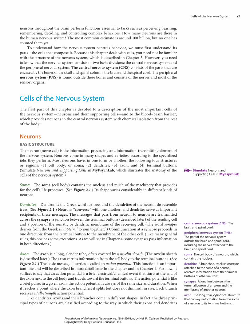
This chapter describes the structure and functions of the most important cells of the nervous system. Information, in the form of light, sound waves, odors, tastes, or contact with objects, is gathered from the environment by specialized cells called sensory neurons. Movements are ac-complished by the contraction of muscles, which are controlled by motor neurons. (The term motor is used here in its original sense to refer to movement, not to a mechanical engine.) And in between sensory neurons and motor neurons come the interneurons—neurons that lie entirely within the central nervous system. Local interneurons form circuits with nearby neurons and analyze small pieces of information. Relay interneurons connect circuits of local interneurons in one region of the brain with those in other regions. Through these connections, circuits of
PROLOGUE | Unresponsive Muscles
Kathryn D. was getting desperate. All her life she had been healthy and active, eating wisely and keeping fit with sports and regular exercise. She went to her health club almost every day for a session of low-impact aerobics, followed by a swim. But several months ago, she began having trouble keeping up with her usual schedule. At first, she found herself getting tired toward the end of her aero-bics class. Her arms, particularly, seemed to get heavy. Then when she entered the pool and started swimming, she found that it was hard to lift her arms over her head; she abandoned the crawl and the backstroke and did the sidestroke and breaststroke instead. She did not have any flulike symptoms, so she told herself that she needed more sleep and perhaps she should eat a little more.
Over the next few weeks, however, things only got worse. Aero-bics classes were becoming an ordeal. Her instructor became con-cerned and suggested that Kathryn see her doctor. She did so, but he could find nothing wrong with her. She was not anemic, showed no signs of an infection, and seemed to be well nourished. He asked how things were going at work.
“Well, lately I’ve been under some pressure,” she said. “The head of my department quit a few weeks ago, and I’ve taken over his job temporarily. I think I have a chance of getting the job perma-nently, but I feel as if my bosses are watching me to see whether I’m good enough for the job.” Kathryn and her physician agreed that increased stress could be the cause of her problem. “I’d prefer not to give you any medication at this time,” he said, “but if you don’t feel better soon we’ll have a closer look at you.”
She did feel better for a while, but then all of a sudden her symp-toms got worse. She quit going to the health club and found that she even had difficulty finishing a day’s work. She was certain that
people were noticing that she was no longer her lively self, and she was afraid that her chances for the promotion were slipping away. One afternoon she tried to look up at the clock on the wall and real-ized that she could hardly see—her eyelids were drooping, and her head felt as if it weighed a hundred pounds. Just then, one of her supervisors came over to her desk, sat down, and asked her to fill him in on the progress she had been making on a new project. As she talked, she found herself getting weaker and weaker. Her jaw was getting tired, even her tongue was getting tired, and her voice was getting weaker. With a sudden feeling of fright she realized that the act of breathing seemed to take a lot of effort. She managed to finish the interview, but immediately afterwards she packed up her briefcase and left for home, saying that she had a bad headache.
She telephoned her physician, who immediately arranged for her to go to the hospital to be seen by Dr. T., a neurologist. Dr. T. listened to a description of her symptoms and examined her briefly. She said to Kathryn, “I think I know what may be causing your symptoms. I’d like to give you an injection and watch your re-action.” She gave some orders to the nurse, who left the room and came back with a syringe. Dr. T. took it, swabbed Kathryn’s arm, and injected the drug. She started questioning Kathryn about her job. Kathryn answered slowly, her voice almost a whisper. As the ques-tions continued, she realized that it was getting easier and easier to talk. She straightened her back and took a deep breath. Yes, she was sure. Her strength was returning! She stood up and raised her arms above her head. “Look,” she said, her excitement growing. “I can do this again. I’ve got my strength back! What was that you gave me? Am I cured?”
(For an answer to her question, see p. 46.)
20
sensory neuron A neuron that detects changes in the external or internal environment and sends information about these changes to the central nervous system.
motor neuron A neuron located within the central nervous system that controls the contraction of a muscle or the secretion of a gland.
interneuron A neuron located entirely within the central nervous system.
Foundations of Behavioral Neuroscience, Ninth Edition, by Neil R. Carlson. Published by Pearson. Copyright © 2013 by Pearson Education, Inc.
Cells of the Nervous System 21
neurons throughout the brain perform functions essential to tasks such as perceiving, learning, remembering, deciding, and controlling complex behaviors. How many neurons are there in the human nervous system? The most common estimate is around 100 billion, but no one has counted them yet.
To understand how the nervous system controls behavior, we must first understand its parts—the cells that compose it. Because this chapter deals with cells, you need not be familiar with the structure of the nervous system, which is described in Chapter 3. However, you need to know that the nervous system consists of two basic divisions: the central nervous system and the peripheral nervous system. The central nervous system (CNS) consists of the parts that are encased by the bones of the skull and spinal column: the brain and the spinal cord. The peripheral nervous system (PNS) is found outside these bones and consists of the nerves and most of the sensory organs.
Cells of the Nervous SystemThe first part of this chapter is devoted to a description of the most important cells of the nervous system—neurons and their supporting cells—and to the blood–brain barrier, which provides neurons in the central nervous system with chemical isolation from the rest of the body.
Neurons
BASIC STRUCTURE
The neuron (nerve cell) is the information-processing and information-transmitting element of the nervous system. Neurons come in many shapes and varieties, according to the specialized jobs they perform. Most neurons have, in one form or another, the following four structures or regions: (1) cell body, or soma; (2) dendrites; (3) axon; and (4) terminal buttons. (Simulate Neurons and Supporting Cells in MyPsychLab, which illustrates the anatomy of the cells of the nervous system.)
Soma The soma (cell body) contains the nucleus and much of the machinery that provides for the cell’s life processes. (See Figure 2.1.) Its shape varies considerably in different kinds of neurons.
Dendrites Dendron is the Greek word for tree, and the dendrites of the neuron do resemble trees. (See Figure 2.1.) Neurons “converse” with one another, and dendrites serve as important recipients of these messages. The messages that pass from neuron to neuron are transmitted across the synapse, a junction between the terminal buttons (described later) of the sending cell and a portion of the somatic or dendritic membrane of the receiving cell. (The word synapsederives from the Greek sunaptein, “to join together.”) Communication at a synapse proceeds in one direction: from the terminal button to the membrane of the other cell. (Like many general rules, this one has some exceptions. As we will see in Chapter 4, some synapses pass information in both directions.)
Axon The axon is a long, slender tube, often covered by a myelin sheath. (The myelin sheath is described later.) The axon carries information from the cell body to the terminal buttons. (See Figure 2.1.) The basic message it carries is called an action potential. This function is an impor-tant one and will be described in more detail later in the chapter and in Chapter 4. For now, it suffices to say that an action potential is a brief electrical/chemical event that starts at the end of the axon next to the cell body and travels toward the terminal buttons. The action potential is like a brief pulse; in a given axon, the action potential is always of the same size and duration. When it reaches a point where the axon branches, it splits but does not diminish in size. Each branch receives a full-strength action potential.
Like dendrites, axons and their branches come in different shapes. In fact, the three prin-cipal types of neurons are classified according to the way in which their axons and dendrites
central nervous system (CNS) Thebrain and spinal cord.
peripheral nervous system (PNS)The part of the nervous system outside the brain and spinal cord, including the nerves attached to the brain and spinal cord.
soma The cell body of a neuron, which contains the nucleus.
dendrite A branched, treelike structure attached to the soma of a neuron; receives information from the terminal buttons of other neurons.
synapse A junction between the terminal button of an axon and the membrane of another neuron.
Simulate Neurons and Supporting Cells in MyPsychLab
axon The long, thin, cylindrical structure that conveys information from the soma of a neuron to its terminal buttons.
Foundations of Behavioral Neuroscience, Ninth Edition, by Neil R. Carlson. Published by Pearson. Copyright © 2013 by Pearson Education, Inc.

22 CHAPTER 2: Structure and Functions of Cells of the Nervous System
leave the soma. The neuron depicted in Figure 2.1 is the most common type found in the cen-tral nervous system; it is a multipolar neuron. In this type of neuron, the somatic membrane gives rise to one axon but to the trunks of many dendritic trees. Bipolar neurons give rise to one axon and one dendritic tree, at opposite ends of the soma. (See Figure 2.2a.) Bipolar neu-
rons are usually sensory; that is, their dendrites detect events occur-ring in the environment and communicate information about these events to the central nervous system.
The third type of nerve cell is the unipolar neuron. It has only one stalk, which leaves the soma and divides into two branches a short distance away. (See Figure 2.2b.) Unipolar neurons, like bi-polar neurons, transmit sensory information from the environment to the CNS. The arborizations (treelike branches) outside the CNS are dendrites; the arborizations within the CNS end in terminal but-tons. The dendrites of most unipolar neurons detect touch, temper-ature changes, and other sensory events that affect the skin. Other unipolar neurons detect events in our joints, muscles, and internal organs.
The central nervous system communicates with the rest of the body through nerves attached to the brain and to the spinal cord. Nerves are bundles of many thousands of individual fibers, all wrapped in a tough, protective membrane. Under a microscope, nerves look something like telephone cables, with their bundles of wires. (See Figure 2.3.) Like the individual wires in a telephone cable, nerve fibers transmit messages through the nerve, from a sense organ to the brain or from the brain to a muscle or gland.
Terminal Buttons Most axons divide and branch many times. The ends of the “twigs” feature little knobs called terminal buttons. (Some neuroscientists prefer the original French word bouton, whereas oth-ers simply refer to them as terminals.) Terminal buttons have a very special function: When an action potential traveling down the axon reaches them, they secrete a chemical called a neurotransmitter. This chemical (there are many different ones in the CNS) either excites or inhibits the receiving cell and thus helps to determine whether an action potential occurs in its axon. Details of this process will be de-scribed later in this chapter.
Cilia are sensitiveto physical stimuli
Dendrites are sensitiveto physical stimuli
Receptor
Dendrite
Axon
Soma ofbipolarneuron
Soma ofunipolarneuron
Axon
To brain To brain
Terminalbuttons
Terminalbuttons
(a) (b)
F I G U R E 2.2 Bipolar and Unipolar Neurons. Pictured here are (a) a bipolar neuron, primarily found in sensory systems (for example, vision and audition) and (b) a unipolar neuron, found in the somatosensory system (touch, pain, and the like).
bipolar neuron A neuron with one axon and one dendrite attached to its soma.
unipolar neuron A neuron with one axon attached to its soma; the axon divides, with one branch receiving sensory information and the other sending the information into the central nervous system.
terminal button The bud at the end of a branch of an axon; forms synapses with another neuron; sends information to that neuron.
Dendrites
Myelin sheath
Terminalbuttons
Direction ofmessages
Axon (insidemyelin sheath)
Soma(cell body)
F I G U R E 2.1 The Principal Parts of a Multipolar Neuron.
neurotransmitter A chemical that is released by a terminal button; has an excitatory or inhibitory effect on another neuron.
multipolar neuron A neuron with one axon and many dendrites attached to its soma.
Foundations of Behavioral Neuroscience, Ninth Edition, by Neil R. Carlson. Published by Pearson. Copyright © 2013 by Pearson Education, Inc.

Cells of the Nervous System 23
An individual neuron receives information from the terminal but-tons of axons of other neurons—and the terminal buttons of its axons form synapses with other neurons. A neuron may receive information from dozens or even hundreds of other neurons, each of which can form a large number of synaptic connections with it. Figure 2.4 illus-trates the nature of these connections. As you can see, terminal buttons can form synapses on the membrane of the dendrites or the soma. (See Figure 2.4.)
INTERNAL STRUCTURE
Figure 2.5 illustrates the internal structure of a typical multipolar neu-ron. (See Figure 2.5.) The membrane defines the boundary of the cell and consists of a double layer of lipid (fatlike) molecules. Embedded in the membrane are a variety of protein molecules that have special func-tions. Some proteins detect substances outside the cell (such as hor-mones) and pass information about the presence of these substances to the cell’s interior. Other proteins control access to the interior of the cell, permitting some substances to enter but barring others. Still other proteins act as transporters, actively carrying certain molecules into or out of the cell. Because the proteins that are found in the neuron’s membrane are especially important in the transmis-sion of information, their characteristics will be discussed in more detail later in this chapter.
The cell is filled with cytoplasm, a jellylike substance that contains small specialized structures, just as the body contains specialized organs. Among these structures are mito-chondria, which break down nutrients such as glucose and provide the cell with energy to perform its functions. Mitochondria produce a chemical called adenosine triphosphate (ATP), which can be used throughout the cell as an energy source. Many eons ago mito-chondria were free-living organisms that came to “infect” larger cells. Because the mito-chondria could extract energy more efficiently than their hosts, they became useful to them and eventually became a permanent part of them. Mitochondria still contain their own genetic information and multiply independently of the cells in which they live. We inherit our mitochondria from our mothers; fathers’ sperms do not contribute any mitochondria to the ova they fertilize.
Deep inside the cell is the nucleus (from the Latin word for “nut”). The nucleus contains the chromosomes. Chromosomes, as you have probably already learned, consist of long strands
BV
A
Bloodvessel Individual
axons
Bundle ofaxons
Nerve
F I G U R E 2.3 Nerves. A nerve consists of a sheath of tissue that encases a bundle of individual nerve fibers (also known as axons).
Synapse on soma
Cell bodyMyelinsheath
Axon Terminalbutton
Synapse ondendrite
F I G U R E 2.4 An Overview of the Synaptic Connections Between Neurons. The arrows represent the directions of the flow of information.
membrane A structure consisting principally of lipid molecules that defines the outer boundaries of a cell and also constitutes many of the cell organelles.
cytoplasm The viscous, semiliquid substance contained in the interior of a cell.
mitochondria An organelle that is responsible for extracting energy from nutrients.
adenosine triphosphate (ATP) (ah deno seen) A molecule of prime importance to cellular energy metabolism; its breakdown liberates energy.
nucleus A structure in the central region of a cell, containing the chromosomes.
chromosome A strand of DNA, with associated proteins, found in the nucleus; carries genetic information.
Foundations of Behavioral Neuroscience, Ninth Edition, by Neil R. Carlson. Published by Pearson. Copyright © 2013 by Pearson Education, Inc.

24 CHAPTER 2: Structure and Functions of Cells of the Nervous System
of deoxyribonucleic acid (DNA). The chromosomes have an important function: They contain the recipes for making proteins. Portions of the chromosomes, called genes, contain the recipes for individual proteins.
Proteins are important in cell functions. If a neu-ron grown in a tissue culture is exposed to a detergent, the lipid membrane and much of the cell’s interior dissolve away, leaving a matrix of insoluble strands of protein. This matrix, called the cytoskeleton, gives the neuron its shape. The cytoskeleton is made of various kinds of protein strands, linked to each other and form-ing a cohesive mass.
Besides providing structure, proteins serve as en-zymes. Enzymes are the cell’s marriage brokers or di-vorce judges: They cause particular molecules to join together or split apart. Thus, enzymes determine what gets made from the raw materials contained in the cell, and they determine which molecules remain intact.
Proteins are also involved in transporting sub-stances within the cell. Axons can be extremely long, relative to their diameter and the size of the soma. For
example, the longest axon in a human stretches from the foot to a region located in the base of the brain. Because terminal buttons need some items that can be produced only in the soma, there must be a system that can transport these items rapidly and efficiently through the axoplasm (that is, the cytoplasm of the axon). This system, axoplasmic transport, is an active process that pro-pels substances from one end of the axon to the other. This transport is accomplished by long pro-tein strands called microtubules, bundles of thirteen filaments arranged around a hollow core. Microtubules serve as railroad tracks, guiding the progress of the substances being transported. Movement from the soma to the terminal buttons is called anterograde axoplasmic transport. (Antero- means “toward the front.”) Retrograde axoplasmic transport carries substances from the terminal buttons back to the soma. (Retro- means “toward the back.”) Anterograde axoplasmic transport is remarkably fast: up to 500 mm per day. Retrograde axoplasmic transport is about half as fast. Energy for both forms of transport is supplied by ATP, produced by the mitochondria.
Supporting Cells
Neurons constitute only about half the volume of the CNS. The rest consists of a variety of support-ing cells. Because neurons have a very high rate of metabolism but have no means of storing nu-trients, they must constantly be supplied with nutrients and oxygen or they will quickly die. Thus, the role played by the cells that support and protect neurons is very important to our existence.
GLIA
The most important supporting cells of the central nervous system are the neuroglia, or “nerve glue.” Glia (also called glial cells) are much more numerous than neurons. They constitute ap-proximately 85 percent of the cells of the brain. Although they glue the CNS together, they do much more than that. Neurons lead a very sheltered existence; they are buffered physically and chemically from the rest of the body by the glial cells. Glial cells surround neurons and hold them in place, controlling their supply of nutrients and some of the chemicals they need to exchange messages with other neurons; they insulate neurons from one another so that neural messages do not get scrambled; and they even act as housekeepers, destroying and removing the carcasses of neurons that are killed by disease or injury.
There are several types of glial cells, each of which plays a special role in the CNS. The three most important types are astrocytes, oligodendrocytes, and microglia. Astrocyte means “star cell,” and this name accurately describes the shape of these cells. Astrocytes (or astroglia)provide physical support to neurons and clean up debris within the brain. They produce some
cytoskeleton Support structure formed of microtubules and other protein fibers that are linked to each other and form a cohesive mass that gives a cell its shape.
enzyme A molecule that controls a chemical reaction, combining two substances or breaking a substance into two parts.
axoplasmic transport An active process by which substances are propelled along microtubules that run the length of the axon.
microtubule (my kro too byool) A long strand of bundles of protein filaments arranged around a hollow core; part of the cytoskeleton and involved in transporting substances from place to place within the cell.
glia (glee ah) The supporting cells of the central nervous system.
astrocyte A glial cell that provides support for neurons of the central nervous system, provides nutrients and other substances, and regulates the chemical composition of the extracellular fluid.
Membrane
Microtubules
Dendrite
Nucleus
Cytoplasm
MitochondriaMyelinsheath
Dendriticspines
F I G U R E 2.5 The Principal Internal Structures of a Multipolar Neuron.
gene The functional unit of the chromosome, which directs synthesis of one or more proteins.
deoxyribonucleic acid (DNA) (dee ox ee ry bo new clay ik) A long, complex macromolecule consisting of two interconnected helical strands; along with associated proteins, strands of DNA constitute the chromosomes.
Foundations of Behavioral Neuroscience, Ninth Edition, by Neil R. Carlson. Published by Pearson. Copyright © 2013 by Pearson Education, Inc.

Cells of the Nervous System 25
chemicals that neurons need to fulfill their functions. They help to control the chemical com-position of the fluid surrounding neurons by actively taking up or releasing substances whose concentrations must be kept within critical levels. Finally, astrocytes are involved in providing nourishment to neurons.
Some of the astrocyte’s processes (the arms of the star) are wrapped around blood vessels. Other processes are wrapped around parts of neurons, so the somatic and dendritic membranes of neurons are largely surrounded by astrocytes. Evidence suggests that astrocytes receive nutri-ents from the capillaries, store them, and release them to neurons when needed (Tsacopoulos and Magistretti, 1996; Brown, Tekkök, and Ransom, 2004). Besides having a role in transporting chemicals to neurons, astrocytes serve as the matrix that holds neurons in place. These cells also surround and isolate synapses, limiting the dispersion of neurotransmitters that are released by the terminal buttons. (See Figure 2.6.)
When cells in the central nervous system die, certain kinds of astrocytes take up the task of cleaning away the debris. These cells are able to travel around the CNS; they extend and retract their processes (pseudopodia, or “false feet”) and glide about the way amoebas do. When these astrocytes contact a piece of debris from a dead neuron, they push themselves against it, finally engulfing and digesting it. We call this process phagocytosis (phagein, “to eat”; kutos, “cell”). If there is a considerable amount of injured tissue to be cleaned up, astrocytes will divide and produce enough new cells to do the task. Once the dead tissue is broken down, a framework of astrocytes will be left to fill in the vacant area, and a specialized kind of astrocyte will form scar tissue, walling off the area.
The principal function of oligodendrocytes is to provide support to axons and to produce the myelin sheath, which insulates most axons from one another. (Very small axons are not myelinated and lack this sheath.) Myelin, which is 80 percent lipid and 20 percent protein, is produced by the oligodendrocytes in the form of a tube surrounding the axon. This tube does not form a continuous sheath; rather, it consists of a series of segments, each approximately 1 mm long, with a small (1–2 μm) portion of uncoated axon between the segments. (A micrometer, ab-breviated μm, is one-millionth of a meter, or one-thousandth of a millimeter.) The bare portion of axon is called a node of Ranvier, after the person who discovered it. The myelinated axon, then, resembles a string of elongated beads. (Actually, the beads are very much elongated—their length is approximately eighty times their width.)
Glucose
Glucose
Glycogen(storage)Lactate
Energy
Lactate
LactateBloodvessel
Astrocyte
Neuron
F I G U R E 2.6 Structure and Location of Astrocytes. The processes of astrocytes surround capillaries and neurons of the central nervous system.
phagocytosis (fagg o sy toe sis) Theprocess by which cells engulf and digest other cells or debris caused by cellular degeneration.
oligodendrocyte (oh li go den droh site) A type of glial cell in the central nervous system that forms myelin sheaths.
myelin sheath (my a lin) A sheath that surrounds axons and insulates them, preventing messages from spreading between adjacent axons.
node of Ranvier (raw vee ay) A naked portion of a myelinated axon, between adjacent oligodendroglia or Schwann cells.
Foundations of Behavioral Neuroscience, Ninth Edition, by Neil R. Carlson. Published by Pearson. Copyright © 2013 by Pearson Education, Inc.

26 CHAPTER 2: Structure and Functions of Cells of the Nervous System
Myelinated axons
Soma ofoligodendrocyte
Microtubule
Node of Ranvier
Mitochondrionin axoplasm
Node ofRanvier
F I G U R E 2.7 Oligodendrocyte. An oligodendrocyte forms the myelin that surrounds many axons in the central nervous system. Each cell forms one segment of myelin for several adjacent axons.
Myelin sheathAxons
Oligodendrocyte
(a)
(b)
Schwann cell
F I G U R E 2.8 Formation of Myelin. During development, a process of an oligodendrocyte or an entire Schwann cell tightly wraps itself many times around an individual axon and forms one segment of the myelin sheath. (a) Oligodendrocyte. (b) Schwann cell.
Dr. C., a retired neurologist, had been afflicted with multiple sclerosis for more than two decades when she died of a heart attack. One evening, twenty-three years previously, she and her husband had had dinner at their favorite restaurant. As they were leaving, she stumbled and almost fell. Her husband joked, “Hey, honey, you shouldn’t have had that last glass of wine.” She smiled at his at-tempt at humor, but she knew better—her clumsiness wasn’t brought on by the two glasses of wine she had drunk with dinner. She suddenly realized that she had been ignoring some symptoms that she should have recognized.
The next day, she consulted with one of her colleagues, who agreed that her own tentative diagnosis was probably correct: Her symptoms fit those of multiple sclerosis. She had experienced fleeting problems with double vision, she sometimes felt unsteady on her feet, and she occasionally noticed tingling sensations in her right hand. None of these symptoms was serious, and they lasted for only a short while, so she ignored them—or perhaps denied to herself that they were important.
A few weeks after Dr. C.’s death, a group of medical students and neurological residents gath-ered in an autopsy room at the medical school. Dr. D., the school’s neuropathologist, displayed a stainless-steel tray on which were lying a brain and a spinal cord. “These belonged to Dr. C.,” he said. “Several years ago she donated her organs to the medical school.” Everyone looked at the brain more intently, knowing that it had animated an esteemed clinician and teacher whom they all knew by reputation, if not personally. Dr. D. led his audience to a set of light boxes on the wall, to which several MRI scans had been clipped. He pointed out some white spots that appeared on one scan. “This scan clearly shows some white-matter lesions, but they are gone on the next one, taken six months later. And here is another one, but it’s gone on the next scan. The immune system attacked the myelin sheaths in a particular region, and then glial cells cleaned up the debris. MRI doesn’t show the lesions then, but the axons can no longer conduct their messages.”
(continued)
A given oligodendrocyte produces up to fifty segments of myelin. During the development of the CNS, oligodendrocytes form processes shaped something like canoe paddles. Each of these paddle-shaped processes then wraps itself many times around a segment of an axon and, while doing so, produces layers of myelin. Each paddle thus becomes a segment of an axon’s myelin sheath. (See Figures 2.7 and 2.8a.)
Foundations of Behavioral Neuroscience, Ninth Edition, by Neil R. Carlson. Published by Pearson. Copyright © 2013 by Pearson Education, Inc.

Cells of the Nervous System 27
As their name indicates, microglia are the smallest of the glial cells. Like some types of astrocytes, they act as phagocytes, engulfing and breaking down dead and dying neurons. But in addition, they serve as one of the representatives of the im-mune system in the brain, protecting the brain from invading microorganisms. They are primarily responsible for the inflammatory reaction in response to brain damage.
SCHWANN CELLS
In the central nervous system the oligodendrocytes support axons and produce my-elin. In the peripheral nervous system the Schwann cells perform the same func-tions. Most axons in the PNS are myelinated. The myelin sheath occurs in segments, as it does in the CNS; each segment consists of a single Schwann cell, wrapped many times around the axon. In the CNS the oligodendrocytes grow a number of paddle-shaped processes that wrap around a number of axons. In the PNS a Schwann cell provides myelin for only one axon, and the entire Schwann cell—not merely a part of it—surrounds the axon. (See Figure 2.8b.)
There is an important difference between oligodendrocytes of the CNS and Schwann cells of the PNS: the chemical composition of the myelin protein they pro-duce. The immune system of people with multiple sclerosis attacks only the myelin protein produced by oligodendrocytes; thus, the myelin of the peripheral nervous system is spared.
The Blood–Brain Barrier
Over one hundred years ago, Paul Ehrlich discovered that if a blue dye is injected into an animal’s bloodstream, all tissues except the brain and spinal cord will be tinted blue. However, if the same dye is injected into the fluid-filled ventricles of the brain, the blue color will spread throughout the CNS (Bradbury, 1979). This experiment demonstrates that a barrier exists between the blood and the fluid that surrounds the cells of the brain: the blood–brain barrier.
Some substances can cross the blood–brain barrier; others cannot. Thus, it is selectively per-meable (from the Latin per, “through,” and meare, “to pass”). In most of the body the cells that line the capillaries do not fit together absolutely tightly. Small gaps are found between them that permit the free exchange of most substances between the blood plasma and the fluid outside the capillaries that surrounds the body’s cells. In the central nervous system the capillaries lack these gaps; therefore, many substances cannot leave the blood. Thus, the walls of the capillaries in the brain constitute the blood–brain barrier. (See Figure 2.9.) Other substances must be actively transported through the capillary walls by special proteins. For example, glucose transporters bring the brain its fuel, and other transporters rid the brain of toxic waste products (Rubin and Staddon, 1999; Zlokovic, 2008).
He put on a pair of surgical gloves, picked up Dr. C.’s brain, and cut it in several slices. He picked one up. “Here, see this?” He pointed out a spot of discoloration in a band of white matter. “This is a sclerotic plaque—a patch that feels harder than the surrounding tissue. There are many of them, located throughout the brain and spinal cord, which is why the disease is called multiple sclerosis.” He picked up the spinal cord, felt along its length with his thumb and forefinger, and then stopped and said, “Yes, I can feel a plaque right here.”
Dr. D. put the spinal cord down and said, “Who can tell me the etiology of this disorder?”One of the students spoke up. “It’s an autoimmune disease. The immune system gets sensitized
to the body’s own myelin protein and periodically attacks it, causing a variety of different neurologi-cal symptoms. Some say that a childhood viral illness somehow causes the immune system to start seeing the protein as foreign.”
“That’s right,” said Dr. D. “The primary criterion for the diagnosis of multiple sclerosis is the pres-ence of neurological symptoms disseminated in time and space. The symptoms don’t all occur at once, and they can be caused only by damage to several different parts of the nervous system, which means that they can’t be the result of a stroke.”
microglia The smallest of glial cells; they act as phagocytes and protect the brain from invading microorganisms.
Schwann cell A cell in the peripheral nervous system that is wrapped around a myelinated axon, providing one segment of its myelin sheath.
Touch, temperature changes, pain, and other sensory events that affect the skin are detected by the dendrites of unipolar neurons.
Jeff Greenberg/The Image Works.
blood–brain barrier A semipermeable barrier between the blood and the brain produced by the cells in the walls of the brain’s capillaries.
Foundations of Behavioral Neuroscience, Ninth Edition, by Neil R. Carlson. Published by Pearson. Copyright © 2013 by Pearson Education, Inc.

28 CHAPTER 2: Structure and Functions of Cells of the Nervous System
What is the function of the blood–brain barrier? As we will see, trans-mission of messages from place to place in the brain depends on a delicate balance between substances within neurons and in the extracellular fluid that surrounds them. If the composition of the extracellular fluid is changed even slightly, the transmission of these messages will be disrupted, which means that brain functions will be disrupted. The presence of the blood–brain barrier makes it easier to regulate the composition of this fluid. In addition, many of the foods that we eat contain chemicals that would interfere with the transmis-sion of information between neurons. The blood–brain barrier prevents these chemicals from reaching the brain.
The blood–brain barrier is not uniform throughout the nervous system. In several places the barrier is relatively permeable, allowing substances that are excluded elsewhere to cross freely. For example, the area postrema is a part of the brain that controls vomiting. The blood–brain barrier is much weaker there, permitting neurons in this region to detect the presence of toxic sub-stances in the blood. A poison that enters the circulatory system from the stomach can thus stimulate this area to initiate vomiting. If the organism is lucky, the poison can be expelled from the stomach before causing too much damage.
Gaps that permit the free flowof substances into and out ofthe blood
Capillary in all of bodyexcept brain
(a) (b)
Capillaryin brain
F I G U R E 2.9 The Blood–Brain Barrier. This figure shows that (a) the cells that form the walls of the capillaries in the body outside the brain have gaps that permit the free passage of substances into and out of the blood and (b) the cells that form the walls of the capillaries in the brain are tightly joined.
area postrema (poss tree ma) A region of the medulla where the blood–brain barrier is weak; poisons can be detected there and can initiate vomiting.
Neurons are the most important cells of the nervous system. The central nervous system (CNS) includes the brain and spinal cord; the peripheral nervous system (PNS) includes nerves and some sensory organs.
Neurons have four principal parts: dendrites, soma (cell body), axon, and terminal buttons. They communicate by means of synapses, junc-tions between the terminal buttons of one neuron and the somatic or dendritic membrane of another. When an action potential travels down an axon, its terminal buttons secrete a chemical that has either an excit-atory or an inhibitory effect on the neurons with which they communi-cate. Ultimately, the effects of these excitatory and inhibitory synapses cause behavior in the form of muscular contractions.
Neurons contain a quantity of clear cytoplasm, enclosed in a mem-brane. Embedded in the membrane are protein molecules that have spe-cial functions, such as the transport of particular substances into and out of the cell. The nucleus contains the genetic information—the recipes for all the proteins that the body can make. Microtubules and other protein filaments compose the cytoskeleton and help to transport chemicals from place to place. Mitochondria serve as the location for most of the chemical reactions through which the cell extracts energy from nutrients.
Neurons are supported by the glial cells of the central nervous sys-tem and the supporting cells of the peripheral nervous system. In the CNS
astrocytes provide support and nourishment, regulate the composition of the fluid that surrounds neurons, and remove debris and form scar tissue in the event of tissue damage. Microglia are phagocytes that serve as the representatives of the immune system. Oligodendrocytes form myelin, the substance that insulates axons, and also support unmyelinated axons. In the PNS, support and myelin are provided by the Schwann cells.
In most organs molecules freely diffuse between the blood within the capillaries that serve them and the extracellular fluid that bathes their cells. The molecules pass through gaps between the cells that line the capillaries. The walls of the capillaries of the CNS lack these gaps; consequently, fewer substances can enter or leave the brain across the blood–brain barrier.
Thought QuestionThe fact that the mitochondria in our cells were originally microorganisms that infected our very remote ancestors points out that evolution can in-volve interactions between two or more species. Most species have other organisms living inside them; in fact, the bacteria in our intestines are necessary for our good health. Some microorganisms can exchange ge-netic information, so adaptive mutations developed in one species can be adopted by another. Is it possible that some of the features of the cells of our nervous system were bequeathed to our ancestors by other species?
SECTION SUMMARY
Cells of the Nervous System
Communication Within a NeuronThis section describes the nature of communication within a neuron—the way an action potential is sent from the cell body down the axon to the terminal buttons, informing them to release some neurotransmitter. The details of synaptic transmission—the communication between neurons—will be described in the next section. As we will see in this section, an action potential consists
Foundations of Behavioral Neuroscience, Ninth Edition, by Neil R. Carlson. Published by Pearson. Copyright © 2013 by Pearson Education, Inc.

Communication Within a Neuron 29
of a series of alterations in the membrane of the axon that permit various substances to move between the interior of the axon and the fluid surrounding it. These exchanges produce electrical currents. (Simulate The Action Potential in MyPsychLab, which illustrates the transmission of information along the axon.)
Neural Communication: An Overview
Before I begin my discussion of the action potential, let’s step back and see how neurons can in-teract to produce a useful behavior. We begin by examining a simple assembly of three neurons and a muscle that controls a withdrawal reflex. In the next two figures (and in subsequent figures that illustrate simple neural circuits), multipolar neurons are depicted in shorthand fashion as several-sided stars. The points of these stars represent dendrites, and only one or two terminal buttons are shown at the end of the axon. The sensory neuron in this example detects painful stimuli. When its dendrites are stimulated by a noxious stimulus (such as contact with a hot object), it sends messages down the axon to the terminal buttons, which are located in the spinal cord. (You will recognize this cell as a unipolar neuron; see Figure 2.10.) The terminal buttons of the sensory neuron release a neurotransmitter that excites the interneuron, causing it to send messages down its axon. The terminal buttons of the interneuron release a neurotransmitter that excites the motor neuron, which then sends messages down its axon. The axon of the motor neu-ron joins a nerve and travels to a muscle. When the terminal buttons of the motor neuron release their neurotransmitter, the muscle cells contract, causing the hand to move away from the hot object. (See Figure 2.10.)
So far, all of the synapses have had excitatory effects. Now let us complicate matters a bit to see the effect of inhibitory synapses. Suppose you have removed a hot casserole from the oven. As you start walking over to the table to put it down, the heat begins to penetrate the rather thin pot-holders you are using. The pain caused by the hot casserole triggers a withdrawal reflex that tends to make you drop it. Yet you manage to keep hold of it long enough to get to the table and put it down. What prevented your withdrawal reflex from making you drop the casserole on the floor?
The pain from the hot casserole increases the activity of excitatory synapses on the motor neurons, which tends to cause the hand to pull away from the casserole. However, this excitation is counteracted by inhibition, supplied by another source: the brain. The brain contains neural circuits that recognize what a disaster it would be if you dropped the casserole on the floor. These neural circuits send information to the spinal cord that prevents the withdrawal reflex from mak-ing you drop the dish.
Figure 2.11 shows how this information reaches the spinal cord. As you can see, an axon from a neuron in the brain reaches the spinal cord, where its terminal buttons form synapses with an inhibitory interneuron. When the neuron in the brain becomes active, its terminal buttons excite
Brain
Spinalcord
Cross sectionof spinal cord
Motorneuron
This interneuron excitesmotor neuron, causingmuscular contraction
This muscle causeswithdrawal fromsource of pain
Axon of sensoryneuron (pain)
Dendrites ofsensory neurondetect painfulstimulus
F I G U R E 2.10 A Withdrawal Reflex. The figure shows a simple example of a useful function of the nervous system. The painful stimulus causes the hand to pull away from the hot iron.
Simulate The Action Potential in MyPsychLab
Foundations of Behavioral Neuroscience, Ninth Edition, by Neil R. Carlson. Published by Pearson. Copyright © 2013 by Pearson Education, Inc.

30 CHAPTER 2: Structure and Functions of Cells of the Nervous System
this inhibitory interneuron. The interneuron releases an inhibitory neurotransmitter, which de-creases the activity of the motor neuron, blocking the withdrawal reflex. This circuit provides an example of a contest between two competing tendencies: to drop the casserole and to hold onto it. (See Figure 2.11.)
Of course, reflexes are more complicated than this description, and the mechanisms that in-hibit them are even more so. In addition, thousands of neurons are involved in this process. The five neurons shown in Figure 2.11 represent many others: Dozens of sensory neurons detect the hot object, hundreds of interneurons are stimulated by their activity, hundreds of motor neurons produce the contraction—and thousands of neurons in the brain must become active if the reflex is to be inhibited. Yet this simple model provides an overview of the process of neural communi-cation, which is described in more detail later in this chapter.
Measuring Electrical Potentials of Axons
Let’s examine the nature of the message that is conducted along the axon. To do so, we obtain an axon that is large enough to work with. Fortunately, nature has provided the neuroscientist with the giant squid axon (the giant axon of a squid, not the axon of a giant squid!). This axon is about 0.5 mm in diameter, which is hundreds of times larger than the largest mammalian axon. (This large axon controls an emergency response: sudden contraction of the mantle, which squirts wa-ter through a jet and propels the squid away from a source of danger.) We place an isolated giant squid axon in a dish of seawater, in which it can exist for a day or two.
To measure the electrical charges generated by an axon, we will need to use a pair of elec-trodes. Electrodes are electrical conductors that provide a path for electricity to enter or leave a medium. One of the electrodes is a simple wire that we place in the seawater. The other one, which we use to record the message from the axon, has to be special. Because even a giant squid axon is rather small, we must use a tiny electrode that will record the membrane potential without damaging the axon. To do so, we use a microelectrode.
A microelectrode, which is simply a very small electrode, can be made of metal or glass. In this case we will use one made of thin glass tubing, which is heated and drawn down to an exceedingly fine point, less than a thousandth of a millimeter in diameter. Because glass will not conduct electricity, the glass microelectrode is filled with a liquid that conducts electricity, such as a solution of potassium chloride.
We place the wire electrode in the seawater and insert the microelectrode into the axon. (See Figure 2.12a.) As soon as we do so, we discover that the inside of the axon is negatively charged with respect to the outside; the difference in charge being 70 mV (millivolts, or thousandths of a volt). Thus, the inside of the membrane is –70 mV. This electrical charge is called the membranepotential. The term potential refers to a stored-up source of energy—in this case, electrical en-ergy. For example, a flashlight battery that is not connected to an electrical circuit has a potentialcharge of 1.5 V between its terminals. If we connect a light bulb to the terminals, the potential
This interneuron inhibitsmotor neuron, preventingmuscular contraction
Cross sectionof spinal cord
Axon fromneuronin brain
Axon of sensoryneuron (pain)
This muscle causeswithdrawal fromsource of painMotor
neuron
This interneuron excitesmotor neuron, causingmuscular contractionNeuron
in brain
Brain
Axon of neuronin brain
Spinal cord
F I G U R E 2.11 The Role of Inhibition. Inhibitory signals arising from the brain can prevent the withdrawal reflex from causing the person to drop the casserole.
electrode A conductive medium that can be used to apply electrical stimulation or to record electrical potentials.
microelectrode A very fine electrode, generally used to record activity of individual neurons.
membrane potential The electrical charge across a cell membrane; the difference in electrical potential inside and outside the cell.
Foundations of Behavioral Neuroscience, Ninth Edition, by Neil R. Carlson. Published by Pearson. Copyright © 2013 by Pearson Education, Inc.

Communication Within a Neuron 31
energy is tapped and converted into radiant energy (light). (See Figure 2.12b.) Similarly, if we connect our electrodes—one inside the axon and one outside it—to a very sensitive voltmeter, we will convert the potential energy to movement of the meter’s needle. Of course, the potential elec-trical energy of the axonal membrane is very weak in comparison with that of a flashlight battery.
As we will see, the message that is conducted down the axon consists of a brief change in the membrane potential. However, this change occurs very rapidly—too rapidly for us to see if we were using a voltmeter. Therefore, to study the message, we will use an oscilloscope. This device, like a voltmeter, measures voltages, but it also produces a record of these voltages, graphing them as a function of time. These graphs are displayed on a screen, much like the one found in a television. The vertical axis represents voltage, and the horizontal axis represents time, going from left to right.
Once we insert our microelectrode into the axon, the oscilloscope draws a straight horizontal line at –70 mV, as long as the axon is not disturbed. This electrical charge across the membrane is called, quite appropriately, the resting potential. Now let us disturb the resting potential and see what hap-pens. To do so, we will use another device: an electrical stimulator that allows us to alter the membrane potential at a specific location. (See Figure 2.13.) The stimulator can pass current through another mi-croelectrode that we have inserted into the axon. Because the inside of the axon is negative, a positive charge applied to the inside of the membrane produces a depolarization. That is, it takes away some of the electrical charge across the membrane near the electrode, reducing the membrane potential.
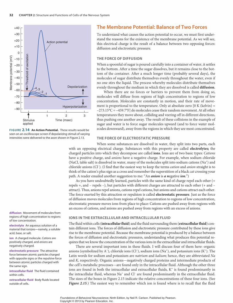
Let us see what happens to an axon when we artificially change the membrane potential at one point. Figure 2.14 shows a graph drawn by an oscilloscope that has been monitoring the effects of brief depolarizing stimuli. The graphs of the effects of these separate stimuli are superimposed on the same drawing so that we can compare them. We deliver a series of depolarizing stimuli, start-ing with a very weak stimulus (number 1) and gradually increasing their strength. Each stimulus briefly depolarizes the membrane potential a little more. Finally, after we present depolarization number 4, the membrane potential suddenly reverses itself, so that the inside becomes positive(and the outside becomes negative). The membrane potential quickly returns to normal, but first it overshoots the resting potential, becoming hyperpolarized—more polarized than normal—for a short time. The whole process takes about 2 msec (milliseconds). (See Figure 2.14.)
This phenomenon, a very rapid reversal of the membrane potential, is called the action po-tential. It constitutes the message carried by the axon from the cell body to the terminal buttons. The voltage level that triggers an action potential—which was achieved only by depolarizing shock number 4—is called the threshold of excitation.
Voltmeter
Battery
Wire electrodeplaced inseawater
Giantsquid axon
Glassmicroelectrodefilled with liquidthat conductselectricity
(a)
(b)
F I G U R E 2.12 Measuring Electrical Charge. This figure shows (a) a voltmeter detecting the charge across a membrane of an axon and (b) a light bulb lit by the charge across the terminals of a battery.
MODEFREQUENCY
Stimulator
Wireelectrodeplaced inseawater
Oscilloscope
Records ofchanges inmembranepotentialdisplayed here
Giant squid axon
Electrical stimulator
Glassmicroelectrodes
F I G U R E 2.13 Studying the Axon. The figure illustrates the means by which an axon can be stimulated while its membrane potential is being recorded.
oscilloscope A laboratory instrument that is capable of displaying a graph of voltage as a function of time on the face of a cathode ray tube.
resting potential The membrane potential of a neuron when it is not being altered by excitatory or inhibitory postsynaptic potentials; approximately –70 mV in the giant squid axon.
depolarization Reduction (toward zero) of the membrane potential of a cell from its normal resting potential.
hyperpolarization An increase in the membrane potential of a cell, relative to the normal resting potential.
action potential The brief electrical impulse that provides the basis for conduction of information along an axon.
threshold of excitation The value of the membrane potential that must be reached to produce an action potential.
Foundations of Behavioral Neuroscience, Ninth Edition, by Neil R. Carlson. Published by Pearson. Copyright © 2013 by Pearson Education, Inc.
32 CHAPTER 2: Structure and Functions of Cells of the Nervous System
The Membrane Potential: Balance of Two Forces
To understand what causes the action potential to occur, we must first under-stand the reasons for the existence of the membrane potential. As we will see, this electrical charge is the result of a balance between two opposing forces: diffusion and electrostatic pressure.
THE FORCE OF DIFFUSION
When a spoonful of sugar is poured carefully into a container of water, it settles to the bottom. After a time the sugar dissolves, but it remains close to the bot-tom of the container. After a much longer time (probably several days), the molecules of sugar distribute themselves evenly throughout the water, even if no one stirs the liquid. The process whereby molecules distribute themselves evenly throughout the medium in which they are dissolved is called diffusion.
When there are no forces or barriers to prevent them from doing so, molecules will diffuse from regions of high concentration to regions of low concentration. Molecules are constantly in motion, and their rate of move-ment is proportional to the temperature. Only at absolute zero [0 K (kelvin) = –273.15°C = –459.7°F] do molecules cease their random movement. At all other temperatures they move about, colliding and veering off in different directions, thus pushing one another away. The result of these collisions in the example of sugar and water is to force sugar molecules upward (and to force water mol-ecules downward), away from the regions in which they are most concentrated.
THE FORCE OF ELECTROSTATIC PRESSURE
When some substances are dissolved in water, they split into two parts, each with an opposing electrical charge. Substances with this property are called electrolytes; the charged particles into which they decompose are called ions. Ions are of two basic types: Cationshave a positive charge, and anions have a negative charge. For example, when sodium chloride (NaCl, table salt) is dissolved in water, many of the molecules split into sodium cations (Na+) and chloride anions (Cl–). (I find that the easiest way to keep the terms cation and anion straight is to think of the cation’s plus sign as a cross and remember the superstition of a black cat crossing your path. A reader emailed another suggestion to me: “An anion is a negative ion.”)
As you have undoubtedly learned, particles with the same kind of charge repel each other (+ repels +, and – repels –), but particles with different charges are attracted to each other (+ and – attract). Thus, anions repel anions, cations repel cations, but anions and cations attract each other. The force exerted by this attraction or repulsion is called electrostatic pressure. Just as the force of diffusion moves molecules from regions of high concentration to regions of low concentration, electrostatic pressure moves ions from place to place: Cations are pushed away from regions with an excess of cations, and anions are pushed away from regions with an excess of anions.
IONS IN THE EXTRACELLULAR AND INTRACELLULAR FLUID
The fluid within cells (intracellular fluid) and the fluid surrounding them (extracellular fluid) con-tain different ions. The forces of diffusion and electrostatic pressure contributed by these ions give rise to the membrane potential. Because the membrane potential is produced by a balance between the forces of diffusion and electrostatic pressures, understanding what produces this potential re-quires that we know the concentration of the various ions in the extracellular and intracellular fluids.
There are several important ions in these fluids. I will discuss four of them here: organic anions (symbolized by A–), chloride ions (Cl–), sodium ions (Na+), and potassium ions (K+). The Latin words for sodium and potassium are natrium and kalium; hence, they are abbreviated Naand K, respectively. Organic anions—negatively charged proteins and intermediate products of the cell’s metabolic processes—are found only in the intracellular fluid. Although the other three ions are found in both the intracellular and extracellular fluids, K+ is found predominantly in the intracellular fluid, whereas Na+ and Cl– are found predominantly in the extracellular fluid. The sizes of the boxes in Figure 2.15 indicate the relative concentrations of these four ions. (See Figure 2.15.) The easiest way to remember which ion is found where is to recall that the fluid
diffusion Movement of molecules from regions of high concentration to regions of low concentration.
electrolyte An aqueous solution of a material that ionizes—namely, a soluble acid, base, or salt.
ion A charged molecule. Cations are positively charged, and anions are negatively charged.
0
1 2 3
4 31
2
Depolarization
Hyperpolarization
Time (msec)Stimulusapplied
+40
+30
+20
+10
–10
–20
–30
–40
–50
–60
–70
–80
–90
Mem
bran
e po
tent
ial (
mV
)
Thr
esho
ld o
f exc
itatio
n
Action potential
F I G U R E 2.14 An Action Potential. These results would be seen on an oscilloscope screen if depolarizing stimuli of varying intensities were delivered to the axon shown in Figure 2.13.
electrostatic pressure The attractive force between atomic particles charged with opposite signs or the repulsive force between atomic particles charged with the same sign.
intracellular fluid The fluid contained within cells.
extracellular fluid Body fluids located outside of cells.
Foundations of Behavioral Neuroscience, Ninth Edition, by Neil R. Carlson. Published by Pearson. Copyright © 2013 by Pearson Education, Inc.

Communication Within a Neuron 33
that surrounds our cells is similar to seawater, which is predominantly a solution of salt, NaCl. The primitive ancestors of our cells lived in the ocean; thus, the seawater was their extracellular fluid. Our extracellular fluid thus resembles seawater, produced and maintained by regulatory mechanisms that are described in Chapter 11.
Let’s consider the ions in Figure 2.15, examining the forces of diffusion and electrostatic pressure exerted on each and reasoning why each is located where it is. A–, the organic anion, is unable to pass through the axon’s membrane; therefore, although the presence of this ion within the cell contributes to the membrane potential, it is located where it is because the membrane is impermeable to it.
The potassium ion K+ is concentrated within the axon; thus, the force of diffusion tends to push it out of the cell. However, the outside of the cell is charged positively with respect to the inside, so electrostatic pressure tends to force this cation inside. Thus, the two opposing forces balance, and potassium ions tend to remain where they are. (See Figure 2.15.)
The chloride ion Cl– is in greatest concentration outside the axon. The force of diffusion pushes this ion inward. However, because the inside of the axon is negatively charged, electrostatic pressure pushes this anion outward. Again, two opposing forces balance each other. (See Figure 2.15.)
The sodium ion Na+ is also in greatest concentration outside the axon, so it, like Cl–, is pushed into the cell by the force of diffusion. But unlike chloride, the sodium ion is positively charged. Therefore, electro-static pressure does not prevent Na+ from entering the cell; indeed, the negative charge inside the axon attracts Na+. (See Figure 2.15.)
How can Na+ remain in greatest concentration in the extracellular fluid, despite the fact that both forces (diffusion and electrostatic pres-sure) tend to push it inside? The answer is this: Another force con-tinuously pushes Na+ out of the axon. This force is provided by a large number of protein molecules embedded in the membrane, driven by energy provided by molecules of ATP produced by the mitochondria. These molecules, known as sodium–potassium transporters, exchange Na+ for K+, pushing three sodium ions out for every two potassium ions they push in. (See Figure 2.16.)
+ + + + + +
K+
Na+Cl–
K+
Na+Cl–
A–
Highconcentration
Electrostaticpressure
Electrostaticpressure
Electrostaticpressure
Force of diffusion
Force of diffusion
Force of diffusion
Lowconcentration
CannotleavecellInside of Cell
Outside of Cell
––––––
F I G U R E 2.15 Control of the Membrane Potential. The figure shows the relative concentration of some important ions inside and outside the neuron and the forces acting on them.
sodium–potassium transporter Aprotein found in the membrane of all cells that extrudes sodium ions from and transports potassium ions into the cell.
K+
K+
Na+Na+ Na+
Sodium–potassiumtransporter
3 sodium ionspumped out
2 potassium ionspumped in
Membrane
Inside of Cell
Outside of Cell
F I G U R E 2.16 A Sodium–Potassium Transporter. Thesetransporters are found in the cell membrane.
Foundations of Behavioral Neuroscience, Ninth Edition, by Neil R. Carlson. Published by Pearson. Copyright © 2013 by Pearson Education, Inc.

34 CHAPTER 2: Structure and Functions of Cells of the Nervous System
Because the membrane is not very permeable to Na+, sodium–potassium transporters very effectively keep the intracellular concentration of Na+ low. By transporting K+ into the cell, they also increase the intracel-lular concentration of K+ a small amount. The membrane is approximately 100 times more permeable to K+ than to Na+, so the increase is slight; but as we will see when we study the process of neural inhibition later in this chapter, it is very important. Sodium–potassium transporters use consider-able energy: Up to 40 percent of a neuron’s metabolic resources are used to operate them. Neurons, muscle cells, glia—in fact, most cells of the body—have sodium–potassium transporters in their membrane.
The Action Potential
As we saw, the forces of both diffusion and electrostatic pressure tend to push Na+ into the cell. However, the membrane is not very permeable to this ion, and sodium–potassium transporters continuously pump out Na+,keeping the intracellular level of Na+ low. But imagine what would happen if the membrane suddenly became permeable to Na+. The forces of diffusion and electrostatic pressure would cause Na+ to rush into the cell. This sud-den influx (inflow) of positively charged ions would drastically change the membrane potential. Indeed, experiments have shown that this mechanism is precisely what causes the action potential: A brief increase in the perme-ability of the membrane to Na+ (allowing these ions to rush into the cell) is
immediately followed by a transient increase in the permeability of the membrane to K+ (allowing these ions to rush out of the cell). What is responsible for these transient increases in permeability?
We already saw that one type of protein molecule embedded in the membrane—the sodium–potassium transporter—actively pumps sodium ions out of the cell and pumps potassium ions into it. Another type of protein molecule provides an opening that permits ions to enter or leave the cells. These molecules provide ion channels, which contain passages (“pores”) that can open or close. When an ion channel is open, a particular type of ion can flow through the pore and thus can enter or leave the cell. (See Figure 2.17.) Neural membranes contain many thousands of ion channels. For example, the giant squid axon contains several hundred sodium channels in each square micrometer of membrane. (There are one million square micrometers in a square millimeter; thus, a patch of axonal membrane the size of a lowercase letter “o” in this book would contain several hundred million sodium channels.) Each sodium channel can admit up to 100 million ions per second when it is open. Thus, the permeability of a membrane to a particular ion at a given moment is determined by the number of ion channels that are open.
Closed ionchannel
Open ionchannel
Lipid moleculesin membrane
IonsProtein subunitsof ion channel
Pore of ionchannel
Outsideof Cell
Insideof Cell
F I G U R E 2.17 Ion Channels. When ion channels are open, ions can pass through them, entering or leaving the cell.
ion channel A specialized protein molecule that permits specific ions to enter or leave cells.
Using the giant axon of the squid, researchers discovered the nature of the message carried by axons.
Lisa Poole/AP Photo.
Foundations of Behavioral Neuroscience, Ninth Edition, by Neil R. Carlson. Published by Pearson. Copyright © 2013 by Pearson Education, Inc.

Communication Within a Neuron 35
The following numbered paragraphs describe the movements of ions through the membrane during the action potential. The numbers on the figure correspond to the numbers of the paragraphs that follow. (See Figure 2.18.)
1. As soon as the threshold of excitation is reached, the sodium channels in the membrane open and Na+ rushes in, propelled by the forces of diffusion and electrostatic pressure. The opening of these channels is triggered by reduction of the membrane potential (depolarization); they open at the point at which an action potential begins: the thresh-old of excitation. Because these channels are opened by changes in the membrane potential, they are called voltage-dependent ion chan-nels. The influx of positively charged sodium ions produces a rapid change in the membrane potential, from –70 mV to +40 mV.
2. The membrane of the axon contains voltage-dependent potassium channels, but these channels are less sensitive than voltage-dependent sodium channels. That is, they require a greater level of depolariza-tion before they begin to open. Thus, they begin to open later than the sodium channels.
3. At about the time the action potential reaches its peak (in approxi-mately 1 msec), the sodium channels become refractory—the chan-nels become blocked and cannot open again until the membrane once more reaches the resting potential. At this time then, no more Na+ can enter the cell.
4. By now, the voltage-dependent potassium channels in the membrane are open, letting K+ ions move freely through the membrane. At this time, the inside of the axon is positively charged, so K+ is driven out of the cell by diffusion and by electrostatic pressure. This outflow of cations causes the membrane potential to return toward its normal value. As it does so, the potassium channels begin to close again.
5. Once the membrane potential returns to normal, the sodium channels reset so that another depolarization can cause them to open again.
6. The membrane actually overshoots its resting value (–70 mV) and only gradually returns to nor-mal as the potassium channels finally close. Eventually, sodium–potassium transporters remove the Na+ ions that leaked in and retrieve the K+ ions that leaked out.
Experiments have shown that an action potential temporarily increases the number of Na+
ions inside the giant squid axon by 0.0003 percent. Although the concentration just inside the membrane is high, the total number of ions entering the cell is very small relative to the number already there. This means that on a short-term basis, sodium–potassium transporters are not very important. The few Na+ ions that manage to leak in diffuse into the rest of the axoplasm, and the slight increase in Na+ concentration is hardly noticeable. However, sodium–potassium transport-ers are important on a long-term basis. Without the activity of sodium–potassium transporters the concentration of sodium ions in the axoplasm would eventually increase enough that the axon would no longer be able to function.
Conduction of the Action Potential
Now that we have a basic understanding of the resting membrane potential and the production of the action potential, we can consider the movement of the message down the axon, or conductionof the action potential. To study this phenomenon, we again make use of the giant squid axon. We attach an electrical stimulator to an electrode at one end of the axon and place recording electrodes, attached to oscilloscopes, at different distances from the stimulating electrode. Then we apply a depolarizing stimulus to the end of the axon and trigger an action potential. We record the action potential from each of the electrodes, one after the other. Thus, we see that the action potential is conducted down the axon. As the action potential travels, it remains constant in size. (See Figure 2.19.)
0
1
1
2
3
3
4
5
6
5+ +
– –
+ +
– –
+ +
– –
– –
+ +
Closed Open Refractory Reset
Sodium channel
Sodium ions enter
Na+ channelsbecomerefractory, nomore Na+enters cell
K+ channelsopen, K+begins to leavecell
Na+ channelsopen, Na+begins to entercell
K+ continues toleave cell,causes membranepotential to returnto resting level
K+ channels close,Na+ channels reset
Extra K+ outsidediffuses away
Threshold ofexcitation
Mem
bran
e po
tent
ial (
mV
)
+40
–70
F I G U R E 2.18 Ion Movements During the Action Potential.The shaded box at the top shows the opening of sodium channels at the threshold of excitation, their refractory condition at the peak of the action potential, and their resetting when the membrane potential returns to normal.
voltage-dependent ion channel An ion channel that opens or closes according to the value of the membrane potential.
Foundations of Behavioral Neuroscience, Ninth Edition, by Neil R. Carlson. Published by Pearson. Copyright © 2013 by Pearson Education, Inc.

36 CHAPTER 2: Structure and Functions of Cells of the Nervous System
This experiment establishes a basic law of axonal conduction: the all-or-none law. This law states that an action potential either occurs or does not occur; and once triggered, it is transmitted down the axon to its end. An action potential always remains the same size, without growing or diminishing. And when an action potential reaches a point where the axon branches, it splits but does not diminish in size. An axon will trans-mit an action potential in either direction, or even in both directions, if it is started in the middle of the axon’s length. However, because action potentials in living animals start at the end attached to the soma, axons normally carry one-way traffic.
As you know, the strength of a muscular contraction can vary from very weak to very forceful, and the strength of a stimulus can vary from barely detectable to very intense. We know that the occurrence of ac-tion potentials in axons controls the strength of muscular contractions and represents the intensity of a physical stimulus. But if the action potential is an all-or-none event, how can it represent information that can vary in a continuous fashion? The answer is simple: A single ac-tion potential is not the basic element of information; rather, variable information is represented by an axon’s rate of firing. (In this context, firing refers to the production of action potentials.) A high rate of fir-
ing causes a strong muscular contraction, and a strong stimulus (such as a bright light) causes a high rate of firing in axons that serve the eyes. Thus, the all-or-none law is supplemented by the rate law. (See Figure 2.20.)
Recall that all but the smallest axons in mammalian nervous systems are myelinated; seg-ments of the axons are covered by a myelin sheath produced by the oligodendrocytes of the CNS or the Schwann cells of the PNS. These segments are separated by portions of naked axon, the nodes of Ranvier. Conduction of an action potential in a myelinated axon is somewhat different from conduction in an unmyelinated axon.
Schwann cells and the oligodendrocytes of the CNS wrap tightly around the axon, leaving no measurable extracellular fluid between them and the axon. The only place where a myelin-ated axon comes into contact with the extracellular fluid is at a node of Ranvier, where the axon is naked. In the myelinated areas there can be no inward flow of Na+ when the sodium channels open, because there is no extracellular sodium. The axon conducts the electrical disturbance from the action potential to the next node of Ranvier. The disturbance is conducted passively, the way an electrical signal is conducted through an insulated cable. The disturbance gets smaller as it passes down the axon, but it is still large enough to trigger a new action potential at the next node. (This decrease in the size of the disturbance is called decremental conduction.) The action potential gets retriggered, or repeated, at each node of Ranvier, and the electrical disturbance that results is conducted decrementally along the myelinated area to the next node. Transmission of this message, hopping from node to node, is called saltatory conduction, from the Latin saltare,“to dance.” (See Figure 2.21.)
Saltatory conduction confers two advantages. The first is economic. Sodium ions enter axons during action potentials, and these ions must eventually be removed. Sodium–potassium trans-porters must be located along the entire length of unmyelinated axons because Na+ enters every-where. However, because Na+ can enter myelinated axons only at the nodes of Ranvier, much less gets in, and consequently, much less has to be pumped out again. Therefore, myelinated axons expend much less energy to maintain their sodium balance.
Depolarizingstimulus
Oscilloscopeshows actionpotentials
Giantsquidaxon
Direction of travel of action potential
F I G U R E 2.19 Conduction of the Action Potential. When an action potential is triggered, its size remains undiminished as it travels down the axon. The speed of conduction can be calculated from the delay between the stimulus and the action potential.
all-or-none law The principle that once an action potential is triggered in an axon, it is propagated, without decrement, to the end of the fiber.
rate law The principle that variations in the intensity of a stimulus or other information being transmitted in an axon are represented by variations in the rate at which that axon fires.
Weak stimulus
Actionpotentials
Actionpotentials
Stimulus Stimulus
Strong stimulus
OnOn OffOff
Time
F I G U R E 2.20 The Rate Law. The strength of a stimulus is represented by the rate of firing of an axon. The size of each action potential is always constant.
saltatory conduction Conduction of action potentials by myelinated axons. The action potential appears to jump from one node of Ranvier to the next.
Foundations of Behavioral Neuroscience, Ninth Edition, by Neil R. Carlson. Published by Pearson. Copyright © 2013 by Pearson Education, Inc.

Communication Between Neurons 37
The second advantage to myelin is speed. Conduction of an action potential is faster in a myelinated axon because the trans-mission between the nodes is very fast. Increased speed enables an animal to react faster and (undoubtedly) to think faster. One of the ways to increase the speed of conduction is to increase size. Because it is so large, the unmyelinated squid axon, with a diam-eter of 500 μm, achieves a conduction velocity of approximately 35 m/sec (meters per second). However, a myelinated cat axon achieves the same speed with a diameter of a mere 6 μm. The fast-est myelinated axon, 20 μm in diameter, can conduct action po-tentials at a speedy 120 m/sec, or 432 km/h (kilometers per hour). At that speed a signal can get from one end of an axon to the other without much delay.
Myelin sheath
Depolarizingstimulus
Decrementalconduction undermyelin sheath
Action potentialis regeneratedat nodes of Ranvier
F I G U R E 2.21 Saltatory Conduction. The figure shows propagation of an action potential down a myelinated axon.
The withdrawal reflex illustrates how neurons can be connected to ac-complish useful behaviors. The circuit responsible for this reflex consists of three sets of neurons: sensory neurons, interneurons, and motor neu-rons. The reflex can be suppressed when neurons in the brain activate inhibitory interneurons that form synapses with the motor neurons.
The message that is conducted down an axon is called an action potential. The membranes of all cells of the body are electrically charged, but only axons can produce action potentials. The resting membrane potential occurs because various ions are located in different concentra-tions in the fluid inside and outside the cell. The extracellular fluid (like seawater) is rich in Na+ and Cl–, and the intracellular fluid is rich in K+ and various organic anions, designated as A–.
The cell membrane is freely permeable to water, but its permeabil-ity to various ions—in particular, Na+ and K+—is regulated by ion chan-nels. When the membrane potential is at its resting value (–70 mV), the voltage-dependent sodium and potassium channels are closed. Some Na+ continuously leaks into the axon but is promptly forced out of the cell again by the sodium–potassium transporters (which also pump po-tassium into the axon). When an electrical stimulator depolarizes the axon’s membrane so that its potential reaches the threshold of excita-tion, voltage-dependent sodium channels open and Na+ rushes into the cell, driven by the force of diffusion and by electrostatic pressure. The entry of these positively charged ions further reduces the membrane potential and, indeed, causes it to reverse, so the inside becomes posi-tive. The opening of the sodium channels is temporary; they soon close again. The depolarization caused by the influx of Na+ activates voltage-dependent potassium channels, and K+ leaves the axon, traveling down
its concentration gradient. This efflux (outflow) of K+ quickly brings the membrane potential back to its resting value.
Because an action potential of a given axon is an all-or-none phe-nomenon, neurons represent intensity by their rate of firing. The action potential normally begins at one end of the axon, where the axon at-taches to the soma. The action potential then travels continuously down unmyelinated axons, remaining constant in size, until it reaches the ter-minal buttons. (If the axon divides, an action potential continues down each branch.) In myelinated axons ions can flow through the membrane only at the nodes of Ranvier, because the axons are covered everywhere else with myelin, which isolates them from the extracellular fluid. Thus, the action potential is conducted passively from one node of Ranvier to the next. When the electrical message reaches a node, voltage-dependent sodium channels open, and a new action potential is triggered. This mechanism saves a considerable amount of energy because sodium–potassium transporters are not needed along the myelinated portions of the axon. Saltatory conduction is also faster than conduction of action potentials in unmyelinated axons.
Thought QuestionsThe evolution of the human brain, with all its complexity, depended on many apparently trivial mechanisms. For example, what if cells had not developed the ability to manufacture myelin? Unmyelinated ax-ons must be very large if they are to transmit action potentials rapidly. How big would the human brain have to be if oligodendrocytes did not produce myelin? Could the human brain as we know it have evolved without myelin?
SECTION SUMMARY
Communication Within a Neuron
Communication Between NeuronsNow that you know about the basic structure of neurons and the nature of the action potential, it is time to describe the ways in which neurons can communicate with each other. These com-munications make it possible for circuits of neurons to gather sensory information, make plans, and initiate behaviors. (Simulate Synapses in MyPsychLab, which illustrates the transmission of information from terminal buttons to postsynaptic cells.)
Simulate Synapses in MyPsychLab
Foundations of Behavioral Neuroscience, Ninth Edition, by Neil R. Carlson. Published by Pearson. Copyright © 2013 by Pearson Education, Inc.
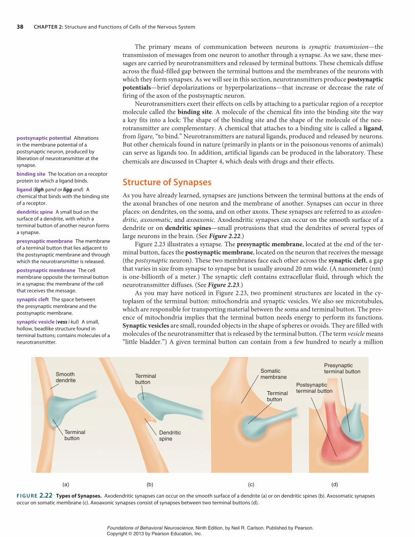
38 CHAPTER 2: Structure and Functions of Cells of the Nervous System
The primary means of communication between neurons is synaptic transmission—thetransmission of messages from one neuron to another through a synapse. As we saw, these mes-sages are carried by neurotransmitters and released by terminal buttons. These chemicals diffuse across the fluid-filled gap between the terminal buttons and the membranes of the neurons with which they form synapses. As we will see in this section, neurotransmitters produce postsynapticpotentials—brief depolarizations or hyperpolarizations—that increase or decrease the rate of firing of the axon of the postsynaptic neuron.
Neurotransmitters exert their effects on cells by attaching to a particular region of a receptor molecule called the binding site. A molecule of the chemical fits into the binding site the way a key fits into a lock: The shape of the binding site and the shape of the molecule of the neu-rotransmitter are complementary. A chemical that attaches to a binding site is called a ligand,from ligare, “to bind.” Neurotransmitters are natural ligands, produced and released by neurons. But other chemicals found in nature (primarily in plants or in the poisonous venoms of animals) can serve as ligands too. In addition, artificial ligands can be produced in the laboratory. Thesechemicals are discussed in Chapter 4, which deals with drugs and their effects.
Structure of Synapses
As you have already learned, synapses are junctions between the terminal buttons at the ends of the axonal branches of one neuron and the membrane of another. Synapses can occur in three places: on dendrites, on the soma, and on other axons. These synapses are referred to as axoden-dritic, axosomatic, and axoaxonic. Axodendritic synapses can occur on the smooth surface of a dendrite or on dendritic spines—small protrusions that stud the dendrites of several types of large neurons in the brain. (See Figure 2.22.)
Figure 2.23 illustrates a synapse. The presynaptic membrane, located at the end of the ter-minal button, faces the postsynaptic membrane, located on the neuron that receives the message (the postsynaptic neuron). These two membranes face each other across the synaptic cleft, a gap that varies in size from synapse to synapse but is usually around 20 nm wide. (A nanometer (nm) is one-billionth of a meter.) The synaptic cleft contains extracellular fluid, through which the neurotransmitter diffuses. (See Figure 2.23.)
As you may have noticed in Figure 2.23, two prominent structures are located in the cy-toplasm of the terminal button: mitochondria and synaptic vesicles. We also see microtubules, which are responsible for transporting material between the soma and terminal button. The pres-ence of mitochondria implies that the terminal button needs energy to perform its functions. Synaptic vesicles are small, rounded objects in the shape of spheres or ovoids. They are filled with molecules of the neurotransmitter that is released by the terminal button. (The term vesicle means “little bladder.”) A given terminal button can contain from a few hundred to nearly a million
postsynaptic potential Alterationsin the membrane potential of a postsynaptic neuron, produced by liberation of neurotransmitter at the synapse.
binding site The location on a receptor protein to which a ligand binds.
ligand (ligh gand or ligg and) Achemical that binds with the binding site of a receptor.
(a) (b) (c) (d)
Terminalbutton
Terminalbutton
Terminalbutton
Smoothdendrite
Dendriticspine
Somaticmembrane
Presynapticterminal button
Postsynapticterminal button
F I G U R E 2.22 Types of Synapses. Axodendritic synapses can occur on the smooth surface of a dendrite (a) or on dendritic spines (b). Axosomatic synapses occur on somatic membrane (c). Axoaxonic synapses consist of synapses between two terminal buttons (d).
dendritic spine A small bud on the surface of a dendrite, with which a terminal button of another neuron forms a synapse.
presynaptic membrane The membrane of a terminal button that lies adjacent to the postsynaptic membrane and through which the neurotransmitter is released.
postsynaptic membrane The cell membrane opposite the terminal button in a synapse; the membrane of the cell that receives the message.
synaptic cleft The space between the presynaptic membrane and the postsynaptic membrane.
synaptic vesicle (vess i kul) A small, hollow, beadlike structure found in terminal buttons; contains molecules of a neurotransmitter.
Foundations of Behavioral Neuroscience, Ninth Edition, by Neil R. Carlson. Published by Pearson. Copyright © 2013 by Pearson Education, Inc.

Communication Between Neurons 39
synaptic vesicles. These vesicles are found in greatest numbers around the part of the presynaptic membrane that faces the synaptic cleft—near the release zone, the region from which the neu-rotransmitter is released. (See Figure 2.23.)
In an electron micrograph the postsynaptic membrane under the terminal button appears somewhat thicker and more dense than the membrane elsewhere. This postsynaptic density is caused by the presence of receptors—specialized protein molecules that detect the presence of neurotransmitters in the synaptic cleft—and protein filaments that hold the receptors in place. (See Figure 2.23.)
Release of Neurotransmitter
When action potentials are conducted down an axon (and down all of its branches), something happens inside all of the terminal buttons: Several synaptic vesicles located just inside the pre-synaptic membrane fuse with the membrane and then break open, spilling their contents into the synaptic cleft.
Heuser and colleagues (Heuser, 1977; Heuser et al., 1979) obtained photomicrographs that illustrate this process. Because the release of neurotransmitter is a very rapid event, tak-ing only a few milliseconds to occur, special procedures are needed to stop the action so that the details can be studied. The experimenters electrically stimulated the nerve attached to an isolated frog muscle and then dropped the muscle against a block of pure copper that had been cooled to 4 K (approximately –453°F). Contact with the supercooled metal froze the outer layer of tissue in 2 msec or less. The ice held the components of the terminal buttons in place until they could be chemically stabilized and examined with an electron microscope. Figure 2.24 shows a portion of the synapse in cross section; note the vesicles that appear to be fused with the presynaptic membrane, forming the shape of an omega (Ω). (See Figure 2.24 and Synapses,in MyPsychLab.)
Activation of Receptors
How do molecules of the neurotransmitter produce a depolarization or hyperpolarization in the postsynaptic membrane? They do so by diffusing across the synaptic cleft and attaching to the binding sites of special protein molecules located in the postsynaptic membrane, called post-synaptic receptors. Once binding occurs, the postsynaptic receptors open neurotransmitter-dependent ion channels, which permit the passage of specific ions into or out of the cell. Thus,
Neuron
Detail of Synapse
Mitochondrion
MicrotubuleSynapticvesicle
Synapticcleft
Synaptic vesiclebeing transportedfrom soma
Terminal button
Presynapticmembrane
Postsynapticmembrane
Postsynapticdensity
Synaptic vesiclelocated at releasezone
F I G U R E 2.23 Details of a Synapse.
release zone A region of the interior of the presynaptic membrane of a synapse to which synaptic vesicles attach and release their neurotransmitter into the synaptic cleft.
postsynaptic receptor A receptor molecule in the postsynaptic membrane of a synapse that contains a binding site for a neurotransmitter.
neurotransmitter-dependent ion channel An ion channel that opens when a molecule of a neurotransmitter binds with a postsynaptic receptor.
Simulate Synapses in MyPsychLab
Foundations of Behavioral Neuroscience, Ninth Edition, by Neil R. Carlson. Published by Pearson. Copyright © 2013 by Pearson Education, Inc.

40 CHAPTER 2: Structure and Functions of Cells of the Nervous System
the presence of the neurotransmitter in the synaptic cleft allows particular ions to pass through the membrane, changing the local membrane potential.
Neurotransmitters open ion channels by at least two different methods, direct and indirect. The direct method is simpler, so I will describe it first. Figure 2.25 illustrates a neurotransmitter-dependent ion channel that is equipped with its own binding site. When a molecule of the appropriate neurotransmitter attaches to it, the ion channel opens. The formal name for this combination receptor/ion channel is an ionotropic receptor. (See Figure 2.25.)
Ionotropic receptors were first discovered in the organ that produces electrical current in Torpedo, the electric ray, where they occur in great number. (The electric ray is a fish that gener-ates a powerful electrical current, not some kind of Star Wars weapon.) These receptors, which are sensitive to a neurotransmitter called acetylcholine, contain sodium channels. When these channels are open, sodium ions enter the cell and depolarize the membrane.
The indirect method is more complicated. Some receptors do not open ion channels directly but instead start a chain of chemical events. These receptors are called metabo-tropic receptors because they involve steps that require the cell to expend metabolic energy. Metabotropic receptors are located in close proximity to another protein attached to the membrane—a G protein. When a molecule of the neurotransmitter binds with the receptor, the receptor activates a G protein situated inside the membrane next to the receptor. When activated, the G protein activates an enzyme that stimulates the production of a chemical called a second messenger. (The neurotransmitter is the first messenger.) Molecules of the second messenger travel through the cytoplasm, attach themselves to nearby ion channels, and cause them to open. Compared with postsynaptic potentials produced by ionotropic receptors, those produced by metabotropic receptors take longer to begin and last longer. (See Figure 2.26.)
The first second messenger to be discovered was cyclic AMP, a chemical that is synthesized from ATP. Since then, several other second messengers have been discovered. As you will see in later chapters, second messengers play an important role in both synaptic and nonsynaptic communication. And they can do more than open ion channels. For example, they can travel to the nucleus or other regions of the neuron and initiate biochemical changes that affect the cell’s
Postsynapticmembrane
PresynapticmembraneOmega
figure
Synapticvesicle
F I G U R E 2.24 Release of Neurotransmitter. This photograph from an electron microscope shows a cross section of a synapse of a terminal button with a muscle of a frog. The omega-shaped figures are synaptic vesicles fusing with the presynaptic membranes.
Heuser, J. E., in Society for Neuroscience Symposia, Vol. II, edited by W. M. Cowan and J. A. Ferrendelli. Bethesda, MD: Society for Neuroscience, 1977.
Binding siteof receptor
Molecule ofneurotransmitterattached tobinding site
Ions
Openion channel
Closedion channel
Insideof Cell
F I G U R E 2.25 Ionotropic Receptors. The ion channel opens when a molecule of neurotransmitter attaches to the binding site. For purposes of clarity the drawing is schematic; molecules of neurotransmitter are actually much larger than individual ions.
ionotropic receptor (eye on oh trowpik) A receptor that contains a binding site for a neurotransmitter and an ion channel that opens when a molecule of the neurotransmitter attaches to the binding site.
metabotropic receptor (meh tab oh trowpik) A receptor that contains a binding site for a neurotransmitter; activates an enzyme that begins a series of events that opens an ion channel elsewhere in the cell’s membrane when a molecule of the neurotransmitter attaches to the binding site.
G protein A protein coupled to a metabotropic receptor; conveys messages to other molecules when a ligand binds with and activates the receptor.
second messenger A chemical produced when a G protein activates an enzyme; carries a signal that results in the opening of the ion channel or causes other events to occur in the cell.
Foundations of Behavioral Neuroscience, Ninth Edition, by Neil R. Carlson. Published by Pearson. Copyright © 2013 by Pearson Education, Inc.

Communication Between Neurons 41
functions. They can even turn specific genes on or off, thus initiating or terminat-ing production of particular proteins.
Postsynaptic Potentials
As I mentioned earlier, postsynaptic potentials can be either depolarizing (ex-citatory) or hyperpolarizing (inhibitory). What determines the nature of the postsynaptic potential at a particular synapse is not the neurotransmitter itself. Instead, it is determined by the characteristics of the postsynaptic receptors—in particular, by the particular type of ion channel they open.
As Figure 2.27 shows, three major types of neurotransmitter-dependent ion channels are found in the postsynaptic membrane: sodium (Na+), potassium (K+),and chloride (Cl–). Although the figure depicts only directly activated (ionotropic) ion channels, you should realize that many ion channels are activated indirectly, by metabotropic receptors coupled to G proteins.
The neurotransmitter-dependent sodium channel is the most important source of excitatory postsynaptic potentials. As we saw, sodium–potassium trans-porters keep sodium outside the cell, waiting for the forces of diffusion and elec-trostatic pressure to push it in. Obviously, when sodium channels are opened, the result is a depolarization—an excitatory postsynaptic potential (EPSP). (See Figure 2.27a.)
We also saw that sodium–potassium transporters maintain a small surplus of potassium ions inside the cell. If potassium channels open, some of these cations will follow this gradient and leave the cell. Because K+ is positively charged, its efflux will hyperpolarize the membrane, producing an inhibitory postsynaptic potential (IPSP). (See Figure 2.27b.)
At many synapses inhibitory neurotransmitters open the chloride channels instead of (or in addition to) potassium channels. The effect of opening chloride channels depends on the membrane potential of the neuron. If the membrane is at the resting potential, nothing hap-pens, because (as we saw earlier) the forces of diffusion and electrostatic pressure balance per-fectly for the chloride ion. However, if the membrane potential has already been depolarized by the activity of excitatory synapses located nearby, then the opening of chloride channels will
Molecule ofneurotransmitter
Surface ofmembrane
Metabotropic receptor
Open ionchannel
Molecule of neurotransmitterattached to binding site ofmetabotropic receptor
Closed ionchannel
F I G U R E 2.26 Metabotropic Receptors. When a molecule of neurotransmitter binds with a receptor, a second messenger (represented by black arrows) is produced that opens nearby ion channels.
a b c
+ + + +
– – – –
Molecule of neurotransmitterattached to binding site
Ion channel
Na+
Na+
K+
K+
Cl-
Cl-
Inflow of Cl- causeshyperpolarization(IPSP)
Outflow of K+ causeshyperpolarization(IPSP)
Inflow of Na+ causesdepolarization(EPSP)
Membrane
F I G U R E 2.27 Ionic Movements During Postsynaptic Potentials.
excitatory postsynaptic potential (EPSP) An excitatory depolarization of the postsynaptic membrane of a synapse caused by the liberation of a neurotransmitter by the terminal button.
inhibitory postsynaptic potential (IPSP) An inhibitory hyperpolarization of the postsynaptic membrane of a synapse caused by the liberation of a neurotransmitter by the terminal button.
Foundations of Behavioral Neuroscience, Ninth Edition, by Neil R. Carlson. Published by Pearson. Copyright © 2013 by Pearson Education, Inc.

42 CHAPTER 2: Structure and Functions of Cells of the Nervous System
permit Cl– to enter the cell. The influx of anions will bring the membrane potential back to its normal resting condition. Thus, the opening of chloride channels serves to neutralize EPSPs. (See Figure 2.27c.)
Termination of Postsynaptic Potentials
Postsynaptic potentials are brief depolarizations or hyperpolarizations caused by the activation of postsynaptic receptors with molecules of a neurotransmitter. They are kept brief by two mecha-nisms: reuptake and enzymatic deactivation.
The postsynaptic potentials produced by most neurotransmitters are terminated by reup-take. This process is simply an extremely rapid removal of neurotransmitter from the synaptic cleft by the terminal button. The neurotransmitter does not return in the vesicles that get pinched off the membrane of the terminal button. Instead, the membrane contains special transporter molecules that draw on the cell’s energy reserves to force molecules of the neu-rotransmitter from the synaptic cleft directly into the cytoplasm—just as sodium–potassium transporters move Na+ and K+ across the membrane. When an action potential arrives, the terminal button releases a small amount of neurotransmitter into the synaptic cleft and then takes it back, giving the postsynaptic receptors only a brief exposure to the neurotransmitter. (See Figure 2.28.)
Enzymatic deactivation is accomplished by an enzyme that destroys molecules of the neurotransmitter. Postsynaptic potentials are terminated in this way for acetylcholine (ACh).Transmission at synapses on muscle fibers and at some synapses between neurons in the central nervous system is mediated by ACh. Postsynaptic potentials produced by ACh are short-lived because the postsynaptic membrane at these synapses contains an enzyme called
reuptake The reentry of a neurotransmitter just liberated by a terminal button back through its membrane, thus terminating the postsynaptic potential.
enzymatic deactivation Thedestruction of a neurotransmitter by an enzyme after its release—for example, the destruction of acetylcholine by acetylcholinesterase.
acetylcholine (ACh) (a see tul kohleen) A neurotransmitter found in the brain, spinal cord, and parts of the peripheral nervous system; responsible for muscular contraction.
Molecules of neurotransmitterreturned to terminal button
Transporter
Presynapticmembrane
Postsynapticmembrane
Synapticcleft
Postsynapticreceptor
“Omega figure”–remnants of synapticvesicle that hasreleased itsneurotransmitter
F I G U R E 2.28 Reuptake. Molecules of a neurotransmitter that has been released into the synaptic cleft are transported back into the terminal button.
Foundations of Behavioral Neuroscience, Ninth Edition, by Neil R. Carlson. Published by Pearson. Copyright © 2013 by Pearson Education, Inc.

Communication Between Neurons 43
acetylcholinesterase (AChE). AChE destroys ACh by cleaving it into its constituents: choline and acetate. Because neither of these substances is capable of activating postsynaptic receptors, the postsynaptic potential is terminated once the molecules of ACh are broken apart. AChE is an extremely energetic destroyer of ACh; one molecule of AChE will chop apart more than 5000 molecules of ACh each second.
Effects of Postsynaptic Potentials: Neural Integration
We have seen how neurons are interconnected by means of synapses, how action potentials trig-ger the release of neurotransmitters, and how these chemicals initiate excitatory or inhibitory postsynaptic potentials. Excitatory postsynaptic potentials increase the likelihood that the post-synaptic neuron will fire; inhibitory postsynaptic potentials decrease this likelihood. (Remember, “firing” refers to the occurrence of an action potential.) Thus, the rate at which an axon fires is determined by the relative activity of the excitatory and inhibitory synapses on the soma and den-drites of that cell. If there are no active excitatory synapses or if the activity of inhibitory synapses is particularly high, that rate could be close to zero.
Let’s look at the elements of this process. (Simulate Postsynaptic Potentials in MyPsychLab,which illustrates the effects of synaptic transmission on postsynaptic cells.) The interaction of the effects of excitatory and inhibitory synapses on a particular neuron is called neural integration.(Integration means “to make whole,” in the sense of combining two or more functions.) Figure 2.29 illustrates the effects of excitatory and inhibitory synapses on a postsynaptic neuron. The top panel shows what happens when several excitatory synapses become active. The release of the neurotransmitter produces depolarizing EPSPs in the dendrites of the neuron. These EPSPs (represented in red) are then transmitted down the dendrites and across the soma to the axonhillock located at the base of the axon. If the depolarization is still strong enough when it reaches this point, the axon will fire. (See Figure 2.29a.)
acetylcholinesterase (AChE) (a see tul koh lin ess ter ace) The enzyme that destroys acetylcholine soon after it is liberated by the terminal buttons, thus terminating the postsynaptic potential.
Activity of excitatorysynapses producesEPSPs (red) inpostsynaptic neuron
Axon hillock reachesthreshold of excitation;action potential istriggered in axon
Activity of inhibitorysynapses producesIPSPs (blue) inpostsynaptic neuron
IPSPs counteract EPSPs;action potential is nottriggered in axon
(b)(a)
F I G U R E 2.29 Neural Integration. (a) If several excitatory synapses are active at the same time, the EPSPs they produce (shown in red) summate as they travel toward the axon, and the axon fires. (b) If several inhibitory synapses are active at the same time, the IPSPs they produce (shown in blue) diminish the size of the EPSPs and prevent the axon from firing.
neural integration The process by which inhibitory and excitatory postsynaptic potentials summate and control the rate of firing of a neuron.
Simulate Postsynaptic Potentials in MyPsychLab
Foundations of Behavioral Neuroscience, Ninth Edition, by Neil R. Carlson. Published by Pearson. Copyright © 2013 by Pearson Education, Inc.

44 CHAPTER 2: Structure and Functions of Cells of the Nervous System
Now let’s consider what would happen if, at the same time, inhibitory synapses also be-come active. Inhibitory postsynaptic potentials are hyperpolarizing—they bring the membrane potential away from the threshold of excitation. Thus, they tend to cancel the effects of excitatory postsynaptic potentials. (See Figure 2.29b.)
The rate at which a neuron fires is controlled by the relative activity of the excitatory and inhibitory synapses on its dendrites and soma. If the activity of excitatory synapses goes up, the rate of firing will go up. If the activity of inhibitory synapses goes up, the rate of firing will go down.
Autoreceptors
Postsynaptic receptors detect the presence of a neurotransmitter in the synaptic cleft and initiate excitatory or inhibitory postsynaptic potentials. But the postsynaptic membrane is not the only location of receptors that respond to neurotransmitters. Many neurons also possess receptors that respond to the neurotransmitter that they themselves release, called autoreceptors.
Autoreceptors can be located on the membrane of any part of the cell, but in this discus-sion we will consider those located on the terminal button. In most cases these autoreceptors do not control ion channels. Thus, when stimulated by a molecule of the neurotransmitter, autoreceptors do not produce changes in the membrane potential of the terminal button. In-stead, they regulate internal processes, including the synthesis and release of the neurotrans-mitter. (As you may have guessed, autoreceptors are metabotropic; the control they exert on these processes is accomplished through G proteins and second messengers.) In most cases the effects of autoreceptor activation are inhibitory; that is, the presence of the neurotransmitter in the extracellular fluid in the vicinity of the neuron causes a decrease in the rate of synthesis or release of the neurotransmitter. Most investigators believe that autoreceptors are part of a regulatory system that controls the amount of neurotransmitter that is released. If too much is released, the autoreceptors inhibit both production and release; if not enough is released, the rates of production and release go up.
Axoaxonic Synapses
As we saw in Figure 2.22, the central nervous system contains three types of synapses. Activity of the first two types, axodendritic and axosomatic synapses, causes postsynaptic excitation or inhibition. The third type, axoaxonic synapses, does not contribute directly to neural integration. Instead, the activity of these synapses alters the amount of neurotrans-mitter released by the terminal buttons of the postsynaptic axon. They can produce presynaptic modulation, presynaptic inhibition, or presyn-aptic facilitation.
As you know, the release of a neurotransmitter by a terminal but-ton is initiated by an action potential. Normally, a particular terminal button releases a fixed amount of neurotransmitter each time an ac-tion potential arrives. However, the release of neurotransmitter can be modulated by the activity of axoaxonic synapses. If the activity of the axoaxonic synapse decreases the release of the neurotransmitter, the effect is called presynaptic inhibition. If it increases the release, it is called presynaptic facilitation. (See Figure 2.30.) By the way, as we will see in Chapter 4, the active ingredient in marijuana exerts its effects on the brain by binding with presynaptic receptors.
Nonsynaptic Chemical Communication
Neurotransmitters are released by terminal buttons of neurons and bind with receptors in the membrane of another cell located a very short
presynaptic inhibition The action of a presynaptic terminal button in an axoaxonic synapse; reduces the amount of neurotransmitter released by the postsynaptic terminal button.
presynaptic facilitation The action of a presynaptic terminal button in an axoaxonic synapse; increases the amount of neurotransmitter released by the postsynaptic terminal button.
Terminalbutton B
Terminalbutton A
Axoaxonicsynapse
Postsynapticdensity
Axodendriticsynapse
Dendriticspine
F I G U R E 2.30 An Axoaxonic Synapse. The activity of terminal button A can increase or decrease the amount of neurotransmitter released by terminal button B.
autoreceptor A receptor molecule located on a neuron that responds to the neurotransmitter released by that neuron.
Foundations of Behavioral Neuroscience, Ninth Edition, by Neil R. Carlson. Published by Pearson. Copyright © 2013 by Pearson Education, Inc.
Communication Between Neurons 45
neuromodulator A naturally secreted substance that acts like a neurotransmitter except that it is not restricted to the synaptic cleft but diffuses through the extracellular fluid.
peptide A chain of amino acids joined together by peptide bonds. Most neuromodulators, and some hormones, consist of peptide molecules.
hormone A chemical substance that is released by an endocrine gland and that has effects on target cells in other organs.
endocrine gland A gland that liberates its secretions into the extracellular fluid around capillaries and hence into the bloodstream.
target cell The type of cell that contains receptors for a particular hormone and is affected by that hormone.
Synapses consist of junctions between the terminal buttons of one neu-ron and the membrane of another neuron, a muscle cell, or a gland cell. When an action potential is transmitted down an axon, the terminal buttons at the end release a neurotransmitter, a chemical that produces either depolarizations (EPSPs) or hyperpolarizations (IPSPs) of the post-synaptic membrane. The rate of firing of the postsynaptic neuron’s axon is determined by the relative activity of the excitatory and inhibitory synapses on the membrane of its dendrites and soma—a phenomenon known as neural integration.
Terminal buttons contain synaptic vesicles, filled with molecules of the neurotransmitter. When an action potential reaches a terminal but-ton, it causes the release of the neurotransmitter: Some of the synaptic vesicles fuse with the presynaptic membrane of the terminal button, break open, and release their contents into the synaptic cleft.
The activation of postsynaptic receptors by molecules of a neu-rotransmitter causes neurotransmitter-dependent ion channels to open, resulting in postsynaptic potentials. Ionotropic receptors contain ion channels, which are directly opened when a ligand attaches to the bind-ing site. Metabotropic receptors are linked to G proteins, which, when activated, open ion channels—usually by producing a chemical called a second messenger.
The nature of the postsynaptic potential depends on the type of ion channel that is opened by the postsynaptic receptors at a particular synapse. Excitatory postsynaptic potentials occur when Na+ enters the cell. Inhibitory postsynaptic potentials are produced when K+ leaves the cell or Cl– enters it.
Postsynaptic potentials are normally brief. They are terminated by two means. The most common mechanism is reuptake: retrieval of
molecules of the neurotransmitter from the synaptic cleft by means of transporters located in the presynaptic membrane, which transport the molecules back into the cytoplasm. Acetylcholine is deactivated by the enzyme acetylcholinesterase.
The presynaptic membrane, as well as the postsynaptic membrane, contains receptors that detect the presence of a neurotransmitter. Pre-synaptic receptors, also called autoreceptors, monitor the quantity of neurotransmitter that a neuron releases and, apparently, regulate the amount that is synthesized and released. Axoaxonic synapses produce presynaptic inhibition or presynaptic facilitation, reducing or enhancing the amount of neurotransmitter that is released.
Neuromodulators and hormones, like neurotransmitters, act on cells by attaching to the binding sites of receptors and initiating electrical or chemical changes in these cells. However, whereas the action of neu-rotransmitters is localized, neuromodulators and hormones have much more widespread effects.
Thought Questions1. Why does synaptic transmission involve the release of chemicals?
Direct electrical coupling of neurons is far simpler, so why do our neurons not use it more extensively? (A tiny percentage of synaptic connections in the human brain do use electrical coupling.) Nor-mally, nature uses the simplest means possible to a given end, so there must be some advantages to chemical transmission. What do you think they are?
2. Consider the control of the withdrawal reflex illustrated in Figure 2.11. Could you design a circuit using electrical synapses that would accomplish the same tasks?
SECTION SUMMARY
Communication Between Neurons
distance away. The communication at each synapse is private. Neuromodulators are chemicals released by neurons that travel farther and are dispersed more widely than are neurotransmitters. Most neuromodulators are peptides, chains of amino acids that are linked together by chemical attachments called peptide bonds (hence their name). Neuromodulators are secreted in larger amounts and diffuse for longer distances, modulating the activity of many neurons in a particular part of the brain. For example, neuromodulators affect general behavioral states such as vigilance, fearfulness, and sensitivity to pain. Chapter 4 discusses the most important neurotransmitters and neuromodulators.
Hormones are secreted by cells of endocrine glands (from the Greek endo-, “within,” and krinein, “to secrete”) or by cells located in various organs, such as the stomach, the intestines, the kidneys, and the brain. Cells that secrete hormones release these chemicals into the extracellular fluid. The hormones are then distributed to the rest of the body through the bloodstream. Hor-mones affect the activity of cells (including neurons) that contain specialized receptors located either on the surface of their membrane or deep within their nuclei. Cells that contain receptors for a particular hormone are referred to as target cells for that hormone; only these cells respond to its presence. Many neurons contain hormone receptors, and hormones are able to affect be-havior by stimulating the receptors and changing the activity of these neurons. For example, a sex hormone, testosterone, increases the aggressiveness of most male mammals.
Foundations of Behavioral Neuroscience, Ninth Edition, by Neil R. Carlson. Published by Pearson. Copyright © 2013 by Pearson Education, Inc.

“Am I cured?” asked Kathryn.Dr. T. smiled ruefully. “I wish it were so simple!” she said. “No, I’m
afraid you aren’t cured, but now we know what is causing your weak-ness. There is a treatment,” she hastened to add, seeing Kathryn’s disappointment. “You have a condition called myasthenia gravis.The injection I gave you lasts only for a few minutes, but I can give you some pills that have effects that last much longer.” Indeed, as she was talking, Kathryn felt herself weakening, and she sat down again.
Myasthenia gravis (MG) was first described in 1672 by Thomas Willis, an English physician. The term literally means “grave mus-cle weakness.” It is not a very common disorder, but most experts believe that many cases—much milder than Kathryn’s, of course—go undiagnosed. Kathryn’s disease involved her face, neck, arm, and trunk muscles, but sometimes only the eye muscles are involved. Before the 1930s, Kathryn would have become bedridden and almost certainly would have died within a few years, probably of pneumonia resulting from difficulty in breathing and coughing. Fortunately, Kathryn’s future is not so bleak. The cause of myasthe-nia gravis is well understood, and it can be treated, if not cured.
The hallmark of myasthenia gravis is fatigability. That is, a patient has reasonable strength when rested but becomes very weak after moving for a little while. For many years, researchers have realized that the weakness occurs in the synapses on the muscles, not in the nervous system or the muscles themselves. In the late nineteenth century, a physician placed electrodes on the skin of a person with myasthenia gravis and electrically stimulated a nerve leading to a muscle. The muscle contracted each time he stimulated the nerve, but the contractions became progressively weaker. However, when he placed the electrodes above the muscle and stimulated it directly, the contractions showed no signs of fatigue. Later, with the develop-ment of techniques of electrical recording, researchers found that the action potentials in the nerves of people with myasthenia gravis were completely normal. If nerve conduction and muscular contrac-tion were normal, then the problem had to lie in the synapses.
In 1934, Dr. Mary Walker remarked that the symptoms of myas-thenia gravis resembled the effects of curare, a poison that blocks neural transmission at the synapses on muscles. The antidote for curare poisoning was a drug called physostigmine, which deacti-vates acetylcholinesterase (AChE). As you learned in this chapter, AChE is an enzyme that destroys the neurotransmitter acetylcho-line (ACh) and terminates the postsynaptic potentials it produces. By deactivating AChE, physostigmine greatly increases and pro-longs the effects of ACh on the postsynaptic membrane. Thus, it increases the strength of synaptic transmission at the synapses on muscles and reverses the effects of curare. (Chapter 4 will say more about both curare and physostigmine.)
Dr. Walker reasoned that if physostigmine reversed the effects of curare poisoning, perhaps it would also reverse the symptoms
EPILOGUE | Myasthenia Gravis
of myasthenia gravis. She tried it, and it did within a matter of a few minutes. Subsequently, pharmaceutical companies discovered drugs that could be taken orally and that produced longer-lasting effects. Nowadays, an injectable drug is used to make the diagnosis and an oral drug is used to treat it.
Researchers turned their efforts to understanding the cause of myasthenia gravis. They discovered that MG is an autoimmune dis-ease. Normally, the immune system protects us from infections by being alert for proteins that are present on invading microorgan-isms. The immune system produces antibodies that attack these foreign proteins, and the microorganisms are killed. However, sometimes the immune system makes a mistake and becomes sensitized against one of the proteins normally present in our bod-ies. As researchers have found, the blood of patients with MG con-tains antibodies against the protein that makes up acetylcholine receptors. Thus, myasthenia gravis is an autoimmune disease in which the immune system attacks and destroys many of the per-son’s ACh receptors, which are necessary for synaptic transmission.
Recently, researchers have succeeded in developing an animal model of MG. An animal model is a disease that can be produced in laboratory animals and that closely resembles a human disease. The course of the disease can then be studied, and possible treat-ments or cures can be tested. In this case, the disease is produced by extracting ACh-receptor protein from electric rays (Torpedo)and injecting it into laboratory animals. The animals’ immune systems become sensitized to the protein and develop antibod-ies that attack their own ACh receptors. The animals exhibit the same muscular fatigability shown by people with MG, and they become stronger after receiving an injection of a drug such as physostigmine.
One promising result that has emerged from studies with the animal model of MG is the finding that an animal’s immune system can be desensitized so that it will not produce antibodies that destroy ACh receptors. If ACh-receptor proteins are modified and then injected into laboratory animals, their immune systems develop an antibody against the altered protein. This antibody does not attack the animals’ own ACh receptors. Later, if they are given the pure ACh-receptor protein, they do not develop MG. Apparently, the pure protein is so similar to the one to which the animals were previously sensitized that the immune system does not bother to produce another antibody. Perhaps a vaccine can be developed to arrest MG in its early stages by inducing the person’s immune system to produce the harmless antibody rather than the one that attacks acetylcholine receptors.
Even with the drugs that are available to physicians today, myas-thenia gravis remains a serious disease. The drugs do not restore a person’s strength to normal, and they can have serious side effects. But the progress made in the laboratory in recent years gives us hope for a brighter future for people like Kathryn.
46
Foundations of Behavioral Neuroscience, Ninth Edition, by Neil R. Carlson. Published by Pearson. Copyright © 2013 by Pearson Education, Inc.

Key Concepts 47
KEY CONCEPTS
CELLS OF THE NERVOUS SYSTEM
1. Neurons have a soma, dendrites, an axon, and terminal but-tons. Circuits of interconnected neurons are responsible for the functions performed by the nervous system. Neurons are supported by glia and by Schwann cells, which provide myelin sheaths, housekeeping services, and physical support. The blood–brain barrier helps to regulate the chemicals that reach the brain.
COMMUNICATION WITHIN A NEURON
2. The action potential occurs when the membrane potential of an axon reaches the threshold of excitation. Although the ac-tion potential is electrical, it is caused by the flow of sodium and potassium ions through voltage-dependent ion channels in the membrane. Saltatory conduction, which takes place in myelinated axons, is faster and more efficient than conduc-tion in unmyelinated axons.
COMMUNICATION BETWEEN NEURONS
3. Neurons communicate by means of synapses, which enable the presynaptic neuron to produce excitatory or inhibitory effects on the postsynaptic neuron. These effects increase or decrease the rate at which the axon of the postsynaptic neu-ron sends action potentials down to its terminal buttons.
4. When an action potential reaches the end of an axon, it causes some synaptic vesicles to release a neurotransmitter into the synaptic cleft. Molecules of the neurotransmitter attach themselves to receptors in the postsynaptic membrane.
5. When they become activated by molecules of the neu-rotransmitters, postsynaptic receptors produce either ex-citatory or inhibitory postsynaptic potentials by opening neurotransmitter-dependent sodium, potassium, or chloride ion channels.
6. The postsynaptic potential is terminated by the destruction of the neurotransmitter or by its reuptake into the terminal button.
7. Autoreceptors help to regulate the amount of neurotransmit-ter that is released.
8. Axoaxonic synapses consist of junctions between two termi-nal buttons. Release of neurotransmitter by the first terminal button increases or decreases the amount of neurotransmitter released by the second.
9. Neuromodulators and hormones have actions similar to those of neurotransmitters: They bind with and activate re-ceptors on or in their target cells.
EXPLORE the Virtual Brain in
NEURAL CONDUCTION
This module reveals the key components of the neural communication system, as well as the pro-cesses of electrical intra-neural and chemical inter-nerual communication. See membrane potentials, synaptic communication, and neurotransmitters in action in detailed animations.
Foundations of Behavioral Neuroscience, Ninth Edition, by Neil R. Carlson. Published by Pearson. Copyright © 2013 by Pearson Education, Inc.