Embed Size (px)
Citation preview
Chapter 2
Structure and Functions of the Cells of the Nervous System
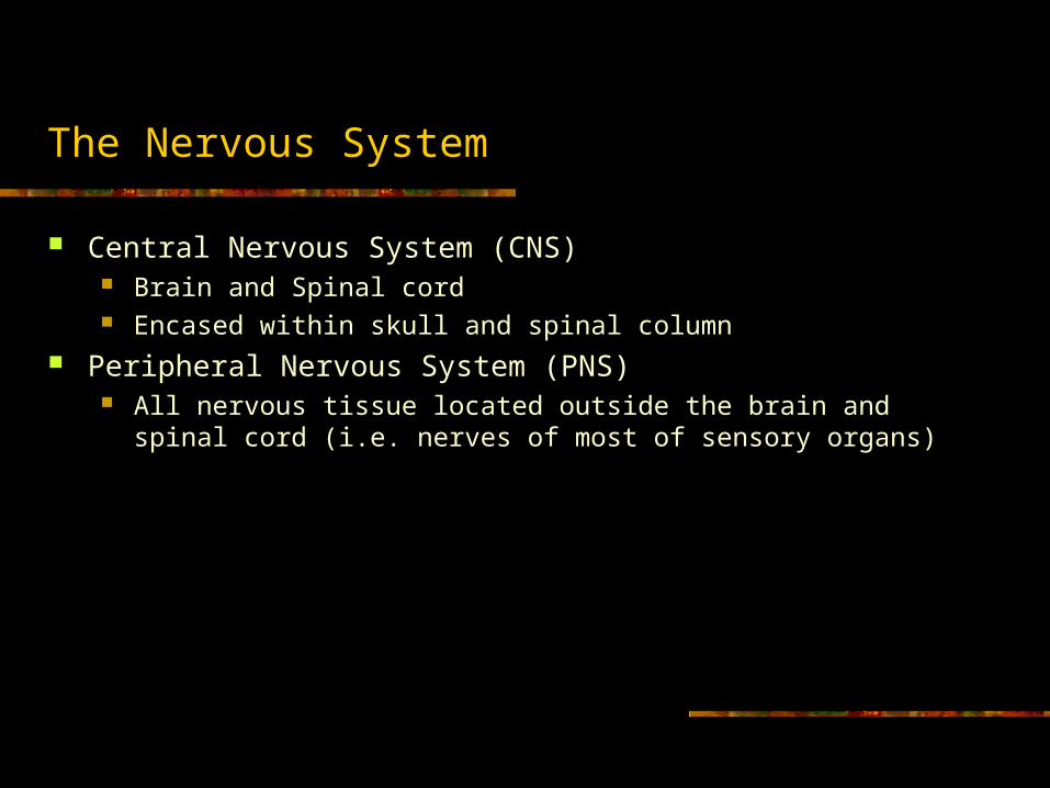
The Nervous System
Central Nervous System (CNS) Brain and Spinal cord Encased within skull and spinal column
Peripheral Nervous System (PNS) All nervous tissue located outside the brain and spinal cord (i.e. nerves
of most of sensory organs)

Types of neurons
Sensory neurons – a neuron that detects changes in the external or internal env’t and sends info about these changes to the CNS
Motor neuron – a neuron located within the CNS that controls the contraction of a muscle or the secretion of a gland
Interneuron – a neuron located entirely within the CNS
brainSpinal cord
Sensory neuron
Motor neuroninterneuron

Cells of the Nervous System: Neurons

Neurons
Neuron types: Multipolar – a neuron with one
axon and many dendrites attached to its soma
Bipolar – a neuron with one axon and one dendrite attached to its soma
Unipolar – a neuron with one axon attached to its soma; the axon divides, with one branch receiving sensory info and the other sending the info to the CNS

Internal structure of the neuron
Membrane – lipid bilayer creates a boundary for the cell’s contents Nucleus – contains nucleolus and chromosomes
Nucleolus – produces ribosomes Ribosomes – a cytoplasmic structure, made of protein, that serves as the site of
production of proteins translated from mRNA Chromosomes – a strand of DNA, with assc. Proteins, found in the nucleus;
carries genetic info Mitochondria – an organelle that is responsible for extracting energy from
nutrients (and thus providing cells with ATP) Endoplasmic reticulum – contains ribosomes (rough) and provides channels
for segregation of molecules involved in cellular processes (smooth); lipid molecules are made here (smooth)
Golgi apparatus – wraps around products of a secretory cell (secretion = exocytosis); also produces lysosomes (breaks down waste products)

Internal structure of the neuron
Cytoskeleton – structural support system of neuron; made of 3 kinds of protein strands (one of these is microtubules)
Microtubule – involved in transporting substances from place to place within cell
Axoplasmic transport – active process by which substances are propelled along microtubules that run the length of the axon
Anterograde – from cell toward terminal buttons Retrograde – from terminal buttons towards cell body


Supporting cells: Glia
Oligodendrocytes Provide support to axons by formation of
myelin sheath Form a non-continuous tube of insulation
along axon Bare, non-myelinated portions called
Nodes of Ranvier In CNS only (Schwann cells form myelin
in PNS) Microglia
Phagocytosis Protect brain from invading
microorganisms Primarily responsible for inflammatory
reaction with brain damage

Supporting cells: Glia
Astrocyte Provide physical support Clean up debris
(phagocytosis) Produce some necessary
compounds Provide nourishment to
neurons

Supporting cells: Glia
Schwann cells Create myelin sheath for axons in PNS Differences from Oligodendrocytes:
With nerve damage, Schwann cells remove dead and dying axons, then help guide regrowth; Oligos don’t aid in regrowth this way
Also, the immune system of individuals with multiple sclerosis attacks only myelin produced by Oligos, not of Schwann cells
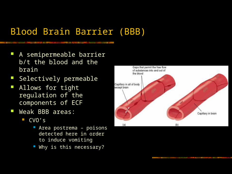
Blood Brain Barrier (BBB)
A semipermeable barrier b/t the blood and the brain
Selectively permeable Allows for tight regulation of
the components of ECF Weak BBB areas:
CVO’s Area postrema – poisons
detected here in order to induce vomiting
Why is this necessary?

Communication within a neuron
Neurons communicate through both chemical and electrical properties Electrical Properties of Axons
By using microelectrodes, we see that the axon is electrically charged: Inside is negatively charged with respect to outside (a difference of 70 mV) Inside membrane of axon charge = -70 mV = membrane potential potential is a stored up source of energy Resting potential – the membrane potential of a neuron when it is not being altered by
excitatory or inhibitory postsynaptic potentials Excitatory vs Inhibitory
Excitatory – causes action potential to happen Inhibitory – inhibits action potential from occurring
Depolarization – reduction (toward zero) of the membrane potential of a cell from normal resting (-70 mV); causes action potential
Hyperpolarization – increase in the membrane potential; occurs after action potential
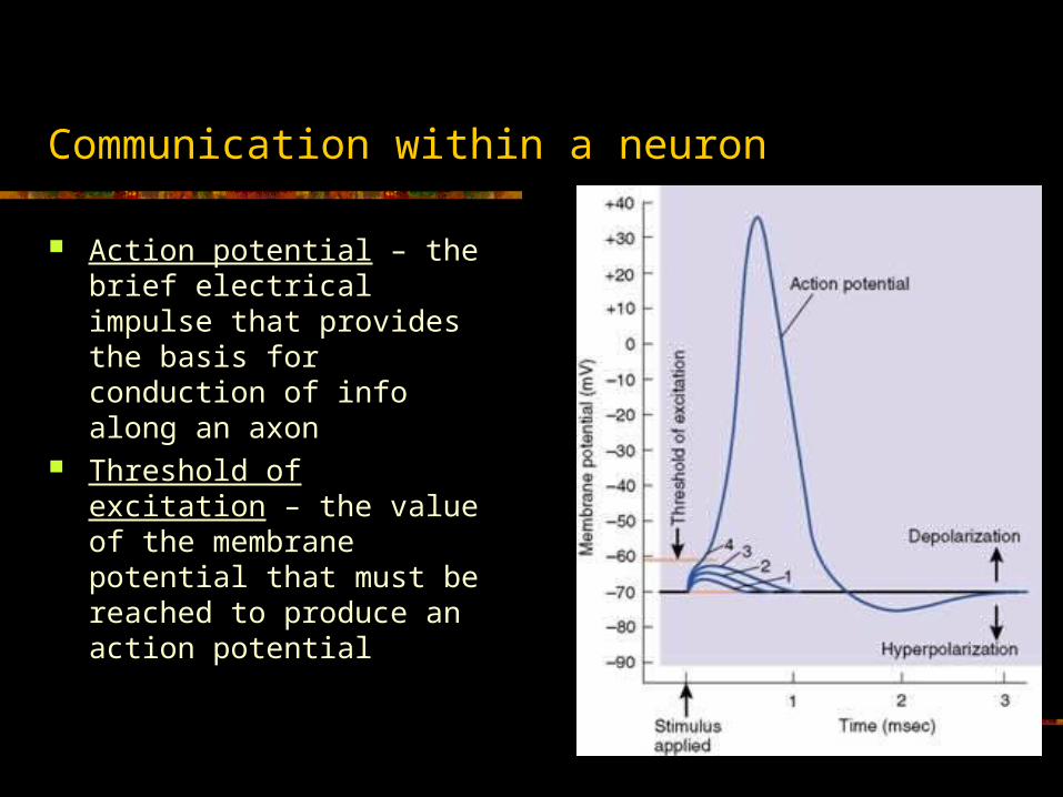
Communication within a neuron
Action potential – the brief electrical impulse that provides the basis for conduction of info along an axon
Threshold of excitation – the value of the membrane potential that must be reached to produce an action potential

Membrane potential
Q: Why is there a membrane potential? A: Result of balance between diffusion and electrostatic pressure Diffusion – movement of molecules from regions of high conc. To
low conc. Substances (electrolytes, i.e. acid, base, or salt) dissolved in water
split into two parts ions (cations and anions) e.g. Na+, K+, Cl-
Electrostatic pressure – the attractive force b/t atomic particles charged with opposite signs or the repulsive force b/t atomic particles charged with the same sign
Na+ K+
Na+ Cl-


Sodium-potassium transporter
A protein found in the membrane of all cells that exchange Na+ for K+ (3 Na+ out, 2 K+ in)
Effectively keep intracellular conc. of Na+ low
Ion channel – a specialized protein molecule that permits specific ions to enter or leave cells

The Action Potential
1. Threshold of excitation is reached, Na+ channels open (voltage dependent), Na+
enters cell 2. K+ channels open, K+ leaves cell (these
open later than Na+ channels)
3. Na+ channels become refractory (i.e. blocked an cannot open again until membrane reaches resting potential), no more Na+ can enter cell
4. K+ keeps leaving cell, causing inside of cell to be positively charged, and return to resting level
5. Resting potential reached (after first overshooting past); K+ channels close, Na+ channels ready again
6. Extra K+ outside diffuses away; axon ready for next action potential!
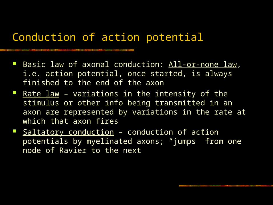
Conduction of action potential
Basic law of axonal conduction: All-or-none law, i.e. action potential, once started, is always finished to the end of the axon
Rate law – variations in the intensity of the stimulus or other info being transmitted in an axon are represented by variations in the rate at which that axon fires
Saltatory conduction – conduction of action potentials by myelinated axons; “jumps” from one node of Ravier to the next

Communication between neurons
Via chemical properties To get info across synapse from presynaptic neuron to postsynaptic neuron:
use of chemical neurotransmission Neurotransmitters produce postsynaptic potentials, either de- or
hyperpolarizations, that affect rate law Neurotransmitters:
Produced in cell Released by terminal buttons Detected by receptors on postsynaptic neuron Also neuromodulators (e.g. peptides) are released, but can travel farther
Hormones, produced by endocrine glands, can affect cell activity also (target cells)
All 3 attach to a receptor molecule called the binding site (lock and key); the chemical that attaches to the binding site is called a ligand
Structure of synapses
3 types: axodendritic, axosomatic, axoaxonic Axodendritic – occur on smooth surface of dendrite or on dendritic
spines Anatomy of synapse:
Presynaptic membrane – synaptic cleft – Postsynaptic membrane In terminal button:
Mitochondria, synaptic vesicles (small or large sacs that contain neurotransmitter), cisternae
Synaptic vesicle production: Small – in Golgi apparatus in soma or in cisternae Large – only in soma, transported trough axoplasm to terminal button
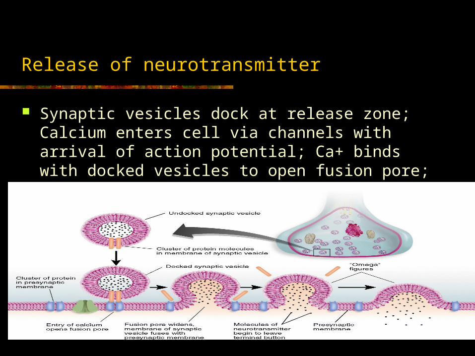
Release of neurotransmitter
Synaptic vesicles dock at release zone; Calcium enters cell via channels with arrival of action potential; Ca+ binds with docked vesicles to open fusion pore; neurotransmitter molecules diffuse from vesicle through fusion pore into synaptic cleft

Activation of receptors
After neurotransmitter release: Cross synaptic cleft to bind to postsynaptic receptors These receptors open neurotransmitter-dependent ion channels, 2 types:
Ionotropic – direct method; contains binding site for neurotransmitter, which when activated, opens an ion channel to allow ions into cell to produce postsynaptic potential (see Fig 2.33 in text); effects do not last long
Metabotropic – indirect method, long-lasting effects; contain neurotransmitter receptors that start a chain of chemical events: (Fig 2.34 in text)
1. Receptor activates G protein (these are called G protein coupled receptors, or GPCRs)
2. α subunit (attached to G protein) breaks away and binds with separate ion channel and opens it (Fig 2.34 a); or attaches to enzyme, which then activates second messenger to open ion channel (Fig 2.34 b)
3. Ions then enter cell to produce postsynaptic potential

Postsynaptic potentials
Action potential is not determined by the neurotransmitter itself, but by the ion channels they open
Ion channel types and effects: Na+ channel: influx causes EPSP K+ channel: efflux (out of cell) causes IPSP Cl- channel: influx causes IPSP Ca2+ channel: influx activates enzyme which has effects on
postsynaptic neuron Buildup of EPSP creates action potential (depolarization) Buildup of IPSP inhibits action potential (hyperpolarization)

Termination of postsynaptic potentials
Almost all NT are terminated by reuptake (transporter protein that moves NT molecules back into presynaptic cell)
Also, by enzymatic deactivation, where an enzyme will break down the NT molecules
e.g. ACh, muscle contractions, broken down by acetylcholinesterase (AChE)

HW for next time
Phew, that was alot! For next class, read Ch 3, and start studying for Quiz 1