Embed Size (px)
Citation preview

HAL Id: cea-01510273https://hal-cea.archives-ouvertes.fr/cea-01510273
Submitted on 19 Apr 2017
HAL is a multi-disciplinary open accessarchive for the deposit and dissemination of sci-entific research documents, whether they are pub-lished or not. The documents may come fromteaching and research institutions in France orabroad, or from public or private research centers.
L’archive ouverte pluridisciplinaire HAL, estdestinée au dépôt et à la diffusion de documentsscientifiques de niveau recherche, publiés ou non,émanant des établissements d’enseignement et derecherche français ou étrangers, des laboratoirespublics ou privés.
Distributed under a Creative Commons Attribution| 4.0 International License
Structure and Function of Adsorbed Hemoglobin onSilica Nanoparticles: Relationship between the
Adsorption Process and the Oxygen Binding PropertiesStéphanie Devineau, Loussiné Zargarian, Jean-Philippe Renault, Serge Pin
To cite this version:Stéphanie Devineau, Loussiné Zargarian, Jean-Philippe Renault, Serge Pin. Structure and Functionof Adsorbed Hemoglobin on Silica Nanoparticles: Relationship between the Adsorption Process andthe Oxygen Binding Properties. Langmuir, American Chemical Society, 2017, 33, pp.3241 - 3252.�10.1021/acs.langmuir.6b04281�. �cea-01510273�

Structure and function of adsorbed hemoglobin on
silica nanoparticles: relationship between the
adsorption process and the oxygen binding
properties
Stéphanie Devineau a,#,*, Loussiné Zargarian b, Jean Philippe Renault a, Serge Pin a
a LIONS, NIMBE, CEA, CNRS, Université Paris-Saclay, CEA Saclay, 91191 Gif-sur-Yvette,
France
b LBPA, ENS de Cachan, CNRS, Université Paris-Saclay, 94235 Cachan Cedex, France
KEYWORDS: Adsorption; porcine hemoglobin; silica nanoparticles; protein structure;
oxygenation; cooperativity.
ABSTRACT. The connection between the mechanisms of protein adsorption on nanoparticles
and the structural and functional properties of the adsorbed protein often remains unclear. We
investigate porcine hemoglobin adsorption on silica nanoparticles and we analyze the structural
and functional modifications of adsorbed hemoglobin by UV-vis spectrophotometry, circular
dichroism and oxygen binding measurement. The structural analysis of adsorbed hemoglobin on
silica nanoparticles reveals a significant loss of secondary structure and a preservation of the
heme electronic structure. However, adsorbed hemoglobin retains its quaternary structure and
exhibits an enhanced oxygen affinity with cooperative binding. Moreover, the structural and
functional modifications are fully reversible after complete desorption from silica nanoparticles
at pH 8.7. The tunable adsorption and desorption of hemoglobin on SNPs with pH change, and

the full control of hemoglobin activity by pH, temperature and the addition of inorganic
phosphate effectors opens the way to an interesting system whereby protein adsorption on
nanoparticles can allow for full control over hemoglobin oxygen binding activity. Our results
suggest that adsorption of hemoglobin on silica nanoparticles leads to a new structural,
functional and dynamic state with full reversibility in a way that significantly differs from
protein denaturation.
TEXT
1. Introduction
Nanotechnology has the potential to revolutionize numerous aspects of our daily life. Engineered
nanomaterials (NMs) have become technological and economic stakes 1–4 for a wide range of
applications, such as construction, plastic processing, fabrics, and energy 5–7. The biological
applications of NMs are expanding in food industry, cosmetics, pharmacology and medicine,
covering a number of different uses from antimicrobial coating and targeted drug delivery to
material biocompatibility and imaging 8–10. In particular, the rapid growth of nanomedicine has
seen a large set of NMs being investigated to evaluate their potential as drug carriers, vaccine
adjuvants, biocompatible implants or diagnostic probes 11–13. The number of products containing
NMs has been multiplied by five between 2006 and 2011 and over 1800 products containing
NMs have been produced in 2014 14,15.
Simultaneously, the field of nanotoxicology has significantly developed to investigate the risks
and the impacts of NM exposure on the environment and health 16–22. The assessment of NM
toxicity has involved substantial evolutions in the field to deal with the specific interactions of
NMs with biological systems. The understanding of the formation of the protein corona and its
key role as an interface between biological systems and NMs has been an essential step 23–26.
Indeed, the adsorption of biomolecules on the surface of NMs in the biological medium leads to
the formation of a complex and dynamic corona, giving NMs their ‘biological identity’ 27,28. The
protein corona determines the NM uptake and its biological outcome 29–31. Moreover, the control
of the conformation and the activity of the adsorbed proteins is necessary to achieve good
material biocompatibility 32,33. A better understanding of the molecular interactions between
NMs, defined by their physical and chemical properties, and endogenous proteins likely to
adsorb on the surface is thus essential to study and predict their biological effects and to design
NMs with excellent biocompatibility 34,35.
It has been known for a long time that protein adsorption on flat surfaces can result in a
modification of the protein structure and activity 36,37. Similarly, protein adsorption on NMs can
impair protein structure and function 38,39. The control of the protein functional state is
particularly important when designing hemoglobin-loaded nanoparticles as blood substitutes 40.
However, the relationship between the NM properties and the adsorbed protein structure,
dynamics and activity is still poorly understood. Based on the adsorption of model proteins on
hydrophobic and hydrophilic particles, Norde and co-workers developed a model to predict
protein adsorption as a function of protein charge and structural stability 41,42. According to this
model, adsorption on a hydrophilic surface is driven by electrostatic forces and the structural
modifications of the adsorbed proteins. Structural rearrangements would significantly contribute
to the adsorption of ‘soft proteins’ with a lower internal stability compared to ‘hard proteins’
with a strong internal stability 36,37. The main interactions and driving forces of protein
adsorption were described by Czeslik 43. More recently, we have developed a strategy to
determine the physico-chemical determinants of protein adsorption based on the identification of
the adsorbed and non-adsorbed proteins in a mixture of hundreds of proteins, and on the

statistical comparison of the structural features of each group 44. This approach revealed the
molecular bases of protein adsorption on silica nanoparticles (SNPs), that is an enrichment in
basic residues (particularly Arg) and a low content in aromatic residues (Phe, Trp, Tyr, and His).
The protein adsorption is thus correlated to the protein flexibility and ability to spread on the
surface.
Protein adsorption on nanoparticles is often associated to a partial loss of secondary structure
36,38,45–47, even if helix formation has also been observed in some cases 48. The structural
modifications depend on the size, shape and surface chemistry of the NMs 38,45. Moreover,
protein adsorption can be used to promote enzyme stability in bioreactors and biosensors for
industrial applications 49–51. The enzymatic activity can be decreased, increased or can remain
constant after adsorption 36,39,45,52,53, highlighting the complex relationship between the structural
and the functional modifications of adsorbed proteins. In Norde’s model, structural
rearrangement contributes to protein adsorption by leading to an increase of the degree of
freedom of the polypeptide chain and finally to an increase in protein flexibility 42. In this model,
the protein structure loss would lead to a favourable gain in entropy 43. However, few
experimental studies have been performed to investigate protein dynamics on surfaces despite its
importance in biological processes. Recent studies rather suggest that protein adsorption can
result in a decrease of the adsorbed protein dynamics 54,55. Using elastic and inelastic neutron
scattering experiments, we showed that myoglobin adsorption on SNPs leads to a decrease of
protein flexibility and to a depletion in low frequency modes, suggesting that the structural loss
of the adsorbed protein may not be the entropic driving force of adsorption 47.
The aim of this study is to investigate the modifications of hemoglobin structure and activity
after adsorption on SNPs in connection with our results on the dynamics of adsorbed myoglobin

47. SNPs are produced and used at an industrial scale, especially in food products. In this study,
we used SNPs as a model hydrophilic inorganic surface 56. Hemoglobin and myoglobin belong to
the same family of hemoproteins. Hemoglobin has several advantages as a model protein: its
biochemical and structural properties are well characterized; and its function of oxygen binding
is particularly well suited to study its activity after adsorption. Indeed, it does not involve any
substrate or cofactor that may interact with the SNPs during the reaction, contrary to most
enzymatic reactions, preventing any interference during the experimental measurement of the
protein activity 30,57. We investigated the mechanisms of hemoglobin adsorption on SNPs and we
analysed the effect of the structural rearrangement on the surface on the activity of hemoglobin.
Our results suggest that adsorption leads to a new state characterized by specific structural and
functional features with full reversibility, in a way that significantly differs from protein
denaturation.
2. Methods
Hemoglobin. Porcine hemoglobin (Sus scrofa domesticus) was purified in the oxygenated form
(HbO2) from fresh blood following standard preparation 58 using erythrocyte membrane
precipitation in 0.28 M Phosphate pH 7.0. HbO2 solution was extensively dialyzed against pure
water at 4°C, stripped by passing hemoglobin desalted solution through a mixed-bed ion-
exchange resin (AG 501-X8 from Bio-Rad) to remove naturally bound 2,3-diphosphoglycerate
(DPG) 59 and centrifuged at 20,000 g for 10 min. HbO2 concentration expressed as heme molar
concentration was measured on a Shimadzu UV-2450 spectrophotometer taking ε576 nm = 15,150
M-1.cm-1 60. Possible iron oxidation and potential protein damages were systematically screened
by measuring the absorbance ratio at 576 nm and 541 nm.
Chemicals. BisTris-HCl, Tris-HCl and Phosphate buffers were prepared at a concentration of 0.1
M by dissolution of BisTris (Sigma-Aldrich B9754), Tris (Sigma-Aldrich T6066), monosodium
phosphate (Sigma-Aldrich 71649) and disodium phosphate (Fisher Scientific S3720). 2,3-
diphosphoglycerate (DPG) (Sigma-Aldrich D5764) and inositol hexaphosphate (IHP) (Sigma-
Aldrich P0109) were dissolved in 0.1 M BisTris-HCl pH 6.0. To avoid DPG hydrolysis, a fresh
solution was prepared before each oxygen binding experiment.
Silica nanoparticles. As explained in the introduction, SNPs (Sigma-Aldrich 637238) come from
the same batch as in our previous studies 44 and were extensively characterized. Their mean
diameter is 26 ± 2 nm and their surface area is 170 m²/g. SNPs were suspended in buffered
solutions and vortexed for 1 min. Small aggregates of 50 ± 2 nm formed in solution.
ζ-potential. Zeta potential (ζ) of SNPs was measured at 25°C on a Zetasizer Nano-ZS (Malvern
Instruments) in 0.1 M BisTris-HCl pH 6.0, Phosphate pH 7.4 and Tris-HCl pH 8.7. SNP
concentration is 0.1 mg/mL. Each measurement was repeated 5 times. The ζ-potential was
calculated by fitting the electrophoretic mobility with the Smoluchowski model.
Adsorption isotherms and protein desorption. Adsorption isotherms of HbO2 on SNPs were
measured by the depletion method 47. HbO2 solution was added to SNP suspension with a final
SNP concentration of 4.5 mg/mL and smoothly mixed overnight at 20°C. The sample was then
centrifuged at 20,000 g for 10 min. HbO2 concentration in the supernatant was measured by
spectrophotometry. The amount of adsorbed protein is expressed as mg/m2. An uncertainty of
5% on the amount of adsorbed protein was determined by repeating 5 times this procedure.
Protein desorption was measured after incubation of 50 µM HbO2 with SNPs at a concentration
of 34 mg/mL overnight. The solution was centrifuged at 20,000 g for 10 min and the supernatant
removed. The SNPs were resuspended in the leaching solution by smooth mixing at 20°C, before
centrifugation at 20,000 g for 10 min and measurement of HbO2 concentration in the supernatant.
To avoid harsh pH change when adding the leaching solution, an additional experiment was
performed by dialysis in 0.1 M Tris-HCl buffer at pH 8.7 at 4°C overnight using a Spectra/Por
membrane with a 50 kDa cut-off. All the measurements were done in triplicate.
Structural analysis. UV-visible spectra were recorded in 1-cm path length quartz cell from 250
to 800 nm at 25°C on a Shimadzu UV-2450 spectrophotometer equipped with an integrating
sphere module. Circular dichroism spectra were recorded from 190 to 260 nm in 1-mm path
length cell at 25°C on a Jobin-Yvon CD6 dichrograph. To minimize UV absorption by the
buffers, HbO2 spectra were recorded in 0.01 M BisTris-HCl, Tris-HCl and Phosphate. Final
HbO2 concentration was 10 µM and SNP concentration was 6.8 mg/mL. Each spectrum is the
average of 4 measurements after baseline correction. The CD spectra of the solutions containing
HbO2 and SNPs were baseline corrected with the SNP solution. However, no signal was
recorded for SNP solutions in these conditions. To control that no particle deposition occurred
during the measurement, the absorbance at 280 nm was recorded before and after the acquisition
of the CD spectrum. No evolution was observed. The alpha-helical content fα was first
determined from the ellipticity value at 222 nm according to eq. 1 61.
𝑓𝛼 = 𝛥𝜀(222 𝑛𝑚) ∙2340
30300 (eq. 1)
The circular dichroism spectra were then fully deconvoluted using CDNN software (Applied
Physics) 62. All the CD analyses were done in triplicate and a single representative spectrum
corresponding to one sample scanned 4 times is shown.
Oxygen binding curves. HbO2 at a concentration of 50 µM was smoothly mixed overnight with
SNPs at a concentration of 34 mg/mL in 0.1 M BisTris-HCl, Tris-HCl or Phosphate. Oxygen
binding was measured in a tonometer by spectrophotometry at 25°C and at 37°C 63. HbO2 was

deoxygenated under an argon flow that bubbled in pure water to prevent drying. Oxygen binding
was recorded by following the absorption at 576 nm. No desorption of hemoglobin from SNPs
was observed during deoxygenation at pH 6.0 and at pH 7.4. All the measurements were done in
triplicate.
3. Results
3.1 Monitoring HbO2 adsorption with pH
The adsorption of hemoglobin on SNPs was quantified by the depletion method where the
concentration of free protein is measured in the supernatant. However, the red color of HbO2
allows for the direct observation of protein adsorption (Fig. 1A). Indeed, two markedly different
behaviors were observed. The red pellet and transparent supernatant indicate strong protein
adsorption in BisTris-HCl at pH 6.0, whereas the white pellet and red supernatant indicate little
or no adsorption in Tris-HCl at pH 8.7. This observation was further confirmed by the
measurement of the adsorption isotherms in BisTris-HCl pH 6.0, in Phosphate pH 7.4 and in
Tris-HCl pH 8.7 (Fig. 1B). The maximum amount of adsorbed HbO2, mads, is high at pH 6.0
(mads = 0.41 ± 0.02 mg/m2) and at pH 7.4 (mads = 0.34 ± 0.02 mg/m2), and null at pH 8.7.

Figure 1. Adsorption behavior of HbO2 on SNPs. A) Images, after centrifugation, of HbO2
solution mixed with SNPs in 0.1 M BisTris-HCl pH 6.0 or Tris-HCl pH 8.7; B) Adsorption
isotherms of HbO2 on SNPs in 0.1 M BisTris-HCl pH 6.0 (circle), Phosphate pH 7.4 (square) and
Tris-HCl pH 8.7 (triangle). Fitting by the Langmuir model is represented by solid lines.
The adsorption constant Kads which reflects the affinity of the protein for the surface was
calculated by fitting the adsorption isotherms with the Langmuir model 64 according to eq. 2
(Fig. 1B).
𝑚𝑎𝑑𝑠 =𝑚∞∗𝐾𝑎𝑑𝑠∗𝐶
1+𝐾𝑎𝑑𝑠∗𝐶 (eq. 2)
Where C is the protein concentration at equilibrium and m∞ the maximum amount of adsorbed
protein. The Langmuir equation is valid for the reversible adsorption of a monolayer of non-
interacting molecules on a surface with identical binding sites. Since protein adsorption often
results in irreversible binding to the surface, the validity of the Langmuir model to depict protein

adsorption has been discussed in the literature 65–67. In the case of hemoglobin, the plateau value
clearly indicates monolayer adsorption on the surface (see §3.5).
The very different adsorption behavior at pH 6.0 and at pH 8.7 suggests a strong effect of
charges on the adsorption of HbO2 on SNPs. The surface chemistry of amorphous SNPs in
aqueous solution is characterized by the hydroxylation of the surface leading to a surface
coverage by silanol groups (Si-OH) of 4.6-4.9 OH/nm2 68. The extent of ionization of the silanol
groups in siloxide (Si-O-) that gives its negative charge to SNPs at neutral pH depends on the
size of the particles and on the ionic strength 69,70. The surface charge density increases when
SNP size decreases and when the ionic strength increases. In our study, the negative charge of
SNPs in the different buffers was confirmed by measuring the ζ-potential of the particles
highlighting the increase in surface charge density with pH (Table S1).
The adsorption behavior of hemoglobin on SNPs as a function of pH was further investigated by
measuring the adsorption isotherms from pH 6.0 to 7.8 in BisTris-HCl, from pH 7.0 to 7.8 in
Phosphate and from pH 7.2 to 8.7 in Tris-HCl (Fig. 2A). The porcine HbO2 tetramer is
structurally similar to that of human oxyhemoglobin 71 and its global isoelectric point is around
7.2 60. The amount of adsorbed HbO2 represented for each condition in Fig. 2B reveals a
maximum level of adsorption from pH 6.2 to pH 7.3 both in BisTris-HCl and Tris-HCl, followed
by a sharp decrease from pH 7.3 to pH 8.5 until zero adsorption. We can notice that for pH > 7.2,
the adsorption isotherms do not reach a plateau in this concentration range. Thus, the maximum
amount of adsorbed HbO2 indicated in Fig. 2B corresponds to the value of the last point of the
adsorption isotherm represented in Fig. 2A and may be underestimated. Alternative models may
represent better fit of the adsorption isotherms in these conditions, as suggested by Findenegg
and coworkers 72. No buffer effect was observed for BisTris-HCl and Tris-HCl although a little
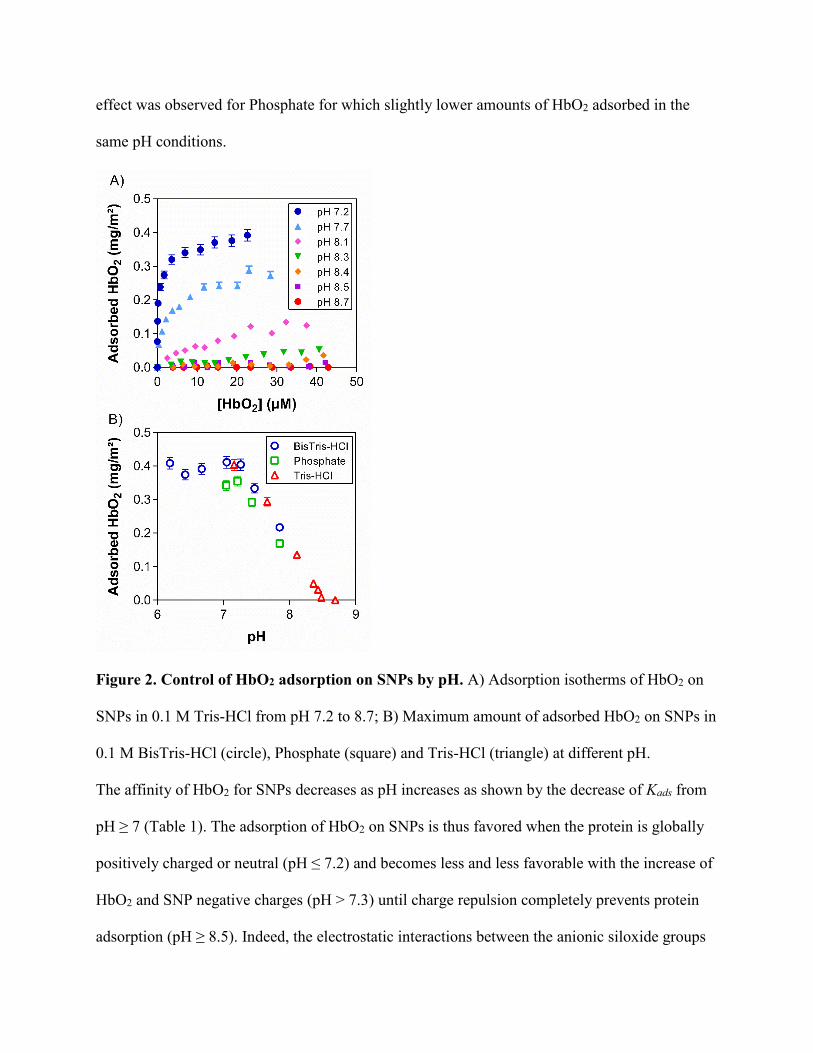
effect was observed for Phosphate for which slightly lower amounts of HbO2 adsorbed in the
same pH conditions.
Figure 2. Control of HbO2 adsorption on SNPs by pH. A) Adsorption isotherms of HbO2 on
SNPs in 0.1 M Tris-HCl from pH 7.2 to 8.7; B) Maximum amount of adsorbed HbO2 on SNPs in
0.1 M BisTris-HCl (circle), Phosphate (square) and Tris-HCl (triangle) at different pH.
The affinity of HbO2 for SNPs decreases as pH increases as shown by the decrease of Kads from
pH ≥ 7 (Table 1). The adsorption of HbO2 on SNPs is thus favored when the protein is globally
positively charged or neutral (pH ≤ 7.2) and becomes less and less favorable with the increase of
HbO2 and SNP negative charges (pH > 7.3) until charge repulsion completely prevents protein
adsorption (pH ≥ 8.5). Indeed, the electrostatic interactions between the anionic siloxide groups

and the basic amino acids (Lys, Arg) are known to play a key role in protein adsorption on SNPs
69 especially when the basic residues form positively charged clusters in the protein structure
44,73. Hemoglobin α chains and β chains contain 14 basic residues (11 Lys, 3 Arg) and 16 basic
residues (11 Lys, 5 Arg) respectively. It suggests that both subunits could contribute to the
electrostatic interactions responsible for HbO2 adsorption and non-adsorption to SNPs. At the
same pH, Kads is smaller for Phosphate compared to BisTris-HCl or Tris-HCl, suggesting that ion
pairing and ion replacement also play a role in HbO2 adsorption on silica.
pH BisTris-
HCl Phosphate Tris-HCl
6.4 2.1 106 - -
6.7 2.1 106 - -
7.0 2.0 106 1.0 106 -
7.3 1.7 106 0.7 106 2.0 106
7.5 1.4 106 0.4 106 0.4 106
7.8 0.2 106 0.1 106 -
8.1 - - 0.05 106
Table 1. Adsorption constant Kads (M-1) of HbO2 on SNPs in 0.1 M BisTris-HCl, Phosphate and
Tris-HCl.
We can conclude that HbO2 adsorption on SNPs is primarily driven by pH with a minor effect of
the ions in solution. HbO2 adsorption can be fully controlled in a small pH range, independently
of the total protein concentration in solution, from a maximum protein coverage at pH 7.3 to no
protein adsorption at pH 8.5.
3.2 Structural analysis of adsorbed HbO2
The structure of HbO2 adsorbed on SNPs was investigated by circular dichroism (CD) (Fig. 3)
and by UV-visible spectroscopy (Fig. S1) to analyze the protein secondary structure and the
oxygenated heme electronic structure respectively. A concentration of 10 µM HbO2 and 6.8
mg/mL SNPs were chosen to achieve almost complete protein adsorption. The amount of
adsorbed HbO2 in this condition is 99% at pH 6.0, 90% at pH 7.4 and <1% at pH 8.7 (Table S2).

The spectra measured at pH 6.0 and at pH 7.4 can thus be attributed to the adsorbed protein with
no contribution or little contribution from the free protein in solution at pH 6.0 and at pH 7.4
respectively. As SNPs may interfere with the optical measurement, a control experiment was
systematically performed to compare the spectra of free HbO2 with SNPs and native HbO2
without SNPs, both in Tris-HCl at pH 8.7. Indeed, no adsorption occurs in this condition so that
any change in the protein spectrum would indicate a bias due to the particles in suspension. The
CD spectra of free and native HbO2 measured at pH 8.7 with and without SNPs respectively
(Fig. 3C) are identical showing that no protein structure perturbation arises from SNPs in
suspension.
The CD spectra of adsorbed HbO2 at pH 6.0 (Fig. 3A) and at pH 7.4 (Fig. 3B) showed a decrease
in ׀Δε׀ compared to native HbO2 indicating a modification of the secondary structure after
adsorption on SNPs. The spectrum of native HbO2 is characterized by one maximum at 192 nm
and two minima at 208 nm and 222 nm corresponding to the predominant α-helix secondary
structure. As reported before, the secondary structure of HbO2 is identical at pH 6.0, at pH 7.4
and at pH 8.7 in the different buffers 60. The percentage of α-helix calculated from the ellipticity
at 222 nm 74 decreases from 80% for native HbO2 to 62% and 49% for adsorbed HbO2 at pH 7.4
and at pH 6.0 respectively. The helicity calculated for adsorbed HbO2 at pH 7.4 was corrected
from the contribution of free HbO2 in solution.
Spectral deconvolution by CDNN 62 confirmed the important loss of α-helix structure together
with an increase in random coil up to 17% at pH 6.0. Therefore HbO2 molecule loses a
significant part of its helical secondary structure after adsorption on SNPs with a gain of
disordered regions, similarly to HbO2 adsorption on quantum dots 75 and on silver nanoparticles
76. Though the formation of some β-sheet cannot be excluded, it has also been shown that a

change in the Δε ratio at 222 nm and 208 nm of α-helical proteins can be related to intra- and
inter-molecular helix interactions and coiled-coil formation which could be indicative of a
modification of adsorbed hemoglobin tertiary structure 77.
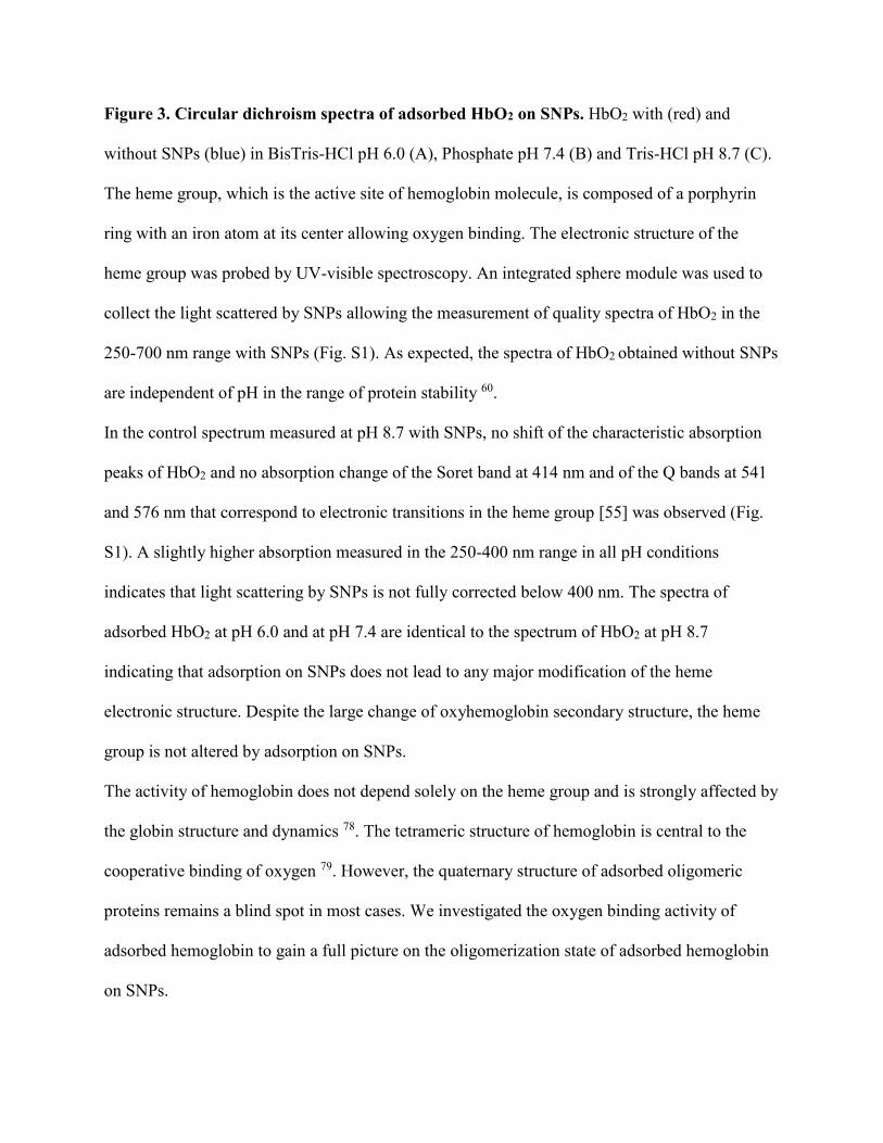
Figure 3. Circular dichroism spectra of adsorbed HbO2 on SNPs. HbO2 with (red) and
without SNPs (blue) in BisTris-HCl pH 6.0 (A), Phosphate pH 7.4 (B) and Tris-HCl pH 8.7 (C).
The heme group, which is the active site of hemoglobin molecule, is composed of a porphyrin
ring with an iron atom at its center allowing oxygen binding. The electronic structure of the
heme group was probed by UV-visible spectroscopy. An integrated sphere module was used to
collect the light scattered by SNPs allowing the measurement of quality spectra of HbO2 in the
250-700 nm range with SNPs (Fig. S1). As expected, the spectra of HbO2 obtained without SNPs
are independent of pH in the range of protein stability 60.
In the control spectrum measured at pH 8.7 with SNPs, no shift of the characteristic absorption
peaks of HbO2 and no absorption change of the Soret band at 414 nm and of the Q bands at 541
and 576 nm that correspond to electronic transitions in the heme group [55] was observed (Fig.
S1). A slightly higher absorption measured in the 250-400 nm range in all pH conditions
indicates that light scattering by SNPs is not fully corrected below 400 nm. The spectra of
adsorbed HbO2 at pH 6.0 and at pH 7.4 are identical to the spectrum of HbO2 at pH 8.7
indicating that adsorption on SNPs does not lead to any major modification of the heme
electronic structure. Despite the large change of oxyhemoglobin secondary structure, the heme
group is not altered by adsorption on SNPs.
The activity of hemoglobin does not depend solely on the heme group and is strongly affected by
the globin structure and dynamics 78. The tetrameric structure of hemoglobin is central to the
cooperative binding of oxygen 79. However, the quaternary structure of adsorbed oligomeric
proteins remains a blind spot in most cases. We investigated the oxygen binding activity of
adsorbed hemoglobin to gain a full picture on the oligomerization state of adsorbed hemoglobin
on SNPs.
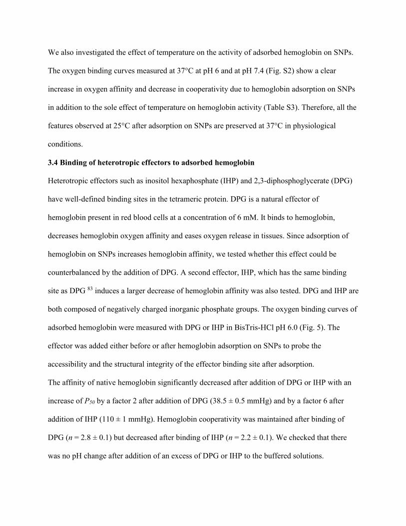
3.3 Functional analysis of adsorbed hemoglobin
Oxygen binding curves of hemoglobin were measured in BisTris-HCl pH 6.0, Phosphate pH 7.4
and Tris-HCl pH 8.7 at 25°C (Fig. 4). Their sigmoidal shape reflects the cooperative binding of
oxygen by tetrameric hemoglobin 60. The variation in hemoglobin affinity for oxygen with pH
reflects the effect of bound or released protons by hemoglobin on its activity known as the Bohr
effect 60,80. Both effects are quantified by the Hill coefficient (n) and the oxygen partial pressure
at half saturation (P50) determined by fitting the experimental curves with the Hill equation (eq.
3), where Y is the fraction of oxyhemoglobin and PO2 the oxygen partial pressure (Table 2).
log𝑌
1−𝑌= n ∙ log 𝑃𝑂2 − log 𝑃50 (eq. 3)
No difference between the oxygen binding curves of hemoglobin at pH 8.7 with and without
SNPs was observed confirming that SNPs did not interfere with oxygen binding or with the
spectroscopic measurement (Fig. 4C). Fig. 4A and Fig. 4B exhibit a more hyperbolic shape of
the oxygen binding curves. This significant increase of the affinity of adsorbed hemoglobin
follows the decrease in cooperativity as shown by the decrease of P50 from 17.1 ± 0.5 to 8.7 ±
0.5 mmHg at pH 6.0 and from 7.7 ± 0.5 to 4.7 ± 0.5 mmHg at pH 7.4. However, adsorbed
hemoglobin still experience cooperative binding as evidenced by the Hill coefficients of 1.6 ±
0.1 and 1.9 ± 0.1 at pH 6.0 and pH 7.4 respectively. This cooperative behavior cancels the
possibility of a dissociation of the tetramer into dimers or monomers during adsorption on SNPs
79,81,82.
pH P50 (mmHg)
- SNPs + SNPs n
- SNPs + SNPs
pH 6.0 17.1 8.7 2.8 1.6
pH 7.4 7.7 4.7 3.0 1.9
pH 8.7 3.2 3.2 3.0 3.0

Table 2. Oxygen partial pressure at half saturation (P50) and Hill coefficient (n) of
hemoglobin oxygen binding in 0.1 M BisTris-HCl pH 6.0, Phosphate pH 7.4 and Tris-HCl pH
8.7 at 25°C with and without SNPs. Values are given with an uncertainty of ± 0.5 mmHg (P50)
and ± 0.1 (n).

Figure 4. Effect of SNPs on hemoglobin activity. Oxygen binding curves of hemoglobin with
(red) and without (blue) SNPs in BisTris-HCl pH 6.0 (A), Phosphate pH 7.4 (B) and Tris-HCl
pH 8.7 (C). Black line represents fitting by the Hill equation.
We also investigated the effect of temperature on the activity of adsorbed hemoglobin on SNPs.
The oxygen binding curves measured at 37°C at pH 6 and at pH 7.4 (Fig. S2) show a clear
increase in oxygen affinity and decrease in cooperativity due to hemoglobin adsorption on SNPs
in addition to the sole effect of temperature on hemoglobin activity (Table S3). Therefore, all the
features observed at 25°C after adsorption on SNPs are preserved at 37°C in physiological
conditions.
3.4 Binding of heterotropic effectors to adsorbed hemoglobin
Heterotropic effectors such as inositol hexaphosphate (IHP) and 2,3-diphosphoglycerate (DPG)
have well-defined binding sites in the tetrameric protein. DPG is a natural effector of
hemoglobin present in red blood cells at a concentration of 6 mM. It binds to hemoglobin,
decreases hemoglobin oxygen affinity and eases oxygen release in tissues. Since adsorption of
hemoglobin on SNPs increases hemoglobin affinity, we tested whether this effect could be
counterbalanced by the addition of DPG. A second effector, IHP, which has the same binding
site as DPG 83 induces a larger decrease of hemoglobin affinity was also tested. DPG and IHP are
both composed of negatively charged inorganic phosphate groups. The oxygen binding curves of
adsorbed hemoglobin were measured with DPG or IHP in BisTris-HCl pH 6.0 (Fig. 5). The
effector was added either before or after hemoglobin adsorption on SNPs to probe the
accessibility and the structural integrity of the effector binding site after adsorption.
The affinity of native hemoglobin significantly decreased after addition of DPG or IHP with an
increase of P50 by a factor 2 after addition of DPG (38.5 ± 0.5 mmHg) and by a factor 6 after
addition of IHP (110 ± 1 mmHg). Hemoglobin cooperativity was maintained after binding of
DPG (n = 2.8 ± 0.1) but decreased after binding of IHP (n = 2.2 ± 0.1). We checked that there
was no pH change after addition of an excess of DPG or IHP to the buffered solutions.

Figure 5. Effect of DPG and IHP effectors on adsorbed hemoglobin activity. Oxygen
binding curves of native hemoglobin (circle) and adsorbed hemoglobin (triangle and square) with
DPG (A) or with IHP (B) in 0.1 M BisTris-HCl pH 6.0 at 25°C. DPG or IHP were added either
before (triangle) or after (square) adsorption of HbO2 on silica NPs. Fitting by Hill equation is
represented by a black line. Hemoglobin concentration is 50 µM, SNP concentration is 34
mg/mL, DPG and IHP concentration is 125 µM.
The affinity of adsorbed hemoglobin significantly decreased after addition of DPG (22.0 ± 0.5
mmHg) or IHP (104 ± 1 mmHg) compared to adsorbed hemoglobin without effectors (8.7 ± 0.5
mmHg). It shows that both effectors can efficiently shift adsorbed hemoglobin affinity towards
lower values (Table 3). Interestingly, the oxygen binding curve of adsorbed hemoglobin with
IHP is identical to the one of native hemoglobin with IHP (Fig. 5B), whereas the oxygen binding

curve of adsorbed hemoglobin with DPG exhibits the characteristic higher affinity and lower
cooperativity compared to native hemoglobin with DPG (Fig. 5A) as observed previously
without effectors. No release of adsorbed hemoglobin was observed after addition of DPG or
IHP. Therefore we can conclude that both effectors efficiently bind to adsorbed hemoglobin and
modulate its activity. In the case of IHP, its effect is predominant over the effect of adsorption to
SNPs: the functional signature of the adsorbed protein is completely cancelled by the addition of
the effector. On the contrary, in the case of DPG, the activity of adsorbed hemoglobin results
from a combination of the effects of adsorption and effector binding.
Effector P50 (mmHg)
- SNPs + SNPs n
- SNPs + SNPs
DPG 38.5 22.0 (a)
2.8 1.7 23.0 (b)
IHP 110 104 (a)
2.2 2.1 104 (b)
Table 3. Oxygen partial pressure at half saturation (P50) and Hill coefficient (n) of free and
adsorbed hemoglobin with DPG or IHP effectors in 0.1 M BisTris-HCl pH 6.0 at 25°C. DPG or
IHP were added either before (a) or after (b) hemoglobin adsorption on SNPs. Values are given
with an uncertainty of 0.5 mmHg (P50, DPG), 1 mmHg (P50, IHP) and 0.1 (n).
The oxygen binding curves of adsorbed hemoglobin are identical whether the effector was added
before or after adsorption on SNPs. It clearly indicates that (i) DPG and IHP binding site is
preserved and accessible after protein adsorption, and (ii) binding of the effector does not
prevent adsorption on SNPs.
These results highlight the possibility of tuning hemoglobin activity to lower or higher affinity at
will via a combination of pH and temperature values, binding of heterotropic effectors as DPG or
IHP and adsorption on SNPs. From this point of view, silica nanoparticles may be considered as

a novel positive heterotropic effector of hemoglobin leading to a specific structural and
functional state.
3.5 Reversibility of the structural and functional modifications of absorbed HbO2
Desorption of HbO2 from SNPs was tested in different conditions starting from adsorbed HbO2
in BisTris-HCl pH 6.0 or in Phosphate pH 7.4 (Table S4). The SNPs were centrifuged and
resuspended in a new solution. No desorption was observed in water, NaCl 1M, CaCl2 1M,
BisTris-HCl pH 6.0 or Phosphate pH 7.4. It means that HbO2 cannot be desorbed by dilution, ion
exchange or washing. On the contrary, almost complete desorption was observed in Tris-HCl at
pH 8.7 after 1h mixing or in softer conditions by dialysis overnight, with up to 90% of the
adsorbed protein released in the solution. This result confirms the key role of electrostatic
interactions in HbO2 adsorption on SNPs and it provides a useful tool to study the reversibility of
the structural and functional modifications of adsorbed HbO2.
Figure 6. Functional analysis of desorbed hemoglobin. Oxygen binding curves of native
(circle) and desorbed (diamond) hemoglobin in 0.1 M Tris-HCl pH 8.7. Fitting by Hill equation
is represented by a black line.
We analyzed the structure and the function of HbO2 desorbed from SNPs by dialysis in 0.01 M
Tris-HCl pH 8.7. The UV-vis and CD spectra of desorbed HbO2 are almost identical to the
spectra of native HbO2 (Fig. S3) indicating almost full reversibility of the structural
modifications after desorption. The slight difference observed in CD spectra may indicate some
remaining secondary and/or tertiary structural modifications 77,84.
The analysis of the oxygen binding curves of desorbed hemoglobin (Fig. 6) confirmed that
desorbed hemoglobin regained its native activity. Thus oxyhemoglobin can switch from one
structural and functional state to another following an adsorption/desorption cycle in a reversible
manner. This result indicates that adsorption of HbO2 on SNPs represents a highly tunable
system in which the adsorption/desorption of the protein can be fully controlled by simple pH
change.
4. Discussion
Molecular mechanisms of HbO2 adsorption on SNPs
Electrostatic interactions play a key role in HbO2 adsorption on SNPs as evidenced by the
control of adsorption and the adsorption/desorption cycle as a function of pH. This is in
agreement with the previous observation by Kondo et al of hemoglobin adsorption on ultrafine
silica particles 85. The electrostatic interactions between the basic residues and the siloxide
groups can counterbalance the loss of H-bonding due to the large loss of the secondary structure
in the enthalpic budget 47. However, other interactions can possibly contribute to HbO2
adsorption such as polar interactions and H-bond formation 43. In particular, H-bonds between
silanol groups and the imidazole ring of His residue are known to favor peptide interaction with
silica and to participate in silica biomineralisation in vivo 86. The molecular mechanisms of
protein adsorption on the same silica NPs were identified in our previous study on a larger
protein population 44. The structural determinants are the electrostatic interactions between
clusters of basic residues (in particular Arg) and the silanol groups on one hand, and protein

flexibility associated to a low content of aromatic residues and π-π interactions on the other hand.
Moreover, clusters of basic residues have also been identified in the binding sites of myoglobin
on silica NPs, a hemoprotein which has a similar primary and secondary structure as
hemoglobin.47
Desorption of HbO2 at pH 8.7 is likely due to electrostatic repulsion which can result from the
deprotonation of silanol groups on the silica surface (surface charging), and/or from the
deprotonation of Arg and Lys residues (loss of positive charges) (Fig. 7). Even though the pKa of
the side chain of the free basic amino acid is 12.0 and 10.4 respectively, it can widely vary inside
the protein structure suggesting that some residues may already be deprotonated at pH 8.7 87,88.
The hydration layer plays an important role in the adsorption of biomolecules on inorganic
surfaces. From an energetic point of view, the displacement of water molecules from the silica
surface can contribute to the adsorption of proteins on SNPs by the entropic gain associated to
the release of water molecules from the surface 47. Water structure at the silica surface depends
on the protonation state of the silanol groups. The orientation of water molecules, e.g. pointing
towards the silica surface or towards the bulk, follows the formation of H-bonds either with the
oxygen atom of the charged Si-O- groups, or with the oxygen or hydrogen atoms of the neutral
Si-OH group 89. A large number of deprotonated charged groups at higher pH also creates an
electric field that contributes to water molecule orientation by charge-dipole interaction in the
electric double layer. Thus, the surface properties to consider to describe protein adsorption on
SNPs at a molecular level include the structure and protonation state of the silanol groups, the
water structure and H-bond network at the interface and the effect of ions on the surface charge
density 47,70,90. A description at a molecular level of the interactions between proteins and
nanoparticles could help to further rationalize and control protein adsorption.

Interestingly, adsorbed hemoglobin is still responsive to the Bohr effect in a similar extent as
native hemoglobin with a P50 divided by two from pH 6.0 to pH 7.4. It indicates that the proton
binding sites involved in this pH range are not affected by the adsorption on SNPs. However,
their position in hemoglobin structure is not well defined because of the large number of possible
residues. For example, hemoglobin tetramer counts 38 histidine residues which all have various
pKa depending on their local environment and the protonation state of the other residues 91.
Reorganization of the secondary structure of the adsorbed protein
The significant loss of the helical structure means that the H-bond network that holds the protein
secondary structure is destabilized during adsorption on SNPs. The difference in structural
modification at pH 6.0 and at pH 7.4 is likely due to the lower affinity of HbO2 for the silica
surface at pH 7.4 where the molecular interactions, in particular the electrostatic interactions
between basic amino acids and siloxide groups, may be fewer and/or weaker, resulting in less
constraints on the adsorbed protein structure.
CD analysis does not allow to identify the protein domains affected by the loss of secondary
structure and to decipher between a short-range effect due to the direct interaction between one
or several residues involved in an α-helix and the SNPs surface; and/or a long-range effect 91
whereby α-helices which are not in close contact with the surface could be destabilized due to
the global reorganization of the protein structure after adsorption. Indeed, if HbO2 is ‘anchored’
on the silica surface through strong electrostatic interactions between basic residues and
negatively charged moieties, then mechanical stress could apply to domains that are not in direct
contact with the surface. Moreover, the extent of the conformational changes of adsorbed
hemoglobin increases with time 92 which suggests further structural reorganization of the
adsorbed protein on the surface following the first interaction and binding step. Such nonlocal
effects would imply that the disordered regions of the proteins would not necessarily correspond
to the sole protein binding sites on SNPs.
Oligomeric state of adsorbed hemoglobin
The binding site of DPG and IHP is in the cavity formed by the two β chains of
deoxyhemoglobin and involves the interactions of the side chains of the residues Val1, His2,
Lys82 and His143 with the negative charges of the phosphate groups of the effectors 93–95.
Therefore, the binding of DPG or IHP to adsorbed hemoglobin evidenced by the decrease of the
oxygen affinity confirms the tetrameric structure of adsorbed hemoglobin. Indeed, if binding of
DPG or IHP to a hemoglobin dimer may be possible, the decrease in affinity is much lower 96. It
also indicates that the cavity between β chains is fully accessible to small molecules after
adsorption. Our conclusion differs from the study of Hallaway and coworkers who concluded
that the increase of hemoglobin affinity after adsorption on a Cab-O-Sil surface was due to the
dissociation into dimers 97.
The distinct effect of DPG and IHP on adsorbed hemoglobin activity remains an open question.
The activity of the adsorbed protein is identical to the activity of native hemoglobin when IHP is
bound. Moreover, the cooperativity of adsorbed hemoglobin bound to IHP (n = 2.1 ± 0.1) is
higher than the cooperativity of adsorbed hemoglobin without effector (n = 1.6 ± 0.1). We
checked the secondary structure of adsorbed HbO2 in presence of IHP by circular dichroism. The
CD spectra of adsorbed HbO2 are identical with and without IHP (Fig. S4) with an important loss
of the helical structure after adsorption on SNPs. Thus, when adsorbed HbO2 binds IHP, it
retains a native activity despite the partial loss of its secondary structure.
Relationship between structural and functional modifications of adsorbed HbO2

Contrary to denatured proteins, the significant loss of HbO2 secondary structure does not induce
a decrease of its activity but a large increase in oxygen binding affinity. Our results demonstrate
that this effect can be rationalized by the preservation of the heme group and of the tetrameric
structure of adsorbed HbO2 with heme-heme interactions (Fig. 7). We can also notice that the
loss of secondary structure is lesser than observed for heat or acidic denaturation of HbO2 98.
Whereas the monomer of absorbed myoglobin has two binding sites for silica 73, the lack of
structural information at a residue level on tetramer structure of adsorbed hemoglobin or on
binding sites prevents deeper analysis. Yet the increase of adsorbed HbO2 activity and the full
reversibility of the structural and functional modifications suggest that the structural state of
adsorbed hemoglobin is different from a denatured state. We previously investigated the
dynamics of myoglobin on SNPs and showed that, contrary to Norde’s prediction, adsorption of
myoglobin on SNPs resulted in a decrease in protein dynamics 47. This result suggests that a
decrease in hemoglobin dynamics could also impact its oxygen binding activity.
Figure 7. Relationship between the adsorption mechanism on SNPs and the structural,
functional and dynamic properties of hemoglobin. The value attributed to each criteria

indicates an increase, a decrease or a preservation of a structural or functional property compared
to native hemoglobin. The decrease in adsorbed hemoglobin dynamics is based on the
experimental results showing a decrease of myoglobin dynamics after adsorption on the SNPs 47.
Numerous models have been proposed to account for hemoglobin cooperativity, such as the
allosteric model proposed by Monod, Wyman and Changeux 99, the stereochemical model
suggested by Perutz 100 or the dynamic allosteric model submitted by Yonetani 78 to cite only a
few of them. Though the aim of this study is not to test the different models, we can discuss our
experimental results on adsorbed hemoglobin structure/function/dynamics in line with the main
models proposed.
In the sequential model of cooperativity based on Perutz hypothesis of two distinct quaternary
structures of hemoglobin, the cooperative oxygen binding is explained by a switch from a low
affinity form to a high affinity form 100. We have demonstrated that hemoglobin tetrameric
structure was preserved after adsorption on SNPs. Though further structural analysis would be
necessary, our results do not support the idea that different quaternary structures are present and
could afford for the increase in affinity of absorbed hemoglobin after a sequential mechanism (as
that proposed by Perutz) given the large structure loss observed after adsorption.
According to Yonetani, thermic fluctuations of hemoglobin ease or prevent oxygen release from
the heme through the globin, a higher dynamic resulting in a lower affinity of hemoglobin 78,101.
If we assume a decrease of adsorbed hemoglobin dynamics as suggested from the analysis of
myoglobin dynamics 47, then this model could possibly account for the increase in oxygen
affinity.
Finally, the tunable adsorption and desorption of hemoglobin on SNPs with pH change, and the
full control of hemoglobin activity by pH, temperature and the addition of inorganic phosphate

effectors opens the way to an interesting system whereby protein adsorption on nanoparticles can
allow for full control over hemoglobin oxygen binding activity.
Conclusion
We investigated porcine hemoglobin adsorption on silica nanoparticles and we analyzed the
structural and functional modifications of the adsorbed protein. HbO2 adsorption can be
controlled by pH from maximum adsorption between pH 6.0 and 7.0 to no adsorption at pH 8.5.
The structural analysis of adsorbed HbO2 on SNPs revealed a significant loss of secondary
structure and a preservation of the heme oxidation state and environment. Despite the large
secondary structure loss, adsorbed hemoglobin exhibited enhanced activity with a higher oxygen
affinity and a lower cooperativity. The affinity of adsorbed hemoglobin could be further
controlled by pH, temperature and the addition of inorganic phosphate effectors. The oxygen
binding properties of adsorbed hemoglobin and its capacity to efficiently bind DPG and IHP
effectors indicate that adsorbed hemoglobin retained its tetrameric structure. Moreover, the
structural and functional modifications of adsorbed hemoglobin are fully reversible after
complete desorption at pH 8.7.
Our results suggest that adsorption leads to a new state characterized by specific structural,
functional and dynamic features with full reversibility in a way that significantly differs from
protein denaturation. Finally, silica nanoparticles may be considered as a new and alternative
positive effector for hemoglobin.
ASSOCIATED CONTENT
Zeta potential of SNPs; UV-visible spectra of HbO2; oxygen binding curves at 37°C; structural
analysis of desorbed HbO2; structural analysis of HbO2 bound to IHP.
AUTHOR INFORMATION
Corresponding Author
* Email: [email protected]
Present Address
# Centre for BioNano Interactions, School of Chemistry, University College Dublin, Belfield,
Dublin 4, Ireland
Funding sources
The authors acknowledge support from the Programme Transversal de Toxicologie of CEA.
ABBREVIATIONS
SNP: silica nanoparticle; NM: nanomaterial; HbO2: oxyhemoglobin; DPG: 2,3-
diphosphoglycerate; IHP: inositol hexaphosphate.
REFERENCES
(1) Delgado, G. C. Economics and Governance of Nanomaterials: Potential and Risks. Technol. Soc.
2010, 32 (2), 137–144.
(2) Hendren, C. O.; Mesnard, X.; Droge, J.; Wiesner, M. R. Estimating Production Data for Five
Engineered Nanomaterials as a Basis for Exposure Assessment. Environ. Sci. Technol. 2011, 45 (7),
2562–2569.
(3) Piccinno, F.; Gottschalk, F.; Seeger, S.; Nowack, B. Industrial Production Quantities and Uses of
Ten Engineered Nanomaterials in Europe and the World. J. Nanoparticle Res. 2012, 14 (9).
(4) Kagan, C. R.; Fernandez, L. E.; Gogotsi, Y.; Hammond, P. T.; Hersam, M. C.; Nel, A. E.; Penner,
R. M.; Willson, C. G.; Weiss, P. S. Nano Day: Celebrating the Next Decade of Nanoscience and
Nanotechnology. ACS Nano 2016, in press.
(5) Cao, G. Nanostructures & Nanomaterials. Synthesis, Properties and Applications; 2004.
(6) Aitken, R. J.; Chaudhry, M. Q.; Boxall, A. B. A.; Hull, M. Manufacture and Use of Nanomaterials:
Current Status in the UK and Global Trends. Occup. Med. (Chic. Ill). 2006, 56 (5), 300–306.
(7) Murty, B. S.; Shankar, P.; Raj, B.; Rath, B. B.; Murday, J. Textbook of Nanoscience and
Nanotechnology; Springer, 2011.
(8) Castner, D. G.; Ratner, B. D. Biomedical Surface Science: Foundations to Frontiers. Surf. Sci. 2002,
500 (1–3), 28–60.
(9) Kasemo, B. Biological Surface Science. Surf. Sci. 2002, 500, 656–677.
(10) Salata, O. V. Applications of Nanoparticles in Biology and Medicine. J. Nanobiotechnology 2004,
6 (3), 1–6.
(11) Ferrari, M. Cancer Nanotechnology: Opportunities and Challenges. Nat. Rev. Cancer 2005, 5 (3),
161–171.
(12) Sanhai, W. R.; Sakamoto, J. H.; Canady, R.; Ferrari, M. Seven Challenges for Nanomedicine. Nat.
Nanotechnol. 2008, 3 (5), 242–244.
(13) Regnier, M.; Metz, B.; Tilstra, W.; Hendriksen, C.; Jiskoot, W.; Norde, W.; Kersten, G. Structural
Perturbation of Diphtheria Toxoid upon Adsorption to Aluminium Hydroxide Adjuvant. Vaccine
2012, 30 (48), 6783–6788.

(14) OECD. Nanomaterials in Waste Streams. Current Knowledge on Risks and Impacts; OECD
Publishing: Paris, 2016.
(15) Vance, M. E.; Kuiken, T.; Vejerano, E. P.; McGinnis, S. P.; Hochella, M. F.; Hull, D. R.
Nanotechnology in the Real World: Redeveloping the Nanomaterial Consumer Products Inventory.
Beilstein J. Nanotechnol. 2015, 6 (1), 1769–1780.
(16) Nel, A.; Xia, T.; Mädler, L.; Li, N. Toxic Potential of Materials at the Nanolevel. Science 2006, 311
(February 2006), 622–627.
(17) Buzea, C.; Pacheco, I. I.; Robbie, K. Nanomaterials and Nanoparticles: Sources and Toxicity.
Biointerphases 2007, 2 (4), MR17-71.
(18) Linkov, I.; Steevens, J. Nato Science for Peace and Security Series - C: Environmental Security.
Nanomaterials: Risks and Benefits, Ed. Spring.; 2009.
(19) Gil, P. R.; Elder, A.; Parak, W. J. Correlating Physico-Chemical with Toxicological Properties of
Nanoparticles : The Present and the Future. 2010, 4 (10), 5527–5531.
(20) Keller, A. A.; McFerran, S.; Lazareva, A.; Suh, S. Global Life Cycle Releases of Engineered
Nanomaterials. J. Nanoparticle Res. 2013, 15 (6), 1–16.
(21) Gunsolus, I. L.; Haynes, C. L. Analytical Aspects of Nanotoxicology. Anal. Chem. 2016, 88 (1),
451–479.
(22) Bettini, S.; Boutet-robinet, E.; Cartier, C.; Coméra, C.; Gaultier, E.; Dupuy, J.; Naud, N.; Taché, S.;
Grysan, P.; Reguer, S.; et al. Food-Grade TiO 2 Impairs Intestinal and Systemic Immune
Homeostasis , Initiates Preneoplastic Lesions and Promotes Aberrant Crypt Development in the Rat
Colon. Sci. Rep. 2017, 7, 1–13.
(23) Nel, A. E.; Mädler, L.; Velegol, D.; Xia, T.; Hoek, E. M. V; Somasundaran, P.; Klaessig, F.;
Castranova, V.; Thompson, M. Understanding Biophysicochemical Interactions at the Nano-Bio
Interface. Nat. Mater. 2009, 8 (7), 543–557.
(24) Lundqvist, M.; Stigler, J.; Elia, G.; Lynch, I.; Cedervall, T.; Dawson, K. A. Nanoparticle Size and
Surface Properties Determine the Protein Corona with Possible Implications for Biological Impacts.
Proc. Natl. Acad. Sci. U. S. A. 2008, 105 (38), 14265–14270.
(25) Lynch, I.; Cedervall, T.; Lundqvist, M.; Cabaleiro-Lago, C.; Linse, S.; Dawson, K. A. The
Nanoparticle-Protein Complex as a Biological Entity; a Complex Fluids and Surface Science
Challenge for the 21st Century. Adv. Colloid Interface Sci. 2007, 134–135, 167–174.
(26) Monopoli, M. P.; Walczyk, D.; Campbell, A.; Elia, G.; Lynch, I.; Baldelli Bombelli, F.; Dawson,
K. A. Physical-Chemical Aspects of Protein Corona: Relevance to in Vitro and in Vivo Biological
Impacts of Nanoparticles. J. Am. Chem. Soc. 2011, 133 (8), 2525–2534.
(27) Lynch, I.; Salvati, A.; Dawson, K. A. What Does the Cell See? Nat. Nanotechnol. 2009, 4 (9), 546–
547.
(28) Monopoli, M. P.; Åberg, C.; Salvati, A.; Dawson, K. A. Biomolecular Coronas Provide the
Biological Identity of Nanosized Materials. Nat. Nanotechnol. 2012, 7 (12), 779–786.
(29) Shaw, C. A.; Mortimer, G. M.; Deng, Z. J.; Carter, E. S.; Connell, S. P.; Miller, M. R.; Duffin, R.;
Newby, D. E.; Hadoke, P. W. F.; Minchin, R. F. Protein Corona Formation in Bronchoalveolar Fluid
Enhances Diesel Exhaust Nanoparticle Uptake and pro-Inflammatory Responses in Macrophages.
Nanotoxicology 2016, 5390 (April), 1–11.
(30) Guadagnini, R.; Halamoda Kenzaoui, B.; Walker, L.; Pojana, G.; Magdolenova, Z.; Bilanicova, D.;
Saunders, M.; Juillerat-Jeanneret, L.; Marcomini, A.; Huk, A.; et al. Toxicity Screenings of
Nanomaterials: Challenges due to Interference with Assay Processes and Components of Classic in
Vitro Tests. Nanotoxicology 2015, 9 (Oecd 2010), 13–24.
(31) Caracciolo, G.; Palchetti, S.; Colapicchioni, V.; Digiacomo, L.; Pozzi, D.; Capriotti, A. L.; La
Barbera, G.; Lagana, A. Stealth Effect of Biomolecular Corona on Nanoparticle Uptake by Immune
Cells. Langmuir 2015, 31 (39), 10764–10773.
(32) Landgraf, L.; Christner, C.; Storck, W.; Schick, I.; Krumbein, I.; Dahring, H.; Haedicke, K.; Heinz-
Herrmann, K.; Teichgraber, U.; Reichenbach, J. R.; et al. A Plasma Protein Corona Enhances the
Biocompatibility of Au@Fe3O4 Janus Particles. Biomaterials 2015, 68, 77–88.
(33) Butcher, N. J.; Mortimer, G. M.; Minchin, R. F. Unravelling the Stealth Effect. Nat. Nanotechnol.
2016, 11, 310–311.
(34) Vroman, L. When Blood Is Touched. Materials (Basel). 2009, 2 (4), 1547–1557.
(35) Aggarwal, P.; Hall, J. B.; McLeland, C. B.; Dobrovolskaia, M. A.; McNeil, S. E. Nanoparticle
Interaction with Plasma Proteins as It Relates to Particle Biodistribution, Biocompatibility and
Therapeutic Efficacy. Adv. Drug Deliv. Rev. 2009, 61 (6), 428–437.
(36) Zoungrana, T.; Findenegg, G.; Norde, W. Structure, Stability, and Activity of Adsorbed Enzymes.
J. Colloid Interface Sci. 1997, 190 (2), 437–448.
(37) Norde, W.; Favier, J. P. Structure of Adsorbed and Desorbed Proteins. Colloids and Surfaces 1992,
64, 87–93.
(38) Lundqvist, M.; Sethson, I.; Jonsson, B. H. Protein Adsorption onto Silica Nanoparticles:
Conformational Changes Depend on the Particles’ Curvature and the Protein Stability. Langmuir
2004, 20 (24), 10639–10647.
(39) Sanfins, E.; Dairou, J.; Hussain, S.; Busi, F.; Chaffotte, A. F.; Rodrigues-Lima, F.; Dupret, J. M.
Carbon Black Nanoparticles Impair Acetylation of Aromatic Amine Carcinogens through
Inactivation of Arylamine N -Acetyltransferase Enzymes. ACS Nano 2011, 5 (6), 4504–4511.
(40) Lu, M.; Zhao, C.; Wang, Q.; You, G.; Wang, Y.; Deng, H.; Chen, G.; Xia, S.; Zhao, J.; Wang, B.;
et al. Preparation, Characterization and in Vivo Investigation of Blood-Compatible Hemoglobin-
Loaded Nanoparticles as Oxygen Carriers. Colloids Surfaces B Biointerfaces 2016, 139, 171–179.
(41) Norde, W.; Anusiem, C. I. Adsorption , Desorption and Re-Adsorption of Proteins on Solid
Surfaces. Colloids And Surfaces 1992, 66 (I 992), 73–80.
(42) Norde, W. My Voyage of Discovery to Proteins in Flatland ...and beyond. Colloids Surfaces B
Biointerfaces 2008, 61 (1), 1–9.
(43) Czeslik, C. Factors Ruling Protein Adsorption. Zeitschrift für Phys. Chemie 2004, 218 (7–2004),
771–801.
(44) Mathé, C.; Devineau, S.; Aude, J. C.; Lagniel, G.; Chédin, S.; Legros, V.; Mathon, M. H.; Renault,
J. P.; Pin, S.; Boulard, Y.; et al. Structural Determinants for Protein Adsorption/non-Adsorption to
Silica Surface. PLoS One 2013, 8 (11), 1–13.
(45) Vertegel, A. A.; Siegel, R. W.; Dordick, J. S. Silica Nanoparticle Size Influences the Structure and
Enzymatic Activity of Adsorbed Lysozyme. Langmuir 2004, 20, 6800–6807.
(46) Cukalevski, R.; Lundqvist, M.; Oslakovic, C.; Linse, S.; Cedervall, T. Structural Changes in
Apolipoproteins Bound to Nanoparticles. Langmuir 2011, 27, 14360–14369.
(47) Devineau, S.; Zanotti, J. M.; Loupiac, C.; Zargarian, L.; Neiers, F.; Pin, S.; Renault, J. P. Myoglobin
on Silica: A Case Study of the Impact of Adsorption on Protein Structure and Dynamics. Langmuir
2013, 29 (44), 13465–13472.
(48) Lundqvist, M.; Nygren, P.; Jonsson, H.; Broo, K. Induction of Structure and Function in a Designed
Peptide upon Adsorption on a Silica Nanoparticle. Angew. Chemie - Int. Ed. 2006, 45, 8169–8173.
(49) Kim, J.; Grate, J. W.; Wang, P. Nanostructures for Enzyme Stabilization. Chem. Eng. Sci. 2006, 61
(3), 1017–1026.
(50) Gupta, M. N.; Kaloti, M.; Kapoor, M.; Solanki, K. Nanomaterials as Matrices for Enzyme
Immobilization. Artif. Cells, Blood Substitutes Biotechnol. 2011, 39 (2), 98–109.
(51) Cipolatti, E. P.; Silva, M. J. A.; Klein, M.; Feddern, V.; Feltes, M. M. C.; Oliveira, J. V.; Ninow, J.
L.; De Oliveira, D. Current Status and Trends in Enzymatic Nanoimmobilization. J. Mol. Catal. B
Enzym. 2014, 99, 56–67.
(52) Fischer, N. O.; McIntosh, C. M.; Simard, J. M.; Rotello, V. M. Inhibition of Chymotrypsin through
Surface Binding Using Nanoparticle-Based Receptors. Proc Natl Acad Sci U S A 2002, 99 (8), 5018–
5023.
(53) Pandey, P.; Singh, S. P.; Arya, S. K.; Gupta, V.; Datta, M.; Singh, S.; Malhotra, B. D. Application
of Thiolated Gold Nanoparticles for the Enhancement of Glucose Oxidase Activity. Langmuir 2007,
23 (6), 3333–3337.
(54) Drobny, G. P.; Long, J. R.; Shaw, W. J.; Cotten, M.; Stayton, P. S. Structure and Dynamics of

Proteins Adsorbed to Biomaterial Interfaces. Encycl. Magn. Reson. 2007, 1–11.
(55) Czeslik, C.; Royer, C.; Hazlett, T.; Mantulin, W. Reorientational Dynamics of Enzymes Adsorbed
on Quartz: A Temperature-Dependent Time-Resolved TIRF Anisotropy Study. Biophys. J. 2003, 84
(4), 2533–2541.
(56) Motzkus, C.; Gaie-Levrel, F.; Ausset, P.; Maille, M.; Baccile, N.; Vaslin-Reimann, S.; Idrac, J.;
Oster, D.; Fischer, N.; Mace, T. Impact of Batch Variability on Physicochemical Properties of
Manufactured TiO2 and SiO2 Nanopowders. Powder Technol. 2014, 267, 39–53.
(57) Schrurs, F.; Lison, D. Focusing the Research Efforts. Nat. Nanotechnol. 2012, 7 (9), 546–548.
(58) Perutz, M. F. Preparation of Haemoglobin Crystals. J. Cryst. Growth 1968, 2 (1), 54–56.
(59) Jelkmann, W.; Baufer, C. What Is the Best Method to Remove 2,3-Diphosphoglycerate from
Hemoglobin? Anal. Biochem. 1976, 75 (2), 382–388.
(60) Antonini, E.; Brunori, M. Hemoglobin and Myoglobin in Their Interactions with Ligands. Frontiers
of Biology; North Holland Publishing Company, 1971.
(61) Krause, E.; Beyermann, M.; Dathe, M.; Rothemund, S.; Bienert, M. Location of an Amphipathic
Alpha-Helix in Peptides Using Reversed-Phase HPLC Retention Behavior of D-Amino Acid
Analogs. Anal. Chem. 1995, 67 (2), 252–258.
(62) Böhm, G.; Muhr, R.; Jaenicke, R. Quantitative Analysis of Protein Far UV Circular Dichroism
Spectra by Neural Networks. Protein Eng. 1992, 5 (3), 191–195.
(63) Giardina, B.; Amiconi, G. Measurement of Binding of Gaseous and Non Gaseous Ligands to
Hemoglobins by Conventional Spectrophotometric Procedures. Methods Enzymol. 1981, 76, 417–
427.
(64) Langmuir, I. The Adsorption of Gases on Plane Surfaces of Glass, Mica and Platinum. J. Am. Chem.
Soc. 1918, 40, 1361–1403.
(65) Johnson, R. D.; Arnold, F. H. The Temkin Isotherm Describes Heterogeneous Protein Adsorption.
Biochim. Biophys. Acta (BBA)/Protein Struct. Mol. 1995, 1247 (2), 293–297.
(66) Mura-Galelli, M. J.; Voegel, J. C.; Behr, S.; Bres, E. F.; Schaaf, P. Adsorption/desorption of Human
Serum Albumin on Hydroxyapatite: A Critical Analysis of the Langmuir Model. Proc. Natl. Acad.
Sci. U. S. A. 1991, 88 (13), 5557–5561.
(67) Latour, R. A. The Langmuir Isotherm: A Commonly Applied but Misleading Approach for the
Analysis of Protein Adsorption Behavior. J. Biomed. Mater. Res. - Part A 2015, 103 (3), 949–958.
(68) Zhuravlev, L. T. The Surface Chemistry of Amorphous Silica. Zhuravlev Model. Colloids Surfaces
A Physicochem. Eng. Asp. 2000, 173 (1–3), 1–38.
(69) Patwardhan, S. V.; Emami, F. S.; Berry, R. J.; Jones, S. E.; Naik, R. R.; Deschaume, O.; Heinz, H.;
Perry, C. C. Chemistry of Aqueous Silica Nanoparticle Surfaces and the Mechanism of Selective
Peptide Adsorption. J. Am. Chem. Soc. 2012, 134 (14), 6244–6256.
(70) Brown, M. A.; Goel, A.; Abbas, Z. Effect of Electrolyte Concentration on the Stern Layer Thickness
at a Charged Interface. Angew. Chemie - Int. Ed. 2016, 55 (11), 3790–3794.
(71) Katz, D. S.; White, S. P.; Huang, W.; Kumar, R.; Christianson, D. W. Structure Determination of
Aquomet Porcine Hemoglobin at 2.8 A Resolution. J. Mol. Biol. 1994, 244, 541–553.
(72) Meissner, J.; Prause, A.; Bharti, B.; Findenegg, G. H. Characterization of Protein Adsorption onto
Silica Nanoparticles: Influence of pH and Ionic Strength. Colloid Polym. Sci. 2015, 293 (11), 3381–
3391.
(73) Devineau, S.; Mathé, C.; Legros, V.; Gonnet, F.; Daniel, R.; Renault, J. P.; Pin, S. The Nano-Bio
Interface Mapped by Oxidative Footprinting of the Adsorption Sites of Myoglobin. Anal. Bioanal.
Chem. 2014, 406 (30), 8037–8040.
(74) Billsten, P.; Wahlgren, M.; Arnebrandt, T.; McGuire, J.; Elwing, H. Structural Changes of T4
Lysozyme upon Adsorption to Silica Nanoparticles Measured by Circular Dichroism. J. Colloid
Interface Sci. 1995, 175, 77–82.
(75) Shen, X.-C.; Liou, X.-Y.; Ye, L.-P.; Liang, H.; Wang, Z.-Y. Spectroscopic Studies on the Interaction
between Human Hemoglobin and CdS Quantum Dots. J. Colloid Interface Sci. 2007, 311 (2), 400–
406.

(76) Mahato, M.; Pal, P.; Tah, B.; Ghosh, M.; Talapatra, G. B. Study of Silver Nanoparticle-Hemoglobin
Interaction and Composite Formation. Colloids Surfaces B Biointerfaces 2011, 88 (1), 141–149.
(77) Choy, N.; Raussens, V.; Narayanaswami, V. Inter-Molecular Coiled-Coil Formation in Human
Apolipoprotein E C-Terminal Domain. J. Mol. Biol. 2003, 334 (3), 527–539.
(78) Yonetani, T.; Laberge, M. Protein Dynamics Explain the Allosteric Behaviors of Hemoglobin.
Biochemistry 2008, 1784, 1146–1158.
(79) Hewitt, J. A.; Kilmartin, J. V; Eyck, L. F.; Perutz, M. F. Noncooperativity of the Alpha Beta Dimer
in the Reaction of Hemoglobin with Oxygen. Proc Natl Acad Sci U S A 1972, 69 (1), 203–207.
(80) Condo, S. G.; Corda, M.; Sanna, M. T.; Pellegrint, M. G.; Ruiz, M. P.; Castagnola, M.; Giardina, B.
Molecular Basis of Low-Temperature Sensitivity in Pig Hemoglobins. Eur. J. Biochem. 1992, 209,
773–776.
(81) Fushitani, K.; Riggs, A. F. The Extracellular Hemoglobin of the Earthworm, Lumbricus Terrestris:
Oxygenation Properties of Isolated Chains, Trimer, and a Reassociated Product. J. Biol. Chem. 1991,
266 (16), 10275–10281.
(82) Venkatesh, B.; Miyazaki, G.; Imai, K.; Morimoto, H.; Hori, H. Oxygen Equilibrium and EPR
Studies on alpha1beta1 Hemoglobin Dimer. J. Biochem. 2004, 136, 595–600.
(83) Laberge, M.; Kövesi, I.; Yonetani, T.; Fidy, J. R-State Hemoglobin Bound to Heterotropic Effectors:
Models of the DPG, IHP and RSR13 Binding Sites. FEBS Lett. 2005, 579 (3), 627–632.
(84) Lundqvist, M.; Sethson, I.; Jonsson, B. H. Transient Interaction with nanoparticles “Freezes” a
Protein in an Ensemble of Metastable near-Native Conformations. Biochemistry 2005, 44 (30),
10093–10099.
(85) Kondo, A.; Mihara, J. Comparison of Adsorption and Conformation of Hemoglobin and Myoglobin
on Various Inorganic Ultrafine Particles. J. Colloid Interface Sci. 1996, 177 (1), 214–221.
(86) Shimizu, K.; Amano, T.; Bari, M. R.; Weaver, J. C.; Arima, J.; Mori, N. Glassin, a Histidine-Rich
Protein from the Siliceous Skeletal System of the Marine Sponge Euplectella , Directs Silica
Polycondensation. Proc. Natl. Acad. Sci. 2015, 112 (37), 11449–11454.
(87) Cocco, M. J.; Kao, Y. H.; Phillips, A. T.; Lecomte, J. T. Structural Comparison of Apomyoglobin
and Metaquomyoglobin: pH Titration of Histidines by NMR Spectroscopy. Biochemistry 1992, 31
(28), 6481–6491.
(88) Isom, D. G.; Castañeda, C. A.; Cannon, B. R.; García-Moreno, B. Large Shifts in pKa Values of
Lysine Residues Buried inside a Protein. Proc. Natl. Acad. Sci. U. S. A. 2011, 108 (13), 5260–5265.
(89) Myalitsin, A.; Urashima, S.; Nihonyanagi, S.; Yamaguchi, S.; Tahara, T. Water Structure at the
Buried Silica/Aqueous Interface Studied by Heterodyne-Detected Vibrational Sum-Frequency
Generation. J. Phys. Chem. C 2016, 120, 9357–9363.
(90) Rimola, A.; Costa, D.; Sodupe, M.; Ugliengo, P. Silica Surface Features and Their Role in the
Adsorption of Biomolecules.pdf. Chem. Rev. 2013, 113, 4216–4313.
(91) Sire, O.; Zentz, C.; Pin, S.; Chinsky, L.; Turpin, P.; Martel, P.; Wong, P. T. T.; Alpert, B.; Kh, O.
Long-Range Effects in Liganded Hemoglobin Investigated by Neutron and UV Raman Scattering ,
FTIR , and CD Spectroscopies. J. Am. Chem. Soc. 1997, 119, 12095–12099.
(92) Kondo, A.; Fukuda, H. Effects of Adsorption Conditions on Kinetics of Protein Adsorption and
Conformational Changes at Ultrafine Silica Particles. J. Colloid Interface Sci. 1998, 198, 34–41.
(93) Arnone, A. X-Ray Diffraction Study of Binding of 2,3-Diphosphoglycerate to Human
Deoxyhemoglobin. Nature 1972, 237, 146–149.
(94) Richard, V.; Dodson, G. G.; Mauguen, Y. Human Deoxyhemoglobin-2,3-Diphosphoglycerate
Complex Low Salt Structure at 2.5 Angstrom Resolution. J. Mol. Biol. 1993, 233, 270–274.
(95) Imai, K. Allosteric Effects in Haemoglobin; Cambridge University Press, Ed.; 1982.
(96) Tsuneshige, A.; Kanaori, K.; Samuni, U.; Danstker, D.; Friedman, J. M.; Neya, S.; Giangiacomo,
L.; Yonetani, T. Semihemoglobins, High Oxygen Affinity Dimeric Forms of Human Hemoglobin
Respond Efficiently to Allosteric Effectors without Forming Tetramers. J. Biol. Chem. 2004, 279
(47), 48959–48967.
(97) Hallaway, B. E.; Hallaway, P. E.; Tisel, W. A.; Rosenberg, A. Changes in Conformation and

Function of Hemoglobin and Myoglobin Induced by Adsorption to Silica. Biochem. Biophys. Res.
Commun. 1979, 86, 689–696.
(98) Franchi, D.; Fronticelli, C.; Bucci, E. Folding Domains as Functional Tools in Allosteric Systems:
A Heme-Dependent Domain in Hemoglobin Beta Subunits. Biochemistry 1982, 21 (0006–2960),
6181–6187.
(99) Monod, J.; Wyman, J.; Changeux, J. P. On the Nature of Allosteric Transitions: A Plausible Model.
J. Mol. Biol. 1965, 12, 88–118.
(100) Perutz, M. F.; Wilkinson, A. J.; Paoli, M.; Dodson, G. G. The Stereochemical Mechanism of the
Cooperative Effects in Hemoglobin Revisited. Annu. Rev. Biophys. Biomol. Struct. 1998, 27, 1–34.
(101) Yonetani, T.; Kanaori, K. How Does Hemoglobin Generate Such Diverse Functionality of
Physiological Relevance? Biochim. Biophys. Acta - Proteins Proteomics 2013, 1834 (9), 1873–
1884.

TABLE OF CONTENT GRAPHIC


![Td Adsorbed (Tetanus and Diphtheria Toxoids …products.sanofi.ca/en/td-adsorbed.pdfTd ADSORBED [Tetanus and Diphtheria Toxoids Adsorbed], is a sterile, cloudy, white, uniform suspension](https://img.dokumen.tips/doc/110x75/5e5ed39d07f6e0285b51c50f/td-adsorbed-tetanus-and-diphtheria-toxoids-td-adsorbed-tetanus-and-diphtheria.jpg)