Embed Size (px)
Citation preview

Subscriber access provided by GEORGE WASHINGTON UNIV
Biochemistry is published by the American Chemical Society. 1155 Sixteenth StreetN.W., Washington, DC 20036Published by American Chemical Society. Copyright © American Chemical Society.However, no copyright claim is made to original U.S. Government works, or worksproduced by employees of any Commonwealth realm Crown government in the courseof their duties.
Article
Structure and Dynamics of the Fish Eye Lens Protein, #M7-crystallinBryon Mahler, Yingwei Chen, Jason Ford, Caleb Thiel, Graeme Wistow, and Zhengrong Wu
Biochemistry, Just Accepted Manuscript • DOI: 10.1021/bi400151c • Publication Date (Web): 18 Apr 2013
Downloaded from http://pubs.acs.org on April 30, 2013
Just Accepted
“Just Accepted” manuscripts have been peer-reviewed and accepted for publication. They are postedonline prior to technical editing, formatting for publication and author proofing. The American ChemicalSociety provides “Just Accepted” as a free service to the research community to expedite thedissemination of scientific material as soon as possible after acceptance. “Just Accepted” manuscriptsappear in full in PDF format accompanied by an HTML abstract. “Just Accepted” manuscripts have beenfully peer reviewed, but should not be considered the official version of record. They are accessible to allreaders and citable by the Digital Object Identifier (DOI®). “Just Accepted” is an optional service offeredto authors. Therefore, the “Just Accepted” Web site may not include all articles that will be publishedin the journal. After a manuscript is technically edited and formatted, it will be removed from the “JustAccepted” Web site and published as an ASAP article. Note that technical editing may introduce minorchanges to the manuscript text and/or graphics which could affect content, and all legal disclaimersand ethical guidelines that apply to the journal pertain. ACS cannot be held responsible for errorsor consequences arising from the use of information contained in these “Just Accepted” manuscripts.

1
Structure and Dynamics of the Fish Eye Lens Protein, γM7-crystallin
Bryon Mahlera#, Yingwei Chenb#, Jason Forda, Caleb Thiela, Graeme Wistowb* and Zhengrong Wua*
a Chemistry and Biochemistry Department, Ohio State University, Columbus, OH 43210;
b Section on Molecular Structure and Function, National Eye Institute, , National Institute of Health, Bethesda, MD 20892; # Contributed equally
* Corresponding author:
Telephone: 614-247-5040
Fax: 614-292-6773
E-mail: [email protected]
Funding: ZW is funded by NIH R21EY018423 and partially by R01AG031903, YC and GW are
funded by the NEI intramural program.
Keywords: γM crystallin, denaturation, cataract, protein aggregation
Page 1 of 34
ACS Paragon Plus Environment
Biochemistry
123456789101112131415161718192021222324252627282930313233343536373839404142434445464748495051525354555657585960

2
Abstract
The vertebrate eye lens contains high concentrations of crystallins. The dense lenses of fish are
particularly abundant in a class called γM-crystallin whose members are characterized by an unusually
high methionine content and partial loss of the four tryptophan residues conserved in all γ-crystallins
from mammals which are proposed to contribute to protection from UV-damage. Here we present the
structure and dynamics of γM7-crystallin from zebrafish (Danio rerio). The solution structure shares
the typical two-domain, four Greek-key motif arrangement of other γ-crystallins, with the major
difference noted in the final loop of the N-terminal domain, spanning residues 65-72. This is likely due
to the absence of the conserved tryptophans. Many of the methionine residues are exposed on the
surface but are mostly well-ordered and frequently have contacts with aromatic side chains. This may
contribute to specialized surface properties of these proteins that exist under high molecular crowding
in the fish lens. NMR relaxation data show increased backbone conformational motions in the loop
regions of γM7 compared to mouse γS-crystallin, and that fast internal motion of the interdomain linker
in γ-crystallins correlates with linker length. Unfolding studies monitored by tryptophan fluorescence
confirms results from mutant mouse γS-crystallin that unfolding of a βγ-crystallin domain likely starts
from unfolding of the variable loop containing the more fluorescently quenched tryptophan residue,
resulting in a native-like unfolding intermediate.
Page 2 of 34
ACS Paragon Plus Environment
Biochemistry
123456789101112131415161718192021222324252627282930313233343536373839404142434445464748495051525354555657585960

3
The related β- and γ-Crystallins (belonging to a βγ-crystallin superfamily) are major protein
components of the fiber cells in vertebrate lenses, where they contribute to the high concentration and
short-range order responsible for providing both transparency and high refractive index to the lens (1).
These proteins share with a common structural of a pair of domains each consisting of two intercalating
Greek Key (GK) motifs. The proteins typically have high thermodynamic stability (2) and a delicate
balance of surface properties for optimal solubility (3).
Fish are the oldest and most diverse group of vertebrates. Due to the lack of corneal refractive
power in water, fish lenses have significantly higher refractive index compared to land vertebrates. The
dominant fish lens proteins, γM-crystallins, are thought to be particularly adapted for dense packing in
these hard lenses (4, 5). Alignment of α, β and γ with other known crystallin sequences indicate that
fish γM-crystallins form a distinct group that differs from those of mammals by lacking conserved
tryptophan pairs in each domain and possessing a very high methionine content (6, 7). The tryptophan
pairs have been proposed to protect the retina from UV damage by absorbing and quenching longer
wavelength ultraviolet (8, 9): an advantage for land vertebrates but perhaps less important for aquatic
species. Even in mammalian γ-crystallins, the content of sulfur-containing residues is high, but this is
greatly exceeded in fish γM-crystallins. It has been speculated that high methionine content might
contribute to protein stability (10), and in mediating intermolecular interactions that may enhance
solubility and dense protein packing in lens (7, 11). Recent studies have also suggested that high
abundance of methionine is consistent with the requirement for high refractive index of crystallins in
lens (12, 13). Proteins with high content of amino acids with high refractive index increments can
achieve higher overall refractive power at relatively lower protein concentration and reduced osmotic
pressure (14). Indeed, other proteins with exceptionally high refractive index such as the S-crystallins
from cephalopods (15, 16) and reflectins from squid (17, 18) are also highly enriched with methionine
and contain limited content of residues with low refractive index increment (12).
Page 3 of 34
ACS Paragon Plus Environment
Biochemistry
123456789101112131415161718192021222324252627282930313233343536373839404142434445464748495051525354555657585960
4
Lens crystallins, upon mutation, post-translational modification and/or encounter with a denaturing
environment, may go through a distinct unfolding pathway by forming partially unfolded
intermediate(s) (19), similar to many serious protein deposition diseases (20), resulting in opacity. For
example, human γD- and γB-mutants can form a partially unfolded intermediate with one domain
unfolded and the other intact (21, 22), while mutant mouse γS can conformationally exchange with a
native-like intermediate under more physiologically relevant conditions (23). Importantly, the partially
unfolded crystallins have been shown to trigger formation of aggregates and/or fibrils in vitro (24).
In these contexts, we have investigated the structure and the unfolding processes of the first γM-
crystallin to be examined. While the solution structure of zebrafish γM7 topologically resembles that
of other γ-crystallins from land vertebrates, significant local structural difference in the variable loop
indicates that the conserved tryptophan residues are critical for the characteristic folding of the βγ-
crystallin domain. By monitoring the distinct quenching properties of each tryptophan residue, we
were able to show that the variable loops of the GK motif are more susceptible to unfolding, possibly
representing a common unfolding pathway for all βγ-crystallin domains.
Page 4 of 34
ACS Paragon Plus Environment
Biochemistry
123456789101112131415161718192021222324252627282930313233343536373839404142434445464748495051525354555657585960

5
MATERIALS AND METHODS
Sample preparation. The DNA encoding γM7 (pI 7.10) and γM2b (pI 8.02) were subcloned into the
pET16b vector (Novagen) between NdeI and HindIII sites and transformed into Escherichia Coli strain
BL21(DE3)pLysS. Protein overexpression and purification has been carried out according to
procedure previously detailed (25, 26). Briefly, cells were grown at 37 ºC until an absorbance at 600
nm of 0.6 and induced by 1 mM IPTG overnight. Cells were lysed by French press and proteins were
first purified by a fast flow Q anion exchange chromatography (GE Healthcare) in 25 mM Tris buffer
(pH 8.5), 1 mM EDTA and 1 mM DTT. Different from γS-crystallin, both γM7 and γM2b were eluted
during extensive wash after flow through. Those protein fractions were dialyzed overnight against 25
mM phosphate buffer (pH 6.2), 1 mM EDTA and 1 mM DTT and further purified by monoS cation
exchange chromatography (GE Healthcare). 15NH4Cl and/or 13C-glucose was used as the sole nitrogen
and carbon source for isotopically labeled γM7 for structure determination by NMR. Mutation of γM7
was achieved by site-directed mutagenesis using QuickChange mutagenesis kit (Stratagene). The
desired nucleotide sequence was confirmed by sequencing, and resulting mutant was further verified by
mass-spectrometry.
Thermal denaturation. 0.5mg/mL protein in buffer was placed in a 1-mm path length cuvette,
equilibrated for 1 min and monitored by CD spectroscopy over a temperature range of 30−90 °C
ramping at 2 °C/min. Each point was an average of three consecutive detections. The apparent fraction
of unfolded protein (Fu) was calculated from the ellipticity as described by Pace (27), using the
expression: Fu = (Y – Yn) / (Yu – Yn) where Y was the observed value, Yu and Yn were the values of
the unfolded and native states, respectively. Peak signal at 218 nm from CD was used for data analysis.
Nuclear Magnetic Resonance Spectroscopy. Protein samples ranging from 0.8 to 2 mM in NMR
buffer (25 mM imidazole-d6, pH 6.2, 20 mM KCl, 2 mM DTT, 0.04% NaN3 and 8% 2H2O) were
obtained in thin-wall Shigemi tube. All spectra were collected at 32 °C with Bruker Avance 600 and
Page 5 of 34
ACS Paragon Plus Environment
Biochemistry
123456789101112131415161718192021222324252627282930313233343536373839404142434445464748495051525354555657585960

6
800 MHz (1H) NMR Spectrometers equipped with z-axis gradient cryogenic probeheads. Backbone
resonance assignments were made with standard heteronuclear triple resonance experiments (28) and,
side chain assignments were determined by 3D HCCH-TOCSY and 15N-TOCSY experiments (29).
The stereo-specific assignments of the methyl side chains was determined from a sample grown in a
media containing 10% 13C-labeled glucose (30) and analyzed by the J-coupling modulated signals in a
2D 1H-13C constant-time HSQC experiment (31). Inter-proton distance restraints were determined
from 3D 15N-edited NOESY-HSQC on a 15N-labeled sample and 3D 13C-edited HMQC-NOESY-HSQC
(tmix 120 ms) on a uniformly 15N/13C-labeled sample in 100% 2H2O NMR buffer. Residual dipolar
couplings 1DNH were measured with an interleaved HSQC and TROSY experiment using a uniformly
15N-labeled sample partially aligned by a stretched 6% polyacrylamide gel (32). Backbone 15N T1, T2
and steady-state heteronuclear Overhauser effects (hnNOE) were collected at 600 MHz with a
repetition delay of 2 s for all experiments. Relaxation delays in the T1 experiments were 0.010, 0.085,
0.205, 0.330, 0.455, 0.605, 0.780, and 1.050 s, and CPMG mixing times in the T2 measurements were
7.344, 14.688, 22.032, 29.376, 36.720, 44.064, 51.408 and 66.096 ms. NOE experiment were collected
in an interleaved mode with and without a 3 s saturation period (33). All data was processed with
NMRPipe (34). Resonance and NOE assignments were made with NMRViewJ (35).
Structure Calculation. Backbone dihedral angle restraints were calculated from chemical shift
assignments using the software package, TALOS+ (36). Distance restraint upper bounds were either
2.70, 3.50 or 5.00 Å as determined by the NOE crosspeak intensity and the lower bound was 1.80 Å.
The structures were calculated from a simulated annealing protocol implemented in XPLOR-NIH (37).
Initial structures were calculated from random structures with decreasing dihedral angle (200 to 20
kcal/rad2) and increasing NOE and hydrogen bonding (0.5 to 50 kcal/Å) force constants, and with van
der Waals radii taken from 0.002 to 0.5 their calculated value, while the temperature was decreased
from 1000 to 300 K in 5 K increments. The resulting structures were then subjected to further
Page 6 of 34
ACS Paragon Plus Environment
Biochemistry
123456789101112131415161718192021222324252627282930313233343536373839404142434445464748495051525354555657585960
7
refinement with slow ramping down of dihedral angle (100 to 10 kcal/rad2), and gradual increasing of
NOE and hydrogen bond (0.002 to 50 kcal/Å) and RDC (0.5 to 1.0 kcal/Hz2) force constants and van
der Waals radii (0.002 to 1.0 their calculated value), while the temperature was decreased from 1000 to
300 K in 5 K increments. The lowest energy structures were checked with PROCHECK-NMR (38).
All structure graphics were made with Chimera (39).
Fluorescence unfolding. Fluorescence emission spectra were taken on a PTI fluorometer (Photon
Technology International, Trenton, NJ) at 20 °C. Chemical unfolding was performed by incubating
proteins at 0.5 mg/ml in 25 mM Tris buffer (pH 8.5), 1 mM EDTA, 1 mM DTT and 0-6 M guanidine
hydrochloride (GuHCl) at 37 °C for 2 hours and 4 °C overnight. Treated proteins in a 1-mm path length
cuvette were then monitored using excitation wavelength of 295 nm and emission spectra over a range
of wavelengths from 310 to 420 nm. Spectra were corrected for background fluorescence from buffer
or the buffer having the same concentration of GuHCl.
Page 7 of 34
ACS Paragon Plus Environment
Biochemistry
123456789101112131415161718192021222324252627282930313233343536373839404142434445464748495051525354555657585960

8
RESULTS
Protein Stability
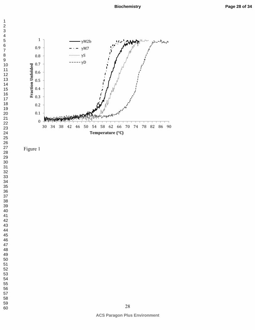
The thermal stability of γM7 and γM2b was measured by monitoring loss of CD signals
corresponding to the 218 nm minimum for β-sheet under increasing temperature. As shown in Figure
1, the melting temperatures for γM7 and γM2b are 58 and 60 °C, respectively, significantly lower than
those of mouse γS and human γD crystallins. Consistently, other γM-crystallins from Antarctic
toothfish and bigeye tuna also showed substantially lower temperature stability than the γ-crystallins
from B. Taurus (40).
Structure Determination
The solution structure of γM7-crystallin from zebrafish has been determined using multi-
dimensional NMR with a total of 1427 restraints, including 961 inter-proton distances, 279 backbone
dihedral angles and 131 residual dipolar coupling constraints. Resonance assignments have been
completed for 164 out of the 169 non-proline residues. Notably, the amide signals are missing from
residues 10 to 13, spanning the region of the first β-hairpin loop between the β-strands 1 and 2. These
vanishingly weak amide signals suggest that this region of γM7-crystallin experiences significant
conformational exchange motion, on the intermediate NMR time-scale that broadens the signals
beyond the detection limit. An ensemble of the 15 lowest energy structures, superpositioned as shown
in Figure 2, exhibits excellent structural convergence with atomic root-mean-square deviations (rmsd)
of 0.48 ± 0.15 and 1.03 ± 0.10 Å respect to the mean coordinate positions for the backbone and all
heavy atoms, respectively. The structural statistics are summarized in Table 1.
Structure Comparison
γM7-crystallin shows a two-domain architecture similar to previously determined γ-crystallin
Page 8 of 34
ACS Paragon Plus Environment
Biochemistry
123456789101112131415161718192021222324252627282930313233343536373839404142434445464748495051525354555657585960
9
structures, with each domain possessing two intercalating Greek-key (GK) motifs. Figure 3A displays
a comparison between the human γD-crystallin X-ray (PDB entry 1HK0) and the present solution
structure, with the associated backbone rms differences being 1.51, 0.88 and 1.67 Å for the N-, C-
terminal domains and the full length protein. With respect to the NMR structure of murine γS-
crystallin (PDB entry 1ZWM), similar backbone rmsd values, 1.53, 1.02 and 2.18 Å respectively, were
also observed. Collectively, this indicates that the main structural difference between γM7 and
mammalian lens crystallins is located in the N-terminal domain, while the C-terminal domains exhibit
high structural similarity. As detailed in Figure 3B, the most noticeable structural difference between
the fish γM7 and γD- or γS-crystallin resides within the variable loop 2 (VL2) in the N-terminal
domain. This loop in γM7 is folded further away from the hydrophobic core compared to that of γD-
crystallin. Evidently, the Hα of Met67 strongly NOE interacts with the HN amide proton of Met72
(Figure 3C), replacing an otherwise i to i+4 NOE pattern observed in the VL4 of γM7 and both VL2
and VL4 of mouse γS (25). As a result, the aromatic side chain of Tyr66 tucks into the core of the N-
terminal domain, making extensive hydrophobic contacts with the side chains of Ile36 and Val76. This
conformation is in sharp contrast to the equivalent residues, His65 in γD- or Tyr69 in γS-crystallin,
which project away from the domain and lack NOE interactions with the hydrophobic core residues.
The burying of Tyr66 is made possible by the absence of a tryptophan residue conservatively
residing in VL2 in all lens γ-crystallins from mammals. This conserved tryptophan makes extensive
contacts with the N-terminal hydrophobic core, so its absence in γM7 allows re-optimization of the
hydrophobic interactions between residues in this loop and the N-terminal core. Consistent with this,
instead of being highly solvent exposed like the equivalent Leu71 in γD-crystallin or Leu75 in γS,
residue Met72 in γM7 protrudes into the hydrophobic core of the N-terminal domain, making extensive
hydrophobic interactions. Leu75 of γS, on the contrary, shows very close methyl proton chemical
Page 9 of 34
ACS Paragon Plus Environment
Biochemistry
123456789101112131415161718192021222324252627282930313233343536373839404142434445464748495051525354555657585960

10
shifts and virtually no inter-residue NOE cross peak with any core residues. It is conceivable that the
increased hydrophobic packing by Tyr66 and Met72 in γM7 may at least in part compensate for the
absence of the Trp residue, resulting in the altered loop conformation. Since all four GK motifs are
structurally similar, we compared VL2 with the equivalent loops VL1 and VL3 that also lack a Trp
residue. In a common sequence Φ(xxx)3-5Φ with the Φ represents a hydrophobic residue, the two
equivalent flanking hydrophobic residues Leu26 and Phe30 in VL1, Leu113 and Met119 in VL3 are all
buried in a similar manner as Tyr66 and Met72 in VL2, further suggesting that a Trp residue is critical
for dictating the VL conformation. A second conserved Trp located at the junction of two GK motifs in
mammalian γ-crystallins is also missing in the N-terminal domain of γM7. It has been shown that this
tryptophan plays an important structural role in stabilizing a corner conformation by forming a
hydrogen bond between its side chain Hε1 (i) to the carbonyl oxygen at the i-3 position (41). It is
plausible that missing such interaction may contribute to decreased protein stability of zebrafish γM7
and other γM-crystallins, as shown in this study and for several examples from other fish (40).
γγγγM7 exhibits increased backbone dynamics in the loop regions
Next we used NMR relaxation experiments to investigate the backbone motions of γM7. Figure 4
shows the comparison of the derived order parameters between γM7 and γS crystallins. Unlike γS,
which exhibits quite uniform order parameter (S2) for almost all residues (except those in the N-, C-
terminal tails and the interdomain linker), γM7 shows much more variability. As shown in Figure 4,
while all β-strands display similar S2, lower values are observed for those primarily located in or near
the loop regions, for example VL1, VL2, VL4 and the first β-hairpin in the C-terminal domain.
Together with the missing NH signal in the first β-hairpin, the relaxation measurement suggests that
γM7 possesses higher magnitude backbone conformational motion in the loop regions. When
Page 10 of 34
ACS Paragon Plus Environment
Biochemistry
123456789101112131415161718192021222324252627282930313233343536373839404142434445464748495051525354555657585960
11
compared to the relaxation results of γD (42), the loop regions of γM7 again appear to be more flexible.
Furthermore, the amplitudes of internal motions of the inter-domain linker among these three proteins
vary significantly, with γS being the most flexible followed by γM7 and then γD, which has similar
order parameters to those of residues in the N-/C-terminal structure core (42). Based on the sequence
alignment of these γ-crystallins (Figure 5A), it appears that the flexibility of the inter-domain linker
correlates with its length, with a three residue-linker for γD, four for γM7 and five for γS. In γD the
short three-residue linker is as rigid as both of the structural domains (42).
Methionine residues in γγγγM crystallins
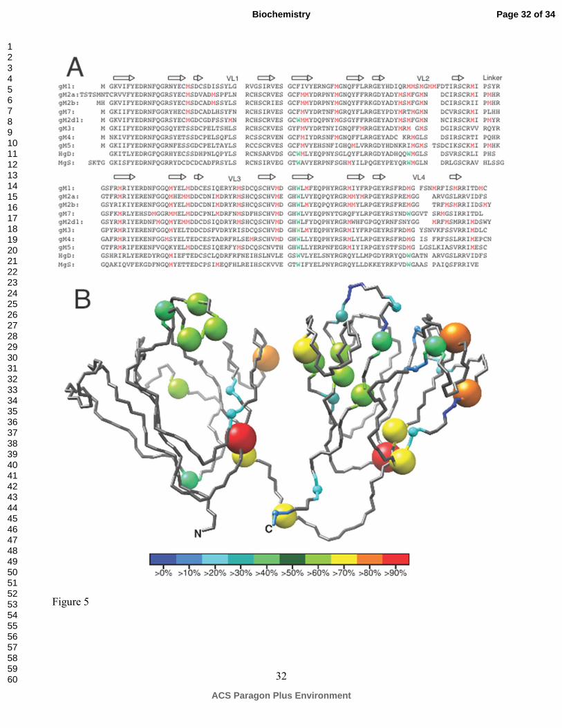
An interesting feature of the fish γM-crystallins is their high methionine content, up to ~15% of the
total amino acid composition. Figure 5A show the alignment of several γM-crystallins with the
methionine residues highlighted, and Figure 5B positions all the methionine residues found in 15 fish
γM crystallins on the structure of γM7. Noticeably, more than 70% of the methionines are located in
the second and the third Greek-Key motifs, whereas less than 10% are found in the first GK. Most of
the methionine substitutions are located in the loops, turns and on the edge of a β-sheet, particularly in
the variable loops and on the second β-strand of the 2nd, 3rd and 4th Greek-Key motifs. In γM7, except
for Met44, Met91 and Met165, which are located in the domain interface and the center of the C-
terminal hydrophobic core respectively, all other methionines are at least partially, if not completely,
solvent accessible. Interestingly, most of the exposed methionine side chains are not fully disordered.
Out of 14 exposed methionine residues, 11 of them make strong to medium NOE contacts to at least
one surface aromatic residue, making those methionine side chains attach to the surface of the protein
instead of freely projecting into solution. For example, as shown in Figure 3B, the side chain of Met67
sits ~5 Å above the aromatic ring of Tyr66 and ~5 Å from that of His34, while that of Met70 makes
simultaneous NOE contacts with Tyr56 and Tyr63, and both Met99 and Met103 interact with the
Page 11 of 34
ACS Paragon Plus Environment
Biochemistry
123456789101112131415161718192021222324252627282930313233343536373839404142434445464748495051525354555657585960

12
aromatic ring of Tyr94 in the first β-hairpin of the C-terminal domain. This partial ordering of the
exposed methionine side chains is consistent with the recent study suggesting that the methionine-
aromatic interaction plays a key role in stabilizing protein structure (43).
Protein unfolding monitored by tryptophan fluorescence
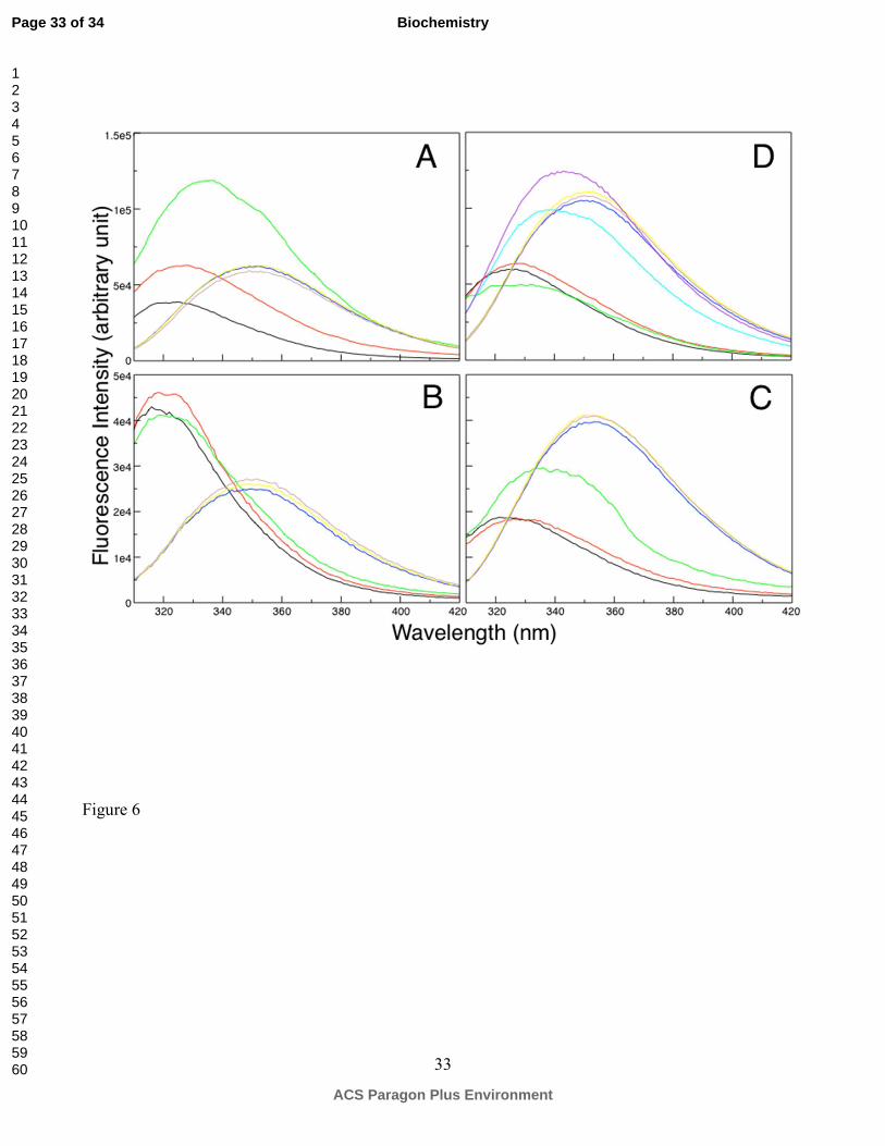
Unfolding of γM7-crystallin was monitored by intrinsic tryptophan fluorescence with increasing
amount of chemical denaturant, guanidinium hydrochloride (GuHCl). Given that the two tryptophan
residues in γM7 are located in the C-terminal domain, their fluorescence change induced by GuHCl
allowed us to monitor how a single βγ-domain unfolds in the content of a native two-domain protein.
Interestingly, as shown in Figure 6A, the Trp fluorescence of γM7 initially increased with increasing
GuHCl, accompanied by a slight red shift of the λmax. The fluorescence reached its maximum intensity
at about ~0.8 M GuHCl, followed by decreasing with increase of the GuHCl concentration until the
protein was completely denatured. To further delineate the individual contribution to the overall
fluorescence change of these two tryptophans, the unfolding of a homologous γM2b and W132F γM7
was monitored in the same manner. γM2b only contains the tryptophan at the conjunction of the 3rd
and 4th GK motifs. The Trp fluorescence profile of γM2b (Figure 6B) showed a continuous decrease
upon denaturation, whereas that of W132F (Figure 6C) exhibited an increasing trend with the increase
of denaturant concentration. A significant fluorescence jump at 0.8 M GuHCl appears to coincide with
the initial fluorescence burst observed in WT γM7. These observations indicate that the local
environments of the two tryptophans in a natively folded crystallin-domain are different. The VL
structure helps to effectively quench the fluorescence of Trp165, whereas Trp132 is more fluorescent
when it is buried in the hydrophobic core. These distinct fluorescence profiles of each tryptophan were
also observed in human γD- and γS crystallins with one behaving as a fluorescence quencher and the
other as a fluorescence enhancer (8, 9). Based on these observations, the initial increase in
Page 12 of 34
ACS Paragon Plus Environment
Biochemistry
123456789101112131415161718192021222324252627282930313233343536373839404142434445464748495051525354555657585960

13
fluorescence of γM7 is likely due to local unfolding of the VL spanning between the β7 and β8,
disrupting the key structure feature responsible for effective fluorescence quenching of Trp165. The
fact that losing the quenching effect of Trp165 precedes any significant fluorescence change from the
more buried Trp132 suggests that under low denaturant concentration a native-like structural core is
maintained while the variable loops can be more readily unfolded.
To test whether the early unfolding of the variable loop observed for γM-crystallins is a common
feature of lens γ-crystallins, we further investigated the denaturation of mouse γS-crystallin. As shown
in Figure 6D, an early loss of fluorescence quenching effect was also observed for γS, followed by a
decrease expected for further exposing the structurally buried Trp36 and Trp132, at higher GuHCl
concentration. The fact that γS has a higher unfolding denaturant concentration again suggests that γM-
crystallins are generally less stable than their counterparts in mammalian lenses. In summary, γ-
crystallin domains may share a common unfolding pathway, starting from the unfolding of the variable
loops on the top of the wedged fold. This could, partially expose the hydrophobic core of the γ-
crystallin domain, potentially leading to protein aggregation.
Page 13 of 34
ACS Paragon Plus Environment
Biochemistry
123456789101112131415161718192021222324252627282930313233343536373839404142434445464748495051525354555657585960

14
DISCUSSION
The solution structure of zebrafish γM7-crystallin reveals the common two-domain, four Greek-key
topology found in other γ- (and β-) crystallins (44). These two domains, linked by a short linker, are
stabilized by inter-domain interactions between conserved hydrophobic residues located in the domain
interface. This evolutionally conserved domain-pairing structure contributes to the high
thermodynamic stability required for lens proteins (2). However, compared to other known lens βγ-
crystallin structures from mammals, significant structural differences were observed in the N-terminal
domain of γM7 (Figure 3), particularly in the variable loop between the β7 and β8 strands (VL2). This
difference seems to be due to the absence of a tryptophan residue, which is conserved in this region in
other βγ-crystallins. As a consequence, the hydrophobic residues flanking VL2 reorganized to
maximize hydrophobic packing, adopting a conformation similar to those structure-equivalent
counterparts VL1 and VL3, which also lack a tryptophan. Apparently, the interaction between the
tryptophan side-chain and the domain core is critical for the specific conformation of the variable loop,
which itself is key for effective fluorescence quenching of the tryptophan residue (45). The C-terminal
domain of γM7, on the other hand, maintains two conserved tryptophans and has a VL structure almost
superimposable on that of γD and γS crystallins. Based on these results, it can be predicted that when
the tryptophan in VL4 is missing, as in many other γM-crystallins, (Figure 5A), the structure of the
variable loop would also closely resemble that of VL2 of γM7.
In γ-crystallin and β-crystallin domains from mammals, the paired tryptophans are conserved in both
N- and C-terminal domains. It has been proposed that these residues to help absorb UV light between
295-400 nm, preventing it from reaching the retina, while escaping photodegradation themselves
through a fast fluorescence quench mechanism (8, 9, 45). Between the two tryptophans, the one
located in the variable loop serves as the major mediator for the fast fluorescence quench (8), whereas
the other one, located at the bottom of the hydrophobic core, is likely to play a more important
Page 14 of 34
ACS Paragon Plus Environment
Biochemistry
123456789101112131415161718192021222324252627282930313233343536373839404142434445464748495051525354555657585960

15
structural role by stabilizing the inter-Greek key connection (25). Consistent with this proposition,
irradiation of bovine γB-crystallin in vitro photodegrades Trp42 and Trp131 three times more
efficiently than Trp68 and Trp157 (46). This UV role may be significant since cumulative exposure to
UV radiation is correlated with the prevalence of senile cataract (47). Compared to mammalian γ-
crystallins, as shown in Figure 5A, only one tryptophan in the core of the C-terminal domain is highly
conserved in all fish γM-crystallins, whereas the other three are completely dispensable. While the
structural tryptophan in the N-terminal core is commonly replaced by a phenylalanine, interestingly,
both tryptophans in the variable loop are frequently substituted by a methionine. The absence of these
tryptophans in aquatic species, especially the high fluorescence quencher, may be related to the low
exposure to UV light under water. Although γM-crystallins do not conserve the Trp pairs, they are
present in fish γS-, γN- and β-crystallins (48). The presence of the four Trp in mammalian γA-F
crystallins, proteins of the lens core, may reflect the greater UV exposure of diurnal, land species.
The striking abundance of methionine residues in the γM-crystallins suggests some importance of
this amino acid for the proteins. The observed lower heat stability of γM7-, γM2b- and some other γM-
crystallins indicates that high methionine content does not itself increase protein stability as speculated
previously (10). In general, the thermal stability of γ-crystallins appears to correlate with the body
temperature (40), with higher stability in mammalian crystallins. Alternatively, the flexibility of
methionine side chains could facilitate intermolecular interactions (11). Interestingly, as shown in
Figure 5B, the VLs and β-strands with high methionine content in the 2nd and 4th GKs contribute to the
domain interface while the equivalent regions on the 3rd GK motif may potentially aid intermolecular
interaction similar to intramolecular domain-domain interaction. Consistent with this notion, in the
crystal structure of the tetrameric βB2-crystallin (2BB2), the same regions from the 2nd, 3rd and 4th GK
motifs are directly involved in interdomain interaction with other crystallin molecules (49). Under the
extreme protein density of the fish lens, the surface flexibility imparted by methionine side chain may
Page 15 of 34
ACS Paragon Plus Environment
Biochemistry
123456789101112131415161718192021222324252627282930313233343536373839404142434445464748495051525354555657585960
16
also contribute to their cold adaptation compared to those in mammals (7) possibly at the expense of
their thermal stability (40). The presence of a high content of methionine residues (and others) in γM-
crystallins has also been suggested to contribute directly to the high refractive index requirement of the
fish lens (12). Consistent with this notion, as shown in Figure 5A, the positions frequently replaced by
a methionine are commonly occupied by those with a lower refractive index increment, such as valine,
alanine, isoleucine and threonine, in mammalian lens γ-crystallins (50). Together with the fact that
other refractive proteins such as the S-crystallins from cephalopods (12) and the reflectins from squid
(17) have also evolved to adopt extremely high methionine content, optimization of the refractive index
seems to be at least one of the driving forces for methionine amino acid selection in lens-related
proteins. Finally, our structure of γM7-crystallin indicates that the majority of the surface exposed
methionines are in close proximity with at least one aromatic residue. Indeed, such interaction seems
to be a common stabilizing factor in many proteins while mutations disrupting the interaction are
associated with several diseases (43). In summary, it is conceivable that high methionine content in
fish γM-crystallins is the result of an accumulative effect from requirement of high refractive
properties, copping with low temperature environment and high protein density. During evolution, the
risk of oxidation of methionine residue, the reduced need for high refractive index and the requirement
for higher protein stability in mammals may have driven the substitution of methionine by other amino
acids.
In order to investigate the consequences of Trp and Met content in fish γM-crystallins for higher
backbone dynamics, NMR relaxation studies of γM7 were performed. These showed a noticeable
difference in dynamics in surface loops among γM7, γS- (25) and γD-crystallins (42), and revealed an
interesting correlation between the fast local internal dynamics in the inter-domain linker and its length.
From a three-residue linker in γD to four-residues in γM7 and five-residues in γS, the motion of the
linker increases. Interestingly, mouse γC has an isoform in mice in which the liker has an extra amino
Page 16 of 34
ACS Paragon Plus Environment
Biochemistry
123456789101112131415161718192021222324252627282930313233343536373839404142434445464748495051525354555657585960

17
acid insertion and it has been proposed that local motions in crystallins could provide entropic
compensation for the restrictions of short-range order in lens at high protein concentration (25).
Tryptophan fluorescence of γM- and γS-crystallins showed a very interesting denaturation profile,
exhibiting a distinct initial increase at low GuHCl concentration followed by a decrease to a level
corresponding to a fully unfolded state. Based on the denaturation profile of each individual
tryptophan in γM2b, W136F-γM7 and the single trp-mutants of γD and γS (8, 9), the initial
fluorescence increase likely reflects loss of quenching by partial unfolding of the variable loop while
the later decrease corresponds to unfolding of the core of the βγ-crystallin domain. This unfolding
mode perfectly agrees with our previous NMR results of the stability-impaired mouse γS-crystallin
mutant, Opj, which revealed a partially unfolded intermediate with a native-like overall structure but a
much more unfolded variable loop (23). Consistent with our results, a single βγ-crystallin domain from
a nonlenticular protein has recently been proposed to unfold through formation of a native-like
intermediate (51), which exhibits elevated flexibility and higher propensity of forming insoluble
aggregates (51). It is noteworthy that these partially unfolded intermediates are structurally different
from those proposed for human γD-crystallin, which sequentially unfolds the four GK motifs under
increasing amount of denaturant (52). It is conceivable that the intermediate observed in current
studies may represent an early stage of unfolding of a βγ-crystallin domain, which may promote further
opening of individual GK motifs and ultimately lead to protein aggregation (53).
Page 17 of 34
ACS Paragon Plus Environment
Biochemistry
123456789101112131415161718192021222324252627282930313233343536373839404142434445464748495051525354555657585960

18
Acknowledgements
We thank Dr. Keith Wyatt for subcloning the γM-crystallin cDNAs.
Page 18 of 34
ACS Paragon Plus Environment
Biochemistry
123456789101112131415161718192021222324252627282930313233343536373839404142434445464748495051525354555657585960

19
REFERENCE
1. Bloemendal, H., de Jong, W., Jaenicke, R., Lubsen, N. H., Slingsby, C., and Tardieu, A. (2004)
Ageing and vision: structure, stability and function of lens crystallins, Prog. Biophys. Mol. Biol.
86, 407-485.
2. Jaenicke, R. (1999) Stability and folding of domain proteins, Progress in Biophysics &
Molecular Biology 71, 155-241.
3. Purkiss, A. G., Bateman, O. A., Wyatt, K., Wilmarth, P. A., David, L. L., Wistow, G. J., and
Slingsby, C. (2007) Biophysical Properties of γC-Crystallin in Human and Mouse Eye Lens:
The Role of Molecular Dipoles, J Mol Biol 372, 205-222.
4. White, H. E., Driessen, H. P., Slingsby, C., Moss, D. S., and Lindley, P. F. (1989) Packing
interactions in the eye-lens. Structural analysis, internal symmetry and lattice interactions of
bovine gamma IVa-crystallin, J Mol Biol 207, 217-235.
5. Slingsby, C. (1985) Structural variation in lens crystallins, Trends Biochem. Sci. 10, 281-284.
6. Pan, F. M., Chang, W. C., Chao, Y. K., and Chiou, S. H. (1994) Characterization of gamma-
crystallins from a hybrid teleostean fish: multiplicity of isoforms as revealed by cDNA
sequence analysis, Biochem Biophys Res Commun 202, 527-534.
7. Kiss, A. J., and Cheng, C. H. (2008) Molecular diversity and genomic organisation of the alpha,
beta and gamma eye lens crystallins from the Antarctic toothfish Dissostichus mawsoni, Comp
Biochem Physiol Part D Genomics Proteomics 3, 155-171.
8. Chen, J., Flaugh, S. L., Callis, P. R., and King, J. (2006) Mechanism of the highly efficient
quenching of tryptophan fluorescence in human gammaD-crystallin, Biochemistry 45, 11552-
11563.
9. Chen, J., Toptygin, D., Brand, L., and King, J. (2008) Mechanism of the efficient tryptophan
fluorescence quenching in human gammaD-crystallin studied by time-resolved fluorescence,
Page 19 of 34
ACS Paragon Plus Environment
Biochemistry
123456789101112131415161718192021222324252627282930313233343536373839404142434445464748495051525354555657585960

20
Biochemistry 47, 10705-10721.
10. Chang, T., Jiang, Y. J., Chiou, S. H., and Chang, W. C. (1988) Carp gamma-crystallins with high
methionine content: cloning and sequencing of the complementary DNA, Biochim Biophys Acta
951, 226-229.
11. Srikanthan, D., Bateman, O. A., Purkiss, A. G., and Slingsby, C. (2004) Sulfur in human
crystallins, Exp Eye Res 79, 823-831.
12. Zhao, H., Brown, P. H., Magone, M. T., and Schuck, P. (2012) The molecular refractive function
of lens gamma-Crystallins, J Mol Biol 411, 680-699.
13. Kappe, G., Purkiss, A. G., van Genesen, S. T., Slingsby, C., and Lubsen, N. H. (2010) Explosive
expansion of betagamma-crystallin genes in the ancestral vertebrate, J Mol Evol 71, 219-230.
14. Zhao, H., Magone, M. T., and Schuck, P. (2012) The role of macromolecular crowding in the
evolution of lens crystallins with high molecular refractive index, Phys Biol 8, 046004.
15. Tomarev, S. I., Zinovieva, R. D., and Piatigorsky, J. (1991) Crystallins of the octopus lens.
Recruitment from detoxification enzymes, J Biol Chem 266, 24226-24231.
16. Siezen, R. J., and Shaw, D. C. (1982) Physicochemical characterization of lens proteins of the
squid Nototodarus gouldi and comparison with vertebrate crystallins, Biochim Biophys Acta
704, 304-320.
17. Crookes, W. J., Ding, L. L., Huang, Q. L., Kimbell, J. R., Horwitz, J., and McFall-Ngai, M. J.
(2004) Reflectins: the unusual proteins of squid reflective tissues, Science 303, 235-238.
18. Kramer, R. M., Crookes-Goodson, W. J., and Naik, R. R. (2007) The self-organizing properties
of squid reflectin protein, Nat Mater 6, 533-538.
19. Mitraki, A., and King, J. (1989) Protein Folding Intermediates and Inclusion Body Formation,
Bio-Technology 7, 690-697.
20. Chiti, F., and Dobson, C. M. (2006) Protein misfolding, functional amyloid, and human disease,
Page 20 of 34
ACS Paragon Plus Environment
Biochemistry
123456789101112131415161718192021222324252627282930313233343536373839404142434445464748495051525354555657585960
21
Annu. Rev. Biochem. 75, 333-366.
21. Rudolph, R., Siebendritt, R., Nesslauer, G., Sharma, A. K., and Jaenicke, R. (1990) Folding of
an all-β protein: independent domain folding in γII-crystallin from calf eye lens, Proc. Natl.
Acad. Sci. USA 87, 4625-4629.
22. Flaugh, S. L., Kosinski-collins, M. S., and King, J. (2005) Interdomain side-chain interactions
in human γD crystallin influencing folding and stability, Protein Sci. 14, 2030-2043.
23. Mahler, B., Doddapaneni, K., Kleckner, I., Yuan, C., Wistow, G., and Wu, Z. (2011)
Characterization of a Transient Unfolding Intermediate in a Core Mutant of gammaS-Crystallin,
J Mol Biol 405, 840-850.
24. Kosinski-collins, M. S., and King, J. (2003) In vitro unfolding, refolding, and polymerization of
human γD crystallin, a protein involved in cataract formation, Protein Sci. 12, 480-490.
25. Wu, Z. R., Delaglio, F., Wyatt, M. K., Wistow, G., and Bax, A. (2005) Solution structure of γS-
crystallin by molecular fragment replacement NMR, Protein Sci. 14, 3101-3114.
26. Sinha, D., Wyatt, M. K., Sarra, R., Jaworski, C., Slingsby, C., Thaung, C., Pannell, L., Robison,
W. G., Favor, J., Lyon, M., and Wistow, G. (2001) A Temperature-sensitive Mutation of Crygs
in the Murine Opj Cataract, J. Biol. Chem. 276, 9308-9315.
27. Pace, C. N., Shirley, B. A., and Thomson, J. A. (1989) Measuring the conformational stability of
a protein, In Protein Structure: A Practical Approach (Creighton, T. E., Ed.), pp 311-329, IRL
Press, Oxford.
28. Grzesiek, S., Dobeli, H., Gentz, R., Garotta, G., Labhardt, A. M., and Bax, A. (1992) 1H, 13C,
and 15N NMR backbone assignments and secondary structure of human interferon,
Biochemistry 31, 8180-8190.
29. Bax, A., and Grzesiek, S. (1993) Methodological advances in protein NMR, Accounts of
Chemical Research 26, 131-138.
Page 21 of 34
ACS Paragon Plus Environment
Biochemistry
123456789101112131415161718192021222324252627282930313233343536373839404142434445464748495051525354555657585960
22
30. Neri, D., Szyperski, T., Otting, G., Senn, H., and Wuthrich, K. (1989) Stereospecific Nuclear
Magnetic Resonance Assignments of the Methyl Groups of Valine and Leucine in the DNA-
Binding Domain of the 434 Repressor by Biosynthetically Directed Fractional 13C Labeling,
Biochemistry 28, 7510-7516.
31. Vuister, G. W., and Bax, A. (1992) Resolution enhancement and spectral editing of uniformly
13C-enriched proteins by homonuclear broadband 13C decoupling., Journal of Magnetic
Resonance 98, 428-435.
32. Chou, J. J., Gaemers, S., Howder, B., Louis, J. M. L., and Bax, A. (2001) A simple apparatus for
generating stretched polyacrylamide gels, yielding uniform alignment of proteins and detergent
micelles, J. Biomol. NMR 21, 377-382.
33. Farrow, N. A., Muhandiram, R., Singer, A. U., Pascal, S. M., Kay, C. M., Gish, G., Shoelson, S.
E., Pawson, T., Forman-Kay, J. D., and Kay, L. E. (1994) Backbone dynamics of a free and a
phosphopeptide-complexed Src homology 2 domain studied by 15N NMR relaxation,
Biochemistry 33, 5984-6003.
34. Delaglio, F., Grzesiek, S., Vuister, G. W., Zhu, G., Pfeifer, J., and Bax, A. (1995) Nmrpipe - a
Multidimensional Spectral Processing System Based On Unix Pipes, J. Biomol. NMR 6, 277-
293.
35. Johnson, B. A., and Blevins, R. A. (1994) NMRView: a computer program for the visualization
and analysis of NMR data, Journal of Biomolecular NMR 4, 603-614.
36. Shirley, B. A., Stanssens, P., Steyaert, J., and Pace, C. N. (1989) Conformational stability and
activity of ribonuclease T1 and mutants. Gln25----Lys, Glu58----Ala, and the double mutant,
The Journal of biological chemistry 264, 11621-11625.
37. Schwieters, C., Kuszewski, J., Tjandra, N., and Clore, G. (2003) The Xplor-NIH NMR
molecular structure determinsation package., J. Magn. Resson. 160, 65-73.
Page 22 of 34
ACS Paragon Plus Environment
Biochemistry
123456789101112131415161718192021222324252627282930313233343536373839404142434445464748495051525354555657585960
23
38. Laskowski, R. A., Rullmann, J. A. C., MacArthur, M. W., Kaptein, R., and Thornton, J. M.
(1996) AQUA and Procheck NMR: Programs for checking the quality of protein structures
solved by NMR, J. Biomol. NMR 8, 477-486.
39. Pettersen, E. F., Goddard, T. D., Huang, C. C., Couch, G. S., Greenblatt, D. M., Meng, E. C.,
and Ferrin, T. E. (2004) UCSF Chimera--a visualization system for exploratory research and
analysis, J Comput Chem 25, 1605-1612.
40. Kiss, A. J., Mirarefi, A. Y., Ramakrishnan, S., Zukoski, C. F., Devries, A. L., and Cheng, C. H.
(2004) Cold-stable eye lens crystallins of the Antarctic nototheniid toothfish Dissostichus
mawsoni Norman, J Exp Biol 207, 4633-4649.
41. Hemmingsen, J. M., Gernert, K. M., Richardson, J. S., and Richarson, D. C. (1994) The
tyrosine corner: A feature of most Greek key (beta)-barrel proteins, Protein Sci. 3, 1927-1937.
42. Jung, J., Byeon, I. J., Wang, Y., King, J., and Gronenborn, A. M. (2009) The structure of the
cataract-causing P23T mutant of human γD-crystallin exhibits distinctive local conformational
and dynamic changes, Biochemistry 48, 2597-2609.
43. Valley, C. C., Cembran, A., Perlmutter, J. D., Lewis, A. K., Labello, N. P., Gao, J., and Sachs, J.
N. (2012) The Methionine-aromatic Motif Plays a Unique Role in Stabilizing Protein Structure,
J Biol Chem 287, 34979-34991.
44. Blundell, T., Lindley, P. F., Miller, L., Moss, D., Slingsby, C., Tickle, I., Turnell, B., and
Wistow, G. (1981) The molecular structure and stability of the eye lens: X-ray analysis of γ-
crystallin II., Nature 289, 771-777.
45. Chen, J., Callis, P. R., and King, J. (2009) Mechanism of the very efficient quenching of
tryptophan fluorescence in human gamma D- and gamma S-crystallins: the gamma-crystallin
fold may have evolved to protect tryptophan residues from ultraviolet photodamage,
Biochemistry 48, 3708-3716.
Page 23 of 34
ACS Paragon Plus Environment
Biochemistry
123456789101112131415161718192021222324252627282930313233343536373839404142434445464748495051525354555657585960

24
46. Tallmadge, D. H., and Borkman, R. F. (1990) The rates of photolysis of the four individual
tryptophan residues in UV exposed calf gamma-II crystallin, Photochem Photobiol 51, 363-368.
47. Robman, L., and Taylor, H. (2005) External factors in the development of cataract, Eye (Lond)
19, 1074-1082.
48. Wistow, G., Wyatt, M. K., David, L., Gao, C., Bateman, O., Bernstein, S., Tomarev, S., Segovia,
L., Slingsby, C., and Vihtelic, T. (2005) γN-crystallin and the evolution of the βγ-crystallin
superfamily in vertebrates., Febs J 272, 2276-2291.
49. Bax, B., Lapatto, R., Nalini, V., Driessen, H., Lindley, P. F., Mahadevan, D., Blundell, T. L., and
Slingsby, C. (1990) X-ray analysis of beta B2-crystallin and evolution of oligomeric lens
proteins, Nature 347, 776-780.
50. Zhao, H., Brown, P. H., and Schuck, P. (2012) On the distribution of protein refractive index
increments, Biophys J 100, 2309-2317.
51. Rajanikanth, V., Srivastava, S. S., Singh, A. K., Rajyalakshmi, M., Chandra, K., Aravind, P.,
Sankaranarayanan, R., and Sharma, Y. (2012) Aggregation-prone near-native intermediate
formation during unfolding of a structurally similar nonlenticular betagamma-crystallin domain,
Biochemistry 51, 8502-8513.
52. Mills, I. A., Flaugh, S. L., Kosinski-Collins, M. S., and King, J. A. (2007) Folding and stability
of the isolated Greek key domains of the long-lived human lens proteins γD-crystallin and γS-
crystallin, Protein Sci 16, 2427-2444.
53. Lee, S., Mahler, B., Toward, J., Jones, B., Wyatt, K., Dong, L., Wistow, G., and Wu, Z. (2010) A
single destabilizing mutation (F9S) promotes concerted unfolding of an entire globular domain
in γS-crystallin, J. Mol. Biol. 399, 320-330.
Page 24 of 34
ACS Paragon Plus Environment
Biochemistry
123456789101112131415161718192021222324252627282930313233343536373839404142434445464748495051525354555657585960
25
FIGURES
Figure 1. Temperature unfolding curves for γM7, γM2b, γS and γD. Loss of secondary structure for
zebrafish γM7, γM2b, mouse γS and human γD-crystallins monitored by loss of CD signals at 218 nm,
indicating lower heat stability for γM-crystallins.
Figure 2. Superposition of 15 lowest energy NMR structures.
Figure 3. Overlay of γM7 NMR structure (cyan) with the X-ray γD-crystallin (1HK0, magenta) (A); A
close view of the variable loop 2 in the N-terminal domain, which lacks a conserved tryptophan residue
in γM7 (B), showing the comparison of the residues Tyr66, Met67, Met72 from γM7 and their
counterpart residues His65, Gln66, Leu71 from γD, and the side chain interactions between Met70 with
aromatic residues Tyr56 and Tyr63 in γM7; Stripes of 3D 15N-edited NOESY showing the unusual i to
i+5 interaction observed in the VL2 of γM7 (C).
Figure 4. Comparison of backbone order parameter (s2) of γM7 (black) and mouse γS (red) derived
from 15N relaxation experiments, indicating larger motions observed in the variable loop regions and
comparatively lower dynamics in the interdomain linker in γM7 than γS. The arrows indicate β-
stranded secondary structure and the variable loops between the 3rd and 4th β-strands in each Greek Key
motif are noted as VL.
Figure 5. (A) Sequence alignment of fish γM- with human γD- and mouse γS-crystallins, with all the
methionine residues colored in red and tryptophan colored in green. (B) Methionine residues in 15 fish
γM-crystallins are positioned on the NMR structure of γM7, with the size and color of the sphere
corresponding to the frequency of the methionine occurrences.
Figure 6. Representative typtophan fluorescence spectra of fish γM7 (A), γM2b (B), γM7 W132F (C)
and γS (D) at 0 M (black), 0.4 M (red), 0.8 M (green), 3.0 M (blue), 4.0 M (yellow), 5.0 M (brown)
GuHCl. Since γS is more stable than γM7, the fluorescence profiles at 1.6 M (cyan) and 2.0 M (purple)
Page 25 of 34
ACS Paragon Plus Environment
Biochemistry
123456789101112131415161718192021222324252627282930313233343536373839404142434445464748495051525354555657585960
26
are shown for γS in (D), showing that the fluorescence jump happened at higher GuHCl concentration
than γM7.
Page 26 of 34
ACS Paragon Plus Environment
Biochemistry
123456789101112131415161718192021222324252627282930313233343536373839404142434445464748495051525354555657585960

27
Table 1. NMR structural statistics of γγγγM7.
NMR distance & dihedral constraints
Total constraints (per monomer) 1427
Long range NOE (5 ≤ i-j) 578
Short range NOE (1 < i-j < 5) 129
Sequential NOE 252
Residual dipolar coupling 131
Dihedral angle restraints: φ a and ψ a 279
Hydrogen bond 56
Structure statistics (20 structures)
Violation statistics
Distance constraints (Å) 0.040 ± 0.001
Maximum distance violation (Å) 0.4
Deviations from idealized covalent geometry
Bond lengths (Å) 0.0029 ± 0.0006
Bond angles (°) 0.473 ± 0.006
PROCHECK (Ramachandran plot)
Most favored region (%) 70.9
Additionally allowed region (%) 26.5
Generously allowed region (%) 2.0
Disallowed region (%) 0.7
R.m.s. deviations from the average structure (Å)b
Backbone atoms 0.48
All heavy atoms 1.03
aThe dihedral angle constraints φ and ψ were derived by using TALOS (36). bR.m.s. deviations from
the average structure were calculated for the residues Lys3-Ile171 of the final 15 structures.
Page 27 of 34
ACS Paragon Plus Environment
Biochemistry
123456789101112131415161718192021222324252627282930313233343536373839404142434445464748495051525354555657585960
28
Figure 1
0
0.1
0.2
0.3
0.4
0.5
0.6
0.7
0.8
0.9
1
30 34 38 42 46 50 54 58 62 66 70 74 78 82 86 90
Fr
ac
tio
n U
nfo
lde
d
Temperature (°C)
γM2b
γM7
γS
γD
Page 28 of 34
ACS Paragon Plus Environment
Biochemistry
123456789101112131415161718192021222324252627282930313233343536373839404142434445464748495051525354555657585960

29
Figure 2
Page 29 of 34
ACS Paragon Plus Environment
Biochemistry
123456789101112131415161718192021222324252627282930313233343536373839404142434445464748495051525354555657585960

30
Figure 3
Page 30 of 34
ACS Paragon Plus Environment
Biochemistry
123456789101112131415161718192021222324252627282930313233343536373839404142434445464748495051525354555657585960

31
Figure 4
Page 31 of 34
ACS Paragon Plus Environment
Biochemistry
123456789101112131415161718192021222324252627282930313233343536373839404142434445464748495051525354555657585960
32
Figure 5
Page 32 of 34
ACS Paragon Plus Environment
Biochemistry
123456789101112131415161718192021222324252627282930313233343536373839404142434445464748495051525354555657585960
33
Figure 6
Page 33 of 34
ACS Paragon Plus Environment
Biochemistry
123456789101112131415161718192021222324252627282930313233343536373839404142434445464748495051525354555657585960
34
For Table of Contents Use Only. Title: Structure and Dynamics of the Fish Eye Lens Protein, γM7-crystallin
Bryon Mahler, Yingwei Chen, Jason Ford, Caleb Thiel, Graeme Wistow and Zhengrong Wu
Page 34 of 34
ACS Paragon Plus Environment
Biochemistry
123456789101112131415161718192021222324252627282930313233343536373839404142434445464748495051525354555657585960


![Changes in zebrafish (Danio rerio) lens crystallin content ... · development and regeneration [12], and more recently has become a model for investigations of lens function and development](https://img.dokumen.tips/doc/110x75/5f0e824d7e708231d43f96a0/changes-in-zebrafish-danio-rerio-lens-crystallin-content-development-and-regeneration.jpg)