Embed Size (px)
Citation preview
Contact Lens and Anterior Eye 40 (2017) 109–115
An observational cross-sectional study on the corneal endothelium ofmedium-term rigid gas permeable contact lens wearers
Michael J. Doughty*Glasgow-Caledonian University, Department of Vision Sciences, Cowcaddens Road, Glasgow G4 OBA, United Kingdom
A R T I C L E I N F O
Article history:Received 14 September 2016Received in revised form 21 November 2016Accepted 1 December 2016
Keywords:HumanCorneal endotheliumRGP lens wearPolymegethismCell pleomorphism
A B S T R A C T
Purpose: To assess if polymegethism and pleomorphism were evident in corneal endothelium aftermedium-term rigid gas permeable (RGP) contact lens wear.Methods: In a cross-sectional observational study over 12 years, single images of the central region of thecorneal endothelium of one eye of 46 subjects were taken with a non-contact specular microscope, alongwith a measure of central corneal thickness (CCT). The images were printed onto A3-sized paper and 100cells/image measured by planimetry.Results: Subjects aged between 20 and 32 years, with an average cumulative RGP wear of 6.0 +/� 1.6 years(range 3–9 years) were assessed; 26 of the subjects were Caucasian and 20 were Asian. The mean CCT was0.515 +/� 0.027 mm. The group cell area value was 401 +/� 42 sq micron to give an estimated endothelialcell density (ECD) of 2520 +/� 273 cells/sq mm. As compared to a historical database, most endothelia(37/46) showed some changes with the mean coefficient of variation on cell area (COV) being 36.7 +/�8.0% and the percentage of 6-sided (HEX) being 51.8 +/� 8.8%. There were modest correlations betweenyears of RGP wear and both COV (p = 0.009, r spearman = 0.424) and HEX (p = 0.025, r spearman = �0.291),but not for ECD or CCT.Conclusions: Corneal endothelial polymegethism appears to be a commonplace consequence of RGP lenswear with the magnitude of the change being related to the cumulative duration of the lens wear.
© 2016 Published by Elsevier Ltd on behalf of British Contact Lens Association.
Contents lists available at ScienceDirect
Contact Lens and Anterior Eye
journal homepage: www.else vie r .com/ locat e/c lae
1. Introduction
As viewed in vivo by specular microscopy, the cornealendothelium of the young healthy adult appears remarkable inbeing composed of cells with uniformity in size and shape [1,2].The cell size, as reported in the vast majority of endothelialassessments, is given as the endothelial cell density, or ECD, incells/mm2. The uniformity of the cell mosaic is less commonlyassessed, but where undertaken has been described by calculationsof the coefficient of variation in cell area (usually referred to as theCOV, CV or polymegethism index) and the relative numbers of 6-sided cells (usually referred to as the hexagonality or HEXmeasure) [3]. For healthy young adults (e.g. aged between 20and 40 years of age) who were not contact lens wearers but ofdifferent ethnic groups, early publications on endothelial mor-phometry over nearly a 20 year period indicated that the COVvalues would likely be between 25 and 30% and the percentage of
* Corresponding author.E-mail address: [email protected] (M.J. Doughty).
http://dx.doi.org/10.1016/j.clae.2016.12.0011367-0484/© 2016 Published by Elsevier Ltd on behalf of British Contact Lens Associat
6-sided cells between 60 and 70% [4–14]. Such reports provided abasis for a recognition, over the same time period, that such anapparently uniform mosaic was not always evident.
It was long ago recognized that contact lens wear could result inthe development of remarkable non-uniformity to the endothelialmosaic, with the appearance of substantial polymegethism(increased variation in cell areas) and pleomorphism (reducedpercentage of 6-sided cells) [15]; this was evident if polymethyl-methacrylate (PMMA) lenses were worn with effect beingattributed to the very limited oxygen permeability of these lenses.With the advent of contact lens materials with higher oxygenpermeability (e.g. rigid gas permeable, RGP lenses) it might beexpected that long-term lens-wear-associated polymegethism ofthe corneal endothelium would be less or not even evident.
In early years of assessments of RGP lens wear and the cornealendothelium, a number of small scale studies (less than 20 eyes),and mostly after only 1 year of RGP lens wear concluded thatsubstantial polymegethism was not evident [14,16–21]. However,one prospective study did report that while the average COV after1 year of RGP lens wear (without prior contact lens wear) was only31%, COV values as high as 40% could be seen in some individuals
ion.
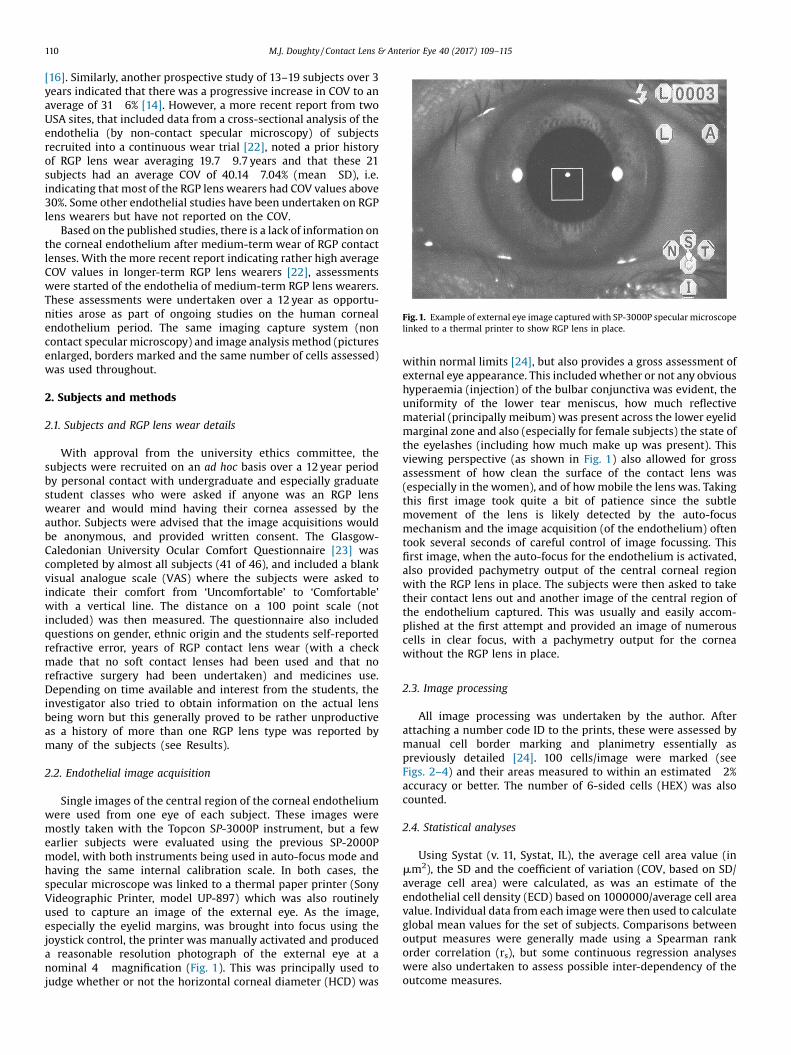
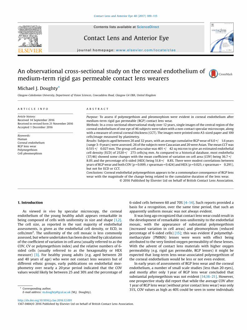
Fig. 1. Example of external eye image captured with SP-3000P specular microscopelinked to a thermal printer to show RGP lens in place.
110 M.J. Doughty / Contact Lens & Anterior Eye 40 (2017) 109–115
[16]. Similarly, another prospective study of 13–19 subjects over 3years indicated that there was a progressive increase in COV to anaverage of 31 �6% [14]. However, a more recent report from twoUSA sites, that included data from a cross-sectional analysis of theendothelia (by non-contact specular microscopy) of subjectsrecruited into a continuous wear trial [22], noted a prior historyof RGP lens wear averaging 19.7 � 9.7 years and that these 21subjects had an average COV of 40.14 � 7.04% (mean � SD), i.e.indicating that most of the RGP lens wearers had COV values above30%. Some other endothelial studies have been undertaken on RGPlens wearers but have not reported on the COV.
Based on the published studies, there is a lack of information onthe corneal endothelium after medium-term wear of RGP contactlenses. With the more recent report indicating rather high averageCOV values in longer-term RGP lens wearers [22], assessmentswere started of the endothelia of medium-term RGP lens wearers.These assessments were undertaken over a 12 year as opportu-nities arose as part of ongoing studies on the human cornealendothelium period. The same imaging capture system (noncontact specular microscopy) and image analysis method (picturesenlarged, borders marked and the same number of cells assessed)was used throughout.
2. Subjects and methods
2.1. Subjects and RGP lens wear details
With approval from the university ethics committee, thesubjects were recruited on an ad hoc basis over a 12 year periodby personal contact with undergraduate and especially graduatestudent classes who were asked if anyone was an RGP lenswearer and would mind having their cornea assessed by theauthor. Subjects were advised that the image acquisitions wouldbe anonymous, and provided written consent. The Glasgow-Caledonian University Ocular Comfort Questionnaire [23] wascompleted by almost all subjects (41 of 46), and included a blankvisual analogue scale (VAS) where the subjects were asked toindicate their comfort from ‘Uncomfortable’ to ‘Comfortable’with a vertical line. The distance on a 100 point scale (notincluded) was then measured. The questionnaire also includedquestions on gender, ethnic origin and the students self-reportedrefractive error, years of RGP contact lens wear (with a checkmade that no soft contact lenses had been used and that norefractive surgery had been undertaken) and medicines use.Depending on time available and interest from the students, theinvestigator also tried to obtain information on the actual lensbeing worn but this generally proved to be rather unproductiveas a history of more than one RGP lens type was reported bymany of the subjects (see Results).
2.2. Endothelial image acquisition
Single images of the central region of the corneal endotheliumwere used from one eye of each subject. These images weremostly taken with the Topcon SP-3000P instrument, but a fewearlier subjects were evaluated using the previous SP-2000Pmodel, with both instruments being used in auto-focus mode andhaving the same internal calibration scale. In both cases, thespecular microscope was linked to a thermal paper printer (SonyVideographic Printer, model UP-897) which was also routinelyused to capture an image of the external eye. As the image,especially the eyelid margins, was brought into focus using thejoystick control, the printer was manually activated and produceda reasonable resolution photograph of the external eye at anominal 4� magnification (Fig. 1). This was principally used tojudge whether or not the horizontal corneal diameter (HCD) was
within normal limits [24], but also provides a gross assessment ofexternal eye appearance. This included whether or not any obvioushyperaemia (injection) of the bulbar conjunctiva was evident, theuniformity of the lower tear meniscus, how much reflectivematerial (principally meibum) was present across the lower eyelidmarginal zone and also (especially for female subjects) the state ofthe eyelashes (including how much make up was present). Thisviewing perspective (as shown in Fig. 1) also allowed for grossassessment of how clean the surface of the contact lens was(especially in the women), and of how mobile the lens was. Takingthis first image took quite a bit of patience since the subtlemovement of the lens is likely detected by the auto-focusmechanism and the image acquisition (of the endothelium) oftentook several seconds of careful control of image focussing. Thisfirst image, when the auto-focus for the endothelium is activated,also provided pachymetry output of the central corneal regionwith the RGP lens in place. The subjects were then asked to taketheir contact lens out and another image of the central region ofthe endothelium captured. This was usually and easily accom-plished at the first attempt and provided an image of numerouscells in clear focus, with a pachymetry output for the corneawithout the RGP lens in place.
2.3. Image processing
All image processing was undertaken by the author. Afterattaching a number code ID to the prints, these were assessed bymanual cell border marking and planimetry essentially aspreviously detailed [24]. 100 cells/image were marked (seeFigs. 2–4) and their areas measured to within an estimated � 2%accuracy or better. The number of 6-sided cells (HEX) was alsocounted.
2.4. Statistical analyses
Using Systat (v. 11, Systat, IL), the average cell area value (inmm2), the SD and the coefficient of variation (COV, based on SD/average cell area) were calculated, as was an estimate of theendothelial cell density (ECD) based on 1000000/average cell areavalue. Individual data from each image were then used to calculateglobal mean values for the set of subjects. Comparisons betweenoutput measures were generally made using a Spearman rankorder correlation (rs), but some continuous regression analyseswere also undertaken to assess possible inter-dependency of theoutcome measures.

Fig. 2. Representative image of central corneal endothelium of a 31 year old male who has never worn contact lenses (A) and the image marking for morphometry (B). Theestimated ECD is 2947 cells/mm2 and COV is 26.90%. The scale between the two broad vertical lines is 200 mm.
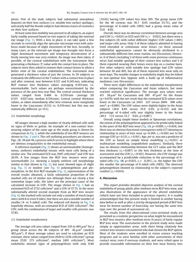
Fig. 3. Representative image of central corneal endothelium of a 25 year old male with 6 years of fairly regular daily wear of an RGP contact lens (A) and the image marking formorphometry (B). The estimated ECD is 2732 cells/mm2 and COV is 37.7%, a value very similar to the average from all of the RGP lens wearing subjects. The scale between thetwo broad vertical lines is 200 mm.
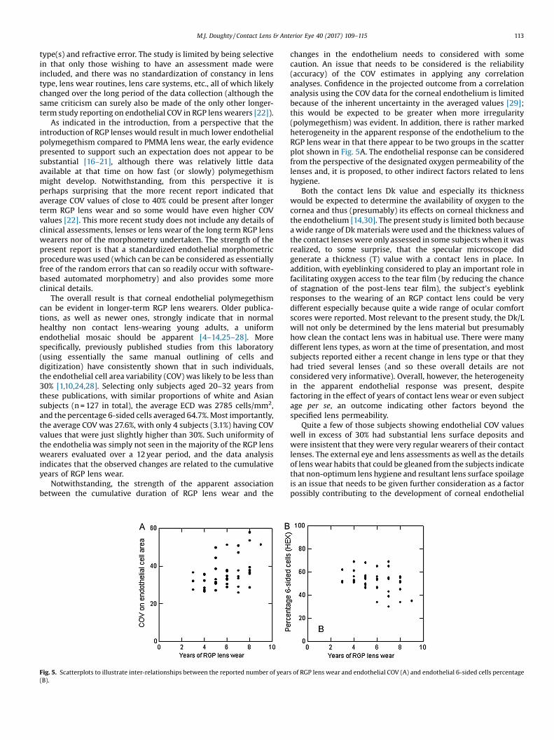
Fig. 4. Image of central corneal endothelium of a 28 year old male with 8 y of largely daily wear of an RGP lens and who had substantial lens spoilage (A) and the imagemarking for morphometry (B). The estimated ECD is 2201 cells/mm2 and the COV is 57.8%. The scale between the two broad vertical lines is 200 mm.
M.J. Doughty / Contact Lens & Anterior Eye 40 (2017) 109–115 111
3. Results
3.1. Subject characteristics
The 46 RGP lens wearing subjects, 31 being male, were agedbetween 20 and 32 years (average 26.0 � 2.4 y), in good generalhealth, and were from different ethnic groups, being identified aswhite Northern European (14), white Southern European (6), fromPakistan (4), SriLanka (1), mainland China (7), Hong Kong (3),Tawian (2), and Malaysia (3). Overall, these were grouped intoCaucasian (26) and Asian (20). There were many different lenstypes, as worn at the time of presentation, and most subjectsreported either a recent change in lens type or that they had triedseveral lenses. Notwithstanding, the reported lens productsincluded Boston 2, Boston 4 (IV), Boston XO2, Comfort HO, AquilaComfort, Menicon EX, Menicon Z, Alpha1, Quantum 2, Fluoroperm30 and 90, Icon Ultra, Metro O2, Paraperm O2 and 02 Plus, i.e.probably including the whole range of RGP lens Dk values from 30to 160. Lens thickness data was not collected simply because mostsubjects had no idea of what this was (although attempts weremade to measure these, see below). All the subjects were myopes(average refractive error of � 3.875 � � 1.166 DS, range � 2.50 to �7.50 DS), and reported an average period of 6.0 � 1.7 years of RGPlens wear (range 3 to 9 years, median 6.0 years).
All subjects indicated that they wore their contact lenses mostdays, and usually on a daily-wear routine (i.e. with overnight
cleaning and disinfection). Only 7 subjects insisted that they woretheir contact lenses every day, with most subjects reporting havingspectacles for wearing on some days especially if their lenses wereuncomfortable for some reason. If comfortable, then lens wear wasgenerally for the whole day. Just 6 subjects admitted having triedextended wear (up to a week) or continuous wear (up to 30 days)for periods of just a few months when trying another lens beforereturning (usually) to daily wear. No subject indicated any seriouscontact lens wear-related adverse events (infection, severe red eyeetc). The average comfort score, at the time of presentation, was79 � 12 (on a scale of 100, range 58–98), with some subjectsobviously tolerating their contact lens wear despite their eyes notfeeling very comfortable yet others reporting being remarkablycomfortable with their RGP lens wear. There was no overalldifference in average age (26.1 vs. 25.8 years, Caucasian versusAsian), refractive error (�3.798 vs. � 3.975 DS), or lens wearexperience (average 5.9 vs. 6.1 years) between the two ethnicgroupings.
External eye evaluations (Fig. 1) did not indicate any substantialredness or any other abnormalities of the bulbar conjunctiva, anysubstantial eyelid margin problems (with uniform or reasonablyuniform inferior tear meniscus), or significant Meibomian glanddysfunction (MGD), although in a few cases (7) the eyelid marginswere considered to have significant amounts of meibum presenton them. These gross external eye assessments indicated a normalpalpebral aperture with no obvious signs of RGP lens wear-related
112 M.J. Doughty / Contact Lens & Anterior Eye 40 (2017) 109–115
ptosis. Five of the male subjects had substantial amorphousdeposits on their lens surfaces (i.e. notable lens surface spoilage),and nearly half the female subjects had obvious traces of cosmeticson their lens surfaces.
At least some lens mobility was present in all subjects, an aspectfairly readily assessed based on two aspects of taking the externaleye image (Fig. 1). With a lens in place, it can take quite a fewseconds to correctly align the specular microsope even with auto-focus mode because of slight movement of the lens. Secondly, inmany cases, as the external eye image was brought into focus aslight downward movement and temporary decentration of afreely moving lens was very evident. An image was then taken, ifpossible, of the corneal endothelium with the instrument thengenerating a thickness (T) value with the contact lens in place. Thesubjects were then asked to remove their contact lenses to allow abetter view of the corneal endothelium. The instrument nowgenerated a thickness value of just the cornea. In 29 subjects soevaluated, the difference in the T values with a contact lens in placeand after removal, was between 0.127 and 0.245 mm, indicatingRGP contact lens thickness values averaging 0.193 mm, i.e.unremarkable. Such values are perhaps overestimated by thepresence of the post-lens tear film. The central corneal thicknessvalues ranged from 0.466 to 0.559 mm (average � SD0.515 � 0.027 mm, median 0.516 mm). Central corneal thicknessvalues, as taken immediately after lens removal, were marginallylower in the Caucasians (0.512 vs. 0.519 mm) but this was notstatistically different (p > 0.5).
3.2. Endothelial morphology
All images showed a high number of clearly-defined cells withno signs of blebs or guttae. An example of a non-contact lens-wearing subject of the same age as the study group is shown forcomparison in Fig. 2, while the endothelia of two RGP wearers areshown in Figs. 3 and 4. The cell outlining procedures are shown inFigs. 2B, 3B and 4B, a process which serves to highlight where thereare obvious irregularities in the endothelial mosaic.
A reference example (Fig. 2) shows an unremarkable (homoge-neous, uniform) endothelial mosaic with most cells being of asimilar size. The estimated ECD was 2947 cells/mm2 and COV was26.9%. A few images from the RGP lens wearers were alsounremarkable (i.e. showing a largely uniform cell morphologysimilar to that shown in Fig. 2), but most showed signs of slight(e.g. Fig. 3) or modest (see Fig. 4) polymegethism and ple-morphism. In the first RGP example (Fig. 3), representative of theoverall results obtained, a fairly substantial proportion of themarked cells are of similar size although there are clearly a fewsomewhat larger cells; the latter are the principal cause of thecalculated increase in COV. The image shown in Fig. 3 had anestimated ECD of 2732 cells/mm2 and a COV of 37.7%. In the moresubstantially altered second example from an RGP lens wearer(Fig. 4), the marked cells include not only a number of much largerones (with 8 or even 9 sides), but there are also a notable number ofsmaller (4- or 5-sided) cells. The reduced cell density in Fig. 3 ishopefully obvious, with an estimated ECD of 2201 cells/mm2. TheCOV, with the combination of larger and smaller cells present, was57.8%.
3.3. Endothelial morphometry
The average cell area values ranged from 310 to 495 mm2 for agroup mean across the 46 subjects of 401 �42 mm2 (median402 mm2). If these average values are used to calculate an ECDestimate, these values ranged from 2020 to 3226 cells/mm2 (groupmean 2520 � 273 cells/mm2, median 2491 cells/mm2). Mostendothelia showed signs of polymegethism with only 9/46
(19.6%) having COV values less than 30%. The group mean COVfor the 46 corneas was 36.7 � 8.0% (median 35.7%), and thepercentage of 6-sided cells (HEX) had a group mean value of51.8 � 8.8%.
Overall, there was no obvious correlation between average areaand COV (rs = 0.023) or ECD and COV (rs = �0.022), but there were afew subjects (8) with rather different (higher) COV values (>40%)and larger cells areas. Only 3 of these subjects admitted to havingtried extended or continuous wear lenses (so these unusualendothelial appearances cannot be obviously attributed to asubstantially different lens wear routine, see Discussion). Howev-er, 6 of them (including 2 of those with some previous continuouswear) had notable spoilage of their contact lens surface and 5 ofthem reported wearing their lenses every day on a routine basis.Five other subjects with notable lens spoilage had COV valuesbetween 30 and 40%, but all of these reported lens wear only onmost days. The notable changes in endothelia might thus be linkedto non-optimal lens hygiene with a build up of inflammatorymediators (see Discussion).
There were slight differences in the endothelial morphometrywhen comparing the Caucasian and Asian subjects, but nonereached statistical significance. The average area values were411 �38 mm2 for Caucasians and 389 � 47 mm2 for the Asians(p = 0.078) with the net result that the estimated ECD values werelower in the Caucasians (at 2455 � 227 versus 2604 � 308 cells/mm2; p = 0.080). The COV values were slightly higher in the Asiansubjects (38.8 � 8.8% versus 35.0 � 6.9%; p = 0.132) and thepercentage of 6-sided cells was slightly lower in the Asians(49.5 � 7.1% versus 53.7 � 9.6%, p = 0.087).
Overall, using simple linear models or Spearman correlations,the extent of the polymegethism appeared to be predictably linkedto the years of the RGP lens wear (Fig. 4A, p = 0.009, rs = 0.424), butthere was no obvious functional consequence with CCT showing norelationship to years of lens wear (p = 0.269, rs = 0.166) nor to theaverage COV (p = 0.121, rs = 0.181). Age, per se, was not a predictor ofthe COV as based on simple linear modelling (rs = 0.031) ormultivariant modelling (unpublished analyses). Similarly, therewas no obvious relationship between the CCT value and the RGPlens thickness (rs = 0.087) or the lens thickness at the time ofassessment and the COV (rs = 0.102). However, the COV change wasaccompanied by a predictable reduction in the percentage of 6-sided cells (Fig. 4B, p = 0.025, rs = �0.291), i.e. the higher the COVthe smaller the percentage of 6-sided cells (HEX). The observedpolymegethism showed no relationship to the subject’s reportedcomfort (rs = 0.014).
4. Discussion
This report provides detailed objective analysis of the cornealendothelia of young adults after medium-term RGP lens wear, andalso illustrations of the appearance of the altered endothelia(which have not been included in previous reports). It is readilyacknowledged that this present study is limited in neither havingdata before as well as after a strictly-designated period of RGP lenswear for known number of hours/day, nor having the same lenstype over the period of assessment etc.
The results from this observational cross-sectional study arepresented as a realistic perspective on what might be encounteredin RGP lens wearers after medium-term (6 years) of fairly regularRGP lens wear. The assessments in the present study were madesimply as opportunities arose, with there being relatively fewcontact lens wearers encountered who had chosen the RGP option.Most of the students were enrolled in vision science teachingprograms and so were familiar with other assessments of theircontact wear, even if overseas students, and were relied upon toprovide reasonable information on their lens wear history, lens
M.J. Doughty / Contact Lens & Anterior Eye 40 (2017) 109–115 113
type(s) and refractive error. The study is limited by being selectivein that only those wishing to have an assessment made wereincluded, and there was no standardization of constancy in lenstype, lens wear routines, lens care systems, etc., all of which likelychanged over the long period of the data collection (although thesame criticism can surely also be made of the only other longer-term study reporting on endothelial COV in RGP lens wearers [22]).
As indicated in the introduction, from a perspective that theintroduction of RGP lenses would result in much lower endothelialpolymegethism compared to PMMA lens wear, the early evidencepresented to support such an expectation does not appear to besubstantial [16–21], although there was relatively little dataavailable at that time on how fast (or slowly) polymegethismmight develop. Notwithstanding, from this perspective it isperhaps surprising that the more recent report indicated thataverage COV values of close to 40% could be present after longerterm RGP lens wear and so some would have even higher COVvalues [22]. This more recent study does not include any details ofclinical assessments, lenses or lens wear of the long term RGP lenswearers nor of the morphometry undertaken. The strength of thepresent report is that a standardized endothelial morphometricprocedure was used (which can be can be considered as essentiallyfree of the random errors that can so readily occur with software-based automated morphometry) and also provides some moreclinical details.
The overall result is that corneal endothelial polymegethismcan be evident in longer-term RGP lens wearers. Older publica-tions, as well as newer ones, strongly indicate that in normalhealthy non contact lens-wearing young adults, a uniformendothelial mosaic should be apparent [4–14,25–28]. Morespecifically, previously published studies from this laboratory(using essentially the same manual outlining of cells anddigitization) have consistently shown that in such individuals,the endothelial cell area variability (COV) was likely to be less than30% [1,10,24,28]. Selecting only subjects aged 20–32 years fromthese publications, with similar proportions of white and Asiansubjects (n = 127 in total), the average ECD was 2785 cells/mm2,and the percentage 6-sided cells averaged 64.7%. Most importantly,the average COV was 27.6%, with only 4 subjects (3.1%) having COVvalues that were just slightly higher than 30%. Such uniformity ofthe endothelia was simply not seen in the majority of the RGP lenswearers evaluated over a 12 year period, and the data analysisindicates that the observed changes are related to the cumulativeyears of RGP lens wear.
Notwithstanding, the strength of the apparent associationbetween the cumulative duration of RGP lens wear and the
Fig. 5. Scatterplots to illustrate inter-relationships between the reported number of year(B).
changes in the endothelium needs to considered with somecaution. An issue that needs to be considered is the reliability(accuracy) of the COV estimates in applying any correlationanalyses. Confidence in the projected outcome from a correlationanalysis using the COV data for the corneal endothelium is limitedbecause of the inherent uncertainty in the averaged values [29];this would be expected to be greater when more irregularity(polymegethism) was evident. In addition, there is rather markedheterogeneity in the apparent response of the endothelium to theRGP lens wear in that there appear to be two groups in the scatterplot shown in Fig. 5A. The endothelial response can be consideredfrom the perspective of the designated oxygen permeability of thelenses and, it is proposed, to other indirect factors related to lenshygiene.
Both the contact lens Dk value and especially its thicknesswould be expected to determine the availability of oxygen to thecornea and thus (presumably) its effects on corneal thickness andthe endothelium [14,30]. The present study is limited both becausea wide range of Dk materials were used and the thickness values ofthe contact lenses were only assessed in some subjects when it wasrealized, to some surprise, that the specular microscope didgenerate a thickness (T) value with a contact lens in place. Inaddition, with eyeblinking considered to play an important role infacilitating oxygen access to the tear film (by reducing the chanceof stagnation of the post-lens tear film), the subject’s eyeblinkresponses to the wearing of an RGP contact lens could be verydifferent especially because quite a wide range of ocular comfortscores were reported. Most relevant to the present study, the Dk/Lwill not only be determined by the lens material but presumablyhow clean the contact lens was in habitual use. There were manydifferent lens types, as worn at the time of presentation, and mostsubjects reported either a recent change in lens type or that theyhad tried several lenses (and so these overall details are notconsidered very informative). Overall, however, the heterogeneityin the apparent endothelial response was present, despitefactoring in the effect of years of contact lens wear or even subjectage per se, an outcome indicating other factors beyond thespecified lens permeability.
Quite a few of those subjects showing endothelial COV valueswell in excess of 30% had substantial lens surface deposits andwere insistent that they were very regular wearers of their contactlenses. The external eye and lens assessments as well as the detailsof lens wear habits that could be gleaned from the subjects indicatethat non-optimum lens hygiene and resultant lens surface spoilageis an issue that needs to be given further consideration as a factorpossibly contributing to the development of corneal endothelial
s of RGP lens wear and endothelial COV (A) and endothelial 6-sided cells percentage
114 M.J. Doughty / Contact Lens & Anterior Eye 40 (2017) 109–115
polymegethism and pleomorphism. This could be a result ofimpairment of oxygen transmissibility of the lenses and/or thedevelopment of a mild inflammatory response to deposits on thelens surface. Assessments of tear film levels of inflammatorymediators in RGP contact lens wearers have been reported [31–34].One earlier report with palpebral assessments noted no detectabledifference in nitric oxide levels after an average of nearly 3 years ofwear [31]. However, three other reports without palpebralassessments indicate possible increases in another type ofinflammatory mediator, interleukins, in the tears of RGP lenswearers [32–34]. Further studies on this aspect of RGP lens wear, toinclude assessments of the endothelium would be useful. Suchincreases may also be accompanied by a decrease in tear film levelsof natural anti-inflammatory molecules such as secretoglobulins inRGP lens wearers [35]
A final comment seems appropriate for the apparent endothe-lial change and its function. In early consideration of thepolymegethism associated with PMMA lens wear, the effect wasattributed to hypoxia and considered to be an indicator of alteredfunction of the endothelial cells [36]. As seen in specular reflection,the endothelial mosaic appears to be remarkably uniform. It shouldbe noted that the specular microscopy method for observation ofthe corneal endothelial cells is limited to the apical surface of thecells (adjacent to the aqueous humour) and not of the whole cell.While this approach to viewing the endothelial cells will likelyremain for clinical studies, laboratory-based imaging of isolatedcorneas at much higher magnification has indicated that theapparent geometric uniformity of the cells may not be evident atthe posterior surface of the endothelium (adjacent to Desçemet’smembrane) [37]. The difference between the cell morphology atthe anterior and posterior surfaces has been proposed to beimportant in determining the net function of the endothelial celllayer [37], although it remains to be established how predictablethis difference might be especially if (anterior) polymegethism orpleomorphism were evident at the apical surface.
In conclusion, mild-to-moderate corneal endothelial polyme-gethism appears to be a commonplace consequence of themedium-term wear of RGP contact lenses, an effect that mightseem at odds with the lens classification. The results in the presentstudy confirm and extend those from previous prospective [14,16]and observational cross-sectional studies [22]. Overall, the presentstudies indicate that further assessments would be usefulespecially to assess the external eye and palpebral conjunctivain more detail in long term RGP lens wearers in addition to thecorneal endothelium.
Funding
The author has no funding or conflicts of interest to disclose.
Acknowledgements
The author has no proprietary interests in any of the equipmentor methods presented, and has no funding or conflicts of interest todisclose.
References
[1] M.J. Doughty, Are there geometric determinants of cell area in rabbit andhuman corneal endothelial cell monolayers? Tissue Cell 30 (1998) 537–544.
[2] M.J. Doughty, Comparative anatomy and physiology of the cornea andconjunctiva, in: R. Martin Herran, R.M. Corrales (Eds.), Ocular Surface, CRCPress, 2012, pp. 32–78.
[3] M.J. Doughty, Towards a quantitative analysis of corneal endothelial cellmorphology � a review of techniques and their application, Optom. Vis. Sci. 66(1989) 626–642.
[4] A. Stefansson, O. Müller, R. Sundmacher, Non-contact specular microscopy ofthe normal corneal endothelium. A statistical evaluation of morphometricparameters, Graefe’s Arch. Clin. Exp. Ophthalmol. 218 (1982) 200–205.
[5] M. Matsuda, R.W. Yee, H.F. Edelhauser, Comparison of the corneal endotheliumin an American and Japanese population, Arch. Ophthalmol. 103 (1985) 68–70.
[6] R.W. Yee, M. Matsuda, R.O. Schultz, H.F. Edelhause, Changes in the normalcorneal endothelial pattern as a function of age, Curr. Eye Res. 4 (1985) 671–678.
[7] K.H. Carlson, W.M. Bourne, J.W. Mclaren, R.F. Brubaker, Variations on humancorneal endothelial cell morphology and permeability to fluorescein with age,Exp. Eye Res. 47 (1988) 27–41.
[8] K.A. Polse, R.J. Brand, S.R. Cohen, M. Guillon, Hypoxic effects on cornealmorphology and function, Invest. Ophthalmol. Vis. Sci. 31 (1990) 1542–1554.
[9] K.-S. Kim, S.-Y. Park, J.-S. Oh, Morphometric analysis of the corneal endothelialcells in normal Koreans [In Korean], J. Korean Ophthalmol. Soc. 33 (1992) 24–28.
[10] M.J. Dought, Prevalence of ‘non-hexagonal’ cells in the corneal endothelium ofyoung Caucasian adults, and their inter-relationships, Ophthal. Physiol. Opt.18(1998) 415–422.
[11] S.W. Cheung, P. Cho, Endothelial cell analysis using the Topcon SP-1000 non-contact specular microscope and IMAGEnet system, Clin. Exp. Optom. 81(1998) 1–7.
[12] S.W. Cheung, P. Cho, Endothelial cells analysis with the TOPCON specularmicroscope SP-2000P and IMAGEnet system, Curr. Eye Res. 21 (2000) 788–798.
[13] S.H. Tseng, F.K. Chen, Morphological and fluorophotometric analysis of thecorneal endothelium after radial keratotomy, Cornea 17 (1998) 471–475.
[14] W.M. Bourne, S.B. Holtan, D.O. Hodge, Morphologic changes in cornealendothelial cells during 3 years of fluorocarbon contact lens wear, Cornea 18(1999) 29–33.
[15] S.M. MacRae, M. Matsuda, S. Shellans, Corneal endothelial changes associatedwith contact lens wear, CLAO J. 15 (1989) 82–87.
[16] G. Liberman, R.B. Mandell, Corneal endothelial polymegethism in high-Dkcontact lens wearers, Int. Contact Lens Clin. 15 (1988) 282–284.
[17] G.N. Orsborn, J.P. Schoessler, Corneal endothelial polymegethism after theextended wear of rigid gas-permeable contact lenses, Am. J. Optom. Physiol.Opt. 65 (1988) 84–90.
[18] H. Shioya, M. Kajita, K. Kobari, H. Yamaguchi, K. Kato, Corneal endothelial cellchange in gas-permeable hard contact lens wearers [in Japanese], J. Jpn.Contact Lens Soc. 33 (1991) 158–161.
[19] C.P. Nieuwendaal, J.H.C. Kok, E.A.M. de Moor, J. Oosting, H.W. Venema, Cornealendothelial cell morphology under permanent wear of rigid contact lenses, Int.Ophthalmol. 15 (1991) 313–319.
[20] K. Ogihara, H. Yamaguchi, Prospective study of corneal endothelial cellchanges in the early stages of contact lens wear [in Japanese], J. Jpn. ContactLens Soc. 37 (1995) 118–122.
[21] Y. Mizutani, S. Mizutani, H. Takahashi, K. Ohara, The effects of contact lenses onthe corneal endothelium [In Japanese], J. Jpn. Contact Lens Soc. 40 (1998) 164–171.
[22] J.T. Barr, B. Pall, L.B. Szczotka, G.L. Mitchell, W. Gleason, Corneal endothelialmorphology results in the Meinicon Z 30-day continuous-wear contact lensclinical trial, Eye Contact Lens 29 (2003) 14–16.
[23] M.J. Doughty, C.-A. Lee, S. Ritchie, T. Naase, An assessment of the discomfortassociated with the use of rose bengal 1% eyedrops on the human eye � acomparison with saline 0.9% and a topical ocular anaesthetic, Ophthal. Physiol.Opt. 27 (2007) 159–167.
[24] M.J. Doughty, A prospective analysis of corneal endothelial polymegethismand cell density in young adult Asians, Clin. Exp. Otom. 97 (2014) 256–263.
[25] M.J. Doughty, B.M. Aakre, Further analysis of assessments of the coefficient ofvariation of corneal endothelial cell areas from specular microscope images,Clin. Exp. Optom. 91 (2008) 438–446.
[26] C.J. Giasson, L. Gosselin, A. Masella, P. Forcier, Does endothelial cell densitycorrelate with corneal diameter in a group of young adults? Cornea 27 (2008)640–643.
[27] M.J. Doughty, A. Müller, M.L. Zaman, Assessment of the reliability of humancorneal endothelial cell-density estimates using a noncontact specularmicroscope, Cornea 19 (2000) 148–158.
[28] M.J. Doughty, Evaluation of possible error sources in corneal endothelialmorphometry with a semi-automated non-contact specular microscope,Cornea 32 (2013) 1196–1203.
[29] M.J. Doughty, D. Fonn, T. Nguyen, Assessment of the reliability of calculationsof the coefficient of variation of normal and poylmegethous cornealendothelium, Optom. Vis. Sci. 70 (1993) 759–770.
[30] H.P. Gardner, B.A. Fink, L.G. Mitchell, R.M. Hill, The effects of High-Dk rigidcontact lens center thickness material permeability, and blinking on theoxygen uptake of the human cornea, Optom. Vis. Sci. 82 (2005) 459–466.
[31] S. Karaküçuk, E. Mirza, I. Karaküçuk, A. Akal, M. Er, Nitric oxide levels in tears ofpatients with mild forms of papillary conjunctivitis induced by rigid gas-permeable contact lenses, CLAO J. 28 (2002) 5–8.
[32] P. Kallinkos, P. Morgan, N. Efron, Assessment of stromal keratocytes and tearfilm inflammatory mediators during extended wear of contact lenses, Cornea25 (2006) 1–10.
[33] I. Lema, J.A. Durán, C. Ruiz, E. Diez-Feijoo, A. Acera, J. Merayo, Inflammatoryresponse to contact lenses in patients with keratoconus compared withmyopic subjects, Cornea 27 (2008) 758–763.

M.J. Doughty / Contact Lens & Anterior Eye 40 (2017) 109–115 115
[34] C.Y. Elgin, G. _Iskeleli, S. Talaz, S. Akyol, Comparative analysis of tear film levelsof inflammatory mediators in contact lens users, Curr. Eye Res. 41 (2015) 441–447.
[35] C. Kramann, N. Boehm, K. Lorenz, N. Wehrwein, B.M. Stoffelns, N. Pfeiffer, F.H.Grus, Effect of contact lenses on the protein composition of tear film: aProteinChip study, Graefes Arch. Clin. Exp. Ophthalmol. 249 (2011) 233–243.
[36] C.G. Connor, M.E. Zagrod, Contact lens-induced corneal endothelialpolymegethism: functional significance and possible mechanisms, Am. J.Optom. Physiol. Opt. 63 (1986) 544–639.
[37] Z. He, F. Forest, P. Gain, D. Rageade, A. Bernard, S. Acquart, M. Peoc’h, D. Defoe,G. Thuret, 3D map of the human corneal endothelial cell, Sci. Rep. 6 (2016)29047.


![MiSight 1day - cahiers-ophtalmologie...Global trends in myopia management attitudes and strategies in clinical practice. Cont Lens Anterior Eye. 2016;39:106-16. [8] Bullimore MA. The](https://img.dokumen.tips/doc/110x75/6102037accd54931976798f6/misight-1day-cahiers-ophtalmologie-global-trends-in-myopia-management-attitudes.jpg)


![Contact Lens and Anterior Eye · [32], the Symptom Assessment in Dry Eye (SANDE) [33] and the Dry Eye Questionnaire (DEQ-5; short version) [34]. 2.2.2. Tear film osmolarity Tear](https://img.dokumen.tips/doc/110x75/5feb70c08fd7cf124839e0cf/contact-lens-and-anterior-eye-32-the-symptom-assessment-in-dry-eye-sande-33.jpg)