Embed Size (px)
Citation preview

Bryn Mawr College Bryn Mawr College
Scholarship, Research, and Creative Work at Bryn Mawr College Scholarship, Research, and Creative Work at Bryn Mawr College
Chemistry Faculty Research and Scholarship Chemistry
2018
Structural features and domain movements controlling substrate Structural features and domain movements controlling substrate
binding and cofactor specificity in class II HMG-CoA reductase binding and cofactor specificity in class II HMG-CoA reductase
Bradley R. Miller Bryn Mawr College, [email protected]
Yan Kung Bryn Mawr College, [email protected]
Follow this and additional works at: https://repository.brynmawr.edu/chem_pubs
Part of the Biochemistry Commons, Other Biochemistry, Biophysics, and Structural Biology Commons,
and the Structural Biology Commons
Let us know how access to this document benefits you.
Custom Citation Custom Citation Miller, B. R. and Kung, Yan. 2018. "Structural Features and Domain Movements Controlling Substrate Binding and Cofactor Specificity in Class II HMG-CoA Reductase." Biochemistry 57.5: 654-662.
This paper is posted at Scholarship, Research, and Creative Work at Bryn Mawr College. https://repository.brynmawr.edu/chem_pubs/27
For more information, please contact [email protected].

Structural Features and Domain Movements Controlling SubstrateBinding and Cofactor Specificity in Class II HMG-CoA ReductaseBradley R. Miller and Yan Kung*
Department of Chemistry, Bryn Mawr College, 101 North Merion Avenue, Bryn Mawr, Pennsylvania 19010, United States
*S Supporting Information
ABSTRACT: The key mevalonate pathway enzyme 3-hydroxy-3-methylglutaryl coenzyme A (HMG-CoA) reduc-tase (HMGR) uses the cofactor NAD(P)H to reduce HMG-CoA to mevalonate in the production of countless metabolitesand natural products. Although inhibition of HMGR by statindrugs is well-understood, several mechanistic details ofHMGR catalysis remain unresolved, and the structural basisfor the wide range of cofactor specificity for either NADH orNADPH among HMGRs from different organisms is alsounknown. Here, we present crystal structures of HMGR fromStreptococcus pneumoniae (SpHMGR) alongside kinetic data ofthe enzyme’s cofactor preferences. Our structure of SpHMGRbound with its kinetically preferred NADPH cofactor suggests how NADPH-specific binding and recognition are achieved. Inaddition, our structure of HMG-CoA-bound SpHMGR reveals large, previously unknown conformational domain movementsthat may control HMGR substrate binding and enable cofactor exchange without intermediate release during the catalytic cycle.Taken together, this work provides critical new insights into both the HMGR reaction mechanism and the structural basis ofcofactor specificity.
HMGR catalyzes the rate-limiting step of the mevalonatepathway, which is found in all kingdoms of life and isresponsible for the biosynthesis of an enormously wide rangeof molecules, from steroids such as cholesterol to isoprenoids,which make up the largest and most diverse class of naturalproducts. Its key role in the biosynthesis of steroids makesHMGR the target of cholesterol-lowering statin drugs. HMGRperforms the four-electron reduction of HMG-CoA tomevalonate and CoA using 2 equiv of the redox cofactorNAD(P)H. The enzyme has evolved into two distinctclasses,1−3 where class I HMGRs are present in eukaryotesand in some bacteria and archaea, while class II enzymes arefound in only bacteria and archaea. Although both HMGRclasses exhibit similar overall folds, with active sites located at ahomodimeric interface, there are many significant differences,including those in catalytic regions of the protein.2
Class I and class II HMGRs also differ in their NAD(P)Hcofactor preferences: although all class I HMGRs, includingthe human enzyme, utilize NADPH exclusively, class IIHMGRs display a wide range of cofactor specificities. Someclass II enzymes use only NADH, including the HMGRs fromPseudomonas mevalonii (PmHMGR)4 and Burkholderia cen-ocepacia,5 while others use only NADPH, such as HMGR fromEnterococcus faecalis.6 Other class II HMGRs are able to useboth NADH and NADPH, often with weak or strongpreferences for one cofactor or the other, including HMGRsfrom Staphylococcus aureus,7 Listeria monocytogenes,8 andArchaeoglobus fulgidus.9
Despite the fact that HMGR inhibition by statins is well-understood, the molecular details of the HMGR catalyticmechanism remain somewhat enigmatic.10 Current proposalssuggest that HMG-CoA is first reduced to a mevaldyl-CoAintermediate using the first equivalent of NAD(P)H. The orderof the next two steps is uncertain. Oxidized NAD(P)+ must beexchanged for a second equivalent of NAD(P)H, andmevaldyl-CoA is also cleaved to form a mevaldehydeintermediate and CoA. Lastly, mevaldehyde is reduced tomevalonate by the second NAD(P)H, and the final productsare released.Interestingly, HMGR can also produce mevalonate if
provided with its mevaldyl-CoA or mevaldehyde intermedi-ates,11,12 skipping the first reduction step. However, whenHMG-CoA is the substrate, neither intermediate is releasedduring the catalytic cycle;10,13−15 instead, the enzyme waitsuntil the second reduction step is complete to release the finalproducts. How HMGR is able trigger and accomplish cofactorexchange during the reaction without releasing the boundintermediates is not known. In addition, the structural basis forclass II HMGR’s wide range of cofactor specificity is alsounclear. Greater knowledge of the structural features thatcontrol substrate and cofactor binding in class II HMGR notonly would provide insight into its catalytic mechanism but
Received: October 3, 2017Published: December 11, 2017
Article
pubs.acs.org/biochemistryCite This: Biochemistry 2018, 57, 654−662
© 2017 American Chemical Society 654 DOI: 10.1021/acs.biochem.7b00999Biochemistry 2018, 57, 654−662

also may enable the development of drugs that target thiscrucial metabolic enzyme in human pathogens.To date, crystal structures for only two class II HMGRs have
been determined, namely, the NADH-specific PmHMGR andHMGR from the major human pathogen Streptococcuspneumoniae (SpHMGR). SpHMGR inhibition has beenpreviously studied,16 but its cofactor preferences have notbeen determined until now. For PmHMGR, many structuresare available,17−19 including the ternary complex depicting theenzyme bound simultaneously with both the cofactor in itsoxidized form, NAD+, and the substrate HMG-CoA [ProteinData Bank (PDB) entry 1QAX] or the substrate analoguedithio-HMG-CoA (PDB entry 4I4B). For SpHMGR, twostructures have been determined in the absence of any boundligands (PDB entries 3QAE and 3QAU). Interestingly, a C-terminal domain that is disordered in the vast majority ofHMGR structures, indicating a high degree of domainmobility, was ordered in structures of the PmHMGR ternarycomplex and apo-SpHMGR. Here, the C-terminal domain inapo-SpHMGR was flipped away from the substrate- andcofactor-binding sites, giving rise to an “open” conformation,while the C-terminal domain in the PmHMGR ternarycomplex was positioned directly over the substrate- andcofactor-binding sites in a “closed” conformation (Figure 1),
where the C-terminal domain contributes a catalyticallyessential histidine, as indicated by mutagenic and structuralstudies.18,20,21 By alternating between “open” and “closed”conformations, the C-terminal domain might be capable ofacting as a “flap” to cover the active site when the substrateand/or cofactor is bound.18 However, when, how, and whattriggers this C-terminal domain movement during the reactionare unknown.To gain mechanistic insight into HMGR catalysis and to
shed light on how substrate binding and cofactor bindinginfluence the enzyme’s structure during the reaction, we firstdetermined the cofactor preferences of SpHMGR and foundthat although the enzyme can use both NADH and NADPH toreduce HMG-CoA, it has a strong kinetic preference forNADPH. We also determined two crystal structures ofSpHMGR, one bound with substrate HMG-CoA and one
bound with its preferred cofactor NADPH, which representsthe first structure of an NADPH-bound class II HMGR. Thesecrystal structures not only provide new structural insight intoHMGR cofactor binding and specificity but also reveal new C-terminal domain conformations, allowing us to illuminate thestructural movements that enable HMGR reactivity.
■ MATERIALS AND METHODSCloning, Expression, and Purification. A codon-
optimized, linear mvaA gene that encodes SpHMGR(Integrated DNA Technologies) was cloned into a modifiedpET28b plasmid termed pSKB3, which encodes an N-terminal,TEV protease-cleavable hexahistidine tag and a kanamycinresistance cassette, using NdeI and BamHI restriction enzymes.The plasmid was transformed into Escherichia coli DH10Bcells, and its gene sequence was confirmed (QuintaraBiosciences) before its transformation into BL21(DE3) cellsfor protein expression.Cells were grown in lysogeny broth supplemented with
kanamycin at 37 °C until OD600 reached ∼0.6. Proteinexpression was induced with 0.5 mM isopropyl β-D-1-thiogalactopyranoside (IPTG) and proceeded for 18 h at 16°C. Cells were harvested by centrifugation at 5000g for 10 min,flash-frozen in liquid nitrogen, and stored at −80 °C. Cellswere resuspended in lysis buffer [50 mM Tris (pH 7.7), 200mM NaCl, 10% glycerol, and 10 mM imidazole] with 0.5 unit/μL benzonase (Millipore) and 0.5 mM phenylmethanesulfonylfluoride (PMSF) and lysed by sonication on ice at 40%amplitude for 9 min with 3 s bursts and 5 s rests. The lysatewas clarified by centrifugation at maximum speed (∼37000g)for 30 min at 4 °C, and the supernatant was applied to a Ni-NTA column equilibrated with lysis buffer. SpHMGR waseluted from the column in fractions using lysis buffer with 300mM imidazole and assessed for purity by SDS−PAGE.Fractions containing the highest purity were pooled;hexahistidine-tagged TEV protease was added to cleave thehexahistidine tag from SpHMGR, and the sample was dialyzedovernight at 4 °C against 50 mM Tris (pH 7.7), 200 mMNaCl, 10% glycerol, and 0.5 mM EDTA. The sample was runover a second Ni-NTA column using lysis buffer to purifycleaved SpHMGR. The protein was further purified by gelfiltration on a HiLoad 16/600 Superdex 200 prep gradecolumn (GE Healthcare Life Sciences) equilibrated with 50mM Tris (pH 7.7), 200 mM NaCl, and 10% glycerol using anAkta Pure chromatography system (GE Healthcare LifeSciences). Purified SpHMGR was concentrated to 15 mg/mL, flash-frozen dropwise in liquid nitrogen, and stored at −80°C.
Kinetic Characterization. The cofactor specificity wasassessed kinetically using a NanoDrop 2000c spectrophotom-eter (ThermoFisher). Each 100 μL reaction mixture at 37 °Ccontained 50 mM Tris (pH 7.4), 50 mM NaCl, 300 μMHMG-CoA, and 25−500 μM NAD(P)H. Reactions wereinitiated by the addition of 20 nM SpHMGR for NADPHreactions or 100 nM SpHMGR for NADH reactions. Enzyme-catalyzed oxidation of NAD(P)H was monitored via a decreasein absorbance at 340 nm, using an extinction coefficient of6200 M−1 cm−1. The Michaelis−Menten constant, Km, and themaximum velocity, Vmax, for the production of mevalonatewere determined using nonlinear regression by fitting thereaction velocities to the Michaelis−Menten equation inGraphPad Prism 6.0. The values for kcat were obtained bydividing Vmax by the molar enzyme concentration. Values are
Figure 1. Structures of “open” and “closed” class II HMGR. Alignedcrystal structures of apo-SpHMGR (PDB entry 3QAU) and thesubstrate- and cofactor-bound PmHMGR ternary complex (PDBentry 1QAX), representing “open” (blue) and “closed” (green)conformations of the C-terminal domain (rmsd for Cα atoms of 0.80).Structures were aligned with their C-terminal domains excluded. Thecofactor and substrate are labeled and shown as sticks, with C coloredgreen, N colored blue, O colored red, P colored orange, and S coloredyellow.
Biochemistry Article
DOI: 10.1021/acs.biochem.7b00999Biochemistry 2018, 57, 654−662
655

given as the means ± the standard error of the mean (SEM)from triplicate experiments.Crystallization. Crystallization conditions for HMG-CoA-
bound SpHMGR were identified by sparse-matrix screening bysitting-drop vapor diffusion using a Crystal Gryphon (ArtRobbins Instruments) with 10 mg/mL SpHMGR and 1 mMHMG-CoA. Crystals were observed under condition 17 ofCrystal Screen 1 (Hampton Research), which contains 100mM Tris (pH 8.5), 200 mM lithium sulfate, and 30%polyethylene glycol (PEG) 4000. Crystallization conditionswere optimized by hanging-drop vapor diffusion with varyinglithium sulfate and PEG 4000 concentrations, and smallcrystals grew overnight. Crystals were washed in mother liquor,crushed via vortexing, and used as seeds for microseeding.Seeded drops contained 1.0 μL of 10 mg/mL SpHMGR with 1mM HMG-CoA, 0.8 μL of crystallization solution, and 0.2 μLof the seed stock. Large crystals grew in 100 mM Tris (pH8.5), 100−250 mM lithium sulfate, and 15−25% PEG 4000.The crystals were cryoprotected using the crystallizationsolution supplemented with 20% glycerol and 1 mM HMG-CoA before being flash-cooled in liquid nitrogen.NADPH-bound SpHMGR crystals grew under the same
crystallization condition that was used for the HMG-CoA-bound SpHMGR crystals. However, a new crystal form thattook several weeks to grow appeared to use the HMG-CoA-bound SpHMGR microcrystals as nucleation sites for crystalgrowth. These rod-shaped crystals were optimized using theseeding protocol described above and co-crystallized with 2.5mM NADPH. Large crystals grew in 100 mM Tris (pH 8.5),100−250 mM lithium sulfate, and 30−40% PEG 4000. Thecrystals were cryoprotected using the crystallization solutionsupplemented with 20% glycerol and 5 mM NADPH beforebeing flash-cooled in liquid nitrogen.X-ray Data Collection, Structure Determination, and
Refinement. X-ray diffraction data were collected atAdvanced Photon Source (APS) beamline 24-ID-E. The datawere indexed, merged, and scaled using iMOSFLM22 in spacegroup P1 with four molecules in the asymmetric unit for theNADPH-bound structure and space group P21 with twomolecules in the asymmetric unit for the HMG-CoA-boundstructure. Structures were determined by molecular replace-ment using Phaser23 in the PHENIX suite,24 where the apo-SpHMGR structure (PDB entry 3QAE) with unresolved C-terminal domains was used as the search model. Electrondensity maps indicated that the C-terminal domains werelocated in novel locations for both structures. Therefore, theywere built manually in Coot25 with iterations of reciprocalspace refinement using phenix.refine.26 After the structureswere determined, we noticed that our protein contained aV355E mutational artifact. As this site is quite distant from thecofactor- and substrate-binding sites (>20 Å) as well as the C-terminal domains (>35 Å), we believe that this is unlikely tohave a significant impact on the main findings of this paper.
■ RESULTSKinetic Characterization of SpHMGR. Previous studies
of SpHMGR focused on enzyme inhibition and characterizedthe activity using NADPH but did not examine cofactorspecificity.16 To determine the cofactor preference ofSpHMGR, we measured steady-state kinetics with varyingconcentrations of either NADH or NADPH (Table 1). Withrespect to NADPH, SpHMGR has a Km of 28.9 ± 5.1 μM anda kcat of 6.85 ± 0.3 s−1. With NADH, the enzyme has a Km of
153 ± 59.3 μM and a kcat of 0.131 ± 0.02 s−1. The resultingcatalytic efficiencies (kcat/Km) are 2.4 × 10⁵ M−1 s−1 forNADPH and 8.6 × 10² M−1 s−1 for NADH. These data showthat although SpHMGR can use either cofactor for HMG-CoAreduction, NADPH is the preferred cofactor by approximately280-fold in terms of kcat/Km, with both a lower Km and a higherkcat for NADPH compared with those of NADH.
Overall Structures. SpHMGR bound to its preferredcofactor NADPH crystallized in the P1 space group with fourmolecules in the asymmetric unit, arranged as two homodimers(chains A and B and chains C and D). The overall structure isnearly identical to the prior apo-SpHMGR structures (PDBentries 3QAE and 3QAU), except for the C-terminal domains,detailed below, with root-mean-square deviations for Cα atoms(rmsd’s) of 0.23−0.25. Clear electron density was observed forall four monomers of the asymmetric unit, except the C-terminal domains (residues 375−424) of chains A and C weredisordered. Electron density in these regions was weak anddiscontinuous, and thus, we did not model the C-terminaldomains for chains A and C. Therefore, in the final model,chains A and C contain residues 3−372 of 424 while chains Band D contain residues 11−424 and 3−424, respectively. Inaddition, positive difference maps showed density in thecofactor-binding sites of both chains A and C, representingNADPH binding. For chain A, clear electron density forNADPH was observed, including for the key 2′-phosphategroup, though it may be noted that the 2′-phosphate density ispartially discontinuous with the rest of the NADPH molecule(Figure 2A), suggesting a small degree of disorder. For chainC, however, electron density for NADPH was significantly
Table 1. SpHMGR Cofactor Preferences
NADPH NADH
Km (μM) 28.9 ± 5.1 153 ± 59.3kcat (s
−1) 6.85 ± 0.3 0.131 ± 0.02kcat/Km (M−1 s−1) 2.4 × 10⁵ 8.6 × 10²
Figure 2. NADPH- and HMG-CoA-binding sites of SpHMGR, withmFO − DFC omit density for (A) NADPH and (B) HMG-CoA boundto SpHMGR (PDB entries 5WPJ and 5WPK, respectively). Theprotein is shown as a gray cartoon; NADPH and HMG-CoA areshown as sticks, with C colored green, N colored blue, O colored red,P colored orange, and S colored yellow. The mFO − DFC polder omitmap is contoured at 3.0σ (pink mesh) and 2.5σ (blue mesh), ascalculated in Phenix.24,33
Biochemistry Article
DOI: 10.1021/acs.biochem.7b00999Biochemistry 2018, 57, 654−662
656

weaker and included significant negative FO − FC differencedensity around the nicotinamide and adenosine rings. A trialrefinement at 70% occupancy for NADPH mostly satisfied thedifference maps (Figure S1), but because the 2FO − FC mapsremained highly discontinuous, we chose to leave NADPH inchain C out of the final model.SpHMGR bound to HMG-CoA crystallized in the P21 space
group with two molecules in the asymmetric unit assembled asa homodimer (chains A and B). The C-terminal domain wasresolved only in chain B; therefore, in the final model, chain Acontains residues 1−379 while chain B contains residues 3−424. Electron density maps showed clear density for HMG-CoA in the substrate-binding sites of both chains (Figure 2B).Complete X-ray diffraction and refinement statistics are listedin Table 2.
In the obligate HMGR homodimer, the larger N-terminaldomain, which includes the active site, forms a majority of thedimer interface. Interestingly, the interface contains aninterlocking motif where residues 1−69 cross and intertwinewith each other through loops formed by residues 38−58,forming an interlocked β-sheet that contains several conservedresidues and has been observed in prior structures of HMGR
(Figure S2A,B).27,28 This region is also involved in substratebinding, as in our HMG-CoA-bound SpHMGR structureGlu50 and Asn51 from the interlocking loop of one monomerinteract with the adenine ring of HMG-CoA bound by theadjacent monomer (Figure S2C).
Substrate- and Cofactor-Binding Sites. The HMG-CoAsubstrate and the NAD(P)H cofactor are both long moleculesthat bind HMGR with their reactive groups pointing towardeach other in the buried active site core at the homodimericinterface and with the rest of the molecules extending out fromthe active site in different directions, together resembling a “V”shape. Correspondingly, in our structure of HMG-CoA-boundSpHMGR (Figure 3A), the substrate binds with its reactive
HMG moiety in the active site, with Arg257 forming a saltbridge with the carboxylate of the HMG moiety, which alsointeracts via a water molecule with the backbone carbonyl ofHis261 and the side chain of Asn362. The pantothenate groupof HMG-CoA then extends out toward the surface of theprotein, interacting via water molecules with Gln361 andAla364. HMG-CoA reaches the protein surface at itsdiphosphate group, which interacts with Lys380 and Lys384,both from the C-terminal domain. Finally, the adenosine groupof HMG-CoA lies on the surface of the protein, with itsadenine ring interacting with the interlocked Glu50 and Asn51of the opposite monomer, as mentioned above. In addition, the3′-phosphate of the adenosine ribose forms hydrogen bondswith the backbone NH group of Gly7 as well as the side chainand backbone NH group of Ser9. Interestingly, Ser9 also formsa hydrogen bond with the same Lys384 of the C-terminaldomain that interacts with the substrate diphosphate, asdescribed. Though distant in primary sequence, this Ser9−Lys384 interaction thus “bridges” the 3′-phosphate and thediphosphate groups of HMG-CoA. In addition, there areseveral hydrophobic interactions between the pantothenate
Table 2. Data Collection and Refinement Statisticsa
NADPH-boundSpHMGR
HMG-CoA-boundSpHMGR
PDB entry 5WPJ 5WPKDiffraction Data
beamline APS, 24-ID-E APS, 24-ID-Ewavelength (Å) 0.9792 0.9792space group P1 P21unit cell dimensions
a, b, c (Å) 58.0, 84.0, 94.2 57.9, 131.2, 57.9α, β, γ (deg) 108.2, 100.6, 109.1 90.0, 102.5, 90.0
resolution range (Å) 72.72−2.00(2.072−2.00)
19.23−2.30(2.382−2.30)
Wilson B (Å2) 20.86 23.56total no. of reflections 201205 (20446) 129322 (12606)no. of uniquereflections
95302 (9432) 36792 (3643)
multiplicity 2.1 (2.2) 3.5 (3.5)completeness (%) 93.23 (92.35) 98.17 (97.98)mean I/σ(I) 5.02 (1.88) 6.48 (1.84)Rmerge 0.1159 (0.4474) 0.1514 (0.5832)Rmeas 0.1556 (0.6014) 0.1788 (0.6913)CC1/2 0.984 (0.436) 0.987 (0.676)CC* 0.996 (0.779) 0.997 (0.898)
RefinementRwork 0.1635 (0.2813) 0.1771 (0.2452)Rfree 0.2236 (0.3245) 0.2333 (0.3234)no. of protein/ligandatoms
13644 6617
rmsd for bonds (Å) 0.008 0.003rmsd for angles (deg) 1.243 0.627average B factor (Å2) 24.0 29.0Ramachandran analysis(%)
favored 97.72 96.87allowed 1.90 3.13outliers 0.38 0
MolProbity Clashscore 9.33 6.19aStatistics for the highest-resolution shell are shown in parentheses.
Figure 3. Interactions between SpHMGR and its substrate andcofactor. (A) HMG-CoA- and (B) NADPH-binding sites ofSpHMGR. Protein amino acids (C colored gray) and all boundligands (C colored green) are shown as sticks, with N colored blue, Ocolored red, P colored orange, and S colored yellow, and water isshown as spheres. Dashed lines represent hydrogen bonds.
Biochemistry Article
DOI: 10.1021/acs.biochem.7b00999Biochemistry 2018, 57, 654−662
657

and β-mercaptoethylamine moieties of HMG-CoA and theenzyme, including its C-terminal domain.In our NADPH-bound SpHMGR structure (Figure 3B),
only one NADPH molecule is included in the final model.Although the active site lies at the dimer interface, the cofactorinteracts almost exclusively with the opposite monomer asHMG-CoA. As with the substrate, the cofactor binds with itsreactive group buried in the active site. Here, the amide groupof the nicotinamide ring hydrogen bonds with Asn212, whilethe 2′-OH group of the nicotinamide’s ribose hydrogen bondswith Asp279. From here, the cofactor, like the substrate,extends out toward the protein surface and reaches the solventat its diphosphate moiety, which interacts directly or throughbridging water molecules with the backbone NH groups ofMet181, Gly182, Ala183, and Asn184. Together, these fourresidues form the N-terminal cap of a helix and its precedingloop. The NADPH adenosine group is solvent-exposed, withthe adenine ring sandwiched between stacking Lys325 andArg150 side chains. Intriguingly, Arg150 also forms a saltbridge with the critical 2′-phosphate of NADPH, which alsointeracts with Ser146 (Figure 3B).C-Terminal Domain. In our NADPH-bound SpHMGR
structure, the resolved C-terminal domains of chains B and Dare both positioned in “open” conformations, in that they areflipped away from the active site, leaving the substrate- andcofactor-binding sites exposed. However, the C-terminaldomains in this structure adopt “open” conformations slightlydifferent from each other (Figure 4A, light and dark orange).Moreover, neither conformation aligns with the previouslyobserved “open” C-terminal domain of the apo-SpHMGRstructure (PDB entry 3QAU) (Figure 4A, blue). These
variations in “open” C-terminal domain conformations appearto be caused by interactions with adjacent molecules in thecrystal.On the other hand, the C-terminal domain in our HMG-
CoA-bound SpHMGR structure is observed in an entirely newposition (Figure 4B). Although flipped more toward the activesite relative to the “open” conformations, the C-terminaldomain is not positioned over the cofactor-binding site, as waspreviously observed in the “closed” structures of thePmHMGR ternary complex (Figure 4B). In the “closed”conformation, the C-terminal domain covers both thesubstrate and the cofactor (Figure 4B, bottom panel), whilealso contacting the N-terminal domain of the adjacentmonomer. Instead, the C-terminal domain in our substrate-bound SpHMGR structure is rotated by approximately 90°from the N-terminal domain of the adjacent monomer andfrom the cofactor-binding site, as well. In this new position, theC-terminal domain still covers the substrate-binding site;indeed, C-terminal domain residues Lys380 and Lys384 bothdirectly interact with the CoA portion of the substrate (Figures3A and 5), as described above. Therefore, we term this novel
position the “partially closed” conformation of the C-terminaldomain, which covers the substrate-binding site while leavingthe cofactor-binding site open (Figures 4B and 5). Despite theC-terminal domain maintaining direct contact with the CoAmoiety in this “partially closed” conformation, the catalytichistidine (His378 in SpHMGR) is lifted slightly and tiltedaway from CoA compared with that in the “closed” PmHMGRternary structure (Figure S3).
■ DISCUSSIONHMGR is a key enzyme in the mevalonate pathway, which isresponsible for the biosynthesis of a wide range of molecules,from cholesterol and other steroids to isoprenoid naturalproducts, many of which have medicinal or other uses ascommodity chemicals. In particular, microbial class II HMGRsdisplay a wide range of NAD(P)H cofactor specificities, wheresome enzymes use either NADH or NADPH exclusively, whileothers can employ both cofactors to reduce HMG-CoA tomevalonate and CoA. Cofactor usage is a great concern in
Figure 4. Conformational movements of the C-terminal domain. (A)Alignment of apo-SpHMGR (PDB entry 3QAU) with chain B andchain D of the NADPH-bound SpHMGR structure, with C-terminaldomains colored blue, light orange, and dark orange (rmsd’s of 0.23and 0.25). (B) Alignment of HMG-CoA-bound SpHMGR, with theC-terminal domain colored teal, with the HMG-CoA- and NAD+-bound PmHMGR ternary complex (PDB entry 1QAX), with the C-terminal domain colored green (rmsd of 0.89). (C) Overlay of panelsA and B. Proteins are shown as gray cartoons, except for C-terminaldomains. All ligands are shown as sticks, with C colored green, Ncolored blue, O colored red, P colored orange, and S colored yellow.
Figure 5. Close-up view of the class II HMGR C-terminal domain inthe “closed” conformation colored green (from the PmHMGR ternarycomplex, PDB entry 1QAX) and the “partially closed” conformationcolored teal (from the HMG-CoA-bound SpHMGR presented here,PDB entry 5WPK) from Figure 4B (bottom panel). Salt bridge andhydrogen bonding interactions between the “partially closed” C-terminal domain and the HMG-CoA substrate are shown as dashedlines.
Biochemistry Article
DOI: 10.1021/acs.biochem.7b00999Biochemistry 2018, 57, 654−662
658

metabolic engineering for the production of commoditychemicals, including isoprenoid-derived drugs and biofuels,29,30
to ensure and maintain redox balance and the availability of thecorrect reductant, either NADH or NADPH. Therefore, abetter understanding of the HMGR reaction mechanism andcofactor specificity may lead to the development of HMGRvariants whose cofactor preferences are optimized to addressissues of redox balance in microorganisms engineered forisoprenoid production via the mevalonate pathway.31 Inaddition, greater insight into the reaction mechanism andcofactor preferences of class II HMGR could lead to thedevelopment of novel antibiotics, as class II HMGRs arepresent only in bacteria and archaea. Indeed, the mevalonatepathway has been shown to be essential for growth in manypathogenic microorganisms, including S. pneumoniae, a majorcause of pneumonia.32
We demonstrate here that class II HMGR from S.pneumoniae can utilize both NADPH and NADH, but with astrong preference for NADPH (kcat/Km of 2.4 × 10⁵ M−1 s−1
for NADPH vs 8.6 × 10² M−1 s−1 for NADH). Therefore, wedetermined the crystal structure of SpHMGR in the presenceof NADPH to better understand the structural basis of cofactorspecificity, representing the first structure of a class II HMGRbound to NADPH.In a prior structure of NADH-bound PmHMGR,18 Asp146
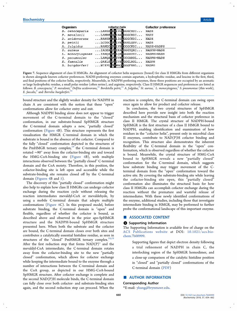
hydrogen bonds with the NADH adenosine 2′-OH group,presumably preventing the larger and negatively charged 2′-phosphate of NADPH from binding to the enzyme. Thisobservation led to the possibility that Asp146 confers cofactorspecificity in PmHMGR; however, its mutation to alanine,glycine, asparagine, or serine did not switch the cofactorpreference,4 as the catalytic efficiency, kcat/KM, for NADH wasstill 10−1000-fold greater than for NADPH, indicating thatAsp146 in PmHMGR is not solely responsible for cofactorspecificity.Surprisingly, in structures of NADPH-preferring SpHMGR,
Asp146 of PmHMGR is not replaced by a smaller or positivelycharged residue to accommodate the 2′-phosphate of NADPH,but with a bulky and neutral residue (Tyr144). In fact, weobserve that Asp146 of PmHMGR and Tyr144 of SpHMGRare both the first amino acids of a short, seven-residueconserved helix that binds the NAD(P)H cofactor at itsadenosine moiety (Figure 6). This helix, which we term the“cofactor helix”, has a completely different sequence inPmHMGR (residues 146−152) and SpHMGR (residues144−150) (DQLLNSL and YPSIVKR, respectively).
Because of its preference for NADPH over NADH, replacingAsp146 of PmHMGR with the bulkier and uncharged Tyr144of SpHMGR may at first seem counterintuitive. However, inour cofactor-bound SpHGMR structure, Tyr144 prevents theNADPH adenosine ribose from occupying the same space asobserved with NADH in PmHMGR (Figure 6). Instead,because of the large size of tyrosine, the NADPH adenosine isshifted in the cofactor-binding site compared to NADH inPmHMGR. Though the nicotinamide rings and thediphosphates of NADH and NADPH align well between thePmHMGR and SpHMGR structures, the phosphoribose inSpHMGR is shifted by ∼3.0 Å, causing the adenine ring also tobe displaced by ∼2.0 Å. Therefore, it appears unlikely thatNADPH of SpHMGR could occupy the same space as NADHin PmHMGR, because of the steric hindrance of Tyr144. As aresult, Tyr144 blocks phosphoribose from getting close to thestart of the cofactor helix, thus providing space for the 2′-phosphate to interact with Ser146 instead, which is locatedtoward the center of the cofactor helix. In fact, this serineresidue appears to be highly conserved among HMGRs thatprefer NADPH, while HMGRs that prefer NADH have ahydrophobic residue at this position (Figure 7), such asLeu148 in PmHMGR. Furthermore, the adenosine ribose ofNADPH has shifted enough to allow Arg150 at the end of theSpHMGR cofactor helix to play a dual role by interacting withboth the 2′-phosphate and the adenine ring, as describedabove. However, in NADH-preferring HMGRs, a hydrophobicresidue is often found in this position instead of arginine(Figure 7), such as Leu152 in PmHMGR.Taken together, these structural observations suggest that
several residues of the cofactor helix contribute to NADPHbinding and recognition in class II HMGRs. In SpHMGR, thebulky Tyr144 causes a shift in the location of the adenosinemoiety of NADPH as compared to that of NADH inPmHMGR (Figure 6). This shift allows the 2′-phosphate ofNADPH to interact with both conserved Ser146 in the centerof the cofactor helix and conserved Arg150 at the end of thehelix, which also stacks with the NADPH adenine ring. Withthese NADPH-binding features now described, future studiesto modify this region may offer additional insight into howcofactor specificity may be controlled or engineered.In addition, our SpHMGR structures reveal large conforma-
tional changes upon substrate binding. The C-terminal domainis disordered and absent in most HMGR crystal structures buthas been visualized in the structures of apo-SpHMGR and theternary PmHMGR complex, bound simultaneously with boththe cofactor and substrate or substrate analogue. These studiesindicated that this domain may act as a flexible flap that canopen and close over the active site at some point during thereaction.18 In our NADPH-bound SpHMGR structure, the tworesolved C-terminal domains are positioned in “open”conformations that are slightly different from each other(Figure 4A), likely because of crystal packing. This suggeststhat the C-terminal domain does not occupy a single, rigid“open” conformation but is flexible and can sample manypossible “open” positions that are all distant from the cofactor-and substrate-binding sites. Such flexibility also explains whythis domain is often unresolved in HMGR crystal structures.Importantly, these “open” conformations are observedregardless of whether the cofactor is bound, as in ourNADPH-bound structure, or unbound, as in the prior apo-SpHMGR structure (PDB entry 3QAE). Indeed, the partialoccupancy for NADPH observed in chain C of our NADPH-
Figure 6. Comparison of NADH and NADPH cofactor-binding sitesof class II HMGR. Alignment of the HMG-CoA- and NAD+-boundPmHMGR ternary complex (green, with residue labels in green) withthe NADPH-bound SpHMGR structure (gray, with residue labels inblack). The “cofactor helix” is labeled, and dashed lines representhydrogen bonds.
Biochemistry Article
DOI: 10.1021/acs.biochem.7b00999Biochemistry 2018, 57, 654−662
659
bound structure and the slightly weaker density for NADPH inchain A are consistent with the notion that these “open”conformations allow for cofactor entry and exit.Although NADPH binding alone does not appear to trigger
movement of the C-terminal domain to the “closed”conformation, in our substrate-bound SpHMGR structurethe C-terminal domain adopts a new, “partially closed”conformation (Figure 4B). This structure represents the firstvisualization the HMGR C-terminal domain in which thesubstrate is bound in the absence of the cofactor. Compared tothe fully “closed” conformation depicted in the structures ofthe PmHMGR ternary complex,19 the C-terminal domain isrotated ∼90° away from the cofactor-binding site and towardthe HMG-CoA-binding site (Figure 4B), with multipleinteractions observed between the “partially closed” C-terminaldomain and the CoA moiety. Therefore, in this position, thecofactor-binding site is left open and accessible while thesubstrate-binding site remains closed off by the C-terminaldomain (Figures 4B and 5).The discovery of this “partially closed” conformation might
also help to explain how class II HMGRs can undergo cofactorexchange during the reaction cycle without releasing thereaction intermediates mevaldyl-CoA or mevaldehyde, byusing a mobile C-terminal domain that adopts multipleconformations (Figure 4C). In this proposed model, beforesubstrate binding, the C-terminal domain is “open” andflexible, regardless of whether the cofactor is bound, asdescribed above and observed in the prior apo-SpHMGRstructure and the NADPH-bound SpHMGR structurepresented here. When both the substrate and the cofactorare bound, the C-terminal domain closes over both sites andcontributes a catalytically essential histidine residue, as seen instructures of the “closed” PmHMGR ternary complex.18,19
After the first reduction step that forms NAD(P)+ and themevaldyl-CoA intermediate, the C-terminal domain rotatesaway from the cofactor-binding site to the new “partiallyclosed” conformation, which allows for cofactor exchangewhile keeping the intermediate bound to the enzyme through anumber of interactions between the C-terminal domain andthe CoA group, as depicted in our HMG-CoA-boundSpHMGR structure. After cofactor exchange is complete andthe second NAD(P)H molecule binds, the C-terminal domaincan fully close over both cofactor- and substrate-binding sitesagain, and the second reduction step can proceed. When the
reaction is complete, the C-terminal domain can swing openonce again to allow for product and cofactor release.In conclusion, the two crystal structures of SpHMGR
described here provide new insight into both the reactionmechanism and the structural basis of cofactor preference inclass II HMGR. The crystal structure of NADPH-boundSpHMGR is the first structure of a class II HMGR bound toNADPH, enabling identification and examination of howresidues in the “cofactor helix”, present only in microbial classII enzymes, contribute to NAD(P)H cofactor binding andrecognition. This structure also demonstrates the inherentflexibility of the C-terminal domain in the “open” con-formation, which is observed regardless of whether the cofactoris bound. Meanwhile, the crystal structure of HMG-CoAbound to SpHMGR reveals a new “partially closed”conformation for the C-terminal domain, which suggestshow substrate binding may trigger movement of the C-terminal domain from the “open” conformation toward theactive site. By covering the substrate-binding site while leavingthe cofactor-binding site open, this “partially closed”conformation also illuminates the structural basis for howclass II HMGRs can accomplish cofactor exchange during thereaction without the premature and wasteful release ofintermediates. With these newly identified conformations ofthe enzyme, additional studies, including those that investigateintermediate binding in HMGR, may be performed to furtherprobe the conformational landscape of this important enzyme.
■ ASSOCIATED CONTENT*S Supporting InformationThe Supporting Information is available free of charge on theACS Publications website at DOI: 10.1021/acs.bio-chem.7b00999.
Supporting figures that depict electron density followinga trial refinement of NADPH in chain C, theinterlocking region of the SpHMGR homodimer, anda close-up comparison of the catalytic histidine positionin “closed” and “partially closed” conformations of theC-terminal domain (PDF)
■ AUTHOR INFORMATIONCorresponding Author*E-mail: [email protected].
Figure 7. Sequence alignment of class II HMGRs. An alignment of cofactor helix sequences (boxed) for class II HMGRs from different organismsis shown alongside known cofactor preferences. NADH-preferring enzymes contain aspartate, a hydrophobic residue, and leucine in the first, third,and final positions of the cofactor helix, respectively. Meanwhile, in NADPH-preferring enzymes, these three positions are occupied by an aromaticor large hydrophobic residue, a small polar residue (often serine), and arginine, respectively. Class II HMGR sequences and preferences are listed asfollows: B. cenocepacia,5 P. mevalonii,4 Delf tia acidovorans,31 Bordetella petrii,31 A. fulgidus,9 St. aureus,7 L. monocytogenes,8 S. pneumoniae (this work),E. faecalis,6 and Borrelia burgdorferi.34
Biochemistry Article
DOI: 10.1021/acs.biochem.7b00999Biochemistry 2018, 57, 654−662
660

ORCIDYan Kung: 0000-0002-6132-7969Author ContributionsY.K. conceived of the project and designed the experimentswith B.R.M., who carried out the kinetic and crystallographicwork and determined and refined the X-ray structures. Y.K.and B.R.M. analyzed the data and wrote the paper.FundingThis work was supported by the National Institutes of Health(GM116029), Bryn Mawr College, the K/G Fund for FacultyResearch, and the Howard Hughes Medical Institute.NotesThe authors declare no competing financial interest.
■ ACKNOWLEDGMENTSThis work is based upon research conducted at theNortheastern Collaborative Access Team beamlines, whichare funded by the National Institute of General MedicalSciences of the National Institutes of Health (P41GM103403). The Eiger 16M detector on beamline 24-ID-Eis funded by a NIH-ORIP HEI grant (S10OD021527). Thisresearch used resources of the Advanced Photon Source, a U.S.Department of Energy (DOE) Office of Science User Facilityoperated for the DOE Office of Science by Argonne NationalLaboratory under Contract DE-AC02-06CH11357.
■ ABBREVIATIONSHMG-CoA, 3-hydroxy-3-methylglutaryl coenzyme A; HMGR,HMG-CoA reductase; PDB, Protein Data Bank; APS,Advanced Photon Source; IPTG, isopropyl β-D-1-thiogalacto-pyranoside; PMSF, phenylmethanesulfonyl fluoride; NTA,nitrilotriacetic acid; SDS−PAGE, sodium dodecyl sulfate−polyacrylamide gel electrophoresis; PEG, polyethylene glycol;rmsd, root-mean-square deviation.
■ REFERENCES(1) Bochar, D. A., Stauffacher, C. V., and Rodwell, V. W. (1999)Sequence comparisons reveal two classes of 3-hydroxy-3-methyl-glutaryl coenzyme A reductase. Mol. Genet. Metab. 66, 122−127.(2) Friesen, J. A., and Rodwell, V. W. (2004) The 3-hydroxy-3-methylglutaryl coenzyme-A (HMG-CoA) reductases,. Genome Biol. 5,248.(3) Hedl, M., Tabernero, L., Stauffacher, C. V., and Rodwell, V. W.(2004) Class II 3-hydroxy-3-methylglutaryl coenzyme A reductases. J.Bacteriol. 186, 1927−1932.(4) Friesen, J. A., Lawrence, C. M., Stauffacher, C. V., and Rodwell,V. W. (1996) Structural determinants of nucleotide coenzymespecificity in the distinctive dinucleotide binding fold of HMG-CoAreductase from Pseudomonas mevalonii. Biochemistry 35, 11945−11950.(5) Schwarz, B. H., Driver, J., Peacock, R. B., Dembinski, H. E.,Corson, M. H., Gordon, S. S., and Watson, J. M. (2014) Kineticcharacterization of an oxidative, cooperative HMG-CoA reductasefrom Burkholderia cenocepacia, Biochim. Biochim. Biophys. Acta,Proteins Proteomics 1844, 457−464.(6) Hedl, M., Sutherlin, A., Wilding, E. I., Mazzulla, M., McDevitt,D., Lane, P., Burgner, J. W., II, Lehnbeuter, K. R., Stauffacher, C. V.,Gwynn, M. N., and Rodwell, V. W. (2002) Enterococcus faecalisacetoacetyl-coenzyme A thiolase/3-hydroxy-3-methylglutaryl-coen-zyme A reductase, a dual-function protein of isopentenyl diphosphatebiosynthesis. J. Bacteriol. 184, 2116−2122.(7) Wilding, E. I., Kim, D. Y., Bryant, M. N., Gwynn, R. D.,Lunsford, D., McDevitt, J. E., Myers, J. E., Jr., Rosenberg, M.,Sylvester, D., Stauffacher, C. V., and Rodwell, V. W. (2000)
Essentiality, expression, and characterization of the class II 3-hydroxy-3-methylglutaryl coenzyme A reductase of Staphylococcusaureus. J. Bacteriol. 182, 5147−5152.(8) Theivagt, A. E., Amanti, E. N., Beresford, J. N., Tabernero, L.,and Friesen, J. A. (2006) Characterization of an HMG-CoA reductasefrom Listeria monocytogenes that exhibits dual coenzyme specificity.Biochemistry 45, 14397−14406.(9) Kim, D.-Y., Stauffacher, C. V., and Rodwell, V. W. (2000) Dualcoenzyme specificity of Archaeoglobus fulgidus HMG-CoA reductase.Protein Sci. 9, 1226−1234.(10) Haines, B. E., Wiest, O., and Stauffacher, C. V. (2013) Theincreasingly complex mechanism of HMG-CoA reductase,. Acc. Chem.Res. 46, 2416−2426.(11) Jordan-Starch, T. C., and Rodwell, V. W. (1989) Pseudomonasmevalonii 3-hydroxy-3-methylglutaryl-CoA reductase: Characteriza-tion and chemical modification. J. Biol. Chem. 264, 17913−17918.(12) Retey, J., von Stetten, E., Coy, U., and Lynen, F. (1970) Aprobable intermediate in the enzymic reduction of 3-hydroxy-3-methylglutaryl coenzyme A. Eur. J. Biochem. 15, 72−76.(13) Bensch, W. R., and Rodwell, V. W. (1970) Purification andproperties of 3-hydroxy-3-methylglutaryl coenzyme A reductase fromPseudomonas. J. Biol. Chem. 245, 3755−3762.(14) Durr, I. F., and Rudney, H. (1960) The reduction of β-hydroxy-β-methylglutaryl coenzyme A to mevalonic acid,. J. Biol. Chem. 235,2572−2578.(15) Ferguson, J. J., Jr., Durr, I. F., and Rudney, H. (1959) Thebiosynthesis of mevalonic acid. Proc. Natl. Acad. Sci. U. S. A. 45, 499−504.(16) Feng, L., Zhou, L., Sun, Y., Gui, J., Wang, X., Wu, P., Wan, J.,Ren, Y., Qiu, S., Wei, X., and Li, J. (2011) Specific inhibitions ofannonaceous acetogenins on class II 3-hydroxy-3-methylglutarylcoenzyme A reductase from Streptococcus pneumoniae. Bioorg. Med.Chem. 19, 3512−3519.(17) Lawrence, C. W., Rodwell, V. W., and Stauffacher, C. V. (1995)Crystal structure of Pseudomonas mevalonii HMG-CoA reductase at3.0 angstrom resolution. Science 268, 1758−1762.(18) Tabernero, L., Bochar, D. A., Rodwell, V. W., and Stauffacher,C. V. (1999) Substrate-induced closure of the flap domain in theternary complex structures provides insights into the mechanism ofcatalysis by 3-hydroxy-3-methylglutaryl-CoA reductase. Proc. Natl.Acad. Sci. U. S. A. 96, 7167−7171.(19) Steussy, C. N., Critchelow, C. J., Schmidt, T., Min, J.-K.,Wrensford, L. V., Burgner, J. W., II, Rodwell, V. W., and Stauffacher,C. V. (2013) A novel role for coenzyme A during hydride transfer in3-hydroxy-3-methylglutaryl-coenzyme A reductase,. Biochemistry 52,5195−5205.(20) Darnay, B. G., Wang, Y., and Rodwell, V. W. (1992)Identification of the catalytically important hisitidine of 3-hydroxy-3-methylglutaryl-coenzyme A reductase. J. Biol. Chem. 267, 15064−15070.(21) Darnay, B. G., and Rodwell, V. W. (1993) His865 is thecatalytically important histidyl residue of Syrian hamster 3-hydroxy-3-methylglutaryl-coenzyme A reductase. J. Biol. Chem. 268, 8429−8435.(22) Battye, T. G., Kontogiannis, L., Johnson, O., Powell, H. R., andLeslie, A. G. (2011) iMOSFLM: a new graphical interface fordiffraction-image processing with MOSFLM. Acta Crystallogr., Sect. D:Biol. Crystallogr. 67, 271−281.(23) McCoy, A. J., Grosse-Kunstleve, R. W., Adams, P. D., Winn, M.D., Storoni, L. C., and Read, R. J. (2007) Phaser CrystallographicSoftware. J. Appl. Crystallogr. 40, 658−674.(24) Adams, P. D., Afonine, P. V., Bunkoczi, G., Chen, V. B., Davis,I. W., Echols, N., Headd, J. J., Hung, L.-W., Kapral, G. J., Grosse-Kunstleve, R. W., McCoy, A. J., Moriarty, N. W., Oeffner, R., Read, R.J., Richardson, D. C., Richardson, J. S., Terwilliger, T. C., and Zwart,P. H. (2010) PHENIX: a comprehensive Python-based system formacromolecular structure solution. Acta Crystallogr., Sect. D: Biol.Crystallogr. 66, 213−221.
Biochemistry Article
DOI: 10.1021/acs.biochem.7b00999Biochemistry 2018, 57, 654−662
661

(25) Emsley, P., and Cowtan, K. (2004) Coot: model-building toolsfor molecular graphics. Acta Crystallogr., Sect. D: Biol. Crystallogr. 60,2126−2132.(26) Afonine, P. V., Grosse-Kunstleve, R. W., Echols, N., Headd, J. J.,Moriarty, N. W., Mustyakimov, M., Terwilliger, T. C., Urzhumtsev, A.,Zwart, P. H., and Adams, P. D. (2012) Towards automatedcrystallographic structure refinement with phenix.refine. ActaCrystallogr., Sect. D: Biol. Crystallogr. 68, 352−367.(27) Istvan, E. S., Palnitkar, M., Buchanan, S. K., and Deisenhofer, J.(2000) Crystal structure of the catalytic portion of human HMG-CoAreductase: insights into regulation of activity and catalysis. EMBO J.19, 819−830.(28) Istvan, E. S. (2001) Bacterial and mammalian HMG-CoAreductase: related enzymes with distinct architectures. Curr. Opin.Struct. Biol. 11, 746−751.(29) Kung, Y., Runguphan, W., and Keasling, J. D. (2012) Fromfields to fuels: recent advances in the microbial production of biofuels.ACS Synth. Biol. 1, 498−513.(30) George, K. W., Alonso-Gutierrez, J., Keasling, J. D., and Lee, T.S. (2015) Isoprenoid drugs, biofuels, and chemicalsartemisinin,farnesene, and beyond. Adv. Biochem. Eng./Biotechnol. 148, 355−389.(31) Ma, S. M., Garcia, D. E., Redding-Johanson, A. M., Friedland,G. D., Chan, R., Batth, T. S., Haliburton, J. R., Chivian, D., Keasling, J.D., Petzold, C. J., Soon Lee, T., and Chhabra, S. R. (2011)Optimization of a heterologous mevalonate pathway through the useof variant HMG-CoA reductases. Metab. Eng. 13, 588−597.(32) Wilding, E. I., Brown, J. R., Bryant, A. P., Chalker, A. F.,Holmes, D. J., Ingraham, K. A., Iordanescu, S., So, C. Y., Rosenberg,M., and Gwynn, M. N. (2000) Indentificaiton, evolution, andessentiality of the mevalonate pathway for isopentenyl diphosphatebiosynthesis in gram-positive cocci. J. Bacteriol. 182, 4319−4327.(33) Liebschner, D., Afonine, P. V., Moriarty, N. W., Poon, B. K.,Sobolev, O. V., Terwilliger, T. C., and Adams, P. D. (2017) Poldermaps: improving OMIT maps by excluding bulk solvent. ActaCrystallogr., Sect. D: Biol. Crystallogr. 73, 148−157.(34) Van Laar, T. A., Lin, Y.-H., Miller, C. L., Karna, S. L. R.,Chambers, J. P., and Seshu, J. (2012) Effect of levels of acetate on themevalonate pathway of Borrelia burgdorferi. PLoS One 7, No. e38171.
Biochemistry Article
DOI: 10.1021/acs.biochem.7b00999Biochemistry 2018, 57, 654−662
662