Embed Size (px)
Citation preview
S1
SupportingInformation
Structuralbasisforexcisionof5-formylcytosinebyThymineDNAGlycosylase
Lakshmi S. Pidugu1, Joshua Flowers2, Christopher T. Coey1, Edwin Pozharski1,3,*, Marc M. Greenberg2,*, and Alexander C. Drohat1,*
1DepartmentofBiochemistryandMolecularBiology,UniversityofMarylandSchoolof
Medicine,Baltimore,MD21201,UnitedStates,2DepartmentofChemistry,JohnsHopkins
University,3400N.CharlesStreet,Baltimore,Maryland21218,UnitedStates,3Centerfor
BiomolecularTherapeutics,InstituteforBioscienceandBiotechnologyResearch,Rockville,
MD20850,UnitedStates
ContentsofSupportingInformation:
ExperimentalMethods S2-S3
TableS1 S4
FigureS1 S5
FigureS2 S6
FigureS3 S6
SynthesisofODNscontaining2′-F-arabino-5-formyl-dCds S7-S19
SupplementaryReferences S20
S2
ExperimentalMethods
Oligodeoxynucleotides.Standardoligodeoxynucleotides(ODNs)wereobtainedfromIDT
andwerepurifiedbyreversephaseHPLC,1exchangedinto0.02MTris-HClpH7.5,0.04M
NaCl,andquantifiedbyabsorbanceasdescribed.2ODNscontaining2´-fluoroarabino-5-
formyldeoxycytidine(hereafter,fdCF)werepreparedasdescribedbelow.TheDNAusedfor
crystalsincludeda28mertargetstrand,5’-AGCTGTCCATCGCTCAxGTACAGAGCTG-3’,
wherexisfdCF,anditscomplement,5’-CAGCTCTGTACGTGAGCGATGGACAGCT-3’,such
thatfdCFispairedwithdGandlocatedinaCpGdinucleotidecontext.2,3Thesame28bp
DNAconstructwasusedforglycosylaseassays(withx=fC).
Enzymes.FulllengthhumanTDG(410residues),anditsN140Avariant,wereexpressedin
Ecoliandpurifiedessentiallyaspreviouslydescribed.2,4TDG82-308,anewconstruct
containingresiduesSer82-Val308ofhumanTDGwasexpressedandpurifiedasdescribed.5
Theenzymepreparationswere>99%pure,asjudgedbySDS-PAGE(Coomassie-stained
gel),andtheenzymeconcentrationwasdeterminedbyabsorbanceat280nm.6,7The
extinctioncoefficientforTDG82-308isidenticaltothatforTDG111-308.8
X-rayCrystallography.Samplesusedforcrystallizationcontained0.35mMenzyme
(TDG82-308)and0.42mMDNAinabufferof5mMTris-HClpH7.5,0.13MNaCl,0.2mMDTT,
0.2mMEDTA.Crystalsweregrownatroomtemperature(~22°C)bysittingdropvapor
diffusion,using1μloftheTDG-DNAsampleand1or2ulofmotherliquor,whichwas30%
(w/v)PEG4000,0.2Mammoniumacetate,0.1Msodiumacetate,pH6.0.Crystalstypically
appearedwithininafewdays.Crystalswerecryo-protectedusingmotherliquor
supplementedwith18%ethyleneglycolandflashcooledinliquidnitrogen.X-ray
diffractiondatawerecollectedattheStanfordSynchrotronRadiationLightsource(SSRL
beamline12-2).ImageswereprocessedusingXDS9andscaledwithAimless10fromthe
CCP4programsuite11withthehelpoftheautoxdsscriptdevelopedbyAnaGonzalezand
YingssuTsai(http://smb.slac.stanford.edu/facilities/software/xds).Resolutioncutoffwas
determinedbasedonCC1/2values.12Structuresweresolvedbymolecularreplacement
usingPhaser,13andapreviouslyreportedstructureofDNA-boundTDG82-308asthesearch
model(PDBID:4Z47).RefinementwasperformedusingBUSTER-TNT,14orREFMAC5,15
andmodelbuildingwasperformedusingCoot.16TLSrefinementprotocolutilizedTLSMD
S3
server,17,18asdescribed.1ThestructuralfiguresweremadewithPyMOL
(http://www.pymol.org).
GlycosylaseAssays.Singleturnoverkineticsreactionswereinitiatedbyaddingenzyme
(TDGorTDG82-308)toG·fCsubstrate(0.5uM)inHEMN.1buffer(0.02MHEPESpH7.5,0.1
MNaCl,0.2mMEDTA,2.5mMMgCl2).Aliquotswereremovedatdesiredtimepoints,
quenchedwith50%(v:v)0.3MNaOH,0.03MEDTA,andheated(3m,85°C)to
quantitativelycleavetheDNAbackboneatabasicsites.TheresultingDNAfragmentswere
resolvedbyHPLCandpeakareaswereusedtodeterminefractionproduct.2Progress
curves(fractionproductvs.time)werefittedbynon-linearregressiontoeq.1:
fractionproduct=A(1−exp(−kobst)) (1)
whereAistheamplitude,kobsistherateconstant,andtisthereactiontime.Experiments
wereperformedwithsaturatingenzyme([E]>>Kd;[E]>[S])suchthattheobservedrate
constantreflectsthemaximalrateofproductformation(kobs≈kmax)andisnotinfluenced
byenzyme-substrateassociationorbyproductreleaseorproductinhibition.4
Acknowledgement
PortionsofthisresearchwerecarriedoutattheStanfordSynchrotronRadiation
Lightsource,aDirectorateofSLACNationalAcceleratorLaboratoryandanOfficeofScience
UserFacilityoperatedfortheU.S.DepartmentofEnergyOffice(DOE)byStanford
University.TheSSRLStructuralMolecularBiologyProgramissupportedbytheDOEOffice
ofBiologicalandEnvironmentalResearch,andbytheNationalInstitutesofHealth(NIH),
NationalInstituteofGeneralMedicalSciences(NIGMS;includingP41GM103393)andthe
NationalCenterforResearchResources(NCRR;P41RR001209).TheBerkeleyCenterfor
StructuralBiologyissupportedinpartbytheNIH,NIGMS,andtheHowardHughesMedical
Institute.TheAdvancedLightSourceissupportedbytheDirector,OfficeofScience,Office
ofBasicEnergySciences,oftheU.S.DOEunderContractNo.DE-AC02-05CH11231.The
contentsofthispublicationaresolelytheresponsibilityoftheauthorsanddonot
necessarilyrepresenttheofficialviewsofNIGMS,NCRRorNIH.

S4
Table S1. Data collection and refinement statistics
TDG82-308 dG:fdCF (PDB ID: 5T2W) Data collection Space Group C2 Cell Dimensions
a, b, c (Å)
β (°)
83.5,51.9,82.0
101.5 Resolution (Å) 39.83-2.20 (2.27-2.20) Rpim 0.170 Mean I/σI 4.3 (0.7) CC1/2 0.982 (0.316) Completeness (%) 99.9 (100.0) Redundancy 10.1 (10.2) Wilson B-factor (Å2) 41.5 Refinement Program Buster 2.10.2 Resolution (Å) 39.83-2.20 No. of reflections 17639 Rwork/Rfree 0.180/0.226 Number of atoms Protein 1541 DNA 1258 Water 156 B-factors (Å2) Protein 34.8 DNA 49.7 Water 43.5 Ramachandran Plot Favoured (%) 97.4 Allowed (%) 2.6 Outliers (%) 0 RMSD from ideal Bond lengths (Å) 0.010 Bond angles (°) 0.99
Values shown in parenthesis are for highest resolution shell. The Ramachandran analysis was performed using Molprobity 19. Wilson B-factor estimated by phenix.xtriage. Number of atoms includes all atom records explicitly included in the model, including alternate positions.
S5
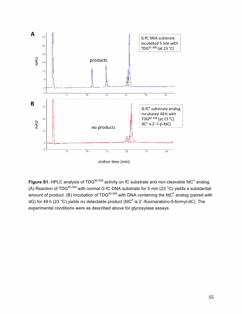
Figure S1. HPLC analysis of TDG82-308 activity on fC substrate and non-cleavable fdCF analog. (A) Reaction of TDG82-308 with normal G·fC DNA substrate for 5 min (23 °C) yields a substantial amount of product. (B) Incubation of TDG82-308 with DNA containing the fdCF analog (paired with dG) for 48 h (23 °C) yields no detectable product (fdCF is 2´-fluoroarabino-5-formyl-dC). The experimental conditions were as described above for glycosylase assays.
S6
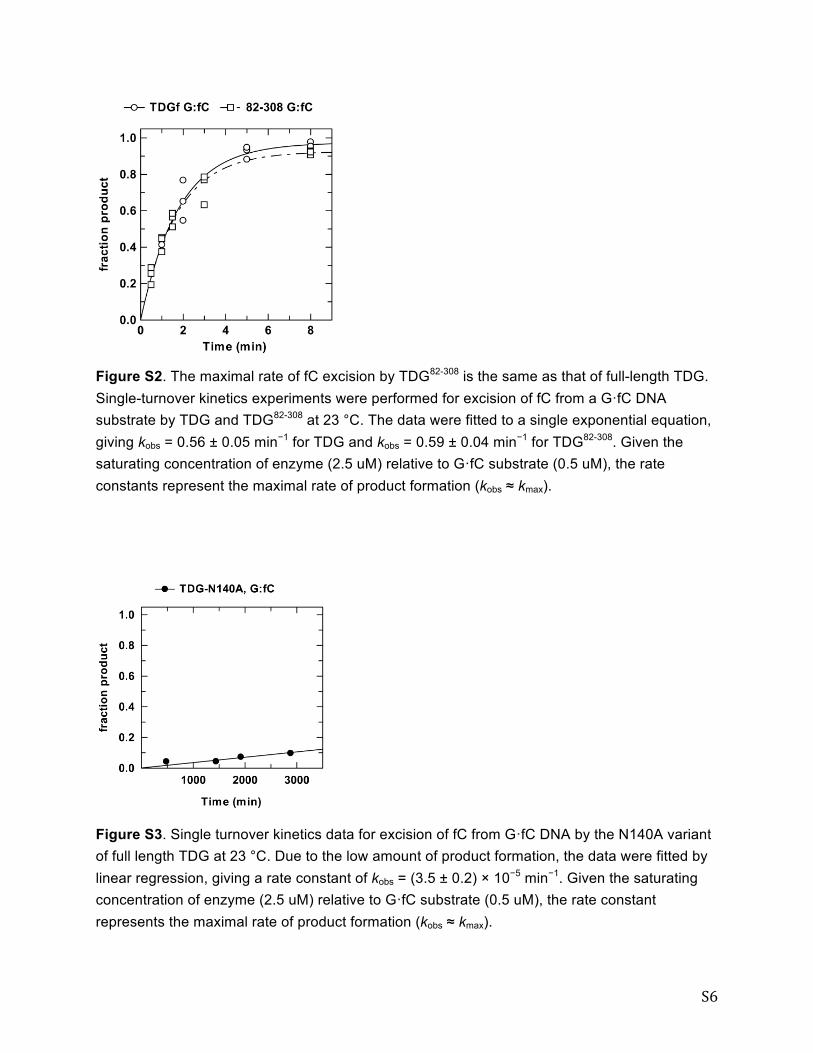
Figure S2. The maximal rate of fC excision by TDG82-308 is the same as that of full-length TDG. Single-turnover kinetics experiments were performed for excision of fC from a G·fC DNA substrate by TDG and TDG82-308 at 23 °C. The data were fitted to a single exponential equation, giving kobs = 0.56 ± 0.05 min−1 for TDG and kobs = 0.59 ± 0.04 min−1 for TDG82-308. Given the saturating concentration of enzyme (2.5 uM) relative to G·fC substrate (0.5 uM), the rate constants represent the maximal rate of product formation (kobs ≈ kmax).
Figure S3. Single turnover kinetics data for excision of fC from G·fC DNA by the N140A variant of full length TDG at 23 °C. Due to the low amount of product formation, the data were fitted by linear regression, giving a rate constant of kobs = (3.5 ± 0.2) × 10−5 min−1. Given the saturating concentration of enzyme (2.5 uM) relative to G·fC substrate (0.5 uM), the rate constant represents the maximal rate of product formation (kobs ≈ kmax).

S7
Synthesis of ODNs containing 2′-deoxy-2′-fluoro-D-arabinofuranosyl-5-formyl-cytosine (2'-
F-fdC). ODNs containing 2'-F-fdC were prepared via solid-phase oligonucleotide synthesis
using phosphoramidite 4, which was synthesized in a manner similar to previously reported
methods.20-22 The 2'-fluoronucleoside core was prepared using a strategy described by Damha
and more recently by He.20,21 The protecting group scheme developed by Carell was employed to
successfully introduce the 5-formylcytosine.22
Preparation of S1. 2′-Deoxy-2′-fluoro-D-arabinofuranosyl-5-iodo-
cytosine21 (1.14 g, 3.07 mmol), imidazole (0.941 g, 13.82 mmol), and
TBDMSCl (1.39 g, 9.21 mmol) were dissolved in DMF (18.3 mL). The
reaction was stirred overnight at 25 °C before being quenched with sat.
NaHCO3 and extracted with CHCl3. The organic layer was then washed
with water and dried over MgSO4. The solution was filtered, concentrated under reduced
pressure, and purified by flash chromatography (5% MeOH/DCM). This yielded 1.56 g (85%)
of S1 as a white solid. 1H-NMR (CDCl3): δ 8.63 (br s, 1H), 7.83 (s, 1H), 6.19 (dd, J = 20, 4 Hz,
1H), 5.63 (br s, 1H), 4.89-5.06 (m, 1H), 4.35-4.43 (m, 1H), 3.92 (m, 1H), 3.79 (m, 2H), 0.93 (s,
9H), 0.89 (s, 9H), 0.11 (m, 12H); 13C-NMR (CDCl3): δ 163.9, 154.3, 147.4, 95.8, 93.9, 85.30,
85.26, 85.1, 75.4, 75.2, 61.5, 56.2, 25.8, 25.5, 18.3, 17.8, -4.9, -5.1, -5.40, -5.42; IR (CDCl3): υ
(cm-1) 3450, 2932, 2859, 2247, 1650, 1490, 1391, 1362, 1335, 1289, 1257, 1107, 1006, 910, 838,
779. MS (ESI-TOF) (m/z): Calculated: 600.1581 [M+H], Observed: 600.1598.
Preparation of S2. Compound S1 (1.56 g, 2.61 mmol) and Pd2(dba)3
(230 mg, 0.250 mmol) were added to a pear flask, which was immediately
purged with argon for 10 min. Toluene that was sparged for 30 min with
argon was added (24.6 mL) to the pear flask. The solution was then
sparged for an additional 15 min with argon, followed by 5 min with CO. Triphenylphosphine
(394 mg, 1.50 mmol) was dissolved in 1 mL of sparged toluene and added to the pear flask. The
solution was then sparged for an additional 5 min with CO. Freshly distilled Bu3SnH (1.10 mL,
3.76 mmol) dissolved in sparged toluene (37.6 mL) was syringed into the pear flask at 50 °C
over a period of 10 h. The reaction was then cooled to 25 °C and filtered through celite before
N
N
NH2
OTBDMSO O
TBDMSO
I
F
H
N
N
NH2
OTBDMSO O
TBDMSO
F
H
H
O
x
S8
being concentrated under reduced pressure. The crude material was purified by flash
chromatography (4:1, 2:1, 1:1 Hex/EtOAc), followed by a second column purification (2%
MeOH/DCM). This yielded 858 mg (66%) of S2 as a slightly yellow solid. 1H-NMR (CDCl3): δ
9.47 (s, 1H), 8.23 (br s, 1H), 8.21 (s, 1H), 7.41 (br s, 0.5 H), 6.22-6.29 (m, 1H), 4.98-5.14 (m,
1H), 4.39-4.46 (m, 1H), 4.00 (m, 1H), 3.82 (m, 2H), 0.91 (s, 9H), 0.90 (s, 9H), 0.11 (m, 12H); 13C-NMR (CDCl3): δ 187.2, 162.8, 154.2, 152.8, 105.3, 95.9, 93.9, 86.1, 86.0, 85.9, 75.6, 75.4,
61.7, 25.8, 25.6, 18.3, 17.9, -4.8, -5.0, -5.4; IR (CDCl3): υ (cm-1) 3395, 2931, 2858, 1672, 1516,
1472, 1418, 1255, 1106, 838, 780. MS (ESI-TOF) (m/z): Calculated: 502.2563 [M+H],
Observed: 502.2586.
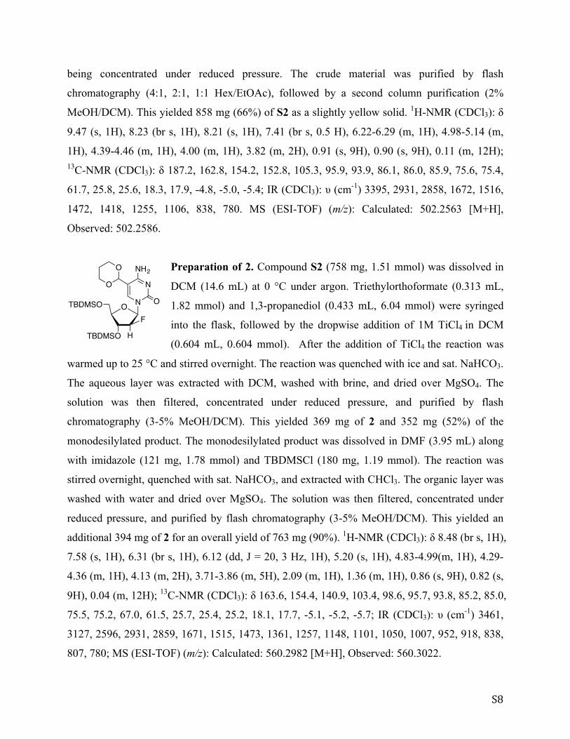
Preparation of 2. Compound S2 (758 mg, 1.51 mmol) was dissolved in
DCM (14.6 mL) at 0 °C under argon. Triethylorthoformate (0.313 mL,
1.82 mmol) and 1,3-propanediol (0.433 mL, 6.04 mmol) were syringed
into the flask, followed by the dropwise addition of 1M TiCl4 in DCM
(0.604 mL, 0.604 mmol). After the addition of TiCl4 the reaction was
warmed up to 25 °C and stirred overnight. The reaction was quenched with ice and sat. NaHCO3.
The aqueous layer was extracted with DCM, washed with brine, and dried over MgSO4. The
solution was then filtered, concentrated under reduced pressure, and purified by flash
chromatography (3-5% MeOH/DCM). This yielded 369 mg of 2 and 352 mg (52%) of the
monodesilylated product. The monodesilylated product was dissolved in DMF (3.95 mL) along
with imidazole (121 mg, 1.78 mmol) and TBDMSCl (180 mg, 1.19 mmol). The reaction was
stirred overnight, quenched with sat. NaHCO3, and extracted with CHCl3. The organic layer was
washed with water and dried over MgSO4. The solution was then filtered, concentrated under
reduced pressure, and purified by flash chromatography (3-5% MeOH/DCM). This yielded an
additional 394 mg of 2 for an overall yield of 763 mg (90%). 1H-NMR (CDCl3): δ 8.48 (br s, 1H),
7.58 (s, 1H), 6.31 (br s, 1H), 6.12 (dd, J = 20, 3 Hz, 1H), 5.20 (s, 1H), 4.83-4.99(m, 1H), 4.29-
4.36 (m, 1H), 4.13 (m, 2H), 3.71-3.86 (m, 5H), 2.09 (m, 1H), 1.36 (m, 1H), 0.86 (s, 9H), 0.82 (s,
9H), 0.04 (m, 12H); 13C-NMR (CDCl3): δ 163.6, 154.4, 140.9, 103.4, 98.6, 95.7, 93.8, 85.2, 85.0,
75.5, 75.2, 67.0, 61.5, 25.7, 25.4, 25.2, 18.1, 17.7, -5.1, -5.2, -5.7; IR (CDCl3): υ (cm-1) 3461,
3127, 2596, 2931, 2859, 1671, 1515, 1473, 1361, 1257, 1148, 1101, 1050, 1007, 952, 918, 838,
807, 780; MS (ESI-TOF) (m/z): Calculated: 560.2982 [M+H], Observed: 560.3022.
N
N
NH2
OTBDMSO O
TBDMSO
F
H
O
O
S9
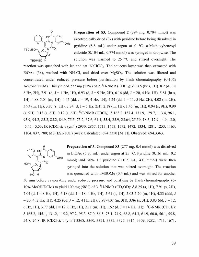
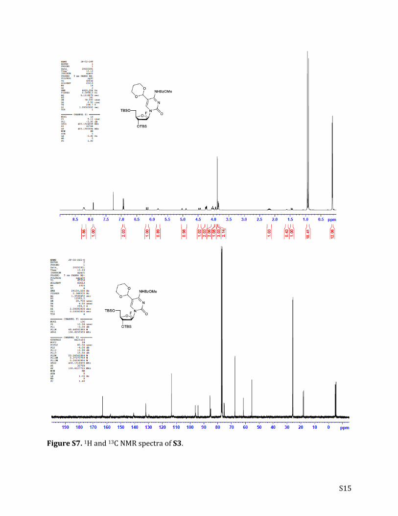
Preparation of S3. Compound 2 (394 mg, 0.704 mmol) was
azeotropically dried (3x) with pyridine before being dissolved in
pyridine (8.8 mL) under argon at 0 °C. p-Methoxybenzoyl
chloride (0.104 mL, 0.774 mmol) was syringed in dropwise. The
solution was warmed to 25 °C and stirred overnight. The
reaction was quenched with ice and sat. NaHCO3. The aqueous layer was then extracted with
EtOAc (3x), washed with NH4Cl, and dried over MgSO4. The solution was filtered and
concentrated under reduced pressure before purification by flash chromatography (0-10%
Acetone/DCM). This yielded 277 mg (57%) of 2. 1H-NMR (CDCl3): δ 13.5 (br s, 1H), 8.2 (d, J =
8 Hz, 2H), 7.91 (d, J = 1 Hz, 1H), 6.93 (d, J = 9 Hz, 2H), 6.16 (dd, J = 20, 4 Hz, 1H), 5.81 (br s,
1H), 4.88-5.04 (m, 1H), 4.45 (dd, J = 19, 4 Hz, 1H), 4.24 (dd, J = 11, 5 Hz, 2H), 4.02 (m, 2H),
3.93 (m, 1H), 3.87 (s, 3H), 3.84 (d, J = 5 Hz, 2H), 2.18 (m, 1H), 1.45 (m, 1H), 0.94 (s, 9H), 0.90
(s, 9H), 0.13 (s, 6H), 0.12 (s, 6H); 13C-NMR (CDCl3): δ 163.2, 157.4, 131.9, 129.7, 113.4, 96.1,
95.9, 94.2, 85.5, 85.2, 84.9, 75.5, 75.2, 67.6, 61.4, 55.4, 25.9, 25.64, 25.59, 18.3, 17.9, -4.9, -5.0,
-5.45, -5.53; IR (CDCl3): υ (cm-1) 2930, 2857, 1713, 1653, 1572, 1472, 1334, 1281, 1253, 1163,
1104, 837, 780; MS (ESI-TOF) (m/z): Calculated: 694.3350 [M+H], Observed: 694.3363.
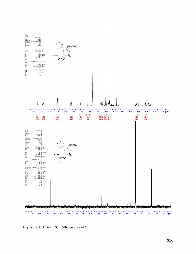
Preparation of 3. Compound S3 (277 mg, 0.4 mmol) was dissolved
in EtOAc (5.70 mL) under argon at 25 °C. Pyridine (0.161 mL, 0.2
mmol) and 70% HF·pyridine (0.105 mL, 4.0 mmol) were then
syringed into the solution that was stirred overnight. The reaction
was quenched with TMSOMe (0.4 mL) and was stirred for another
30 min before evaporating under reduced pressure and purifying by flash chromatography (6-
10% MeOH/DCM) to yield 109 mg (58%) of 3. 1H-NMR (CD3OD): δ 8.25 (s, 1H), 7.91 (s, 2H),
7.04 (d, J = 8 Hz, 1H), 6.18 (dd, J = 18, 4 Hz, 1H), 5.61 (s, 1H), 5.03-5.20 (m, 1H), 4.33 (ddd, J
= 20, 4, 2 Hz, 1H), 4.25 (dd, J = 12, 4 Hz, 2H), 3.98-4.07 (m, 3H), 3.86 (s, 3H), 3.83 (dd, J = 12,
6 Hz, 1H), 3.77 (dd, J = 12, 6 Hz, 1H), 2.11 (m, 1H), 1.52 (d, J = 14 Hz, 1H); 13C-NMR (CDCl3):
δ 165.2, 145.1, 131.2, 115.2, 97.2, 95.3, 87.0, 86.5, 75.1, 74.9, 68.8, 64.3, 61.9, 60.0, 56.1, 55.8,
54.8, 26.8; IR (CDCl3): υ (cm-1) 3368, 3360, 3351, 3337, 3325, 3316, 3309, 3282, 1711, 1671,
N
N
HN
OTBDMSO O
TBDMSO
F
H
O
O
O
OMe
x
N
N
HN
OHO O
HO
F
H
O
O
O
OMe

S10
1605, 1576, 1484, 1456, 1253, 1177, 1092, 1066, 1046, 1026; MS (ESI-TOF) (m/z): Calculated:
466.1620 [M+H], Observed: 466.1643.
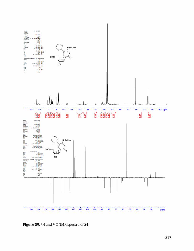
Preparation of S4. Compound 3 (109 mg, 0.234 mmol) was
azeotropically dried (3x) with distilled pyridine. Pyridine (5.1 mL)
was then used to dissolve 3 and DMTCl (95 mg, 0.28 mmol) was
added. The reaction stirred overnight at 25 °C before quenching
with MeOH and evaporating under reduced pressure. The crude
material was redissolved in DCM, washed with NaHCO3 (5% v/v), and brine, before drying over
MgSO4. The solution was filtered, evaporated under reduced pressure, and purified by flash
chromatography (70% EtOAc/Hex) to yield 98 mg (55%) of S4. 1H-NMR (CDCl3): δ 8.22 (d, J
= 1 Hz, 1H), 8.09 (s, 2H), 7.50 (m, 2H), 7.18-7.41 (m, 7H), 6.93 (d, J = 9 Hz, 2H), 6.84 (m, 4H),
6.30 (d, J = 20 Hz, 1H), 5.51 (s, 1H), 5.19 (d, J = 52 Hz, 1H), 4.52 (dd, J = 19, 3 Hz, 1H), 4.11
(m, 2H), 3.95 (m, 1H), 3.68-3.90 (m, 13H), 3.51 (d, J = 10 Hz, 1H), 3.30 (dd, J = 11, 5 Hz, 1H),
1.75 (s, 1H), 1.18 (d, J = 14 Hz, 1H); 13C-NMR (CDCl3): δ 163.2, 158.4, 144.8, 136.0, 135.9,
130.1, 128.2, 127.9, 126.7, 113.6, 113.2, 96.0, 94.1, 86.3, 84.0, 75.7, 75.4, 67.3, 67.2, 62.4, 60.4,
55.4, 55.2, 29.7, 25.2, 21.1, 14.2; IR (CDCl3): υ (cm-1) 3308, 2932, 2838, 1709, 1667, 1604,
1568, 1508, 1480, 1305, 1247, 1174, 1093, 1029, 950, 915, 828, 760, 727, 701, 646, 584; MS
(ESI-TOF) (m/z): Calculated: 768.2927 [M+H], Observed: 768.2950.
Preparation of 4. Compound S4 (98 mg, 0.128 mmol) was
dissolved in a solution of DCM (5.4 mL) and DIPEA (0.15 mL,
0.85 mmol) at 0 °C under argon. Phosphoramidic chloride
(0.043 mL, 0.192 mmol) was syringed in dropwise and the
reaction was stirred for 2 h. The reaction was diluted with DCM,
washed with NaHCO3 (5% v/v) and then brine before drying
over MgSO4. The solution was filtered, dried under reduced pressure, and purified by flash
chromatography (30-50% EtOAc/Hex). This yielded 85 mg (69%) of 4. 1H-NMR (CDCl3): δ
13.48 (br s, 1H), 8.22 (m, 3H), 7.51 (m, 2H), 7.19-7.43 (m, 7H), 6.93 (d, J = 9 Hz, 2H), 6.86 (m,
4H), 6.24 (m, 1H), 5.73 (s, 1H), 5.04-5.27 (m, 1H), 4.63 (m, 1H), 4.08 (m, 1H), 3.71-3.96 (m,
14H), 3.56 (m, 4H), 3.29 (dd, J = 11, 6 Hz, 1H), 2.59 (t, J = 6 Hz, 1H), 2.35 (m, 1H), 0.98-1.20
N
N
HN
ODMTO O
HO
F
H
O
O
O
OMe
x
N
N
HN
ODMTO O
O
F
H
O
O
O
OMe
PONCN(iPr)2
S11
(m, 12H); 31P-NMR (CDCl3): δ 151.50, 150.67; MS (ESI-TOF) (m/z): Calculated: 968.4005
[M+H], Observed: 968.3994.
Oligonucleotide synthesis. Oligonucleotide synthesis was carried out on an Applied
Biosystems 394 DNA/RNA Synthesizer. Oligonucleotides containing native nucleotides only
were prepared via standard conditions. Extended coupling (45 s), capping (25 s), and oxidation
(15 s) times were used for coupling unmodified phosphoramidites in oligonucleotides that
contained 2'-F-fdC. Phosphoramidite 4 was coupled for 5 min. Oligonucleotides containing 2'-F-
fdC were deprotected using concentrated NH4OH for 24 h at 25 °C. The deprotection was then
flash frozen using liquid nitrogen and sublimated on the speed vac. The resin was then
redissolved in 80% AcOH/H2O and was placed in a thermocycler at 20 °C for 24 h. The solution
was again flash frozen and sublimated on the speed vac. The resin was then redissolved in 90%
formamide loading buffer and purified by gel electrophoresis (20% denaturing PAGE).
S12
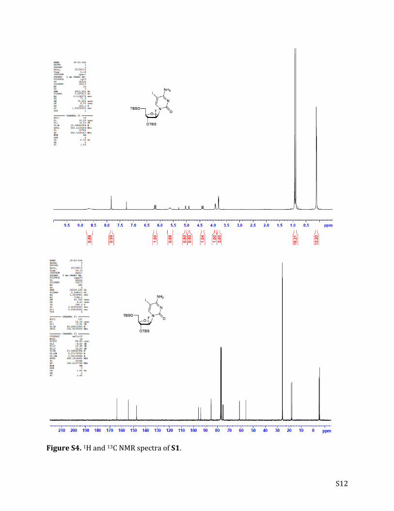
FigureS4.1Hand13CNMRspectraofS1.
S13
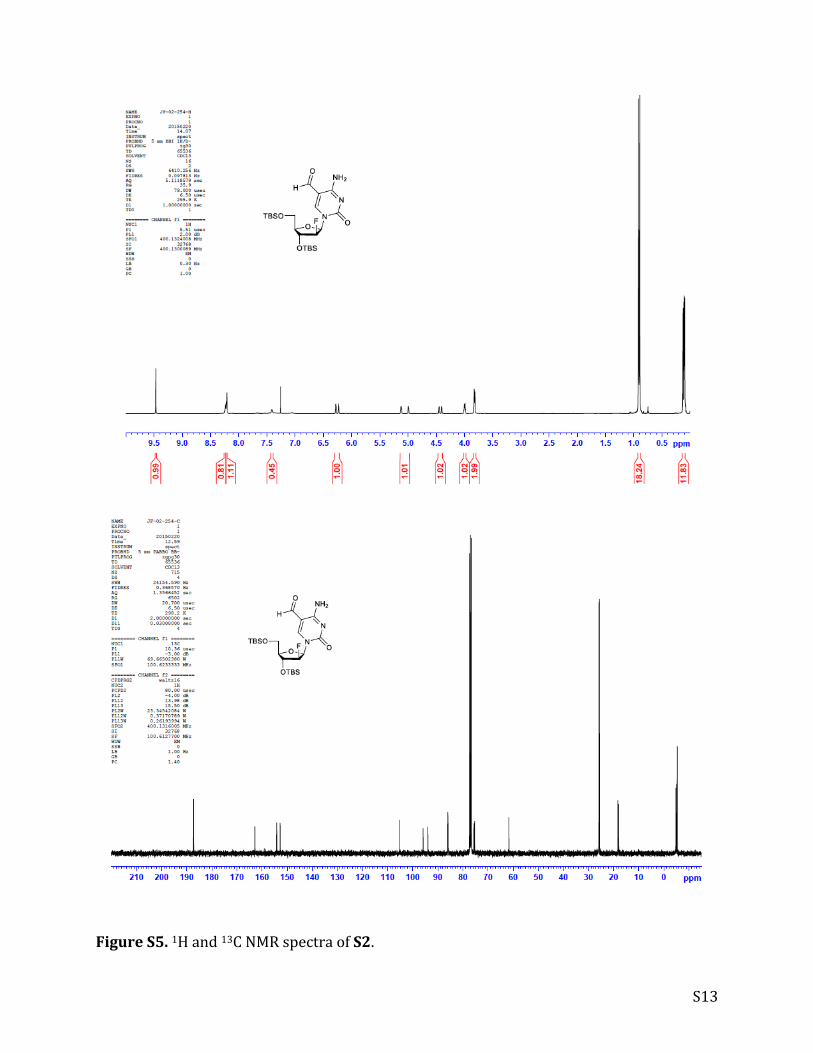
FigureS5.1Hand13CNMRspectraofS2.
S14
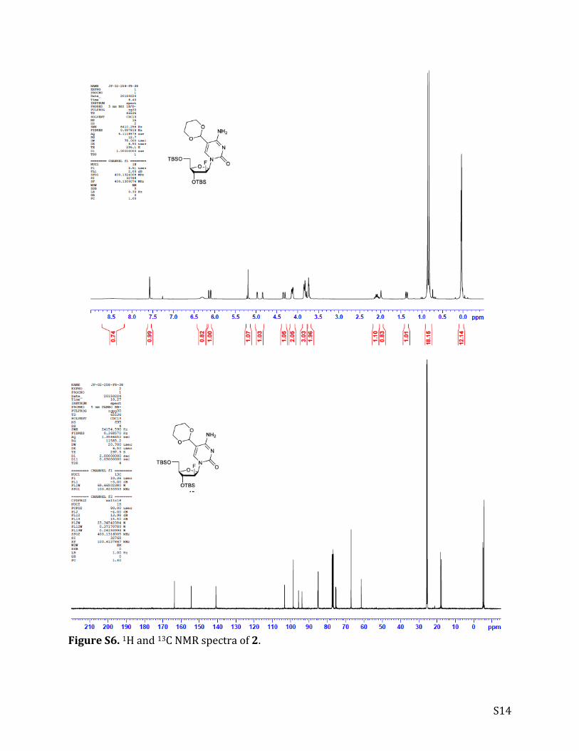
FigureS6.1Hand13CNMRspectraof2.
S15
FigureS7.1Hand13CNMRspectraofS3.
S16
FigureS8.1Hand13CNMRspectraof3.
S17
FigureS9.1Hand13CNMRspectraofS4.

S18
FigureS10.1Hand31PNMRspectraof4.
S19
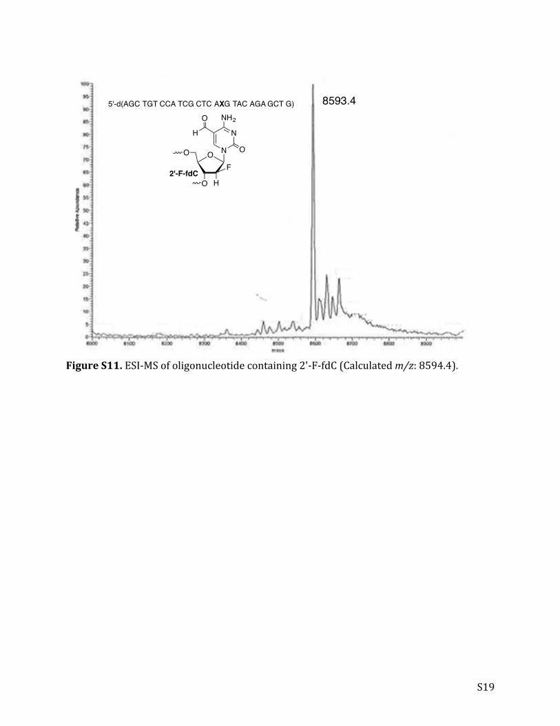
FigureS11.ESI-MSofoligonucleotidecontaining2'-F-fdC(Calculatedm/z:8594.4).
8593.45'-d(AGC TGT CCA TCG CTC AXG TAC AGA GCT G)
2'-F-fdC
N
N
NH2
OO O
O
F
H
O
H

S20
References(1) Malik, S. S., Coey, C. T., Varney, K. M., Pozharski, E., and Drohat, A. C. (2015) Nucleic Acids Res 43,
9541-9552.
(2) Morgan, M. T., Bennett, M. T., and Drohat, A. C. (2007) J Biol Chem 282, 27578-27586.
(3) Waters, T. R., and Swann, P. F. (1998) J Biol Chem 273, 20007-20014.
(4) Maiti, A., Morgan, M. T., and Drohat, A. C. (2009) J Biol Chem 284, 36680-36688.
(5) Coey, C. T., Malik, S. S., Pidugu, L. S., Varney, K. M., Pozharski, E., and Drohat, A. C. (2016) Nucleic
Acids Res, doi: 10.1093/nar/gkw768.
(6) Gill, S. C., and von Hippel, P. H. (1989) Anal. Biochem. 182, 319-326.
(7) Morgan, M. T., Maiti, A., Fitzgerald, M. E., and Drohat, A. C. (2011) Nucleic Acids Res 39, 2319-2329.
(8) Maiti, A., Morgan, M. T., Pozharski, E., and Drohat, A. C. (2008) Proc Natl Acad Sci USA 105, 8890-
8895.
(9) Kabsch, W. (2010) Acta Crystallogr D Biol Crystallogr 66, 125-132.
(10) Evans, P. R. (2011) Acta Crystallogr D Biol Crystallogr 67, 282-292.
(11) Winn, M. D., Ballard, C. C., Cowtan, K. D., Dodson, E. J., Emsley, P., Evans, P. R., Keegan, R. M.,
Krissinel, E. B., Leslie, A. G., McCoy, A., McNicholas, S. J., Murshudov, G. N., Pannu, N. S.,
Potterton, E. A., Powell, H. R., Read, R. J., Vagin, A., and Wilson, K. S. (2011) Acta Crystallogr D
Biol Crystallogr 67, 235-242.
(12) Karplus, P. A., and Diederichs, K. (2012) Science 336, 1030-1033.
(13) McCoy, A. J., Grosse-Kunstleve, R. W., Storoni, L. C., and Read, R. J. (2005) Acta Crystallographica
Section D-Biological Crystallography 61, 458-464.
(14) Bricogne, G., Blanc, E., Brandl, M., Flensburg, C., Keller, P., Paciorek, W., Rovers, i. P., Sharff, A.,
Smart, O. S., Vonrhein, C., and Womack, T. O. (2011) BUSTER version 2.10.2, Global Phasing Ltd.,
Cambridge, United Kingdom.
(15) Winn, M. D., Isupov, M. N., and Murshudov, G. N. (2001) Acta Crystallographica Section D-Biological
Crystallography 57, 122-133.
(16) Emsley, P., and Cowtan, K. (2004) Acta Crystallographica Section D-Biological Crystallography 60,
2126-2132.
(17) Painter, J., and Merritt, E. A. (2006) Acta Crystallogr D Biol Crystallogr 62, 439-450.
(18) Painter, J., and Merritt, E. A. (2006) Journal of Applied Crystallography 39, 109-111.
(19) Chen, V. B., Arendall, W. B., III, Headd, J. J., Keedy, D. A., Immormino, R. M., Kapral, G. J., Murray,
L. W., Richardson, J. S., and Richardson, D. C. (2010) Acta Crystallographica Section D 66, 12-21.
(20) Wilds, C. J., and Damha, M. J. (2000) Nucl. Acids. Res. 28, 3625-3635.
(21) Dai, Q., Lu, X., Zhang, L., and He, C. (2012) Tetrahedron 68, 5145-5151.
(22) Schroder, A. S., Steinbacher, J., Steigenberger, B., Gnerlich, F. A., Schiesser, S., Pfaffeneder, T.,
and Carell, T. (2014) Angew Chem Int Ed Engl 53, 315-318.