Embed Size (px)
Citation preview
Cell Reports, Volume 18
Supplemental Information
Stratum, a Homolog of the Human GEF Mss4,
Partnered with Rab8, Controls the Basal Restriction
of Basement Membrane Proteins in Epithelial Cells
Olivier Devergne, Gina H. Sun, and Trudi Schüpbach
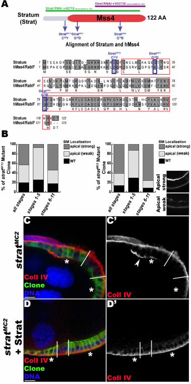

Figure S1. Generation of stratum mutant alleles. Related to Figure 1.
(A) Strat protein schematic and amino acid sequence alignment of Drosophila Strat and human Mss4 (hMss4/RabIF). Mss4 domain is in red. The strat mutations and the parts of the mRNA targeted by the two RNAi transgenes are indicated. The four mutants that we isolated fail to complement a deficiency uncovering the strat locus as well as each other, suggesting they belong to the same complementation group. The missense mutations identified in the newly isolated strat alleles are shown. For MF13 and MA2, mutations change conserved amino acids localized inside (MA2) or outside (MF13) of the Mss4 domain. (B) Quantification of the BM mislocalization phenotype observed in stratMF13 and stratMC2 mutant clones. Representative images are shown. A strong apical localization is observed more frequently in older egg chambers, indicating that secretion at the apical side is progressive during egg chamber maturation (C-D). Lg-sections through FC layers of egg chambers containing stratMC2 mutant clones (C) or stratMC2 mutant clones expressing Strat under control of UAS/Gal4 using the MARCM system (D). Clonal boundaries are indicated by dashed lines; and homozygous mutant FCs are indicated by asterisks (*) and the expression of the membrane marker mCD8-GFP (green). Egg chambers immunostained for α1-Coll IV (red) and stained for DNA (blue). No apical accumulation of Coll IV is detected in strat mutant cells when Strat is expressed (D’, compare to C’). This confirms that the BM deposition defect is due to mutation of strat. Thus, we successfully generated strat mutant alleles. Bars, 10µm.
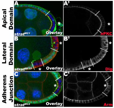
Figure S2. stratum mutant FCs maintain normal apical-basal polarity. Related to Figures 1 and 2.
(A-C) Lg-sections through FC layers of egg chambers containing strat mutant clones expressing Pcan-GFP (green). Clonal boundaries are indicated by dashed lines; homozygous mutant FCs are indicated by asterisks (*) and a loss of intracellular GFP (green). Egg chambers are immunostained for markers of epithelial polarity, including atypical Protein Kinase C (aPKC, A, red), Discs large (Dlg, B, red) and Armadillo (Arm, C, red), and stained for DNA (blue). strat mutant FCs do not show any differences in the distributions of the apical marker aPKC (A’), the lateral marker Dlg (B’) or the adherens junction marker Arm (C’). Note the apical mislocalization of Pcan-GFP in strat mutant FCs (green, A-C). Altogether, these data indicate Strat is specifically required for restriction of BM deposition to the basal side of the cell. Bars, 10µm.
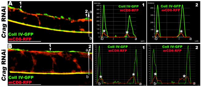
Figure S3. FCs depleted for Crag secrete apically BM proteins. Related to Figure 2.
(A-B) 3D-SIM reconstructions and micrographs of Lg-sections through Crag RNAi FE expressing Coll IV-GFP (green) and the membrane marker mCD8-RFP (red) exclusively expressed in the FCs. Histograms show the distributions of green (Coll IV-GFP) and red (mCD8-RFP) pixels along the basal-apical axis (b-a) at different positions of the epithelium (arrowheads). The x-axis represents the distance along the b-a axis (µm), while the y-axis represents arbitrary pixel intensity. The basal and apical plasma membranes are marked with asterisks (*). In Crag RNAi FCs, Coll IV accumulates both apically and basally in the FE. As shown by the micrographs and histograms, Coll IV and the plasma membrane are closely associated apically. However, Coll IV also accumulates apically outside of cells (green peaks are apical to red peaks). These data suggest that in Crag RNAi cells, as in strat RNAi cells (Figure 2), Coll IV is apically secreted outside of epithelial cells and stays associated with the plasma membrane. Bars, 10µm.


Figure S4. Subcellular distribution of Rab8 in the FE. Related to Figure 4.
(A-D) Lg-sections through egg chambers (Confocal ER) expressing an endogenously tagged YFPMYC-Rab8 (green), stained for F-Actin (white), DNA (blue) and for the Golgi marker Lava lamp (Lva, A), a cis-Golgi marker (GM130, B), Rab5 (C) or Rab11 (D). Rab8 partially co-localizes with the Golgi (arrowheads, A’ and B’) and with the early endosome (E.E., Rab5, arrowheads, C’) and the recycling endosome (R.E., Rab11, arrowheads, D’). Bars, 10µm.

Supplemental Experimental Procedures
Naming of CG7787
The basement membrane mislocalization phenotype observed in CG7787 mutant cells is similar to the phenotype observed in Crag mutant cells. Thus, we chose a name for the gene CG7787 emphasizing its relation to Crag, which in geology defines a steep rugged rock or cliff. Since BM proteins accumulate as a continuous apical sheet of BM proteins in CG7787-depleted epithelial cells, we named the gene stratum (strat), which defines a layer of sedimentary rock.
Additional antibodies used
The following additional primary antibodies were used: guinea pig anti-Coll IV α1-chain (Cgc25c, 1:500, (Shahab et al., 2015)), rabbit anti-aPKC (1:1000, Santa Cruz), mouse anti-Armadillo (N2 7A1, 1:50, DSHB), mouse anti-Discs large (4F3, 1:100, DSHB), rabbit anti-Rab5 (1:50, (Wucherpfennig et al., 2003)), rabbit anti-Rab11 (1:100, (Satoh et al., 2005)), rabbit anti Lava lamp (1:500, (Sisson et al., 2000)) and rabbit anti-GM130 (1:200, Abcam).
Generation of Crag Antibodies
For antibody production a fragment encoding aa 919–1666 of Crag-PA was used to generate rat anti-Crag polyclonal antibodies. The serum was used at 1:200 dilution for immunostaining.
Generation and isolation of mutant alleles of stratum
To generate strat mutant alleles, males of the genotypes FRT40A cn or FRT40A cn bw were mutagenized with EMS and their progeny tested using standard procedures (Chen and Schüpbach, 2006). To isolate strat mutant alleles, the potential mutants were mapped using different deficiency lines uncovering genomic regions around the strat locus: Df(2L)BSC20 and Df(2L)BSC229, which both overlap with strat, and Df(2L)ED578, which does not overlap with strat. Deficiency lines used for mapping were obtained from the Bloomington Stock Center. We isolated four strat mutant alleles (stratMA2, stratMC1, stratMC2 and stratMF13) that belong to the same complementation group. To identify the molecular lesions, each strat mutant line was balanced using CyO, Kr::GFP and genomic DNA from non-GFP-expressing strat-/strat-
embryos was isolated. PCR products of the strat gene were sequenced and compared with those of FRT40A control products (Figure S1). Rescue of the BM proteins mislocalization phenotype observed in strat mutant FCs was performed by generating strat mutant cell clones that express UAS-Strat using the MARCM method (Lee and Luo, 1999) (Figure S1).
Molecular cloning and generation of transgenic lines
A full-length cDNA clone for stratum (clone #RE45155, Drosophila Genomics Resource Center) and a full-length cDNA clone for hMss4/RabIF (clone #4475846, GE Healthcare) were used as templates to generate transgenic lines. To generate pUASp-strat or pUASp-hMss4, the coding regions of strat or hMss4, respectively, were cloned into pTIGER, a derivative of the pUASp vector (Ferguson et al., 2012). To generate pUASp-strat-HA, the coding region of strat was cloned into pTIGER in frame with a C-terminal HA tag. These different constructs were inserted in the Drosophila genome via site-specific recombination (ΦC31 targeted integration (Markstein et al., 2008)) into attP40 (Chromosome II) and/or attP2 (Chromosome III) sites.
Co-Immunoprecipitation
The physical interaction between Strat and Rab8 was confirmed using ovary lysates expressing YFPMYC-Rab8 under Rab8 endogenous promoter (Dunst et al., 2015) and/or Strat-HA using the UAS/Gal4 system. Ovaries were dissected in ice-cold PBS and homogenized in lysis buffer (50mM Tris pH 7.5, 100mM Nacl, 1mM EDTA, 1% NP40, protease inhibitor cocktail) using a pestle. Myc IP reactions were performed using anti-cMyc antibody (9E10, DHSB) and Protein A/G PLUS-Agarose beads (Santa Cruz) at 4°C for 2h. Total lysate and immunoprecipitates were separated via SDS-PAGE and analyzed by Western Blot using anti-

Myc (9E10, 1:100, DHSB) and anti-HA (3F10, 1:3000, Roche) antibodies followed by HRP-conjugated secondary antibodies (Jackson ImmunoResearch). Blots were imaged using a FluorChem HD2 system (Alpha Innotech).
Supplemental References
Chen, Y., and Schüpbach, T. (2006). The role of brinker in eggshell patterning. Mech. Dev. 123, 395–406.
Ferguson, S.B., Blundon, M.A., Klovstad, M.S., and Schupbach, T. (2012). Modulation of gurken translation by insulin and TOR signaling in Drosophila. J. Cell Sci. 125, 1407–1419.
Lee, T., and Luo, L. (1999). Mosaic Analysis with a Repressible Cell Marker for Studies of Gene Function in Neuronal Morphogenesis. Neuron 22, 451–461.
Markstein, M., Pitsouli, C., Villalta, C., Celniker, S.E., and Perrimon, N. (2008). Exploiting position effects and the gypsy retrovirus insulator to engineer precisely expressed transgenes. Nat. Genet. 40, 476–483.
Satoh, A.K., O’Tousa, J.E., Ozaki, K., and Ready, D.F. (2005). Rab11 mediates post-Golgi trafficking of rhodopsin to the photosensitive apical membrane of Drosophila photoreceptors. Development 132, 1487–1497.
Sisson, J.C., Field, C., Ventura, R., Royou, A., and Sullivan, W. (2000). Lava Lamp, a Novel Peripheral Golgi Protein, Is Required for Drosophila melanogaster Cellularization. J. Cell Biol. 151, 905–918.
Wucherpfennig, T., Wilsch-Bräuninger, M., and González-Gaitán, M. (2003). Role of Drosophila Rab5 during endosomal trafficking at the synapse and evoked neurotransmitter release. J. Cell Biol. 161, 609–624.