Embed Size (px)
Citation preview

5129
J. Dairy Sci. 96 :5129–5145http://dx.doi.org/ 10.3168/jds.2013-6741 © American Dairy Science Association®, 2013 .
ABSTRACT
Streptococcus uberis is an important cause of intra-mammary infection in dairy cattle. Strains of Strep. uberis appear to differ in their ability to cause disease based on previous epidemiological studies. We explored the pathogenicity of 2 strains of Strep. uberis, where one strain represented a putatively host-adapted type based on its ability to cause persistent infection and to spread from cow to cow in a lactating herd. This type was part of a clonal complex that is commonly associ-ated with bovine mastitis. The other strain, which was isolated from a transient infection in a single animal in the same herd and did not belong to any known clonal complex, was selected as putatively nonadapted type. Cows (6 per strain) were experimentally challenged in a single hind quarter and the adjacent hind quarter was used as mock challenged control quarter. Both strains showed an equal ability to grow in the milk of chal-lenge animals in vitro. All cows that were challenged with the putatively host-adapted strain developed clinical signs of mastitis, including fever and milk yield depression as well as elevated somatic cell count due to influx of polymorphonuclear leucocytes and lympho-cytes. The cytokine response followed a specific order, with an increase in IL-1β, IL-6, and IL-8 levels at the time of first SCC elevation, followed by an increase in IL-10, IL-12p40, and tumor necrosis factor-α levels approximately 6 h later. In 4 of 6 animals, IL-17A was detected in milk between 57 and 168 h postchallenge. The increase in IL-17A levels coincided with inversion of the prechallenge CD4+-to-CD8+ T lymphocyte ratio,
which was observed from 96 h postchallenge. This was followed by normalization of the CD4+-to-CD8+ ratio due to continued increase of the CD8+ concentration up to 312 h postchallenge. Spontaneous resolution of infection was observed in 5 animals and coincided with a measurable IL-17A response in 4 animals, suggesting that IL-17 may be involved in the resolution of intrama-mmary infection. With the exception of minor elevation of IL-8 levels, no clinical, cytological, or immunological response was detected in quarters challenged with the nonadapted strain. The observed strain-specific patho-genicity was consistent across animals, implying that it is determined by pathogen factors rather than host factors. Key words: mastitis , Streptococcus uberis , lympho-cyte , interleukin-17A
INTRODUCTION
Streptococcus uberis is a major mastitis pathogen in dairy cattle. It is one of the most common causes of clinical and subclinical mastitis in the United Kingdom, New Zealand, and elsewhere (Milne et al., 2002; Zadoks, 2007; Petrovski et al., 2011). Streptococcus uberis causes mastitis in primiparous heifers, dry cows, and lactating cows, and measures to control contagious mastitis have limited effect on the prevalence and incidence of Strep. uberis mastitis (Hill, 1988; Leigh, 1999). For those reasons, Strep. uberis is generally considered an envi-ronmental pathogen. However, epidemiological studies indicate that predominant strains of Strep. uberis occur in many herds, suggesting that these strains may be ca-pable of cow-to-cow transmission or that these strains are highly effective at colonizing the bovine mammary gland (Gillespie et al., 1998; Phuektes et al., 2001b; Zad-oks et al., 2003). These putatively host-adapted strains are in contrast to other mastitis-causing Strep. uberisstrains, which appear to be less suited to the mammary gland environment. Such nonadapted strains are usu-ally isolated sporadically from 1 or 2 animals within
Strain-specific pathogenicity of putative host-adapted and nonadapted strains of Streptococcus uberis in dairy cattle R. Tassi ,*† T. N. McNeilly ,* J. L. Fitzpatrick ,* M. C. Fontaine ,* D. Reddick ,‡ C. Ramage ,‡ M. Lutton ,*1 Y. H. Schukken ,§ and R. N. Zadoks *†§2,3
* Moredun Research Institute, Pentlands Science Park, Bush Loan, Penicuik EH26 0PZ, United Kingdom † Royal (Dick) School of Veterinary Studies, University of Edinburgh, Easter Bush Veterinary Centre, Roslin EH25 9RG, United Kingdom ‡ Moredun Scientific, Pentlands Science Park, Bush Loan, Penicuik EH26 0PZ, United Kingdom § Department of Population Medicine and Diagnostic Sciences, Cornell University, Ithaca, NY 14853
Received February 28, 2013. Accepted April 29, 2013. 1 Current address: Liverpool School of Tropical Medicine, Pembroke
Place, Liverpool, L3 5QA, UK. 2 Current address: Institute of Biodiversity, Animal Health and
Comparative Medicine, College of Medical, Veterinary and Life Sciences, University of Glasgow, 464 Bearsden Rd., Glasgow G61 1QH, UK.
3 Corresponding author: [email protected]
5130 TASSI ET AL.
Journal of Dairy Science Vol. 96 No. 8, 2013
a herd. Why host-adapted and nonadapted strains exhibit different capacities to colonize the bovine mam-mary gland is unknown, although it is possible that, in addition to inherent bacterial factors, these strains may induce different host immune responses. Currently, no experimental data exist comparing the pathogenicity of putatively host-adapted and nonadapted Strep. uberis strains or the clinical and immunological host response following intramammary challenge with such strains.
The early host response to Strep. uberis infection is characterized by a relatively slow but massive influx of PMNL in the infected mammary gland. In experi-mental challenge studies, this influx of PMNL does not generally result in effective reduction in the number of bacteria (Pedersen et al., 2003; Rambeaud et al., 2003; Bannerman et al., 2004a). In one study, Strep. uberis was observed in macrophages after experimental challenge rather than in neutrophils (Thomas et al., 1994), suggesting that macrophages may play a role in bacterial clearance. However, as macrophages from the milk of lactating cows have low killing activity and allow for intracellular replication of Strep. uberis, their role in the protective immune response against Strep. uberis may be limited (Denis et al., 2006). Most ex-perimental challenge studies focus on the early stages of infection, and resolution of infection is rarely described in those studies (Rambeaud et al., 2003; Bannerman et al., 2004a; Harp et al., 2006). In addition, few ex-perimental challenge studies have been used to examine the role of lymphocytes, which are involved in the im-mune response to IMI (Riollet et al., 2000). In cows with natural IMI due to Streptococcus spp., the level of CD4+, CD8+, and γ or δ cells in milk is elevated (Taylor et al., 1997; Soltys and Quinn, 1999). It has been suggested that CD8+ cells, in particular, may play a role in the control of Strep. uberis infection through bactericidal activity (Denis et al., 2011). In addition to the cellular response, the immune response to Strep. uberis involves production of several cytokines, includ-ing the proinflammatory cytokines tumor necrosis fac-tor α (TNF-α) and IL-1β, the chemoattractant IL-8 (Rambeaud et al., 2003; Bannerman et al., 2004a), the anti-inflammatory cytokine IL-10, and T helper type 1 (Th1)-associated cytokines, such as IFN-γ and IL-12 (Bannerman et al., 2004a). Somatic cells obtained from mammary glands with chronic streptococcal or enterococcal infections had elevated levels of mRNA encoding IL-17A compared with cells from uninfected control quarters (Bruno et al., 2010). Interleukin-17A also modulates the response of mammary epithelial cells to staphylococci in vitro (Bougarn et al., 2011). To date, the IL-17A profile of milk during development or resolution of IMI with Strep. uberis has not been described yet.
The aims of our study were to provide simultane-ous characterization of the clinical, bacteriological, and cellular response to intramammary challenge with a putatively host-adapted and a nonadapted strain of Strep. uberis, and to characterize local cytokine profiles during onset and resolution of infection with inclusion of the prototypic member of the IL-17 cytokine family, IL-17A.
MATERIALS AND METHODS
Challenge Strains
Challenge strains of Strep. uberis were selected using epidemiological, clinical, and molecular data. Strain FSL Z1–048 was selected to represent characteristics of putatively host-adapted Strep. uberis, such as chro-nicity of infection and contagious transmission within a herd, whereas strain FSL Z1–124 was selected to represent characteristics of nonadapted Strep. uberis, such as transient infection and environmental origin (Zadoks et al., 2003; Zadoks, 2007). The 2 strains were originally isolated from the same herd during the same time period so that differences in clinical manifestation and transmission could not be attributed to differences in management conditions on the farm of origin. Strain FSL Z1–048 was isolated from several cases of persis-tent subclinical mastitis (median duration = 50 d) in lactating cows during a mastitis outbreak, whereas FSL Z1–124 belonged to a strain that was isolated from a single case of mastitis of short duration (observed duration < 9 d) in a heifer at calving (i.e., without prior exposure to the milking machine; Zadoks et al., 2003). The strains are genetically distinct by a large number of open-reading frames (Lang et al., 2009). Based on multilocus sequence typing, FSL Z1–048 be-longs to sequence type 385 and clonal complex 143, a clonal complex that is predominantly associated with subclinical mastitis (Tomita et al., 2008). Strain FSL Z1–124 belongs to ST383, which is a singleton (i.e., it is not associated with a known clonal complex; Lang et al., 2009; http://pubmlst.org/suberis/).
Growth of Challenge Strains in Milk
The ability of both challenge strains to grow in milk was assessed in vitro, using milk from the animals enrolled in the challenge study (see Challenge Study Design section). The bacterial inoculum for the in vitro growth experiment was prepared from stock cultures of FSL Z1–048 and FSL Z1–124 stored at −80°C. Stock cultures were thawed, plated on 5% horse blood agar (E&O Laboratories, Bonnybridge, UK) and incubated overnight at 37°C to check for viability and purity. Ap-
Journal of Dairy Science Vol. 96 No. 8, 2013
STRAIN-SPECIFIC PATHOGENICITY OF STREPTOCOCCUS UBERIS 5131
proximately 5 colonies per strain were then inoculated into 15 mL of brain heart infusion broth (Difco, Cam-bridge, UK) and incubated for 20 h at 37°C. Bacterial concentration of the incubated broth was determined by viable count method as detailed at the end of this section, whereas the remainder was stored at 4°C, a temperature that does not permit growth of Strep. uberis (Dogan and Boor, 2004). The next day, stored broth cultures were serially diluted in sterile PBS to obtain the target inoculum concentration of 1,000 cfu/mL for each strain. The actual inoculum concentration was checked by viable count method.
To assess the growth potential of challenge strains in milk from study animals, milk was collected asepti-cally from both hind quarters of each animal just before milking, 1 d before experimental challenge. Milk sam-ples were kept on ice and transported to the laboratory for inoculation of culture plates within 1 h of sample collection. For each cow, equal volumes of milk from both hind quarters were comingled, and 10 μL were plated onto horse blood agar (E&O Laboratories) for overnight incubation at 37°C to check for the absence of contaminants and pathogens associated with IMI. For each cow, one 14-mL aliquot of comingled milk was then inoculated with strain FSL Z1–048 and 1 aliquot of 14 mL with FSL Z1–124, using approximately 1,000 cfu of Strep. uberis in 1 mL of PBS (see Intramammary Challenge section). Inoculated milk was incubated for 24 h at 37°C under shaking (200 rpm). At 0, 3, 6, 12, and 24 h post inoculation, 100 μL of milk were taken off and bacterial concentration was determined by viable counting, that is, 1:10 serial dilution series were prepared in sterile PBS and three 10-μL drops per dilution were plated onto horse blood agar plates (E&O Laboratories) and incubated overnight at 37°C. Colonies were counted when they were in the range of 5 to 50 cfu per spot, and the bacterial concentration for each time point (cfu/mL) was calculated based on average colony counts for the appropriate dilution.
Challenge Study Design
Twelve nonpregnant, clinically healthy Holstein cows with no history of clinical mastitis were used in this experiment. Cows were in midlactation (60–160 DIM on the day of challenge) and parity ranged from 1 to 5. Composite SCC as measured during 4 weekly milk recordings was <100,000 cells/mL at the time of pur-chase, with the exception of 1 animal that had compos-ite SCC of 152,000 cells/mL. This animal tested posi-tive for CNS in 1 hind quarter and the culture negative contralateral quarter was used as challenge quarter. On the day before challenge, quarter level SCC was <100,000 cells/mL for 22 of 24 challenge quarters and
between 100,000 and 200,000 cells/mL for the remain-ing 2 quarters. Quarter milk samples from all animals were negative for Strep. uberis before purchase and be-tween purchase and challenge. Challenge studies were conducted using 4 cows at a time, with cows housed in loose housing (group 1) or cubicles (groups 2 and 3), with straw bedding in both housing systems. Cows had access to water and grass silage ad libitum and concentrate was fed 3 times a day based on milk yield. Cows were milked twice a day, at 0600 and 1500 h, using a dedicated milking unit for each cow. After each milking, all teats were dipped in iodine-based teat dis-infectant. Each cow was challenged with Strep. uberis in 1 hind quarter and mock challenged with PBS in the other hind quarter as detailed in the next section. In each group of 4 animals, strain-quarter combinations (FSL Z1–048/right hind, FSL Z1–124/ right hind, FSL Z1–048/left hind, FSL Z1–124/ left hind) were allo-cated to individual cows based on random sampling without replacement. Clinical data and samples were collected for 14 d postchallenge. All experiments were conducted at the Moredun Research Institute (Peni-cuik, UK) with approval of the Institute’s Experiments and Ethical Review Committee in accordance with the Animals (Scientific Procedures) Act 1986.
Intramammary Challenge
The bacterial inoculum for challenge experiments was prepared as described for the growth curve experi-ment, with the exception that the target concentration for animal challenge was 100 cfu/mL. Challenge doses were kept at 4°C (in the laboratory) or on ice (in the milking parlor) until infusion. On the day of challenge, cows were milked per normal procedures, including the use of postmilking teat disinfection. Subsequently, teat ends were disinfected by scrubbing with cotton wool swabs with 70% (vol/vol) ethanol. Two milliliters of inoculum preparation (target dose = 200 cfu/quarter) were infused into each challenge quarter via a J-12 teat infusion cannula (Jorgens Laboratories, Loveland, CO). Control quarters were mock challenged by infusion of 2 mL of sterile PBS. After infusion, teats were dipped in postmilking teat disinfectant and cows were returned from the milking parlor to their pen, as per normal procedures. The actual challenge dose for each strain in each challenge round was determined by viable count method using spare doses of the inoculum preparation.
Sample and Data Collection
Milk samples and clinical data were collected at vari-ous time points following infusion. For the first 48 h, clinical data and milk samples for SCC determination,

5132 TASSI ET AL.
Journal of Dairy Science Vol. 96 No. 8, 2013
bacterial culture, and cytokine ELISA were collected every 6 h. From 2 to 11 d postchallenge, milk samples for SCC and culture were collected twice a day, and from 11 to 13 d postchallenge once a day. On d 3 and 4 postchallenge, milk samples for cytokine ELISA were collected twice a day, and on d 5 through 7 postchal-lenge samples were collected once a day. Additional milk samples for cytokine ELISA were collected on d 10 and 13 postchallenge. Samples for flow cytometric analysis of lymphocyte populations were collected on d 1, 2, 4, 6, 8, 10, and 13 postchallenge. Clinical scores were assigned to each quarter and cow based on a 3-point scale where 0 = no clinical signs; 1 = milk alteration only, such as alteration of color or presence of clots; 2 = local signs, such as redness of the udder, pain on palpation, or swelling; 3 = systemic signs, for example, fever, anorexia or lethargy. Quarter milk samples for bacteriological analysis were collected using aseptic technique (National Mastitis Council, 1999) and stored on ice until refrigeration at 4°C. Separate receptacles were used to collect quarter milk samples for SCC mea-surement, cytokine ELISA, and flow cytometry.
Bacteriological Analysis
Qualitative and quantitative bacteriological analysis was performed by Moredun Scientific Ltd. (Penicuik, UK). For qualitative analysis, 10 μL of milk from each quarter (challenged, mock-challenged, and unchal-lenged quarters) were plated on horse blood agar (E&O Laboratories) and incubated at 37°C for 24 h. Colonies were identified based on morphology and standard biochemical tests (Gram staining, catalase test, esculin splitting; National Mastitis Council, 1999). For quanti-tative analysis, milk samples from challenged quarters were serially diluted with 10-fold dilutions from neat to 10−8 in sterile PBS. Triplicate 10-μL aliquots of each dilution were spotted on blood agar plates (E&O Labo-ratories), allowed to air dry, and incubated overnight at 37°C. Colonies with morphology consistent with Strep. uberis were counted, if possible in the range of 5 to 50 cfu per spot, and the bacterial concentration in milk (cfu/mL) was calculated based on average colony counts for appropriate dilutions.
Molecular Typing
Polymerase chain reaction and pulsed-field gel elec-trophoresis (PFGE) were used to confirm species and strain identity, respectively. Where possible, 3 Strep. uberis isolates from each challenged quarter were analyzed (i.e., 1 isolate each from the first and last positive sample postchallenge and from the time point with peak bacterial shedding). A total of 18 isolates
were analyzed from the 6 cows challenged with strain FSL Z1–048, with each cow represented by 3 isolates. Five isolates, representing 3 cows, were available from animals challenged with strain FSL Z1–124. Species identity of isolates was confirmed using PCR primers and parameter settings as described by Phuektes et al. (2001a). For PFGE of confirmed Strep. uberis isolates, 1.5 mL of overnight culture in brain heart infusion was centrifuged for 10 min at 13,400 × g at room tem-perature. Supernatants were discarded and cell pellets were resuspended in 0.5 mL of Tris-EDTA (TE) buf-fer (10 mM Tris hydrochloric acid, 1 mM EDTA, pH 8). Bacterial suspensions were mixed with 2% (wt/vol) low-melting-point agarose (BioRad Laboratories, Hertfordshire, UK) in TE buffer at 56°C. Plugs were allowed to solidify and incubated overnight at 37°C in 2 mL of TE buffer containing 4 mg/mL lysozyme (Sigma-Aldrich, Dorset, UK). Supernatants were removed and plugs were incubated for 48 h at 56°C in 2 mL of 0.5 M of EDTA containing 2 mg/mL of proteinase K (Sigma-Aldrich) and 1% wt/vol n-laurylsarcosine sodium. Pro-teinase K solution was removed and plugs were washed for 1 h in 36 mL of TE buffer under shaking (100 rpm). Washing was repeated 6 times. Plugs were pre-incubated in 300 μL of reaction buffer containing (1% wt/vol) BSA, 20 mM Tris-acetate, 50 mM potassium acetate, 10 mM magnesium acetate, and 1 mM dithio-threitol (pH 7.9) for 1 h at 37°C. Reaction buffer was removed, 100 μL of enzyme buffer containing 30 units of restriction enzyme SmaI (New England Biolabs, Hitchin, UK) was added to each plug, and plugs were incubated overnight at 25°C. Plugs were then inserted into a gel prepared from 1% (wt/vol) PFGE-certified agarose (BioRad Laboratories) in 0.5 × Tris-borate-EDTA (TBE) buffer (45 mM Tris base, 45 mM boric acid, 1 mM EDTA, pH 8.0). Restriction fragments were separated by PFGE at 14°C for 24 h, with initial switch time 3 s, final switch time 55 s, and linear ramp us-ing a CHEF mapper (BioRad Laboratories). Gels were stained with SYBR Gold stain (Invitrogen, Paisley, UK) according manufacturer’s instruction, rinsed, and photographed under UV light.
Somatic Cell Counting and Flow Cytometry
Approximately 25 mL of milk were collected in plas-tic vials preloaded with preservative and shipped to National Milk Laboratories (Hillington Park, Glasgow, UK) for determination of SCC by Fossomatic milk cell counter (Foss Electronic, Hillerød, Denmark). For flow cytometry, approximately 50 mL of milk per quarter was collected in plastic Falcon vials (BD Bioscience, Oxford, UK). Milk was kept on ice and transported to the laboratory within 30 min, where it was diluted

Journal of Dairy Science Vol. 96 No. 8, 2013
STRAIN-SPECIFIC PATHOGENICITY OF STREPTOCOCCUS UBERIS 5133
with an equal volume of cold PBS and centrifuged at 800 × g for 20 min. Using a sterile pastette, cell pellets were transferred into 30 mL of PBS and resuspended. Cell suspensions were filtered through a 70-μm cell strainer (BD Bioscience) and centrifuged at 400 × g for 15 min. Supernatants were discarded and cell pellets resuspended in 25 mL of PBS. Washing was repeated once and pellets were resuspended in 1 mL of FACS buffer (PBS supplemented with 1% wt/vol BSA). Ten microliters of cell suspension were stained with an equal volume of trypan blue (Sigma-Aldrich) and viable cell concentration was determined using a microscope cell counting chamber, with cell concentration adjusted to 1 × 10⁶ cells/mL. Aliquots of milk cells were labeled with a panel of monoclonal antibodies (Table 1) to identify T-lymphocyte populations and subpopulations (i.e., CD4, CD8, and γ/δ T cells). Fifty microliters of cell suspension were mixed with 50 μL of the appropriate blocking buffer (20% vol/vol heat-inactivated normal mouse serum in FACS buffer for CD4 or CD8 stain-ing; 20% vol/vol heat-inactivated normal goat serum in FACS buffer for CD3 or γ/δ staining). Samples were incubated for 30 min at room temperature and subse-quently centrifuged at 700 × g for 1 min. Supernatants were discarded and 100 μL of antibody diluted with the appropriate buffer (10% vol/vol heat-inactivated normal mouse serum in FACS buffer for CD4 or CD8 staining; 10% vol/vol heat-inactivated normal goat serum in FACS buffer for CD3 or γ/δ staining) were added. Samples were incubated for 30 min at 4°C. Af-ter staining with anti-CD4 or anti-CD8 antibody, cells were washed twice with FACS buffer and once with PBS. Finally, they were resuspended in PBS with 1% wt/vol paraformaldehyde for 10 min. Cells stained with anti-CD3 or anti-γ/δ antibodies were washed twice with FACS buffer. Then, 100 μL of a 1 μg/mL dilution
of goat anti-mouse IgG conjugated to Alexa Fluor 488 (Invitrogen) was added and the cells were incubated at 4°C for 30 min. After staining, cells were washed and fixed in PBS with paraformaldehyde as described for CD4 and CD8 staining. Cells were acquired with a 2 laser Cyan flow cytometer (Beckman Coulter, High Wycombe, UK). Cell debris was differentiated from cells by means of gating and data were collected for a minimum of 10,000 cells for each sample. Data were analyzed using FlowJo software (Tree Star, Ashland, OR). The lymphocyte population was gated based on forward and side scatter and the percentage of positive cells for each staining was calculated within the lym-phocyte gate. Cytocentrifuge preparations of selected samples were prepared using a Shandon Cytospin 4 cytocentrifuge (Thermo Electron Corporation, Milford, MA) and stained using a Reastain Quick-Diff Kit (Rea-gena, Toivala, Finland) for subsequent differential cell counting. Lymphocyte concentrations were calculated based on SCC and the percentage of lymphocytes as determined by flow cytometry.
Cytokine Measurements
Milk samples for cytokine measurement were col-lected in sterile 50-mL plastic Falcon tubes. Skim milk was prepared by centrifuging 50 mL of milk at 2,800 × g at 4°C for 20 min. The fat layer was discarded and the supernatant was transferred to a new 50-mL Falcon tube. Centrifugation was repeated and the supernatant was stored at −20°C until analysis. Microtiter plates (Immunolon 2 HB, Thermo Electron Corporation, Langenselbold, Germany) were coated overnight at 4°C with 100 μL/well of the appropriate coating antibody (Table 2) diluted in 0.5 M carbonate buffer (0.5 M Na2CO3, 0.5 M NaHCO3, pH 9.6). Wells were washed
Table 1. Details of mouse monoclonal antibodies used for flow cytometry
Clone Specificity Isotype ConjugationCellular expression Source
CC8 Bovine CD4 IgG2a Alexa Fluor 647 Helper T cells AbD Serotec1
CC63 Bovine CD8 IgG2a Fluorescein isothiocyanate
Cytotoxic T cells AbD Serotec1
MM1A Bovine CD3 IgG1 NA Pan-T cells VMRD Inc.2
GB21A Bovine γδ TCR3 IgG2b NA Pan-γδ T cells VMRD Inc.2
eBM2A NA4 IgG2a Alexa Fluor 647/FITC Isotype control eBioscience5
VPM21 BDV6 p125/p80 IgG1 NA Isotype control MRI7
VPM22 BDV p125/p80 IgG2b NA Isotype control MRI7
1AbD Serotec (Kidlington, UK).2VMRD Inc. (Pullman, WA).3TCR = T cell receptor.4NA = not applicable.5eBioscience (Hatfield, UK).6BDV = border disease virus (Entrican et al., 1995).7Provided by Sean Wattegedera, Moredun Research Institute (Penicuik, UK).
5134 TASSI ET AL.
Journal of Dairy Science Vol. 96 No. 8, 2013
with washing buffer [PBS at pH 7.4 with 0.05% vol/vol Tween 20 (Sigma-Aldrich)] and nonspecific binding sites were blocked at room temperature for 1 h with 300 μL/well of PBS containing 3% (wt/vol) BSA and 0.05% (vol/vol) Tween 20 (Sigma-Aldrich). Plates were washed again with washing buffer and incubated for 1 h at room temperature with 100 μL/well of skim milk. Each sample was tested in duplicate. When nec-essary, samples were diluted as appropriate with PBS supplemented with 0.05% (vol/vol) Tween 20 (Sigma-Aldrich) and 1% (wt/vol) BSA (reagent diluent). A standard curve of known cytokine concentrations was assayed for each plate using appropriate standard di-luted in reagent diluent. Recombinant bovine standards were used for IL-1β (AbD Serotec, Kidlington, UK), IL-8 (Kingfisher Biotech, Upper Heyford, UK), and TNF-α (R&D systems, Minneapolis, MN). For IL-10 and IL-12p40, recombinant ovine standards were used (provided by Sean Wattegedera, Moredun Research Institute, Penicuik, UK). Plates were washed with washing buffer and 100 μL/well of appropriate detec-tion antibody (Table 2) in reagent diluent were added, followed by incubation for 1 h at room temperature. For IL-1β, IL-10, IL-12p40, and TNF-α plates, 100 μL/well of horseradish peroxidase (HRP)-streptavidin diluted 1:500 in reagent diluent (Sigma-Aldrich) were added, followed by incubation for 45 min at room tem-perature. Interleukin-8 plates were incubated with 100 μL/well of HRP-conjugated polyclonal goat antirabbit immunoglobulins (Dako, Ely, UK) diluted 1:1,000 in reagent diluent for 1 h at room temperature. After incubation with HRP-streptavidin or HRP-conjugated antibody, plates were washed and incubated for 20 min at room temperature with 100 μL per well of o-Phenyl-enediamine dihydrochloride substrate (Sigma-Aldrich). The reaction was stopped with 25 μL per well of 2.5 M of H2SO4 and optical density was measured at 492
nm using a sunrise absorbance reader (Tecan, Theale, UK). Cytokine concentrations in skim milk samples were calculated from the standard curve. Concentra-tions of IFN-γ, IL-6, and IL-17A in milk samples were measured with commercial kits (R&D Systems; Pierce biotechnology, Rockford, IL; and Kingfisher Biotech, respectively) according to the manufacturers’ instruc-tions.
Statistical Analysis
All data were entered into a database and checked for outliers and missing values using SAS 9.2 (SAS Institute Inc., Cary, NC). Outliers were re-evaluated where necessary, but no outliers were removed from the data set. Simple descriptive and graphical analyses were performed for all parameters. Where necessary, transformations were performed to normalize the data (e.g., for SCC and cfu counts). To evaluate the effect of strain and time postinfusion (PI) on any of the mea-sured parameters, a generalized linear mixed model was fit to the data. The general format of the model was
f(y) = incpt + time + strain
+ time × strain + Cow + Re,
where f(y) is a function of the parameter of interest (i.e., a transformation or identity function, as appropri-ate); incpt is the intercept; time is the time PI; strain is an indicator variable indicating either FSL Z1–048 or FSL Z1–124; time × strain is the interaction between time and strain; Cow is a random cow effect; and Re is a complex error term where R indicates the within quarter correlation of the repeated measures and e in-dicates a random error term, assumed to be normally distributed with mean 0 and variance σ2. The model fit
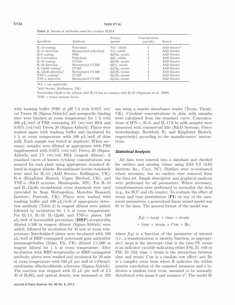
Table 2. Details of antibodies used for cytokine ELISA
Specificity AntibodyIsotype, species
Concentration (μg/mL) Source
IL-1β coating Polyclonal NA,1 rabbit 5 AbD Serotec2
IL-1β detection Biotinylated polyclonal NA, rabbit 1 AbD SerotecIL-8 coating 8M6 IgG2a, mouse 5 AbD SerotecIL-8 secondary Polyclonal IgG, rabbit 2 AbD SerotecIL-10 coating CC318 IgG2b, mouse 4 AbD SerotecIL-10 detection Biotinylated CC230 IgG1, mouse 1 AbD SerotecIL-12p40 coating3 CC301 IgG2a, mouse 1 AbD SerotecIL-12p40 detection3 Biotinylated CC326 IgG2b, mouse 2 AbD SerotecTNF-α coating4 CC327 IgG2b, mouse 2 AbD SerotecTNF-α detection Biotinylated CC328 IgG2a, mouse 1 AbD Serotec1NA = not applicable.2AbD Serotec (Kidlington, UK).3Interleukin-12p40 is the subunit that IL-12 has in common with IL-23 (Oppmann et al., 2000).4TNF = tumor necrosis factor.

Journal of Dairy Science Vol. 96 No. 8, 2013
STRAIN-SPECIFIC PATHOGENICITY OF STREPTOCOCCUS UBERIS 5135
was assessed using the ratio of the deviance to the re-maining degrees of freedom. Least squares means from the models were calculated and used for graphing of the data. Significance testing was done to evaluate the differences between strains over time using contrasts of the least squares means. A Bonferroni correction was used in case of comparisons against prechallenge values in infected and control quarters at multiple time points postchallenge. Statistical significance was declared at P < 0.05. Postinoculation least squares means values were compared with the preinoculation (Time 0) val-ues, with the exception of lymphocyte concentrations and ratios. The latter were compared between challenge quarters and control quarters within time points to allow for significance testing despite lack of complete data for individual quarters.
RESULTS
In Vitro Growth of Challenge Strains in Milk
The ability of strains FSL Z1–048 (putatively host-adapted) and FSL Z1–124 (nonadapted) to grow in fresh milk in vitro was tested using milk samples from 8 individual animals. Results from 1 sample inocu-lated with strain FSL Z1–048 were discarded because of bacterial contamination of the sample. The actual inoculum dose ranged between 700 and 1,280 cfu of Strep. uberis per sample. Both challenge strains were able to grow in fresh milk in vitro with significantly higher levels of bacterial growth observed with strain FSL Z1–124 compared with strain FSL Z1–048 at 3, 6, and 12 h postinoculation (Figure 1A). Strain FSL Z1–124 was first detected at 3 h postinoculation, with more than half of the milk samples testing positive, whereas strain FSL Z1–048 was first detected in milk at 6 h postinoculation, with only 1 sample testing positive (Figure 1B). At 24 h postinoculation, all milk samples tested positive. The maximal bacterial concentration at 24 h posinoculation did not differ between FSL Z1–048 and FSL Z1–124 (7.68 ± 0.13 and 6.68 ± 0.19 log10 cfu/mL respectively).
Clinical Response
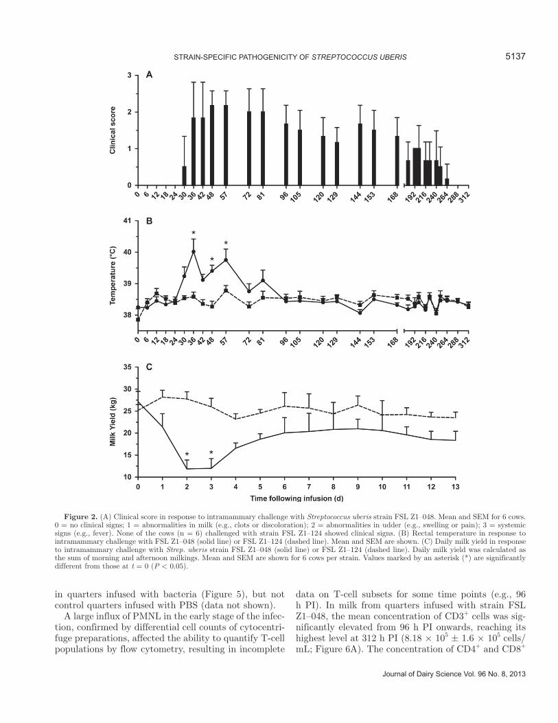
All 6 cows challenged with Strep. uberis strain FSL Z1–048 developed clinical mastitis. In contrast, none of the 6 cows challenged with strain FSL Z1–124 de-veloped clinical signs of mastitis. Clinical signs were first observed 30 to 48 h PI (Table 3). The peak in average clinical score (2.2 ± 0.4) occurred between 48 and 57 h PI (Figure 2A). Increased rectal temperature was observed in all animals challenged with strain FSL Z1–048. The first increase in rectal temperature was
detected between 24 and 72 h PI (Table 3). The average temperature reached a peak of 40.0 ± 1.0°C at 36 h PI (Figure 2B) and remained elevated at 48 and 57 h PI (P < 0.05). At 72 h, average body temperature was not significantly different from prechallenge levels, despite delayed onset of fever in 1 individual (Table 3; Figure 2B). The body temperature of cows challenged with FSL Z1–124 remained at prechallenge level throughout the study. Milk production decreased in all animals challenged with strain FSL Z1–048. Mean production decreased by almost 50% on the second day PI and remained significantly depressed on d 3 PI (P < 0.05; Figure 2C). Milk production started to increase again at d 4 PI. No change in milk output was observed in animals challenged with strain FSL Z1–124 (Figure 2C).
Bacterial Culture and Molecular Typing
Culture of unused doses of the challenge inoculum showed that quarters had been infused with 53 to 712 cfu of strain FSL Z1–048 or 80 to 700 cfu of FSL Z1–124. In the last round of the experiment, the dose of FSL Z1–124 was increased approximately 100-fold, to 14,800 cfu, to determine whether a high inoculum dose would
Figure 1. (A) In vitro growth curves of Streptococcus uberis strain FSL Z1–048 (solid line) and FSL Z1–124 (dashed line) in milk from individual cows. Mean bacterial concentrations and SEM are shown for culture positive samples. Asterisks (*) indicate significant differ-ences between strains. (B) Proportion of milk samples from individual cows inoculated with Strep. uberis strain FSL Z1–048 (solid bars; n = 7) or FSL Z1–124 (hashed bars; n = 8) in which bacteria were detected (detection limit = 333 cfu/mL).

5136 TASSI ET AL.
Journal of Dairy Science Vol. 96 No. 8, 2013
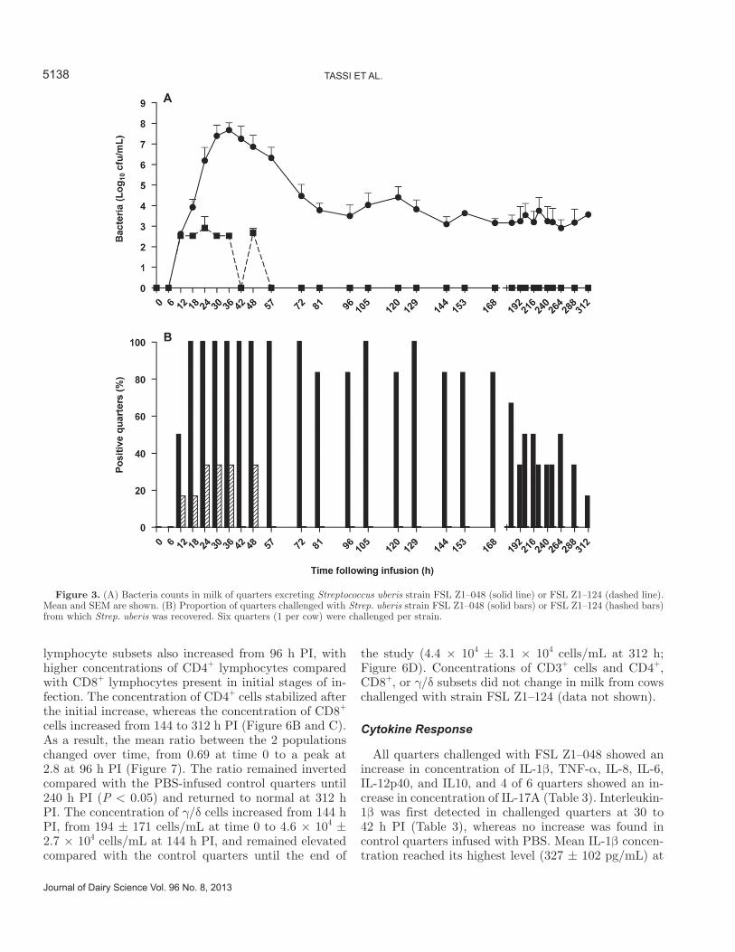
induce a clinical response; however, no difference in re-sponse was observed between animals challenged with either the low or the high dose of this strain. Viable Strep. uberis was isolated from milk from all mammary quarters (n = 6) infused with FSL Z1–048 (Figure 3A). Bacteria were first detected at 12 h PI, and all quarters were positive at 18 h PI (Figure 3B). By 81 h PI, the number of culture positive quarters started to fluctuate. Five of 6 animals had cleared the infection spontane-ously by the end of the study, at 312 h postchallenge (Figure 3B), as confirmed by culture of milk samples collected on a daily basis for 10 d after the end of the study. The maximum average concentration of Strep. uberis in positive quarters was observed at 36 h PI (7.67 ± 0.35 log10 cfu/mL; Figure 3A). By 96 h PI, bacterial concentration had decreased to 3.49 ± 0.54 log10 cfu/mL, and it remained at a similar level in culture posi-tive quarters for the rest of the follow-up period (Figure 3A). Streptococcus uberis was recovered in milk from 4 quarters challenged with strain FSL Z1–124 between 12 and 48 h PI, of which 3 had been challenged with the low dose and 1 with the high dose. No more than 2 quarters were positive at any given time (Figure 3B). Average concentration in positive quarters reached its maximum at 24 h postchallenge (2.91 ± 0.55 log10 cfu/mL; Figure 3A) and all quarters were negative for FSL Z1–124 by 57 h PI.
From each quarter challenged with strain FSL Z1–048, 3 isolates were used for PCR and PFGE (i.e., 1 isolate representing the first isolation, peak concentra-tion, and last isolation from each quarter, respectively, for a total of 18 isolates). All isolates were confirmed to be Strep. uberis by PCR; PFGE patterns of all iso-lates matched that of the challenge strain (Figure 4). Only a limited number of isolates from quarters chal-
lenged with FSL Z1–124 were available for analysis, that is, 1 isolate from 1 quarter and 3 isolates obtained at 3 different time points (12, 18, and 24 h PI) from a different quarter. Polymerase chain reaction us-ing species-specific primers yielded a band of 470 bp rather than the expected 330 bp. Sequence analysis of the PCR amplicon revealed a 160 bp insertion in the 16S-23S rRNA intergenic spacer region. The inser-tion fragment showed 99% sequence homology with the 16S-23S intergenic spacer region of Streptococcus porcinus (ATCC35647). Otherwise, the 16S-23S rRNA intergenic spacer amplicon showed 100% sequence ho-mogeneity with that of Strep. uberis reference strains O140J, ATCC70047, and ATCC19436. Sequencing of a 740 bp fragment of a second housekeeping gene, rpoB (Drancourt et al., 2004), confirmed the species identity of FSL Z1–124 as Strep. uberis. The PFGE patterns of isolates from quarters challenged with FSL Z1–124 were indistinguishable from the PFGE pattern of the challenge strain (Figure 4).
SCC and Flow Cytometry
All quarters challenged with strain FSL Z1–048 showed an increase in SCC. In individual quarters, el-evation of SCC was first observed 30 to 42 h PI (Table 3). Mean SCC reached its peak (7.40 ± 0.07 log10 cells/mL) at 42 h PI and remained elevated throughout the study (Figure 5). Phosphate-buffered saline-infused con-trol quarters from animals challenged with FSL Z1–048 showed a significant increase in SCC compared with prechallenge levels at several time points postchallenge (e.g., during peak milk yield depression and toward the end of the study; Figure 5). The elevation in SCC was observed in animals challenged with strain FSL Z1–124
Table 3. Time (hours postinfusion) of first detection (bacteria; clinical signs other than fever) or first detected increase (temperature, SCC, cytokine levels) of several parameters in 6 cows challenged in 1 mammary quarter with Streptococcus uberis strain FSL Z1–048; apart from body temperature, all parameters were observed or measured at quarter level
Item
Cow identification
1 2 3 4 5 6
Clinical signs 30 30 36 36 48 36Temperature 30 24 24 30 72 36SCC 30 30 30 30 42 36Bacteria 12 18 12 12 18 18IL-1β 30 36 30 30 42 36IL-6 30 36 30 30 42 30IL-8 30 30 30 30 42 36IL-10 36 36 30 36 48 42IL-12p40 36 36 36 36 48 42TNF-α1 36 36 42 36 48 36IL-17A 144 ND2 72 57 ND 721TNF = tumor necrosis factor.2ND = not detected.
Journal of Dairy Science Vol. 96 No. 8, 2013
STRAIN-SPECIFIC PATHOGENICITY OF STREPTOCOCCUS UBERIS 5137
in quarters infused with bacteria (Figure 5), but not control quarters infused with PBS (data not shown).
A large influx of PMNL in the early stage of the infec-tion, confirmed by differential cell counts of cytocentri-fuge preparations, affected the ability to quantify T-cell populations by flow cytometry, resulting in incomplete
data on T-cell subsets for some time points (e.g., 96 h PI). In milk from quarters infused with strain FSL Z1–048, the mean concentration of CD3+ cells was sig-nificantly elevated from 96 h PI onwards, reaching its highest level at 312 h PI (8.18 × 10⁵ ± 1.6 × 10⁵ cells/mL; Figure 6A). The concentration of CD4+ and CD8+
Figure 2. (A) Clinical score in response to intramammary challenge with Streptococcus uberis strain FSL Z1–048. Mean and SEM for 6 cows. 0 = no clinical signs; 1 = abnormalities in milk (e.g., clots or discoloration); 2 = abnormalities in udder (e.g., swelling or pain); 3 = systemic signs (e.g., fever). None of the cows (n = 6) challenged with strain FSL Z1–124 showed clinical signs. (B) Rectal temperature in response to intramammary challenge with FSL Z1–048 (solid line) or FSL Z1–124 (dashed line). Mean and SEM are shown. (C) Daily milk yield in response to intramammary challenge with Strep. uberis strain FSL Z1–048 (solid line) or FSL Z1–124 (dashed line). Daily milk yield was calculated as the sum of morning and afternoon milkings. Mean and SEM are shown for 6 cows per strain. Values marked by an asterisk (*) are significantly different from those at t = 0 (P < 0.05).
5138 TASSI ET AL.
Journal of Dairy Science Vol. 96 No. 8, 2013
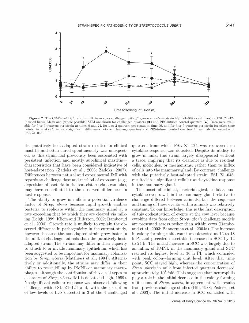
lymphocyte subsets also increased from 96 h PI, with higher concentrations of CD4+ lymphocytes compared with CD8+ lymphocytes present in initial stages of in-fection. The concentration of CD4+ cells stabilized after the initial increase, whereas the concentration of CD8+ cells increased from 144 to 312 h PI (Figure 6B and C). As a result, the mean ratio between the 2 populations changed over time, from 0.69 at time 0 to a peak at 2.8 at 96 h PI (Figure 7). The ratio remained inverted compared with the PBS-infused control quarters until 240 h PI (P < 0.05) and returned to normal at 312 h PI. The concentration of γ/δ cells increased from 144 h PI, from 194 ± 171 cells/mL at time 0 to 4.6 × 10⁴ ± 2.7 × 10⁴ cells/mL at 144 h PI, and remained elevated compared with the control quarters until the end of
the study (4.4 × 10⁴ ± 3.1 × 10⁴ cells/mL at 312 h; Figure 6D). Concentrations of CD3+ cells and CD4+, CD8+, or γ/δ subsets did not change in milk from cows challenged with strain FSL Z1–124 (data not shown).
Cytokine Response
All quarters challenged with FSL Z1–048 showed an increase in concentration of IL-1β, TNF-α, IL-8, IL-6, IL-12p40, and IL10, and 4 of 6 quarters showed an in-crease in concentration of IL-17A (Table 3). Interleukin-1β was first detected in challenged quarters at 30 to 42 h PI (Table 3), whereas no increase was found in control quarters infused with PBS. Mean IL-1β concen-tration reached its highest level (327 ± 102 pg/mL) at
Figure 3. (A) Bacteria counts in milk of quarters excreting Streptococcus uberis strain FSL Z1–048 (solid line) or FSL Z1–124 (dashed line). Mean and SEM are shown. (B) Proportion of quarters challenged with Strep. uberis strain FSL Z1–048 (solid bars) or FSL Z1–124 (hashed bars) from which Strep. uberis was recovered. Six quarters (1 per cow) were challenged per strain.

Journal of Dairy Science Vol. 96 No. 8, 2013
STRAIN-SPECIFIC PATHOGENICITY OF STREPTOCOCCUS UBERIS 5139
48 h PI (Figure 8A). Tumor necrosis factor-α was first detected between 36 and 48 h PI and the highest mean concentration (1,219 ± 599 pg/mL) was observed at 36 h PI (Figure 8B). Interleukin-8 was first detected at 30 to 42 h PI (Table 3), and the highest mean IL-8 concen-tration (4.2 ± 0.2 log10 pg/mL) was reached at 48 h PI (Figure 8C). Five of 6 control quarters in FSL Z1–048 challenged animals showed a moderate increase in IL-8 concentration (peak of mean concentration 1.72 ± 0.36 log10 pg/mL), which coincided with peak concentra-tions in challenged quarters (Figure 8C). Interleukin-6 was first detected at 30 to 42 h PI (Table 3). The IL-6 curve was bimodal, with peaks at 48 and 72 h PI (11.3 ± 2.8 and 11.2 ± 2.9 ng/mL, respectively; Figure 8D). Increased IL-12p40 concentration was first detected at 30 to 48 h PI (Table 3). Mean IL-12p40 concentration peaked at 48 h PI [124 ± 35 biological units (bU)/
mL; Figure 8E]. Increased IL-10 concentration was first detected at 30 to 42 h PI (Table 3), and the peak in mean concentration (35.6 ± 8.2 bU/mL) occurred at 48 h PI (Figure 8F). Interferon-γ was detected at 1 or 2 isolated time points in milk from some challenged quarters, whereas in other quarters, the concentration was elevated for up to 10 consecutive time points. This variability was also observed in PBS-infused control quarters (Figure 8G). The first increase in IL-17A con-centration was detected 57 to 144 h PI (Table 3), and mean IL-17A concentration reached its peak (463 ± 345 pg/mL) at 81 h postchallenge (Figure 8H). The elevation in concentration of IL-17A coincided with the decrease in concentration of Strep. uberis bacteria in milk from infected quarters (Figure 9). All 4 animals that showed an increase in levels of milk IL-17A cleared the infection before the end of the study, whereas 1 of 2 animals without a detectable IL17-A response did not clear the infection spontaneously.
For quarters challenged with strain FSL Z1–124, cy-tokines were measured in milk samples collected at all time points for 1 animal, and at time points up to 81 h PI for the remaining 5 animals. No increase in con-centration of IL-1β, TNF-α, IL-6, IL-10, IL-12p40, or IL-17A was observed in challenged or control quarters of cows challenged with strain FSL Z1–124. Low levels of IL-8 were detected in 3 of 6 quarters challenged with strain FSL Z1–124 (data not shown). Elevations in IL-8 concentration were observed between 12 and 48 h PI, ranging from 32.2 to 72.6 pg/mL, and only occurred in quarters in which the challenge strain was recovered.
DISCUSSION
We observed clear differences in the ability of a pu-tatively host-adapted and nonadapted strain of Strep. uberis (FSL Z1–048 and FSL Z1–124, respectively) to cause mastitis in lactating cows, with clinical signs, PMNL and lymphocyte influx, and multiple cytokine responses developing in all of the quarters challenged with the putatively host-adapted strain and none of the quarters challenged with the nonadapted strain, even when the dose of the nonadapted strain was increased 100-fold. By associating low ability to cause IMI with the nonadapted strain and high ability to cause IMI with the putatively host-adapted strain, which were identified as such using epidemiological and molecular data, our results support the hypothesis that strains of Strep. uberis that predominate within herds are more adapted to colonizing the mammary gland than those which are only sporadically identified as a cause of mas-titis. Thus, differences in incidence of strain-specific IMI are not simply due to different levels of exposure. The observation in the current study that IMI caused by
Figure 4. Examples of pulsed-field gel electrophoresis patterns of Streptococcus uberis isolates from quarters challenged with strain FSL Z1–048 (lanes 6 and 8) or FSL Z1–124 (lanes 3 and 5). The respec-tive challenge strains were included for comparison (lane 4 and lane 7). Lanes 1 and 2 show a DNA ladder (concatamers of λ DNA) and a marker strain (Streptococcus agalactiae STIR-CD-25), respectively.

5140 TASSI ET AL.
Journal of Dairy Science Vol. 96 No. 8, 2013
Figure 5. Somatic cell count in response to challenge with Streptococcus uberis strain FSL Z1–048 (solid lines) or FSL Z1–124 (dashed line). Main and SEM are shown for challenge quarters () and PBS-infused control quarters () of animals challenged with strain FSL Z1–048. The PBS-infused control quarters of animals challenged with FSL Z1–124 did not show elevation of SCC compared with prechallenge levels (data not shown). Significant differences (P < 0.05) between pre- and postchallenge levels are shown for challenge and PBS-infused control quarters of animals challenged with FSL Z1–048 (* and #, respectively).
Figure 6. Concentration in milk of CD3+ (A), CD4+ (B), CD8+ l (C), and γ/δ (D) lymphocytes in milk. Mean and SEM are shown for quarters infused with strain FSL Z1–048 (solid line) and PBS-infused control quarters (dashed line) of the same animals. Asterisks (*) indicate significant differences between challenged quarters and PBS-infused control quarters. Models for γ/δ lymphocytes did not converge.
Journal of Dairy Science Vol. 96 No. 8, 2013
STRAIN-SPECIFIC PATHOGENICITY OF STREPTOCOCCUS UBERIS 5141
the putatively host-adapted strain resulted in clinical mastitis and often cured spontaneously was unexpect-ed, as this strain had previously been associated with persistent infection and mostly subclinical mastitis—characteristics that have been considered indicative of host-adaptation (Zadoks et al., 2003; Zadoks, 2007). Differences between natural and experimental IMI with regards to challenge dose and method of exposure (e.g., deposition of bacteria in the teat cistern via a cannula), may have contributed to the observed differences in host response.
The ability to grow in milk is a potential virulence factor of Strep. uberis because rapid growth enables bacteria to replicate within the mammary gland at a rate exceeding that by which they are cleared via milk-ing (Leigh, 1999; Kliem and Hillerton, 2002; Rambeaud et al., 2004). Growth rate is unlikely to explain the ob-served difference in pathogenicity in the current study, however, because the nonadapted strain grew faster in the milk of challenge animals than the putatively host-adapted strain. The strains may differ in their capacity to attach to or invade mammary epithelium, which has been suggested to be important for mammary coloniza-tion by Strep. uberis (Matthews et al., 1994). Alterna-tively or additionally, the strains may differ in their ability to resist killing by PMNL or mammary macro-phages, although the contribution of those cell types to clearance of Strep. uberis IMI is debated (Leigh, 1999). No significant cellular response was observed following challenge with FSL Z1–124 and, with the exception of low levels of IL-8 detected in 3 of the 4 challenged
quarters from which FSL Z1–124 was recovered, no cytokine response was detected. Despite its ability to grow in milk, this strain largely disappeared without a trace, implying that its clearance is due to resident cells, molecules, or mechanisms, rather than to influx of cells into the mammary gland. By contrast, challenge with the putatively host-adapted strain, FSL Z1–048, resulted in a significant cellular and cytokine response in the mammary gland.
The onset of clinical, bacteriological, cellular, and cytokine events within the mammary gland relative to challenge differed between animals, but the sequence and timing of these events within animals was relatively constant. To our knowledge, this is the first description of this orchestration of events at the cow level because cytokine data from other Strep. uberis challenge models are presented across rather than within cows (Rambe-aud et al., 2003; Bannerman et al., 2004a). The increase in colony-forming units count was detected at 12 to 18 h PI and preceded detectable increases in SCC by 12 to 24 h. The initial increase in SCC was largely due to an influx of PMNL in the mammary gland and SCC reached its highest level at 36 h PI, which coincided with peak colony-forming unit level. After that time point, SCC stayed high, whereas the concentration of Strep. uberis in milk from infected quarters decreased approximately 104-fold. This suggests that neutrophils play a role in the initial decrease in the colony-forming unit count of Strep. uberis, in agreement with results from previous challenge studies (Hill, 1988; Pedersen et al., 2003). The initial increase in SCC coincided with
Figure 7. The CD4+-to-CD8+ ratio in milk from cows challenged with Streptococcus uberis strain FSL Z1–048 (solid lines) or FSL Z1–124 (dashed lines). Mean and (where possible) SEM are shown for challenged quarters () and PBS-infused control quarters (). Data were avail-able for 5 or 6 quarters per strain at times 0 and 24, for 1 or 2 quarters per strain at time 96, and for 3 or 5 quarters per strain for other time points. Asterisks (*) indicate significant differences between challenge quarters and PBS-infused control quarters for animals challenged with FSL Z1–048.

5142 TASSI ET AL.
Journal of Dairy Science Vol. 96 No. 8, 2013
the first detectable increase in IL-8 in all animals. In a challenge model using Strep. uberis strain O140J, SCC increases also coincided with increase in IL-8, and both were detected at 30 h postchallenge (Bannerman et al., 2004a). These observations are consistent with the role of IL-8 in neutrophil chemotaxis (Harada et al., 1994). In a third challenge study, SCC increase preceded the first detected increase in IL-8 by approximately 6 h (Rambeaud et al., 2003). In all 3 challenge studies with
Strep. uberis, the increase in IL-8 levels was sustained for days, which was not the case in challenge studies with different mastitis pathogens (i.e., Escherichia coli, Klebsiella pneumoniae, Serratia marcescens, or Staphy-lococcus aureus; Bannerman et al., 2004a,b,c), where IL-8 levels did not increase measurably (Staph. aureus) or only for the first 18 to 72 h postchallenge (gram-negative pathogens). The early inflammatory response was also characterized by increased levels of the proin-
Figure 8. Cytokine concentration in milk from cows challenged with Streptococcus uberis strain FSL Z1–048. Mean and SEM are shown for challenge quarters (solid line) and, where postchallenge levels were above the detection limit, for PBS-infused control quarters (dashed line). (A) Concentration of IL-1β (detection limit 31.25 pg/mL). (B) Concentration of tumor necrosis factor-α (TNF-α detection limit 125 pg/mL). (C) Concentration of IL-8 (detection limit 0.031 ng/mL). (D) Concentration of IL-6 (detection limit 78.25 pg/mL). (E) Concentration of IL-12p40 (detection limit 0.366 biological units/mL). (F) Concentration of IL-10 (detection limit 0.825 bU). (G) Concentration of IL-17A (detection limit 188 pg/mL). (H) Concentration of IFN-γ (detection limit 156.125 pg/mL). Asterisks (*) indicate significant elevation compared with prechal-lenge levels (P < 0.05).

Journal of Dairy Science Vol. 96 No. 8, 2013
STRAIN-SPECIFIC PATHOGENICITY OF STREPTOCOCCUS UBERIS 5143
flammatory cytokines IL-1β and IL-6, which were first detected at 30 to 42 h PI. Levels of IL-1β, which is both proinflammatory and pyrogenic, showed a single peak and remained significantly elevated until 57 h PI, which is consistent with the observed increase in body temperature in our study, but earlier and shorter than described for other Strep. uberis strains (Rambeaud et al., 2003; Bannerman et al., 2004a). Thus, the immune response to mammary pathogens differs between bacte-rial species as well as bacterial strains. The IL-6 peak appeared to be bimodal, with peaks at 48 and 72 h PI, and levels remained significantly elevated until 96 h PI. This may reflect the role of IL-6, which has both pro-inflammatory and anti-inflammatory activity and plays a role in the transition from innate to adaptive immu-nity, for example by modulating T cell polarization or promoting T cell trafficking into tissues (Jones, 2005). After intramammary challenge with Strep. uberis, IL-6 mRNA expression is upregulated in blood PMNL (Moyes et al., 2010), and its detection in milk may be due to migration of PMNL to the mammary gland. Alternatively, upregulation of IL-6 mRNA expression may occur locally, as both milk cells and mammary
epithelial cells can express the cytokine (Taylor et al., 1997; Zhu et al., 2012). The IL-6 response to intra-mammary challenge with Strep. uberis has not been described before. In field studies, the IL-6 response was similar between cows with clinical mastitis due to streptococci, staphylococci, or E. coli (Taylor et al., 1997). In humans and mice, IL-6 attracts lymphocytes (Weissenbach et al., 2004; McLoughlin et al., 2005); it is unknown whether IL-6 has a similar function in cattle, but IL-6 levels increased before a lymphocyte influx was observed, making this a possibility. An increase in levels of TNF-α, IL-10, and IL-12p40 was generally first detected 6 h after the increase in IL-8 (Table 3), and levels remained elevated for up to 72 to 81 h PI. The observed increase in IL-12p40 levels may indicate the presence of IL-12 or IL-23, because they share the p40 subunit (Oppmann et al., 2000) After challenge with Strep. uberis strain O140J, IL-10 and IL-12 levels increased at the same time as IL-8 levels, and remained high for up to 168 h (Bannerman et al., 2004a). A major difference between our study and the study by Bannerman et al. (2004a) is that colony-forming unit counts continued to increase in their study, whereas,
Figure 9. Concentration of Streptococcus uberis bacteria (dashed line) and IL-17A (solid line) in milk from mammary quarters challenged with strain FSL Z1–048. Quarters with detectable IL-17A are shown. Elevation of IL-17A coincided with decreased concentration of bacteria after the initial peak. All the quarters with detectable IL-17A cleared the infection by the end of the study (312 h postinfusion). Cow identifica-tion numbers match those in Table 3. LH = left hind quarter; RH = right hind quarter.
5144 TASSI ET AL.
Journal of Dairy Science Vol. 96 No. 8, 2013
in our study, colony-forming unit counts stabilized around 72 to 81 h PI and spontaneous resolution of infection was first observed around that time (Figure 3). At this stage in the process, the first detectable rise in IL-17A occurred and so did the influx of CD4+ and CD8+ T lymphocytes, although the exact time of onset of lymphocyte influx was not measured (Figure 6, 8). Interleukin-17A is produced primarily by CD4+TH17 cells; in vitro, the differentiation of naive T-cells in CD4+Th17 cells is supported by IL-6 and TGF-β1. This effect is enhanced by the presence of IL-1β and TNF-α, and subsequent maintenance of Th17 cells is promoted by IL-23 (Veldhoen et al., 2006; Stritesky et al. 2008). The same cytokines may play a role in the re-sponse of cows to intramammary challenge with Strep. uberis, given that elevation of IL-1β and IL-6 levels was followed by elevation of IL-12p40 or IL-23 and TNF-α levels and, finally, elevation of the IL-17A level. Indeed, at this stage in the inflammatory process, the number of CD4+ cells increased more rapidly than the CD8+ cell population, resulting in an inversion of the CD4-to-CD8 ratio (Figure 6). Similar inversion of the CD4-to-CD8 ratio occurs in cows with naturally occurring mastitis due to Streptococcus spp. (Taylor et al., 1997; Soltys and Quinn, 1999). The CD4+ level remained high but, due to continued increases in CD8+ level, the ratio normalized after 240 h PI. Spontaneous resolution of infection occurred in 5 animals. Resolution of infection was preceded by measurable levels of IL-17A in 4 of 6 animals, and was temporally associated with increased lymphocyte levels. Although the number of observa-tions in the current study is too limited to draw solid conclusions, it raises interesting questions with regard to the role of IL-17A or lymphocytes in Strep. uberis IMI. Interleukin-17 may be important for control of ex-tracellular bacterial infections (Curtis and Way, 2009); in mice, IL-17A mediates acquired immunity to strep-tococcal colonization and infection, possibly through enhanced pneumococcal killing by PMNL (Lu et al., 2008). In vitro, IL-17 upregulates genes that encode antimicrobial proteins in bovine mammary epithelial cells, providing an alternative mechanism for bacterial killing (Bougarn et al., 2011). Our observations after experimental challenge are compatible with a role of IL-17A in the control of Strep. uberis infection, and the temporal association of increased levels of CD4+ T cells and IL17-A within the milk suggests that CD4+ T cells may be the principal cellular source of IL17-A. Bacterial killing may also be affected by lymphocytes. A recent study suggested that Strep. uberis-specific CD8+ cells are present in most cows, regardless of prior IMI with Strep. uberis, and that they have a direct killing activity against Strep. uberis in vitro (Denis et al., 2011). The observed sequence of events in our study
is compatible with a role of lymphocytes in clearance of infection.
CONCLUSIONS
In conclusion, our study demonstrates that puta-tively host-adapted strain FSL Z1–048 and nonadapted strain FSL Z1–124, which caused IMI with different epidemiological patterns in a field study, reproducibly elicit distinct clinical and immune responses after ex-perimental challenge of lactating dairy cows. Moreover, this study suggests that neutrophils, lymphocytes, and IL-17A may play important roles in reduction of bacte-rial load in the mammary gland and in clearance of Strep. uberis IMI, which merits further investigation.
ACKNOWLEDGMENTS
We thank Moredun Research Institute’s Bioservices division, and in particular the stockmen, for their sup-port during the challenge experiments. This study was financially supported by Pfizer Animal Health (Ka-lamazoo, MI).
REFERENCES
Bannerman, D. D., M. J. Paape, J. P. Goff, K. Kimura, J. D. Lippolis, and J. C. Hope. 2004a. Innate immune response to intramammary infection with Serratia marcescens and Streptococcus uberis. Vet. Res. 35:681–700.
Bannerman, D. D., M. J. Paape, W. R. Hare, and J. C. Hope. 2004b. Characterization of the bovine innate immune response to intra-mammary infection with Klebsiella pneumoniae. J. Dairy Sci. 87:2420–2432.
Bannerman, D. D., M. J. Paape, J. W. Lee, X. Zhao, J. C. Hope, and P. Rainard. 2004c. Escherichia coli and Staphylococcus aureus elicit differential innate immune responses following intramammary in-fection. Clin. Diagn. Lab. Immunol. 11:463–472.
Bougarn, S., P. Cunha, F. B. Gilbert, A. Harmache, G. Foucras, and P. Rainard. 2011. Staphylococcal-associated molecular patterns enhance expression of immune defense genes induced by IL-17 in mammary epithelial cells. Cytokine 56:749–759.
Bruno, D. R., P. V. Rossitto, R. G. Bruno, M. T. Blanchard, T. Sitt, B. V. Yeargan, W. L. Smith, J. S. Cullor, and J. L. Stott. 2010. Differential levels of mRNA transcripts encoding immunologic me-diators in mammary gland secretions from dairy cows with sub-clinical environmental Streptococci infections. Vet. Immunol. Im-munopathol. 138:15–24.
Curtis, M. M., and S. S. Way. 2009. Interleukin-17 in host defence against bacterial, mycobacterial and fungal pathogens. Immunol. 126:177–185.
Denis, M., N. A. Parlane, S. J. Lacy-Hulbert, E. L. Summers, B. M. Buddle, and D. N. Wedlock. 2006. Bactericidal activity of macro-phages against Streptococcus uberis is different in mammary gland secretions of lactating and drying off cows. Vet. Immunol. Immu-nopathol. 114:111–120.
Denis, M., S. J. Lacy-Hulbert, B. M. Buddle, J. H. Williamson, and D. N. Wedlock. 2011. Streptococcus uberis-specific T cells are present in mammary gland secretions of cows and can be activated to kill Strep. uberis. Vet. Res. Commun. 35:145–156.
Dogan, B., and K. J. Boor. 2004. Short communication: Growth char-acteristics of Streptococcus uberis in UHT-treated milk. J. Dairy Sci. 87:813–815.
Journal of Dairy Science Vol. 96 No. 8, 2013
STRAIN-SPECIFIC PATHOGENICITY OF STREPTOCOCCUS UBERIS 5145
Drancourt, M., V. Roux, P.-E. Fournier, and D. Raoult. 2004. rpoB gene sequence-based identification of aerobic gram-positive cocci of the genera Streptococcus, Enterococcus, Gemella, Abiotrophia, and Granulicatella. J. Clin. Microbiol. 42:497–504.
Entrican, G., A. Dand, and P. F. Nettleton. 1995. A double monoclo-nal antibody ELISA for detecting pestivirus antigen in the blood of viraemic cattle and sheep. Vet. Microbiol. 43:65–74.
Gillespie, B. E., B. M. Jayarao, J. W. Pankey, and S. P. Oliver. 1998. Subtyping of Streptococcus dysgalactiae and Streptococcus uberis isolated from bovine mammary secretions by DNA fingerprinting. Zentralbl. Veterinarmed. B. 45:585–593.
Harada, A., N. Sekido, T. Akahoshi, T. Wada, N. Mukaida, and K. Matsushima. 1994. Essential involvement of interleukin-8 (IL-8) in acute inflammation. J. Leukoc. Biol. 56:559–564.
Harp, J. A., T. E. Waters, J. P. Goff, D. D. Bannerman, and M. J. Paape. 2006. Expression of lymphocyte homing and adhesion mol-ecules during intramammary infection of cows with Serratia marc-escens or Streptococcus uberis: Correlation with bacterial coloniza-tion and clinical signs. Vet. Immunol. Immunopathol. 109:13–21.
Hill, A. W. 1988. Pathogenicity of two strains of Streptococcus uberis infused into lactating and non-lactating bovine mammary glands. Res. Vet. Sci. 45:400–404.
Jones, S. A. 2005. Directing transition from innate to acquired immu-nity: Defining a role for IL-6. J. Immunol. 175:3463–3468.
Kliem, K. E., and J. E. Hillerton. 2002. Possible labile inhibition of the growth of Streptococcus uberis in milk from cows free from mastitis. J. Dairy Res. 69:375–382.
Lang, P., T. Lefébure, W. Wang, R. N. Zadoks, Y. Schukken, and M. J. Stanhope. 2009. Gene content differences across strains of Strep-tococcus uberis identified using oligonucleotide microarray com-parative genomic hybridization. Infect. Genet. Evol. 9:179–188.
Leigh, J. A. 1999. Streptococcus uberis: A permanent barrier to the control of bovine mastitis? Vet. J. 157:225–238.
Lu, Y.-J., J. Gross, D. Bogaert, A. Finn, L. Bagrade, Q. Zhang, J. K. Kolls, A. Srivastava, A. Lundgren, S. Forte, C. M. Thompson, K. F. Harney, P. W. Anderson, M. Lipsitch, and R. Malley. 2008. Interleukin-17A mediates acquired immunity to pneumococcal colonization. PLoS Pathog. 4:e1000159.
Matthews, K. R., R. A. Almeida, and S. P. Oliver. 1994. Bovine mam-mary epithelial cell invasion by Streptococcus uberis. Infect. Im-mun. 62:5641–5646.
McLoughlin, R. M., B. J. Jenkins, D. Grail, A. S. Williams, C. A. Fielding, C. R. Parker, M. Ernst, N. Topley, and S. A. Jones. 2005. IL-6 trans-signaling via STAT3 directs T cell infiltration in acute inflammation. Proc. Natl. Acad. Sci. USA 102:9589–9594.
Milne, M. H., D. C. Barrett, J. L. Fitzpatrick, and A. M. Biggs. 2002. Prevalence and aetiology of clinical mastitis on dairy farms in Devon. Vet. Rec. 151:241–243.
Moyes, K. M., J. K. Drackley, D. E. Morin, and L. L. Loor. 2010. Greater expression of TLR2, TLR4, and IL6 due to negative en-ergy balance is associated with lower expression of HLA-DRA and HLA-A in bovine blood neutrophils after intramammary masti-tis challenge with Streptococcus uberis. Funct. Integr. Genomics 10:53–61.
National Mastitis Council. 1999. Laboratory Handbook on Bovine Mastitis. National Mastitis Council, Madison, WI.
Oppmann, B., R. Lesley, B. Blom, J. C. Timans, Y. Xu, B. Hunte, F. Vega, N. Yu, J. Wang, K. Singh, F. Zonin, E. Vaisberg, T. Chura-kova, M. Liu, D. Gorman, J. Wagner, S. Zurawski, Y. Liu, J. S. Abrams, K. W. Moore, D. Rennick, R. de Waal-Malefyt, C. Han-num, J. F. Bazan, and R. A. Kastelein. 2000. Novel p19 protein engages IL-12p40 to form a cytokine, IL-23, with biological activi-ties similar as well as distinct from IL-12. Immunity 13:715–725.
Pedersen, L. H., B. Aalbaek, C. M. Røntved, K. L. Ingvartsen, N. S. Sorensen, P. M. Heegaard, and H. E. Jensen. 2003. Early patho-
genesis and inflammatory response in experimental bovine mastitis due to Streptococcus uberis. J. Comp. Pathol. 128:156–164.
Petrovski, K. R., N. B. Williamson, N. Lopez-Villalobos, T. J. Par-kinson, and I. G. Tucker. 2011. Culture results from milk samples submitted to veterinary diagnostic laboratories from August 2003 to December 2006 in New Zealand. N. Z. Vet. J. 59:317–322.
Phuektes, P., P. D. Mansell, and G. F. Browning. 2001a. Multiplex polymerase chain reaction assay for simultaneous detection of Staphylococcus aureus and streptococcal causes of bovine mastitis. J. Dairy Sci. 84:1140–1148.
Phuektes, P., P. D. Mansell, R. S. Dyson, N. D. Hooper, J. S. Dick, and G. F. Browning. 2001b. Molecular epidemiology of Streptococ-cus uberis isolates from dairy cows with mastitis. J. Clin. Micro-biol. 39:1460–1466.
Rambeaud, M., R. A. Almeida, and S. P. Oliver. 2004. Growth of Streptococcus uberis in skim milk obtained from Holstein and Jer-sey dairy cows during different stages of lactation. J. Vet. Med. B. Infect. Dis. Vet. Public Health 51:143–145.
Rambeaud, M., R. A. Almeida, G. M. Pighetti, and S. P. Oliver. 2003. Dynamics of leukocytes and cytokines during experimentally induced Streptococcus uberis mastitis. Vet. Immunol. Immuno-pathol. 96:193–205.
Riollet, C., P. Rainard, and B. Poutrel. 2000. Cells and cytokines in inflammatory secretions of bovine mammary gland. Adv. Exp. Med. Biol. 480:247–258.
Soltys, J., and M. T. Quinn. 1999. Selective recruitment of T-cell sub-sets to the udder during staphylococcal and streptococcal mastitis: Analysis of lymphocyte subsets and adhesion molecule expression. Infect. Immun. 67:6293–6302.
Stritesky, G. L., N. Yeh, and M. H. Kaplan. 2008. IL-23 pmromotes maintenance but not commitment to the Th17 Lineage. J. Im-munol. 181:5948–5955.
Taylor, B. C., R. G. Keefe, J. D. Dellinger, Y. Nakamura, J. S. Cullor, and J. L. Stott. 1997. T cell populations and cytokine expression in milk derived from normal and bacteria-infected bovine mammary glands. Cell. Immunol. 182:68–76.
Thomas, L. H., W. Haider, A. W. Hill, and R. S. Cook. 1994. Patholog-ic findings of experimentally induced Streptococcus uberis infection in the mammary gland of cows. Am. J. Vet. Res. 55:1723–1728.
Tomita, T., B. Meehan, N. Wongkattiya, J. Malmo, G. Pullinger, J. Leigh, and M. Deighton. 2008. Identification of Streptococcus uberis multilocus sequence types highly associated with mastitis. Appl. Environ. Microbiol. 74:114–124.
Veldhoen, M., R. J. Hocking, C. J. Atkins, R. M. Locksley, and B. Stockinger. 2006. TGF-b in the context of an inflammatory cyto-kine milieu supports de novo differentiation of IL-17-producing T cells. Immunity 24:179–189.
Weissenbach, M., T. Clahsen, C. Weber, D. Spitzer, D. Wirth, D. Vestweber, P. C. Heinrich, and F. Schaper. 2004. Interleukin-6 is a direct mediator of T cell migration. Eur. J. Immunol. 34:2895–2906.
Zadoks, R. N. 2007. Sources and epidemiology of Streptococcus uberis, with special emphasis on mastitis in dairy cattle. CAB Reviews: Perspectives in Agriculture, Veterinary Science, Nutrition and Natural Resources, 2007. CAB International, Wallingford, UK.
Zadoks, R. N., B. E. Gillespie, H. W. Barkema, O. C. Sampimon, S. P. Oliver, and Y. H. Schukken. 2003. Clinical, epidemiological and molecular characteristics of Streptococcus uberis infections in dairy herds. Epidemiol. Infect. 130:335–349.
Zhu, Y. H., P. Q. Liu, X. G. Weng, Z. Y. Zhuge, R. Zhang, J. L. Ma, X. Q. Qiu, R. Q. Li, X. L. Zhang, and J. F. Wang. 2012. Short communication: Pheromonicin-SA affects mRNA expression of toll-like receptors, cytokines, and lactoferrin by Staphylococcus aureus-infected bovine mammary epithelial cells. J. Dairy Sci. 95:759–764.