Embed Size (px)
Citation preview

Strabismus Research Update
ARVO Asia Brisbane 2017Lionel Kowal Melbourne
With help from recent FellowsDrs Sheth Kini Mitchell
No financial conflicts
Today Some of the advances in recent years that have changed or are changing
the understanding amp treatment protocols and options in sensory and
motor strabismus
חננו מאתך דעה בינה והשכל
AMBLYOPIAPEDIG amp MOTAS
bull Rx often based on quantifying the sensory asymmetry amp treating it with asymmetric treatments
bull Glasses alone sometimes effectivebull Less treatment is often as effective as morebull Atropine [used for 100 yrs] amp opaque occlusion [used for 300
yrs] equivalent effect for many ptsbull There are NO other treatments still used in medicine
that are as old as these
21st Century Amblyopia RxHigh Tech Asymmetric Rx 1
Binocular treatment of amblyopia using videogames (BRAVO) study protocol for a randomised controlled trialGuo CX hellipKowal LhellipTrials 2016 Oct 1817(1)504
Using asymmetric high tech inputs
bull Blurred video game to good eye
bull Clear video game to amblyopic eye
Results expected in next few weeks
21st Century Amblyopia Rx 2
bull Effect of a Binocular iPad Game vs Part-time Patching in Children Aged 5 to 12 Years With Amblyopia A Randomized Clinical Trial
bull Holmes JM helliphellipPEDIG
bull JAMA Ophthalmol 2016 Dec 1134(12)1391-1400
helliphelliphelliphelliphelliphelliphelliphelliphellip
bull Binocular Treatment of Amblyopia in Children Teething Problems on the Path to Clinical Practice
bull Dahlmann-Noor A1 JAMA Ophthalmol 2016 Nov 3
21st Century Amblyopia Rx 3
bull Invited Commentarybull New Treatments for AmblyopiamdashTo Patch or Playbull John SloperMoorfields Londonbull JAMA Ophthalmol Published online November 10 2016
doi101001jamaophthalmol20164296
EditorialJ AAPOSFeb 2015
bull Some published data Many more papers presented meetings
bull I expect this will a popular 1st treatment for amblyopia
bull Commercial competition ++ expected
Radiology of strabismus Orbital pulleys
bull Orbital pulleys have been recognised gt100 yrs [lsquopoulesrsquo in 19C French literature]
bull Clinical relevance has been appreciated for ~20 years and practical application of the knowledge is growing fast
Todaybull Childhood pulley disordersbull Acquired pulley disorders ndash elderly and high myopesPulley surgeriesbull The medial rectus pulleybull The lateral rectus
Radiology of strabismusRecent findings orbital pulleys
Middle East African Journal of Ophthalmology July-Sept 2015 pp 279-285
bull Abnormal location of the pulleys could explain many cases of incomitant strabismus conventionally [amp without scientific justification] attributed to lsquooblique muscle dysfunctionrsquo
ldquoVrdquo pattern ETInf displacement of LR (RgtL)As if orbital contents EXtorted
ldquoArdquopattern ETLR displaced sup to MR and SR displaced nasal to IRAs if orbital contents INtorted
J AAPOS
Demer Clark Miller Advances in Strabismology
12 yo
previous LR amp IO weakening for V -
XT
Recurrent ResidualV- XT [UG 30 DG 10]Minimal IO OANo fundus torsion
Childhood pulley disorders
Coronal MRI T1 inf positioning of LR (LgtR) and nasal shift of IR
Infraplaced Lateral rectus seen before the medial rectus
At upper edge of MR LR no longer seen
Childhood pulley disorders unknown of childhood strabismus esp incomitant strabismus
Acquired pulley disordersCommon(est) cause of small angle ET +- vertical in the
healthy elderly
Acquired L ET Sagging LLR
J AAPOS
Downward displacement of the LLR changes itrsquos vector and causes an aBduction deficit
Postoperative (52 days after surgery)
Preoperative
Acquired pulley disorders Extreme Esotropia of High Myopia[aka Heavy Eye]
Dr Yokoyamarsquos case
Extreme myopia
LR displaced down SR nasal
Preoperative
1811 deg
LR
SR
Postoperative
1036 deg
LR
SR
From Tsuranu Yokoyama
Pulley surgeriesThe medial rectus pulley suture
a safer alternative to posterior scleral fixation
J AAPOS 2012
Each half of each EOM has a unique non-overlapping nerve supply
Compartmentalization not [yet] demonstrated for superior rectus
Clinical implications of EOM compartmentalization
bull 1 Sup compartment LR atrophy Esotropia ndash of ndash obscure - cause not- quite LR palsyNot rare bull 2 Sup compartment MR atrophyProgressive exotropia ndash of- obscure- causeRare ndash no published cases yetbull 3 Medial Lateral Sup Obl compartment atrophyProbably explains why some have vertical diplopia vs torsional diplopia vs both V amp T4 Probably Many more incomitant clinical scenarios waiting to beappreciated
1 Esotropia amp compartment hemi-atrophy
bull Superior compartment atrophy of the LR produces a clinical condition that resembles LR paresis
bull Clinical picture more LR function than complete palsy Treatment implications uncertain
Clark R A amp Demer J L (2014) Lateral rectus superior compartment palsy American Journal of Ophthalmology 157(2)
Total RLR atrophy = palsy Sup compartment RLR palsy
Clinical picture more LR function than complete palsy Treatment implications uncertain
Adult ET of uncertain causeDelayed diagnosis of superior compartment LR paresis
9 years after first presentation and 3 years after the last MRI and the 3rd
horizontal rectus surgery the diagnosis is clearer
Clark amp Demer Lateral Rectus Sup compartment palsyAmer J Ophth 2014 157 Fig 2 on page 481
2012 MRI re-read for purpose of a talk RLR has a triangular appearance
2 Exotropia and compartment hemi-atrophy
bull Diplopia onset 66 yo
bull 68yo increased prism to 10Δ
bull 69yo hellipto 24Δ
bull 70yo D50Δ N 60Δ
bull MR -2mm OU
Bilateral asymmetric atrophy of the superior half of medical rectus compared to inferior
Surgery and CourseMR plicateresect OU LR recess x1 Adjustables 10 w followup single vision small phorias
Asymmetry can be expected to produce a small vertical
Effective lowering of the MR vector might cause an lsquoArsquo pattern
Different types of Superior Oblique PalsyParesis
= A
= B
= C D
Medial compartment of SO [=SOm] controls torsionLateral hellip[=SOl] controls vertical movement
BCD 20 develop floppy tendons requiring tendon tightening surgery
Flap tear of the inferior rectusCommon Commonest cause of vertical diplopia after orbital trauma
Normal [contralateral] IR Affected IROuter frac12 has been shaved off
Superficial layer of inferior rectus lsquoshavedrsquo off
From Irene Ludwig
Example where lsquodivotrsquo of muscle has been lsquoslicedrsquo off
Large divot ~50 Normal contralateral IR
Flap Tear HypothesisFrom Irene Ludwig
bull Blunt trauma causes outward traction on orbital septae
bull Orbital connective tissue which attaches onto EOM pulls away portion of muscle amp weakens it
bull Flap can acts as a tether further restricting amp complicating motility
2016 update ARVO amp AAPOSMRI images of the flap tear
Validates Irene Ludwigrsquos observations first publishedin 2001
Intramuscular injection of 3 Bupivacaine BP with Botox BT to treat strabismus
bull The unwanted changes caused by accidental injection of local anaesthetic agents like Bupivacaine into EOM during ocular anaesthetic procedures can be exploited to treat the lsquoweakrsquo muscle in strabismus eg the MR in consecutive XT [combined with Botox BT to the LR]
J AAPOS 2016
Time post injection
Evolution of RLR changes (post BT)
Evolution of RMR changes (post BP)
Evolution of alignment
PRE INJECTION
4 days
2 weeks
3 weeks
4 weeks
5 weeks
6 weeks
7 weeks
9 weeks
14 weeks
17 weeks
65 months
ET 4 Near 6Distance
XT 12 Near Ortho Distance
ET 1 Near(Variable) Ortho Distance
BT yet to show effect BP showing full anesthetic effectFull effect of BT Anesthetic effect of BP worn off secondary changes to RMR begin
BT effect worn off fully
Why does this transposition often work so well
Innovator Dr Earl CrouchValidated by this paper from Harvard
Technique of SR transposition amp LMR Rc
WHY DOES SUCH AN ASYMMETRIC OPERATION HAVE SO FEW UNEXPECTED CYCLOVERTICAL COMPLICATIONS
Useful in traumatic 6ths
WHY DOES SUCH AN ASYMMETRIC OPERATION HAVE SO FEW UNEXPECTED CYCLOVERTICAL COMPLICATIONSSO MUCH MORE TO KNOW
J AAPOS Dec 2016
bull THANK YOU
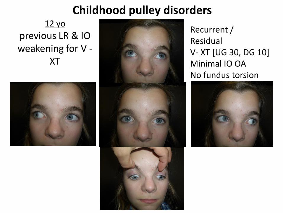
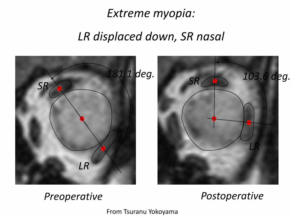
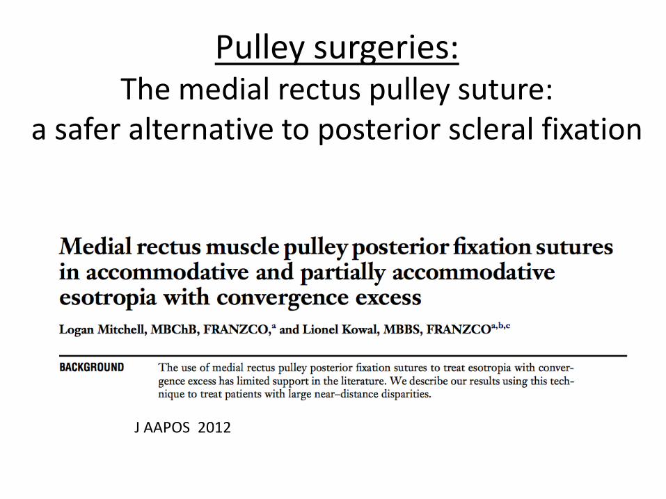
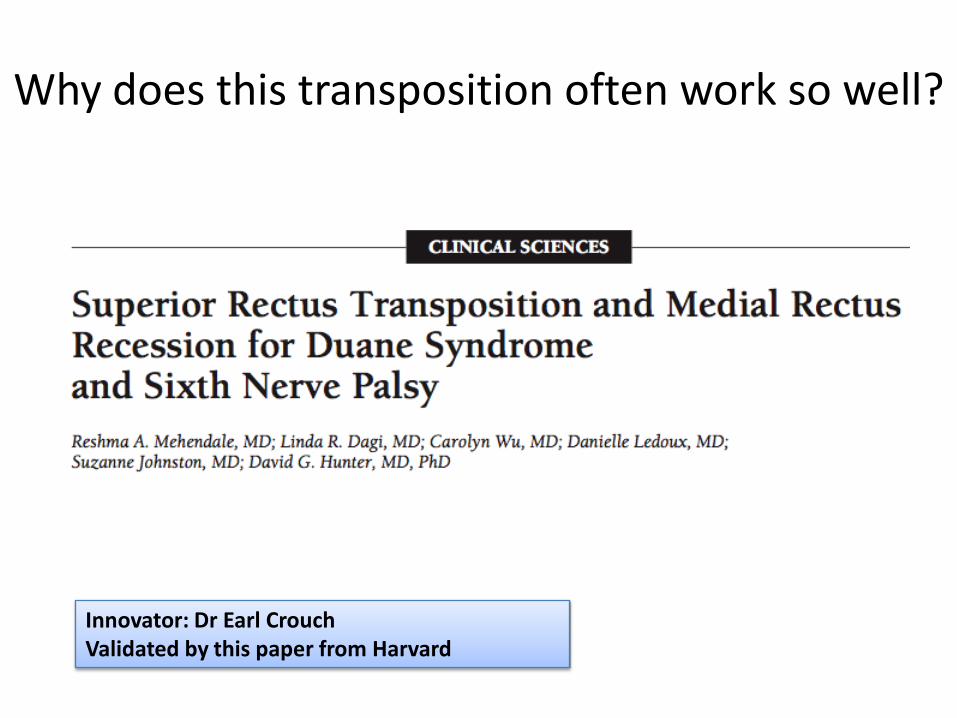
Technique of SR transposition amp LMR Rc
WHY DOES SUCH AN ASYMMETRIC OPERATION HAVE SO FEW UNEXPECTED CYCLOVERTICAL COMPLICATIONS
Useful in traumatic 6ths
WHY DOES SUCH AN ASYMMETRIC OPERATION HAVE SO FEW UNEXPECTED CYCLOVERTICAL COMPLICATIONSSO MUCH MORE TO KNOW
J AAPOS Dec 2016
bull THANK YOU

Useful in traumatic 6ths
WHY DOES SUCH AN ASYMMETRIC OPERATION HAVE SO FEW UNEXPECTED CYCLOVERTICAL COMPLICATIONSSO MUCH MORE TO KNOW
J AAPOS Dec 2016
bull THANK YOU

bull THANK YOU