Embed Size (px)
Citation preview
Section 1 Ultrasound fundamentals
Introduction
S1 Stephanie J. Doniger
Pediatric Emergency Medicine is a relatively new field of med-icine developed in the 1980s. Since its inception, severaladvancements have been made in order to improve the emer-gency care of children. In 2007, an Institute of Medicine reportdescribed the deficit of pediatric-centered emergency care, call-ing for the need for more resources to be dedicated towards theemergency care of children. The mere existence of this field ofmedicine embodies the idea that children are not just “smalladults” but have distinct pathology, and need care and equip-ment tailored to their size and physiology.
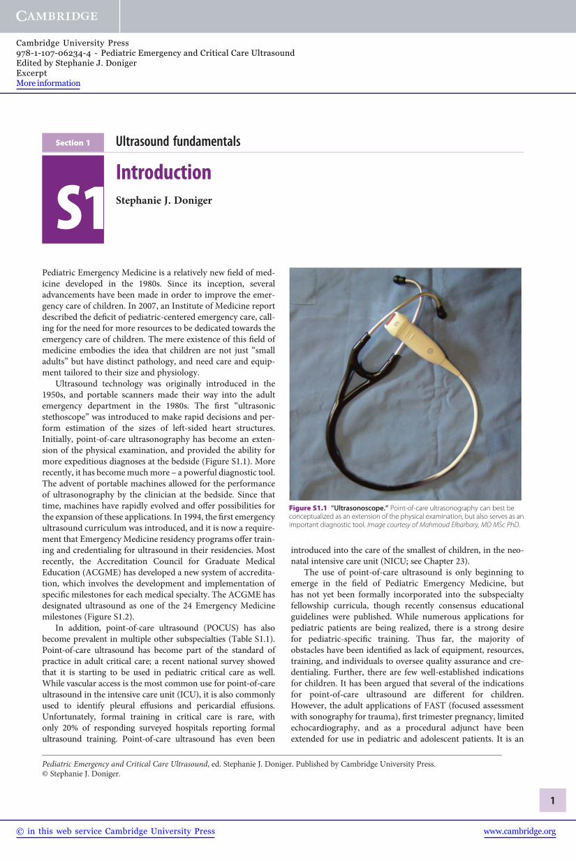
Ultrasound technology was originally introduced in the1950s, and portable scanners made their way into the adultemergency department in the 1980s. The first “ultrasonicstethoscope” was introduced to make rapid decisions and per-form estimation of the sizes of left-sided heart structures.Initially, point-of-care ultrasonography has become an exten-sion of the physical examination, and provided the ability formore expeditious diagnoses at the bedside (Figure S1.1). Morerecently, it has becomemuchmore – a powerful diagnostic tool.The advent of portable machines allowed for the performanceof ultrasonography by the clinician at the bedside. Since thattime, machines have rapidly evolved and offer possibilities forthe expansion of these applications. In 1994, the first emergencyultrasound curriculum was introduced, and it is now a require-ment that Emergency Medicine residency programs offer train-ing and credentialing for ultrasound in their residencies. Mostrecently, the Accreditation Council for Graduate MedicalEducation (ACGME) has developed a new system of accredita-tion, which involves the development and implementation ofspecific milestones for each medical specialty. The ACGME hasdesignated ultrasound as one of the 24 Emergency Medicinemilestones (Figure S1.2).
In addition, point-of-care ultrasound (POCUS) has alsobecome prevalent in multiple other subspecialties (Table S1.1).Point-of-care ultrasound has become part of the standard ofpractice in adult critical care; a recent national survey showedthat it is starting to be used in pediatric critical care as well.While vascular access is the most common use for point-of-careultrasound in the intensive care unit (ICU), it is also commonlyused to identify pleural effusions and pericardial effusions.Unfortunately, formal training in critical care is rare, withonly 20% of responding surveyed hospitals reporting formalultrasound training. Point-of-care ultrasound has even been
introduced into the care of the smallest of children, in the neo-natal intensive care unit (NICU; see Chapter 23).
The use of point-of-care ultrasound is only beginning toemerge in the field of Pediatric Emergency Medicine, buthas not yet been formally incorporated into the subspecialtyfellowship curricula, though recently consensus educationalguidelines were published. While numerous applications forpediatric patients are being realized, there is a strong desirefor pediatric-specific training. Thus far, the majority ofobstacles have been identified as lack of equipment, resources,training, and individuals to oversee quality assurance and cre-dentialing. Further, there are few well-established indicationsfor children. It has been argued that several of the indicationsfor point-of-care ultrasound are different for children.However, the adult applications of FAST (focused assessmentwith sonography for trauma), first trimester pregnancy, limitedechocardiography, and as a procedural adjunct have beenextended for use in pediatric and adolescent patients. It is an
Figure S1.1 “Ultrasonoscope.” Point-of-care ultrasonography can best beconceptualized as an extension of the physical examination, but also serves as animportant diagnostic tool. Image courtesy of Mahmoud Elbarbary, MD MSc PhD.
Pediatric Emergency and Critical Care Ultrasound, ed. Stephanie J. Doniger. Published by Cambridge University Press.© Stephanie J. Doniger.
1
www.cambridge.org© in this web service Cambridge University Press
Cambridge University Press978-1-107-06234-4 - Pediatric Emergency and Critical Care UltrasoundEdited by Stephanie J. DonigerExcerptMore information

Table S1.1 Selected applications of point-of-care ultrasonography, according to medical specialty.
Specialty Ultrasound applications
Anesthesia Guidance for vascular access, regional anesthesia, intraoperative monitoring of fluid status and cardiac function
Cardiology Echocardiography, intracardiac assessment
Critical care medicine Procedural guidance, pulmonary assessment, focused echocardiography
Dermatology Assessment of skin lesions and tumors
Emergency medicine FASTa, focused emergency assessment, procedural guidance
Endocrinology and endocrine surgery Assessment of thyroid and parathyroid, procedural guidance
General surgery Ultrasonography of the breast, procedural guidance, intraoperative assessment
Gynecology Assessment of cervix, uterus, and adnexa; procedural guidance
Obstetrics, maternal–fetal medicine Assessment of pregnancy, detection of fetal abnormalities, procedural guidance
Neonatology Cranial and pulmonary assessments
Nephrology Vascular access for dialysis
Neurology Transcranial Doppler, peripheral-nerve evaluation
Ophthalmology Corneal and retinal assessment
Orthopedic surgery Musculoskeletal applications
Otolaryngology Assessment of thyroid, parathyroid, and neck masses; procedural guidance
Pediatrics Assessment of bladder, procedural guidance
Pulmonary medicine Transthoracic pulmonary assessment, endobronchial assessment, procedural guidance
Radiology, interventional radiology Ultrasonography taken to the patient with interpretation at the bedside, procedural guidance
Rheumatology Monitoring of synovitis, procedural guidance
Trauma surgery FASTa, procedural guidance
Urology Renal, bladder, and prostate assessment; procedural guidance
Vascular surgery Carotid, arterial, and venous assessment; procedural assessment
a FAST denotes focused assessment of sonography with trauma.Source (copyright): Moore, C. L., Copel, J. A. (2011) Point-of-care ultrasonography. N Engl J Med 364: 749–57.
Uses ultrasound for the bedside diagnostic evaluation of emergency medical conditions and diagnoses, resuscitation of the acutely ill or injured patient. and proceduralguidance
Level 1
Describes the indicationsand limitations oflimited, goal-directedemergency ultrasound
Explains how to optimize ultrasoundimages and identify the properprobe for each of the focusedultrasound applications
Performs an E-FAST
Comments:
Performs focused ultrasoundexaminations such as intrauterinepregnancy, AAA, cardiac, biliary,urinary tract, softtissue/musculoskeletal, and thoracicprocedures, and procedures forocular complaints
Performs a minimum of 150 focusedultrasound examinations
Expands ultrasonographyskills to include: advancedecho, TEE, bowel, adnexaland testicular pathology, andtranscranial Doppler
Uses ultrasound for proceduralguidance for central venous access
Correctly interprets acquired images
5 leveL4 leveL3 leveL2 leveL
Figure S1.2 Emergency medicinemilestones. The ACGME named ultrasound as one of the 24 milestones in Emergency Medicine. Therefore, Emergency Medicineresidentsmust attain competency through their training, through various evaluationmethods. E-FAST, extended focused assessment with sonography for trauma; AAA,abdominal aortic aneurysm; TEE, transesophageal echocardiogram. Used with permission by the American Board of Emergency Medicine and the Accreditation Council forGraduate Medical Education.
Section 1: Ultrasound fundamentals
2
www.cambridge.org© in this web service Cambridge University Press
Cambridge University Press978-1-107-06234-4 - Pediatric Emergency and Critical Care UltrasoundEdited by Stephanie J. DonigerExcerptMore information

especially attractive modality for children, since it involves noionizing radiation and has the potential to decrease the use ofradiographs and computed tomography (CT) scans.
This textbook is meant to serve as a comprehensive reviewof the techniques to perform point-of-care or point-of-care
ultrasonography. Each chapter involves a literature reviewand details of the techniques of how to perform theultrasound examinations, and highlights important pathology.Finally, case presentations will help serve to incorporatethe point-of-care ultrasound assessment into the care of patients.
Selected referencesAmerican College of Emergency Physicians.
(2009) Emergency ultrasound guidelines.Ann Emerg Med 53: 550–70.
Beeson, M., Christopher, T., Heidt, J., et al.(2012) Emergency Medicine Milestones.Chicago, IL: Accreditation Council forGraduate Medical Education.
Chamberlain, M. C., Reid, S. R., Madhok, M.(2011) Utilization of emergencyultrasound in pediatric emergencydepartment. Pediatr Emerg Care 27:628–32.
Chen, L., Baker, M. (2007) Novelapplications of ultrasound in pediatric
emergency medicine. Pediatr Emerg Care23: 115–25.
Hegenbarth, M. (2004) Bedside ultrasound inthe pediatric emergency department: basicskill or passing fancy? Clin Pediatr EmergMed 5: 201–16.
Institute of Medicine’s Committee on theFuture of Emergency Care in the UnitedStates Health System. (2007) EmergencyCare for Children: Growing Pains.Washington, DC: The NationalAcademies Press.
Logtvoet, C., Rusterborgh, H., Kappen, L.,Bom, N. (1978) Real time ultrasonic
imaging with a hand-held scanner. Part I –technical description. Ultrasound MedBiol 4: 91–2.
Ludwig, S., Fleisher, G. (1985) Pediatricemergency care: a new journal. PediatrEmerg Care 1: 1–2.
Mateer, J., Plummer, D., Heller, M., et al.(1994) Model curriculum for physiciantraining in emergency ultrasonography.Ann Emerg Med 23: 95–102.
Roelandt, J., Bom, K., Hugenholtz, P. (1980)The ultrasound cardioscope: a hand-heldscanner for real-time cardiac imaging. JClin Ultrasound 8: 221–5.
Chapter S1: Introduction
3
www.cambridge.org© in this web service Cambridge University Press
Cambridge University Press978-1-107-06234-4 - Pediatric Emergency and Critical Care UltrasoundEdited by Stephanie J. DonigerExcerptMore information
Chapter Physics and “knobology”
1 Alyssa Abo
Ultrasound, as an imaging modality, has been in existence fordecades (Figure 1.1a,b). Over the last half century, technologyhas significantly improved, providing enhanced image qualitywith compact machines at more affordable pricing(Figure 1.1c). These advances have broadened its spectrum ofuse in the medical field, and expanded its applicability.Ultrasound is an integral part of not only radiology but alsoother specialties including obstetrics and gynecology, anesthesi-ology, and cardiology to name a few. In the acute care setting,such as the emergency and critical care departments, the field ofbedside or point-of-care ultrasound is rapidly expanding.
Point-of-care ultrasound provides the clinician with real-time, non-invasive data, making it an asset in the emergencyand critical care departments. Its lack of ionizing radiationmakes it particularly valuable for use in pediatric patients.For the abovementioned reasons, ultrasound is gainingmomentum in the pediatric acute care setting as an adjunctto patient evaluation, as well as an adjunct for invasiveprocedures.
It is important for providers performing ultrasound tounderstand the basic concepts behind the complicated technol-ogy. Simply stated, ultrasound is based on sound waves andtheir physical principles. While audible sound is in the range of20–20,000 Hertz (Hz), medical ultrasound uses sound waves inthe range of 2–20 mega or million Hertz, MHz. Appreciatingthe fundamentals of ultrasound as well as the system functions,or “knobology,” will guide the clinician in better imageacquisition.
Basic ultrasound principles, physicsThe general principle of medical ultrasound is the “pulse echoprinciple,” which is best described using the analogy of sonar.SONAR, which is the acronym for sound navigation and ranging,is a technique based on sound propagation where devices gen-erate and receive sound. For example, in submarine navigation,sonar capability allows a submarine to emit an acoustic pulse andthen receive the returning sound after it strikes an object. Sincethe acoustic pulse has a known speed in a known medium(water), the elapsed “flight” time, the time from when thesound is transmitted to when it is received, can be used tocalculate how far away the object of interest is located.
In medical ultrasound, the transducer emits pulsed longi-tudinal sound waves into the patient’s body and “listens” forreturning echoes. The transducer contains ceramic crystals that,via the “piezoelectric effect” (Figure 1.2), convert electricalenergy into mechanical energy (i.e. sound waves). Electricityis generated that vibrates the crystals to release sound waves at aspeed of 1540 m/s, which is the average speed of sound inhumans at body temperature. The transducer then listens forreturning echoes, accounting for the time elapsed as well asintensity of the returning echoes. The returning sound vibratesthe crystals and is converted back to electrical energy, to finallyproduce an image.
Sound is a form of mechanical energy that is characterizedas a cycle of an upward and downward deflection. Frequency(Hertz, Hz) is the number of cycles that are repeated in onesecond. The amplitude (decibels, dB) is the height of the deflec-tion and correlates with the “loudness” or intensity of the echo.The wavelength (mm) is the distance traveled in a single cycle(Figure 1.3).
AttenuationAs ultrasonic waves travel through the body, the path andintensity of the sound change. Attenuation, which is the lossof energy, weakens the sound waves. Some energy is absorbedby the surrounding tissues and released as heat. Sound wavesare also attenuated through reflection, refraction, and scatter-ing. The degree to which the path changes is related to theacoustic impedance of the neighboring tissues through whichit is traveling. Reflection is the redirection of the sound waveback to its source. Refraction is the redirection of part of thesound wave, as it crosses a boundary of two mediums withdifferent propagation speeds. Scattering occurs when the soundwaves encounter an irregular interface or one that is smallerthan the sound beam. As sound waves encounter tissues ofdifferent densities, the path may be bent or refracted. Thesound waves that are reflected back to the transducer are ulti-mately translated into an image.
Acoustic impedanceAcoustic impedance is the resistance of the tissue to molecularmovement. Acoustic impedance is directly related to the density
Pediatric Emergency and Critical Care Ultrasound, ed. Stephanie J. Doniger. Published by Cambridge University Press.© Stephanie J. Doniger.
4
www.cambridge.org© in this web service Cambridge University Press
Cambridge University Press978-1-107-06234-4 - Pediatric Emergency and Critical Care UltrasoundEdited by Stephanie J. DonigerExcerptMore information

(a)
(b)
Figure 1.1 The evolution of ultrasound. When ultrasound was introduced, it was (a) a large apparatus that required (b) the immersion of a patientinto a waterbath. (c) and (d) Current point-of-care ultrasound machines are portable and may be used at the patient’s bedside.
Chapter 1: Physics and “knobology”
5
www.cambridge.org© in this web service Cambridge University Press
Cambridge University Press978-1-107-06234-4 - Pediatric Emergency and Critical Care UltrasoundEdited by Stephanie J. DonigerExcerptMore information

as well as the propagation of sound through that specific tissue.The greater the difference in acoustic impedance from one tissueto another, the louder the echo produced. Greater changes inacoustic impedance require more energy; thus, sound waves willhave less energy to interrogate deeper structures.When the differ-ence in acoustic impedance is the largest (i.e. soft tissue next tobone or air) this is referred to as acoustic mismatch. In thisscenario, sound waves are scattered, and little information isreflected back to the transducer. In contrast, tissues with similaracoustic impedance allow sound to penetrate and interrogatedeeper structures and are referred to as acoustic windows. Thebladder is an example of an acoustic window.
Equipment
TransducersUltrasound transducers, or probes, are manufactured inassorted shapes and sizes (Figure 1.4). They are broadbanddevices that work over many frequencies. For example, the“abdominal probe” has a range from 2 to 5 MHz. The user
can switch that transducer’s frequency to 2MHz, 3MHz, 4MHz, or 5MHz. The lower frequencies offer penetration,while the higher frequencies offer resolution.
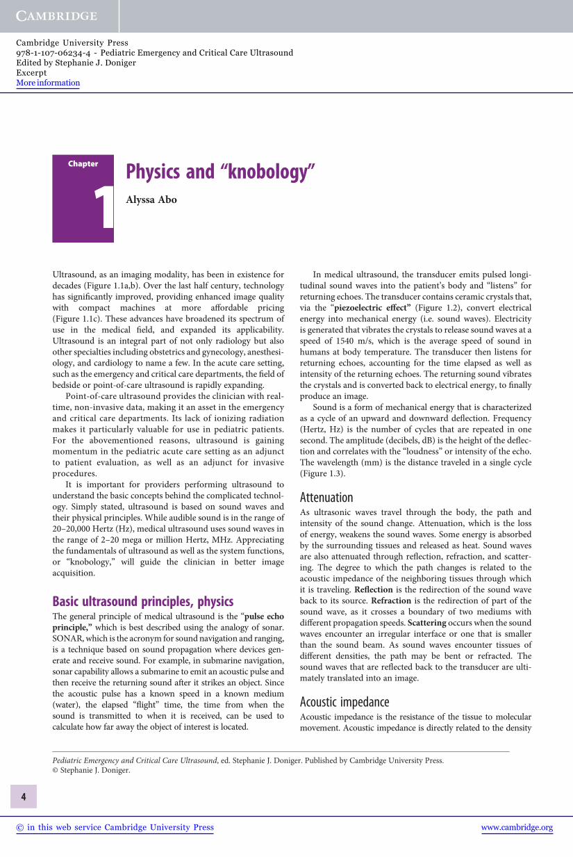
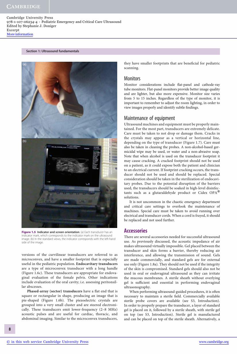
The choice of a particular transducer should reflect theclinical indication and the patient population. Each transducerhas a marker, or indicator, on it, correlating to a marker on thescreen. This allows for proper orientation by the sonographerwhen performing and interpreting ultrasound (Figure 1.5).Spatial orientation is directly related to the transducer positionand the two-dimensional plane being interrogated (see S2,Introduction).
A thorough investigation requires movement of the trans-ducer. Fanning or sweeping of the transducer is accomplishedby moving the transducer along an imaginary arc. Rocking theprobe will tilt it, while rotating the probe is achieved by twistingthe transducer clockwise or counterclockwise. Applying
(c)
Figure 1.1 (cont.)
(d)
Figure 1.1 (cont.)
Section 1: Ultrasound fundamentals
6
www.cambridge.org© in this web service Cambridge University Press
Cambridge University Press978-1-107-06234-4 - Pediatric Emergency and Critical Care UltrasoundEdited by Stephanie J. DonigerExcerptMore information

pressure on the transducer and pushing downward may dis-place bowel gas that interferes with image acquisition.
There are different types of ultrasound transducers: linear,curvilinear (or convex), phased array/sector, and endocavitary.They are characterized by shape, arrangement of the piezo-electric crystals, frequency range, footprint, and shape of theimage produced. The footprint is the area of the transducer thatcomes in contact with the patient.
Linear transducers have a flat footprint that is rectangularin shape, and they therefore produce a rectangular image. Thepiezoelectric crystals are arranged in a linear fashion. There arevarious types of linear transducers that have different sizedfootprints; there is a particular linear transducer known as the
“hockey stick” (Figure 1.6a). The frequency range varies amongtransducers and is approximately 5–10MHz. Linear trans-ducers often have higher frequencies, providing better resolu-tion for shallow structures. However, with higher frequenciespenetration and depth are sacrificed. These transducers areappropriate for visualizing superficial structures and as anadjunct for performing procedures such as vascular access,musculoskeletal applications, and abscess evaluation (seeSection 3, Procedural ultrasound).
Convex (curvilinear)-array transducers have a curvedend (Figure 1.6b). They produce a sector-shaped image,with a curved area at the top. The piezoelectric crystals arearranged alongside one another along the curved face. Thesetransducers have lower frequencies (2–5MHz), allowing forbetter penetration of deeper structures. They are commonlyused for the examination of the abdomen and pelvis. Smaller
Figure 1.2 The piezoelectric effect. Electricity causes the crystals withinthe transducer to vibrate, creating sound. The sound reaches the organ ofinterest and returns to the transducer. The transducer spends 99% of the time“listening” to the returning sound waves. These are then converted into animage that can be seen on the ultrasound monitor. Illustration by Laura Berg, MD.
Amplitude
Wavelength
Figure 1.3 The cycle of sound. Frequency (Hz) is the number of cyclesrepeated in one second, amplitude (dB) is the height of the deflection, and thewavelength (mm) is the distance traveled in a single cycle.
Figure 1.4 Various types of transducers.
Chapter 1: Physics and “knobology”
7
www.cambridge.org© in this web service Cambridge University Press
Cambridge University Press978-1-107-06234-4 - Pediatric Emergency and Critical Care UltrasoundEdited by Stephanie J. DonigerExcerptMore information
versions of the curvilinear transducers are referred to asmicroconvex, and have a smaller footprint that is especiallyuseful in the pediatric population. Endocavitary transducersare a type of microconvex transducer with a long handle(Figure 1.6c). These transducers are appropriate for endova-ginal evaluation of the female pelvis. Other indicationsinclude evaluation of the oral cavity; i.e. assessing peritonsil-lar abscesses.
Phased-array (sector) transducers have a flat end that issquare or rectangular in shape, producing an image that ispie-shaped (Figure 1.6b). The piezoelectric crystals aregrouped into a very small cluster and are steered electroni-cally. These transducers emit lower-frequency (2–8 MHz)acoustic pulses and are useful for cardiac, thoracic, andabdominal imaging. Similar to the microconvex transducers,
they have smaller footprints that are beneficial for pediatricscanning.
MonitorsMonitor considerations include flat-panel and cathode-raytube monitors. Flat-panel monitors provide better image qualityand are lighter, but also more expensive. Monitor size variesfrom 5 to 15 inches. Regardless of the type of monitor, it isimportant to remember to adjust the room lighting, in order toview images properly and identify subtle findings.
Maintenance of equipmentUltrasound machines and equipment must be properly main-tained. For the most part, transducers are extremely delicate.Care must be taken to not drop or damage them. Cracks inthe crystals may appear as a vertical or horizontal line,depending on the type of transducer (Figure 1.7). Care mustalso be taken in cleaning the probes. A non-alcohol-based ger-micidal wipe may be used, or water and a non-abrasive soap.Note that when alcohol is used on the transducer footprint itmay cause cracking. A cracked footprint should not be usedon a patient, as it could expose both the patient and clinicianto an electrical current. If footprint cracking occurs, the trans-ducer should not be used and should be replaced. Specialconsideration should be taken in the sterilization of endocavi-tary probes. Due to the potential disruption of the barriersused, the transducers should be soaked in high-level disinfec-tants such as a glutaraldehyde product or Cidex OPA®solutions.
It is not uncommon in the chaotic emergency departmentand critical care settings to overlook the maintenance ofmachines. Special care must be taken to avoid running overelectrical and transducer cords. When a cord is frayed, it shouldbe replaced and not used further.
AccessoriesThere are several accessories needed for successful ultrasounduse. As previously discussed, the acoustic impedance of airmakes ultrasound virtually impossible. Gel placed between thetransducer and skin forms a barrier, thereby reducing airinterference, and allowing the transmission of sound. Gelsare made commercially, and standard gels are for externaluse only (Figure 1.8a). They should not be used if the integrityof the skin is compromised. Standard gels should also not beused in oral or endovaginal ultrasound as they can irritatethe mucous membranes. A sterile sheath without overlyinggel is sufficient and essential in performing endovaginalultrasonography.
When performing ultrasound-guided procedures, it is oftennecessary to maintain a sterile field. Commercially availablesterile probe covers are available (see S3, Introduction).In order to properly prepare the transducer, a layer of standardgel is placed on it, followed by a sterile sheath, with sterile gelon top (see S3, Introduction). Sterile gel is manufacturedand can be placed on top of the sterile sheath. Alternatively, a
(a)
(b)
Figure 1.5 Indicator and screen orientation. (a) Each transducer has anindicator mark, which corresponds to the indicator mark on the ultrasoundimage. (b) In the standard views, the indicator corresponds with the left-handside of the image.
Section 1: Ultrasound fundamentals
8
www.cambridge.org© in this web service Cambridge University Press
Cambridge University Press978-1-107-06234-4 - Pediatric Emergency and Critical Care UltrasoundEdited by Stephanie J. DonigerExcerptMore information

sterile lubricant may be utilized (Figure 1.8b). This type ofset up is used for sterile procedures, including central lineplacement. In addition, many manufacturers offer a needleguide that attaches to the transducer to assist with ultrasound-guided procedures. These may be beneficial but are not anecessity.
Image storageThere are several options for image storage of both video clipsand still images. Through a USB port, images can either be
transferred to a flash drive, external hard drive or anothercomputer. Some machines have a burner for CDs or DVDs,to which images can be stored. There are also the options ofnetwork storage, where images and videos can be transferredvia an Ethernet port or WiFi and stored on a remote com-puter. Digital imaging and communication in medicine(DICOM) is the format that allows for transmission andstorage of the images. This format is a standard amongmany departments and allows the user to manage the imageswithin the available software applications. Disadvantagesinclude the relative cost associated with DICOM packagesand associated software. Lastly, images can be printed andstored as hard copies. Machines may come with either a blackand white or color printer (Figure 1.9).
Modes of scanningIn order to produce an image, the transducer accounts for thetime–distance relationship as well as intensity of the returningsound. The data can be displayed differently depending on themode of scanning.
A-modeA-mode, or “amplitude” mode, is one of the original modes ofmedical ultrasound. In A-mode, sound travels along a pathwhere the ultrasound beam encounters different tissues ofdifferent acoustic impedances. The information received bythe transducer is plotted on an x–y graph where the y-axis isamplitude or intensity and the x-axis is time. The image pro-duced is a tracing. This mode has been utilized in medicine forevaluation of the eye as well as the fetal head.
(b)(a)
(c)
Figure 1.6 Transducers. (a) Linear high-frequency transducers. Note the different-sized footprints. The transducer on the right is a “hockey stick” linear transducer.(b) Low-frequency transducers used for cardiac and abdominal examinations. The transducer on the left is a phased array and the transducer on the right is thecurvilinear. (c) Endocavitary transducer which is used for transvaginal ultrasonography and for the evaluation of peritonsillar abscesses.
Figure 1.7 A damaged transducer. For the most part, transducers areextremely delicate. Dropping a transducer may result in a cracked crystal. In thislinear transducer, there is a crack in a crystal, creating a vertical black line in thecenter of the image. When crystals are cracked, the transducer needs to bereplaced.
Chapter 1: Physics and “knobology”
9
www.cambridge.org© in this web service Cambridge University Press
Cambridge University Press978-1-107-06234-4 - Pediatric Emergency and Critical Care UltrasoundEdited by Stephanie J. DonigerExcerptMore information

B-modeB-mode, or “brightness”mode, is the primary imagingmodal-ity in medical ultrasound. B-mode is similar to A-mode inthat the time–distance relationship is fundamental in inter-preting the returning sound waves. It differs, however, inhow it translates the intensity of that sound. The returningecho is translated into a dot where the amplitude or intensityof the sound determines the brightness of the dot. Thegreater the intensity, the brighter the dot appears. Structures
that produce stronger echoes are termed hyperechoic andappear “brighter” or “whiter,” as opposed to structures thatdo not produce echoes and are termed anechoic, appearingblack. Hypoechoic is a relative term describing structuresthat give off weaker echoes than the surrounding tissues,and isoechoic refers to tissues of similar echogenicity(Figure 1.10). The standard two-dimensional image producedis therefore based on a series of dots and a gray scale of 256shades of gray (Figure 1.11).
Figure 1.9 Ultrasound printer. Images may bestored digitally. However, a printed copy is oftenrequired for billing purposes.
(a) (b)
Figure 1.8 Gel. In order to transmit ultrasound waves, gel must be placed on the transducer footprint. (a) Commercially available gels are available, but are not sterile.(b) In order to maintain a sterile field while performing procedures, sterile lubricant may be utilized as an alternative.
Section 1: Ultrasound fundamentals
10
www.cambridge.org© in this web service Cambridge University Press
Cambridge University Press978-1-107-06234-4 - Pediatric Emergency and Critical Care UltrasoundEdited by Stephanie J. DonigerExcerptMore information