Embed Size (px)
Citation preview

Stem Cell Research Products & Services

2
Accelerating discovery
We are scientists who strive to help other scientistsIn stem cell research, new questions arise as rapidly as new discoveries are generated. Your innovative ideas have no boundaries when backed by our tools—cells, media, differentiation systems, gene editing systems, and reagents—and custom services. With over 15 years of stem cell experience behind our Cellartis® brand, we test the boundaries of knowledge to facilitate your exploration of health and wellness.
We strive to support and improve the workflows used by all scientists who employ stem cells in their research.
Stem cell modificationStem cell cultureStem cell generation

takarabio.com/stemcells3
Resources for stem cell research Visit our website to explore products for stem cell research, find technical information, and get help from our technical support scientists.
• Selection guides
• Product information
• FAQs
• Technical notes
• Webinars
• Protocols
Products and expertise for any stage of your researchWhether you want to derive and expand pluripotent cells, to tightly control (or prevent) differentiation, to edit your cells, or to differentiate them along a specific lineage, our products and services support your aim. At every step, our tools for reprogramming, culture, engineering, differentiation, and analysis will remove experimental hurdles, allowing you to push your experiments forward—and that’s good science!
Basic research
Disease modeling
Toxicity testing
Cell therapy development
Drug discovery
Regenerative medicine R&D
Stem cell characterizationStem cell differentiation

4
Pluripotent cell derivationIn basic and translational research, high-quality starting material is critical for downstream experimental validity. Embryonic stem (ES) cell lines derived from blastocysts can be studied as pluripotent cells, or differentiated in vitro into somatic cell types. To generate induced pluripotent stem (iPS) cell populations, reprogramming factors can be delivered via viral infection or by transfection using our Xfect™ Transfection Reagent.
Characteristics of Cellartis human iPS cells
Product name Age Confirmed differentiation
Karyotype (from banked cells) HLA typification
Cellartis human iPS cell line 7
(ChiPSC7)
20 Beta cells Cardiomyocytes
Hepatocytes
46, XX HLA-A*03:01 HLA-B*07:02, HLA-B*35:01 HLA-C*04:01, HLA-C*07:02
HLA-DRB1*01:01, HLA-DRB1*15:01 HLA-DQB1*05:01, HLA-DQB1*06:02 HLA-DPB1*04:01, HLA-DPB1*04:02
Cellartis human iPS cell line 12
(ChiPSC12)
24 Beta cells Cardiomyocytes
Hepatocytes Neural progenitors
46, XY HLA-A*01:01 HLA-B*08:01, HLA-B*37:01 HLA-C*06:02, HLA-C*07:01
HLA-DRB1*03:01, HLA-DRB1*11:04 HLA-DQB1*02:01, HLA-DQB1*03:01HLA-DPB1*01:01, HLA-DPB1*04:01
Cellartis human iPS cell line 18
(ChiPSC18)
32 Cardiomyocytes Hepatocytes
Neural progenitors
46, XY HLA-A*23:01HLA-B*07:02, HLA-B*49:01HLA-C*07:01, HLA-C*07:02
HLA-DRB1*04:06, HLA-DRB1*07:01 HLA-DQB1*02:02, HLA-DQB1*04:02HLA-DPB1*03:01, HLA-DPB1*04:01
Cellartis human iPS cell line 22
(ChiPSC22)
32 Beta cells Cardiomyocytes
Hepatocytes Neural progenitors
46, XY HLA-A*02:01HLA-B*07:02, HLA-B*40:01HLA-C*03:04, HLA-C*07:02
HLA-DRB1*13:02, HLA-DRB1*14:01 HLA-DQB1*05:03, HLA-DQB1*06:04HLA-DPB1*03:01, HLA-DPB1*04:01
KLF4
C-MYC
OCT4
SOX2
EndodermEctoderm
Embryonic stem (ES) cells Induced pluripotent stem (iPS) cells
DIFFERENTIATION
Mesoderm
Pancreatic beta cellsCardiomyocytes Adipocytes HepatocytesSkin cellsNeurons Erythrocytes
Selfrenewal
Human iPS cellsOur ready-made human iPS cell lines were created from samples sourced under stringent requirements, including donor consent, and were characterized according to the highest industry standards. Cells were derived from human skin fibroblasts from healthy donors (see table below) using defective polycistronic retrovirus technology to deliver OCT4, SOX2, KLF4, and c-myc. Cells were tested for purity and stem cell characteristics, including recovery after thawing, absence of mycoplasma and bacteria, expression of stem cell-specific markers (Oct-4, Nanog, SSEA-3, SSEA-4, TRA-1-60, and TRA-1-81), absence of differentiation markers (beta tubulin-III, Foxa2, and α-SMA), expected karyotype, and confirmation of pluripotency through differentiation into specific cell lineages.
Stem cell generation and culture
takarabio.com/stemcells5
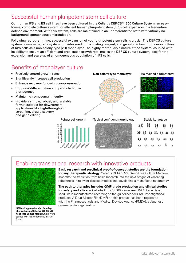
Successful human pluripotent stem cell cultureOur human iPS and ES cell lines have been cultured in the Cellartis DEF-CS™ 500 Culture System, an easy-to-use, complete culture system for efficient human pluripotent stem (hPS) cell expansion in a feeder-free, defined environment. With this system, cells are maintained in an undifferentiated state with virtually no background spontaneous differentiation.
Following reprogramming, successful expansion of your pluripotent stem cells is crucial. The DEF-CS culture system, a research-grade system, provides medium, a coating reagent, and growth factors for the easy culture of hPS cells as a non-colony type (2D) monolayer. The highly reproducible nature of the system, coupled with its ability to ensure an efficient and predictable growth rate, makes the DEF-CS culture system ideal for the expansion and scale-up of a homogeneous population of hPS cells.
Benefits of monolayer culture• Precisely control growth rates
• Significantly increase cell production
• Enhance recovery following cryopreservation
• Suppress differentiation and promote higher pluripotency
• Maintain chromosomal integrity
• Provide a simple, robust, and scalable format suitable for downstream applications like high-throughput screening, drug discovery, and gene editing
100 µm
Gen
erat
ion
tim
e (h
r)
05
1015202530354045
Non-colony type monolayer Maintained pluripotency
Robust cell growth Typical confluent morphology Stable karyotype
Enabling translational research with innovative productsBasic research and preclinical proof-of-concept studies are the foundation for any therapeutic strategy. Cellartis DEF-CS 500 Xeno-Free Culture Medium smooths the transition from basic research into the next stages of validating robustness in relevant disease models and developing a manufacturing strategy.
The path to therapies includes GMP-grade production and clinical studies for safety and efficacy. Cellartis DEF-CS 500 Xeno-Free GMP Grade Basal Medium is manufactured according to the guidelines for GMP investigational products. A Drug Master File (DMF) on this product has been registered with the Pharmaceuticals and Medical Devices Agency (PMDA), a Japanese governmental organization.
hiPS cell aggregates after four days of growth using Cellartis DEF-CS 500 Xeno-Free Culture Medium. Cells were stained with the pluripotency marker Oct-4.
Culturing as a hiPS cell monolayer in
the DEF-CS system
Editing using electroporation or
another delivery method
Expanding and scaling up into edited clonal linesSingle-cell cloningusing serial dilution,
FACS, or clone picking
6
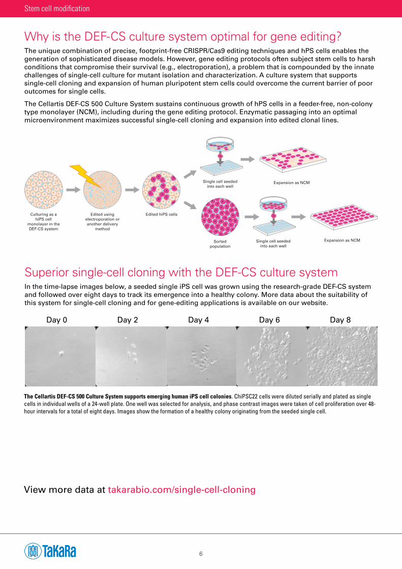
Why is the DEF-CS culture system optimal for gene editing?The unique combination of precise, footprint-free CRISPR/Cas9 editing techniques and hPS cells enables the generation of sophisticated disease models. However, gene editing protocols often subject stem cells to harsh conditions that compromise their survival (e.g., electroporation), a problem that is compounded by the innate challenges of single-cell culture for mutant isolation and characterization. A culture system that supports single-cell cloning and expansion of human pluripotent stem cells could overcome the current barrier of poor outcomes for single cells.
The Cellartis DEF-CS 500 Culture System sustains continuous growth of hPS cells in a feeder-free, non-colony type monolayer (NCM), including during the gene editing protocol. Enzymatic passaging into an optimal microenvironment maximizes successful single-cell cloning and expansion into edited clonal lines.
Superior single-cell cloning with the DEF-CS culture systemIn the time-lapse images below, a seeded single iPS cell was grown using the research-grade DEF-CS system and followed over eight days to track its emergence into a healthy colony. More data about the suitability of this system for single-cell cloning and for gene-editing applications is available on our website.
The Cellartis DEF-CS 500 Culture System supports emerging human iPS cell colonies. ChiPSC22 cells were diluted serially and plated as single cells in individual wells of a 24-well plate. One well was selected for analysis, and phase contrast images were taken of cell proliferation over 48-hour intervals for a total of eight days. Images show the formation of a healthy colony originating from the seeded single cell.
View more data at takarabio.com/single-cell-cloning
10X
Day 0 Day 2 Day 4 Day 6 Day 8
Stem cell modification
Culturing as ahiPS cell
monolayer in theDEF-CS system
Edited hiPS cellsEdited usingelectroporation oranother delivery
method
Sortedpopulation
Single cell seededinto each well
Single cell seededinto each well
Expansion as NCM
Expansion as NCM
takarabio.com/stemcells7
View hPS cell gene editing kits at takarabio.com/edit-hPSC
Guiding gene editing in stem cells The CRISPR/Cas9 system is leading the way as an easy, robust editing mechanism in stem cells. No matter which gene-editing protocol you choose (transgene delivery via electroporation, viral vectors, or cell-derived nanovesicles called gesicles), we have the tools to enable successful knockins and knockouts. While the DEF-CS culture system provides the foundation for human iPS cell survival and the formation of edited clonal lines, our Guide-it™ tools support the overall editing workflow.
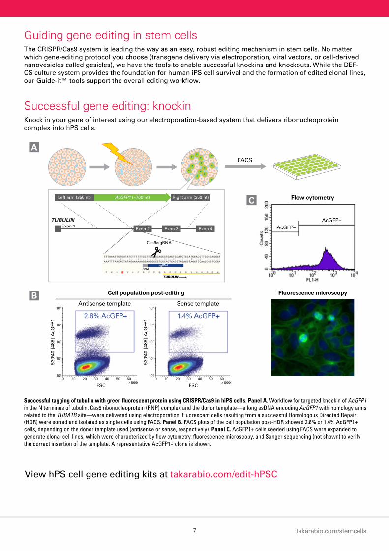
Successful gene editing: knockinKnock in your gene of interest using our electroporation-based system that delivers ribonucleoprotein complex into hPS cells.
Successful tagging of tubulin with green fluorescent protein using CRISPR/Cas9 in hiPS cells. Panel A. Workflow for targeted knockin of AcGFP1 in the N terminus of tubulin. Cas9 ribonucleoprotein (RNP) complex and the donor template—a long ssDNA encoding AcGFP1 with homology arms related to the TUBA1B site—were delivered using electroporation. Fluorescent cells resulting from a successful Homologous Directed Repair (HDR) were sorted and isolated as single cells using FACS. Panel B. FACS plots of the cell population post-HDR showed 2.8% or 1.4% AcGFP1+ cells, depending on the donor template used (antisense or sense, respectively). Panel C. AcGFP1+ cells seeded using FACS were expanded to generate clonal cell lines, which were characterized by flow cytometry, fluorescence microscopy, and Sanger sequencing (not shown) to verify the correct insertion of the template. A representative AcGFP1+ clone is shown.
104
103
102
101
100
0 10 20 30 40 50 60
104
103
102
101
100
0 10 20 30 40 50 60
104
103
102
101
100
0 10 20 30 40 50 60
ssDNA (S)Negative control ssDNA (A)
530/
40 [
488]
-AcG
FP1
FSCx1000
0%
530/
40 [
488]
-AcG
FP1
FSCx1000
2.8% AcGFP+
530/
40 [
488]
-AcG
FP1
FSCx1000
1.4% AcGFP+
Cell population post-editing
Antisense template Sense template
A
B Fluorescence microscopy
FACS
Cas9/sgRNA
Left arm (350 nt) AcGFP1 (~700 nt) Right arm (350 nt)
TUBULINExon 1
Exon 4Exon 3Exon 2
TUBULIN
PAMsgRNA
AcGFP+AcGFP–
Flow cytometryC

8
The DEF-CS culture system prepares cells for directed differentiationOnce you have a highly pure, pluripotent population, you can efficiently direct its differentiation into any of the three germ layers: endoderm, mesoderm, or ectoderm. Successful differentiation depends on the quality of the starting material: a homogeneous, undifferentiated stem cell population is ideal.
Human iPS cell-derived cellsWe specialize in the generation of high-quality cardiomyocytes, hepatocytes, beta cells, and definitive endoderm cells—enabling you to easily obtain ready-made, iPS cell-derived cells. If customization is what you’re after, differentiate your own pluripotent cells down your desired lineage, or use Cellartis Human Pluripotent Stem Cell Services to source, generate, and differentiate lines for you (read more about services on page 12).
Try our:• Cardiomyocytes
• Hepatocytes
• Definitive endoderm cells
• Beta cells
Media for neural differentiationDirecting neural differentiation from ES cells or neural stem (NS) cells requires optimized reagents in order to ensure a reliable outcome.
NDiff® 227 neural differentiation medium supports straightforward differentiation of pluripotent stem cells into the neural lineage. Using a traditional formulation supplemented with N2 and B-27, this medium enables simple and efficient neural differentiation.
RHB-A® neural stem cell culture medium enables derivation, maintenance, and expansion of NS cells. By sequentially withdrawing growth factors, differentiation of NS cells into functional neurons can be achieved.
Pluripotent stem cells differentiated into neurons using NDiff 227. These cells express neuron-specific class III beta-tubulin (Tuj1), which stains green.
Stem cell differentiation
Endoderm (HNF4A) Mesoderm (ASMA) Ectoderm (Beta-tubulin III)
Cellartis beta cells fixed 14 days post-thaw. These cells express C-peptide (green) and MAFA (red), indicators of insulin production.

takarabio.com/stemcells9
Create your own human iPS cell-derived hepatocytesHepatocytes derived from human iPS cells are an alternative to primary hepatocytes as they express major hepatic markers and demonstrate stable cytochrome P450 (CYP) activities over time in culture.
The Cellartis iPS Cell to Hepatocyte Differentiation System simplifies the production of large panels of iPS cell-derived, functional hepatocytes with your desired genotypes/phenotypes for disease modeling, drug discovery, drug metabolism research, and hepatotoxicity studies.
• Highly reproducible, robust system—the same protocol has been shown to work across 25 different iPS and ES cell lines. There is no need to optimize for your lines.
• Ideal for drug metabolism and safety studies—consistently generate panels of functional, iPS cell-derived hepatocytes with diverse genetic backgrounds.
• Customized starting materials—start with any patient- or disease-specific human iPS cell lines and create accurate liver disease models.
CYP activity of human iPS cell-derived hepatocytes recapitulates the inter-individual variation of the human population. CYP activity was measured by LC/MS and normalized to the protein content per well in iPS cell-derived hepatocytes (29 days after the start of differentiation). Activities were comparable with cryopreserved human hepatocytes (cryo hphep) from four different donors. Hepatocytes derived from five different hiPS cell lines show diverse CYP activity profiles, reflecting the metabolic diversity found in human primary hepatocytes from different donors. For example, CYP2C19 activity is low in ChiPSC18, but high in ChiPSC6b, reflecting naturally occurring interindividual variation.
View more data at takarabio.com/Power-medium
0
1
2
3
4
5
6
7
8
9
10
ChiPSC18 ChiPSC4 ChiPSC6b P11012 P11025 cryo hphep (n=4)
pm
ol /
mg
pro
tein
/ m
in
Paracetamol = CYP1A1-OH-Midazolam = CYP3A4-OH-Diclofenac = CYP2C94-OH-Mephenytoin = CYP2C19
Working with human primary hepatocytes?Extend the culture time of your human primary hepatocytes. Cellartis Power™ Primary HEP Medium maintains primary hepatocyte viability and metabolic activity for four weeks as measured by CYP activities, albumin secretion, and CYP induction capabilities.
Get started on your own cell model at takarabio.com/DIY-hepatocytes
Definitive EndodermDay 7
Ventral ForegutDays 8–10
HepatoblastDays 11–14
Fetal-Like HepatocyteDays 15–20
HepatocyteDay 21+
hiPS CellsDay 0
10
Antibodies to detect pluripotency, engraftment, and differentiationYour experiments may require sensitive methods to identify and characterize differentiated cells derived from embryonic and induced pluripotent stem cells. We offer a variety of antibodies for characterizing pluripotency, monitoring differentiation, identifying and sorting differentiated cells, and tracking transplanted stem cells.
Want to monitor the fate of engrafted human stem cells?• STEM101® and STEM121® monoclonal antibodies detect
engraftment, migration, and differentiation of human-derived stem cells after transplantation into mouse or rat.
Want to identify, characterize, and isolate undifferentiated human ES and iPS cells? • hES-Cellect™ and ES-Cellect™antibodies recognize human
pluripotent stem cells and can be used to separate human ES cells from feeder cells or differentiated progeny.
• hFF-Cellect antibody recognizes human fibroblasts, and can be used to assess human feeder cell depletion and identify non-reprogrammed fibroblasts during human iPS cell derivation.
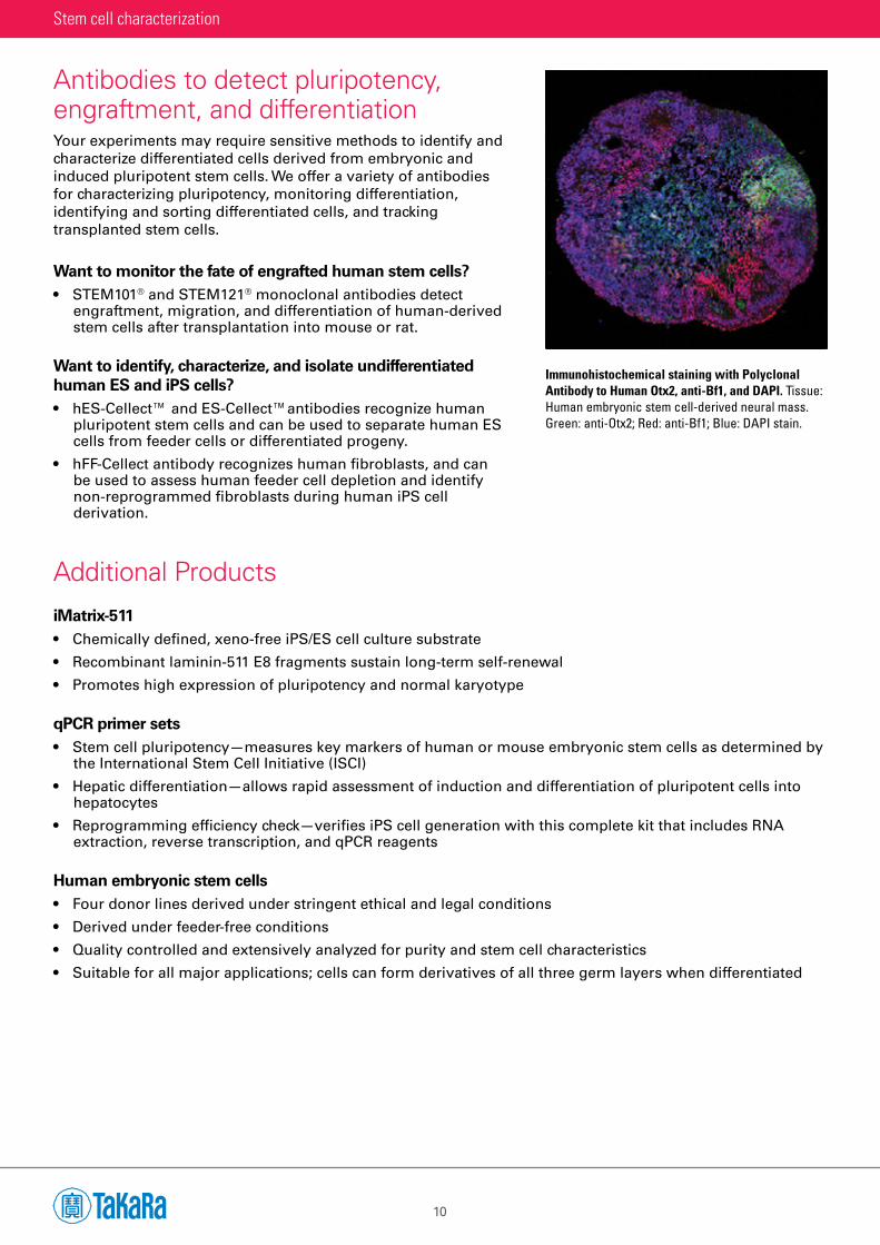
Immunohistochemical staining with Polyclonal Antibody to Human Otx2, anti-Bf1, and DAPI. Tissue: Human embryonic stem cell-derived neural mass. Green: anti-Otx2; Red: anti-Bf1; Blue: DAPI stain.
iMatrix-511• Chemically defined, xeno-free iPS/ES cell culture substrate
• Recombinant laminin-511 E8 fragments sustain long-term self-renewal
• Promotes high expression of pluripotency and normal karyotype
qPCR primer sets• Stem cell pluripotency—measures key markers of human or mouse embryonic stem cells as determined by
the International Stem Cell Initiative (ISCI)
• Hepatic differentiation—allows rapid assessment of induction and differentiation of pluripotent cells into hepatocytes
• Reprogramming efficiency check—verifies iPS cell generation with this complete kit that includes RNA extraction, reverse transcription, and qPCR reagents
Human embryonic stem cells• Four donor lines derived under stringent ethical and legal conditions
• Derived under feeder-free conditions
• Quality controlled and extensively analyzed for purity and stem cell characteristics
• Suitable for all major applications; cells can form derivatives of all three germ layers when differentiated
Additional Products
Stem cell characterization

takarabio.com/stemcells11
Antibody selection guidePluripotent stem cell markersIdentify stemness and characterize your pluripotent cells using antibodies against a variety of stem cell markers. The antibodies below can be used to characterize pluripotency and purify ES and iPS cell lines from contaminating feeder cells and non-stem cells.
Differentiated cell markersIdentify and characterize differentiated cell types derived from embryonic and induced pluripotent stem cells. The antibodies below can be used to monitor differentiation, identify and sort differentiated cells, and track transplanted stem cells.
Target Identifies Species reactivity Product name
Oct-4 Human pluripotent stem cells Human Oct4 (Human), Monoclonal
Sox-2 Human pluripotent stem cells Human Sox2 (Human), Monoclonal
Lin-28 Human pluripotent stem cells Human Lin28 (Human), Monoclonal
Surface epitope on human ES and iPS cells
Human pluripotent stem cells Human hES-Cellect
Surface epitope on human and mouse ES and iPS cells
Pluripotent stem cells Human Mouse
ES-Cellect
Target Identifies Species reactivity Product name
Nuclear protein Human cells transplanted into rodents Human STEM101
Cytoplasmic protein Human cells transplanted into rodents Human STEM121
Glial fibrillary acidic protein (GFAP)
Astrocytes derived from human neural stem cells transplanted into rodents
Human STEM123®
Human fibroblasts Human feeder cells, to distinguish non-reprogrammed fibroblasts
Human hFF-Cellect
Bf1 Cerebral neural progenitor cells in the telencephalon
Human Mouse
Anti-Human/Mouse Bf1, Polyclonal
Crx Retinal photoreceptor cells (cone and rod cells) during embryonic development
Human Anti-Human Crx, Polyclonal
Emx1 Cerebral cortex neurons during embryonic development
Mouse Polyclonal Antibody to Mouse Emx1
Irx3 Neural plate progenitor cells involved in caudal nerve development
Mouse Anti-Mouse Irx3, Polyclonal
L7/Pcp2 Purkinje progenitor cells Human Mouse
Polyclonal Antibody to Human (or Mouse) L7/Pcp2
Otp Hindbrain and hypothalamic neurons during embryonic development
Mouse Polyclonal Antibody to Mouse Otp
Otx2 Retinal photoreceptor cells during embryonic development
Human Mouse
Polyclonal Antibody to Human (or Mouse) Otx2
Rx Retinal progenitor cells Human/mouse Mouse
Anti-Human/Mouse Rx, Polyclonal (Guinea Pig); Anti-Mouse Rx, Polyclonal
Six3 Rostral brain progenitors; forebrain and retina, early embryonic and CNS development
Mouse Polyclonal Antibody to Mouse Six3
AFP Hepatocytes Human Monoclonal Antibody to Human Alpha Fetoprotein
Albumin Hepatocytes Human Monoclonal Antibody to Human Albumin

12
Cellartis Human Pluripotent Stem Cell Services
With more than 15 years of experience in stem cell research, including genome engineering and differentiation, our services team offers a variety of services for your iPS cell-based project. Our services range from donor material sourcing and reprogramming to cell differentiation and gene editing. You can choose the level of support required for your project, from start to finish at every step. And you will get data you can trust—all our procedures are performed with the highest quality standards and appropriate controls.
From the design to the delivery of your project, we provide world-renowned scientific and technical expertise. When you use Cellartis Human Pluripotent Stem Cell Services, your project is in the hands of a dedicated and enthusiastic team of expert stem cell scientists.
takarabio.com
STEMCELL-BR-0818
Interested in how our services can support the goals of your project? Please visit: takarabio.com/stem-cell-services
Contact us
Clinical-grade hES cell line derivationGenerate clinical-grade human ES cell lines per your specifications.
Materials are sourced according to FDA guidelines, and the ES cell lines are generated under xeno-free, GMP-grade conditions.
SourcingObtain patient- or disease-specific cells, according to your requirements, for later reprogramming into iPSCs.
Specify detailed donor requirements, such as gender, age, ethnic background, health status, genotype, blood type, and HLA type.
ReprogrammingGet high-quality, highly pure iPS cells from your samples or sourced samples.
Footprint-free reprogramming of your samples (PBMC or fibroblasts) or sourced PBMC samples using Sendai virus technology.
Cell bankingGenerate a Master Cell Bank from your iPS or ES cells.
Highly pluripotent cells are efficiently expanded in the monolayer-based, feeder-free Cellartis DEF-CS 500 Culture System and cryopreserved.
Gene editingGenetic engineering of your iPS or ES cell lines using CRISPR/Cas9 (RNP complex).
Gene knockin or knockout to create unique dis-ease models for your research.
Directed differentiationMake hepatocytes, beta cells, or definitive endoderm cells from your own patient- or disease-specific iPS or ES cell lines.
Our 15+ years’ experience with endodermal lineage differentiation means you can count on us to deliver high-quality, functional cells.
Takara Bio [email protected] • [email protected] • [email protected] • +33 (0)1 3904 6880
For Research Use Only. Not for use in diagnostic procedures. © 2018 Takara Bio Inc. All Rights Reserved. All trademarks are the property of Takara Bio Inc. or its affiliate(s) in the U.S. and/or other countries or their respective owners. Certain trademarks may not be registered in all jurisdictions. Additional product, intellectual property, and restricted use information is available at takarabio.com.