Embed Size (px)
Citation preview

International Journal of
Molecular Sciences
Review
Statin Treatment-Induced Development of Type 2Diabetes: From Clinical Evidence toMechanistic Insights
Unai Galicia-Garcia 1, Shifa Jebari 2,3, Asier Larrea-Sebal 1,2, Kepa B. Uribe 4 , Haziq Siddiqi 5,Helena Ostolaza 1,2, Asier Benito-Vicente 1,2 and César Martín 1,2,*
1 Fundación Biofisika Bizkaia, Barrio Sarriena s/n, 48940 Leioa, Spain; [email protected] (U.G.-G.);[email protected] (A.L.-S.); [email protected] (H.O.); [email protected] (A.B.-V.)
2 Biofisika Institute (UPV/EHU, CSIC), Barrio Sarriena s/n, 48940 Leioa, Spain; [email protected] Department of Biochemistry and Molecular Biology, Universidad del País Vasco UPV/EHU, Apdo. 644,
48080 Bilbao, Spain4 Center for Cooperative Research in Biomaterials (CIC biomaGUNE), Basque Research and Technology
Alliance (BRTA), Paseo de Miramon 182, 20014 Donostia San Sebastián, Spain; [email protected] Harvard Medical School, Harvard University, 25 Shattuck St, Boston, MA 02115, USA;
[email protected]* Correspondence: [email protected]; Tel.: +34-94-601-8052
Received: 9 June 2020; Accepted: 30 June 2020; Published: 2 July 2020�����������������
Abstract: Statins are the gold-standard treatment for the prevention of primary and secondarycardiovascular disease, which is the leading cause of mortality worldwide. Despite the safetyand relative tolerability of statins, observational studies, clinical trials and meta-analyses indicatean increased risk of developing new-onset type 2 diabetes mellitus (T2DM) after long-term statintreatment. It has been shown that statins can impair insulin sensitivity and secretion by pancreaticβ-cells and increase insulin resistance in peripheral tissues. The mechanisms involved in theseprocesses include, among others, impaired Ca2+ signaling in pancreatic β-cells, down-regulation ofGLUT-4 in adipocytes and compromised insulin signaling. In addition, it has also been described thatstatins’ impact on epigenetics may also contribute to statin-induced T2DM via differential expressionof microRNAs. This review focuses on the evidence and mechanisms by which statin therapy isassociated with the development of T2DM. This review describes the multifactorial combination ofeffects that most likely contributes to the diabetogenic effects of statins. Clinically, these findingsshould encourage clinicians to consider diabetes monitoring in patients receiving statin therapy inorder to ensure early diagnosis and appropriate management.
Keywords: statin; type 2 diabetes mellitus; clinical trial; insulin resistance; microRNA
1. Introduction
Statins are a guideline-directed, first line therapy for prevention of primary and secondarycardiovascular disease (CVD), which is the leading cause of mortality worldwide [1,2]. Although theprincipal mechanism of the action of statins is inhibition of 3-hydroxy-3-methyl-glutaryl coenzyme-A(HMG-CoA) reductase, statins have been implicated in several other beneficial pleiotropic effectsincluding improving endothelial function, stabilization of atherosclerotic plaques and anti-inflammatoryactivities [3]. Despite the safety and relative tolerability of statins, observational studies [4–8], clinicaltrials [9,10] and meta-analyses [11–16] have found that statins can increase the risk of new-onsettype 2 diabetes mellitus (T2DM). These studies implicated statins in negatively impacting insulinsensitivity, decreasing secretion by pancreatic β-cells and increasing insulin resistance [11,17,18]. While
Int. J. Mol. Sci. 2020, 21, 4725; doi:10.3390/ijms21134725 www.mdpi.com/journal/ijms

Int. J. Mol. Sci. 2020, 21, 4725 2 of 25
the lipid-lowering mechanism of statins is relatively well understood, the mechanisms underlyingstatin-induced T2DM development seem to be multifactorial and remain unclear. Among experimentalstudies, multiple works have indicated that statins diminish pancreaticβ-cell function via Ca2+ signalingpathways impairment [19,20], compromise insulin signaling and down-regulate the insulin-responsiveglucose transporter 4 (GLUT-4) [21,22]. In addition, it has also been described that statins impact onepigenetics may also contribute to statin-induced T2DM via differential expression of microRNAs [23].
This review focuses on the evidence and mechanisms by which statin therapy is associated withthe development of T2DM. Here, we will describe the existing data from clinical studies as well asexperimental results that shed some light on the mechanisms of this association.
2. Primary Action of Statins: Cholesterol Biosynthetic Pathway
Statins are reversible and competitive inhibitors of HMG-CoA reductase, which is therate-determining enzyme in the cholesterol biosynthetic pathway [24]. The HMG-like portion of statins,which is a modified 3,5-dihydroxyglutaric acid moiety, is structurally similar to HMG-CoA and causesthe inhibition of HMG-CoA reduction reactions [25]. Through this mechanism, the mevalonate pathwayis inhibited along with a consequent decrease in downstream products and cholesterol synthesis(Figure 1A). In addition, this statin-mediated decrease in intracellular cholesterol content leads toup-regulation of the LDL receptor (LDLR) in the liver and peripheral tissues, resulting in decreasedblood LDL cholesterol (LDL-C) [26]. LDLR is the primary route by which LDL-C is removed fromcirculation, and its synthesis has been shown to be inversely correlated to the amount of cholesterolsynthesized by a cell [27]. Through the action of statins, the cellular cholesterol concentration decreases,stimulating production of more LDLR and promoting LDL-C removal from the bloodstream, ultimatelyreducing CVD risk [27].
Statins are classified according to their hydrophobicity into hydrophilic statins (pravastatin androsuvastatin) and lipophilic statins (atorvastatin, cerivastatin, fluvastatin, lovastatin, pitavastatinand simvastatin) [28,29]. The solubility and pharmacological properties of statins are determinedby the substituents on the ring attached to the active moiety [29]. Hydrophilicity originates frompolar substituents added to the active site while the addition of nonpolar substituents leads tolipophilicity [25,29] (Figure 1B). Although the target of both types of statins is HMG-CoA reductase,the inhibitory mechanisms are distinct. Hydrophilic statins target the liver more efficiently becausetheir uptake is carrier-mediated, while lipophilic statins passively diffuse through the hepatocellularmembrane and similarly are also able to diffuse in extrahepatic tissues, thus showing reducedhepatoselectivity [29,30]. Their diffuse influence on extrahepatic tissues may explain the higherincidence of adverse effects observed with lipophilic statins. The notable exception to this is rosuvastatin,which is a hydrophilic statin but has a similar activity profile to lipophilic statins [31].

Int. J. Mol. Sci. 2020, 21, 4725 3 of 25Int. J. Mol. Sci. 2020, 21, x FOR PEER REVIEW 3 of 25
Figure 1. Statin-induced inhibition of the mevalonate pathway and structure of statins. (A) Inhibition of HMG-CoA reductase significantly blocks the production of mevalonate, a necessary precursor for cholesterol synthesis. Mevalonate is the building block for a variety of other compounds. (B) Structural formulas of statins and HMG-CoA. The HMG-like moiety (in red) is conserved in all statins. The polar substituents responsible of pravastatin and rosuvastatin are colored in green.
3. Beneficial Effects of Statins on Diabetic Complication and/or Inflammation in T2DM
There are many factors that contribute to the development of atherosclerotic cardiovascular disease, the main mortality cause in T2DM patients. These include dyslipidemia, increased oxidative stress, enhanced protein glycation or chronic inflammatory state all of them worsen in T2DM [32]. Statins are the gold standard treatment for the prevention and management of cardiovascular disease and their use in T2DM patients is recommended by The American Diabetes Association 2019 guidelines [33]. In addition to the reduction of cholesterol levels and dyslipidemia improvement by reducing lipoprotein levels in plasma, the pleiotropic effects of statins reduce high sensitive C-reactive protein and other pro-inflammatory markers [34], improve endothelial function and reduce oxidative stress [35], which together contribute to a significant CVD reduction in T2DM patients.
Several clinical trials have pointed out the beneficial effects of statins in diabetic patients [36]. The collaborative atorvastatin diabetes study (CARDS) showed nearly 40% reduction in relative risk of cardiovascular events in diabetic patients aged 45–70 years old with high cholesterol levels and
A B
Figure 1. Statin-induced inhibition of the mevalonate pathway and structure of statins. (A) Inhibitionof HMG-CoA reductase significantly blocks the production of mevalonate, a necessary precursor forcholesterol synthesis. Mevalonate is the building block for a variety of other compounds. (B) Structuralformulas of statins and HMG-CoA. The HMG-like moiety (in red) is conserved in all statins. The polarsubstituents responsible of pravastatin and rosuvastatin are colored in green.
3. Beneficial Effects of Statins on Diabetic Complication and/or Inflammation in T2DM
There are many factors that contribute to the development of atherosclerotic cardiovascular disease,the main mortality cause in T2DM patients. These include dyslipidemia, increased oxidative stress,enhanced protein glycation or chronic inflammatory state all of them worsen in T2DM [32]. Statins arethe gold standard treatment for the prevention and management of cardiovascular disease and theiruse in T2DM patients is recommended by The American Diabetes Association 2019 guidelines [33].In addition to the reduction of cholesterol levels and dyslipidemia improvement by reducing lipoproteinlevels in plasma, the pleiotropic effects of statins reduce high sensitive C-reactive protein and otherpro-inflammatory markers [34], improve endothelial function and reduce oxidative stress [35], whichtogether contribute to a significant CVD reduction in T2DM patients.
Several clinical trials have pointed out the beneficial effects of statins in diabetic patients [36].The collaborative atorvastatin diabetes study (CARDS) showed nearly 40% reduction in relative risk ofcardiovascular events in diabetic patients aged 45–70 years old with high cholesterol levels and treatedwith atorvastatin during 4 years [37]. A meta-analysis of 14 randomized trials including more than

Int. J. Mol. Sci. 2020, 21, 4725 4 of 25
18,000 patients confirmed the beneficial effects of statins in diabetic patients showing a 21% reductionin major vascular events per mmol/L LDL-C reduction [38]. Further studies, confirmed the benefits ofstatin treatment in diabetic patients independently of LDL-C baseline [39].
Unfortunately, in some cases, statin treatment leads to adverse effects such as the decreased insulinsensitivity shown by atorvastatin, simvastatin and rosuvastatin [40]. For atorvastatin and simvastatin,one proposed explanation is that the higher diffusion rate of lipophilic statins to the intracellular spacecan interfere with cellular processes, leading to decreased intracellular insulin secretion in response toglucose [41]. For rosuvastatin, despite its hydrophilicity, the higher affinity and efficient transport ofrosuvastatin into cells, which can underlie it effects on insulin sensitivity [29].
4. Statin Therapy and Risk of Developing T2DM: Observational Studies, Clinical Trialsand Meta-Analysis
Statins, discovered in the early 70s and commercially available in the mid-80s, havewell-characterized benefits in terms of lowering LDL-C and cardiovascular risk reduction. However, 20years after becoming commonly prescribed, findings from observational studies showed an increasedT2DM risk upon statin administration in several populations. Despite the considerable variabilityamong these studies and the statin administered, hazard ratios (HR) were statistically significantranging from 1.19 to 1.57, after follow-up durations of 3–6 years [4,6,7]. Observational studies carriedout in Canada, Taiwan and Ireland examining the association between statin administration andT2DM development, showed 10–22%, 15% and 20% increases in the risk of T2DM associated withstatin therapy, respectively [42–44]. Later on, the effects of statin treatment on the risk of T2DMand hyperglycemia deterioration were assessed in the metabolic syndrome in men (METSIM) studycohort, which found that statin therapy was associated with a 46% increased risk of T2DM along withworsening of hyperglycemia [45]. In addition, the study found statin use to be associated with a 24%reduction in insulin sensitivity and a 12% decrease in β-cell count compared to individuals not takingstatin therapy [45]. Notably, treatment with both simvastatin and atorvastatin was associated withreductions in insulin sensitivity and secretion in a dose-dependent manner [5].
Collectively, statin randomized control trials (RCT) were designed and, large, long-term, doubleblind, placebo-controlled studies were conducted to evaluate the effects of statins in a variety of clinicalsituations. Although most statin RCTs, including the largest statin RCT trial, were designed primarilyto evaluate efficacy in a variety of clinical situations, several RCTs also evaluated the relationshipbetween stain treatment and T2DM development. Among them, the justification for the use of statinin prevention: an intervention trial evaluating rosuvastatin (JUPITER), study of the effectiveness ofadditional reductions in cholesterol and homocysteine (SEARCH) and Cholesterol Treatment Trialiststrials were un-confounded regarding the intervention and aimed to recruit at least 1000 participantswith treatment duration of at least 2 years.
The justification for the use of statin in prevention: an intervention trial evaluating rosuvastatin(JUPITER) trial showed a small but significant increase in diabetes incidence rates in patients whoreceived statin treatment when compared to placebo over a median of 1.9 years (absolute increase of0.6%; relative increase of 24%; p = 0.01) [46]. Subsequent meta-analyses of the available randomizedcontrolled trials showed that standard statin dose regimens were associated with a proportionalincrease of about 10% in reported T2DM. According to the results of the JUPITER trial, treatmentwith high statin concentrations resulted in a further increase by 10% [16,47]. In addition, a post-hocanalysis of the JUPITER trial showed that participants with one or more major diabetes risk factorwere at higher risk of developing T2DM than were those without a major risk factor. Of note, however,benefits of statin therapy exceeded the diabetes hazard even in participants at high risk of developingdiabetes [10]. In patients who had risk factors for diabetes (e.g., elevated body-mass index or HbA1c,or impaired fasting glucose), the excess of T2DM diagnoses appeared soon after the start of statintherapy, and did not appear to get larger as treatment continued [10,48,49].

Int. J. Mol. Sci. 2020, 21, 4725 5 of 25
Another RCT carried out by the study of the effectiveness of additional reductions in cholesteroland homocysteine (SEARCH) collaborative group found that the simvastatin treatment was associatedwith a dose-dependent increased risk of diabetes, with diabetes found in 11.6% participants whoreceived 80 mg simvastatin compared to 10.9% in participants receiving 20 mg simvastatin [50].Collectively, the findings of multiple RCTs indicate that statin therapy may lead to the developmentof diabetes [51]. Although results from individual RCTs have shown substantial variability in theassociation between statin therapy and incident diabetes, they generated a large amount of data thatcould be more powerfully analyzed in meta-analysis. For the most relevant insights, meta-analysesthat compile data from several RCTs represent a powerful tool for understanding the impacts ofstatin therapy.
Consistent with the aforementioned RCTs, The Cholesterol Treatment Trialists’ Collaboratorsmeta-analysis (CTT) showed that LDL-C reduction is associated with a 21% reduction in the incidenceof any major vascular event in both patients with or without diabetes [38]. In the study, randomizedtrials were eligible for inclusion if: (i) the main effect of at least one of the trial interventions was tomodify lipid levels; (ii) the trial was unconfounded with respect to this intervention (i.e., no otherdifferences in modification of risk factors between the relevant treatment groups were intended) and(iii) the trial aimed to recruit 1000 or more participants with treatment duration of at least 2 years [38].The study assessed possible variation in the proportional effects of allocation to a statin in differentcircumstances only for major vascular events. Trial participants were considered to have diabetes ifthey had a recorded history of diabetes at randomization, and subdivision of diabetes subtypes wasdone according to the definitions used in the individual trials [38]. The study showed that statins aredirectly correlated with an increased risk of developing T2DM. Interestingly, multiple meta-analyseshave found that the risk of statin-associated T2DM is higher in participants taking higher doses whencompared to patients taking lower doses [15,16,47]. Accordingly, the data obtained indicated an excessrisk ranging from 9% to 13%, with the highest risk of T2DM seen in patients taking high-intensitystatin therapy [13,15,16,43,47,52]. Specifically, a recent meta-analysis showed that atorvastatin 80 mg isassociated with the highest risk of T2DM, followed by rosuvastatin and simvastatin 80 mg, indicatingthat statins have varying effects on the risk of T2DM [53]. Overall, meta-analysis studies found aclear association between diabetes and statins across multiple statins, indicating that the diabetogenicproperty of statins is a class effect. Most importantly, despite the increase of T2DM, it is importantto emphasize that the benefits of statin administration in reducing myocardial infarction, stroke andcardiovascular deaths in high CVD risk patients are enough to warrant statin treatment, althoughT2DM prevention and screening is important to take into consideration.
As listed above, clinical trials, meta-analyses and observational studies highlight that patientswho received statin treatment had a 10–12% increase in T2DM risk [17]. However, the risk is evenhigher in patients receiving high-intensity statin therapy and among patients with pre-existing riskfactors for diabetes. Recent studies indicate a clear correlation between statin type and treatmentintensity with T2DM development. Specifically, pravastatin 40 mg/day treatment has been associatedthe lowest risk of T2DM, while rosuvastatin 20 mg/day and atorvastatin 80 mg/day treatment areassociated with increased risks of T2DM. Between rosuvastatin and atorvastatin, rosuvastatin has beenassociated with the higher risk of T2DM [14].
However, even if statin type and treatment intensity clearly correlate with T2DM development,individual’s risk factors should not be overlooked. Development of T2DM during statin treatmentis more frequent among individuals with pre-existing risk factors, including increased adiposity,predisposing dietary patterns, sedentary lifestyle, psychosocial factors and previous medical history [54],as well as age and gender [55]. In fact, for patients with none to 1 risk factor, the incidence of T2DM issimilar between those receiving high dose and moderate dose of statins (3.22% and 3.35%, respectively).Conversely, for patients with 2–4 risk factors the incidence is 14.3% in the high dose group and 11.9%in the moderate dose group [17].
Int. J. Mol. Sci. 2020, 21, 4725 6 of 25
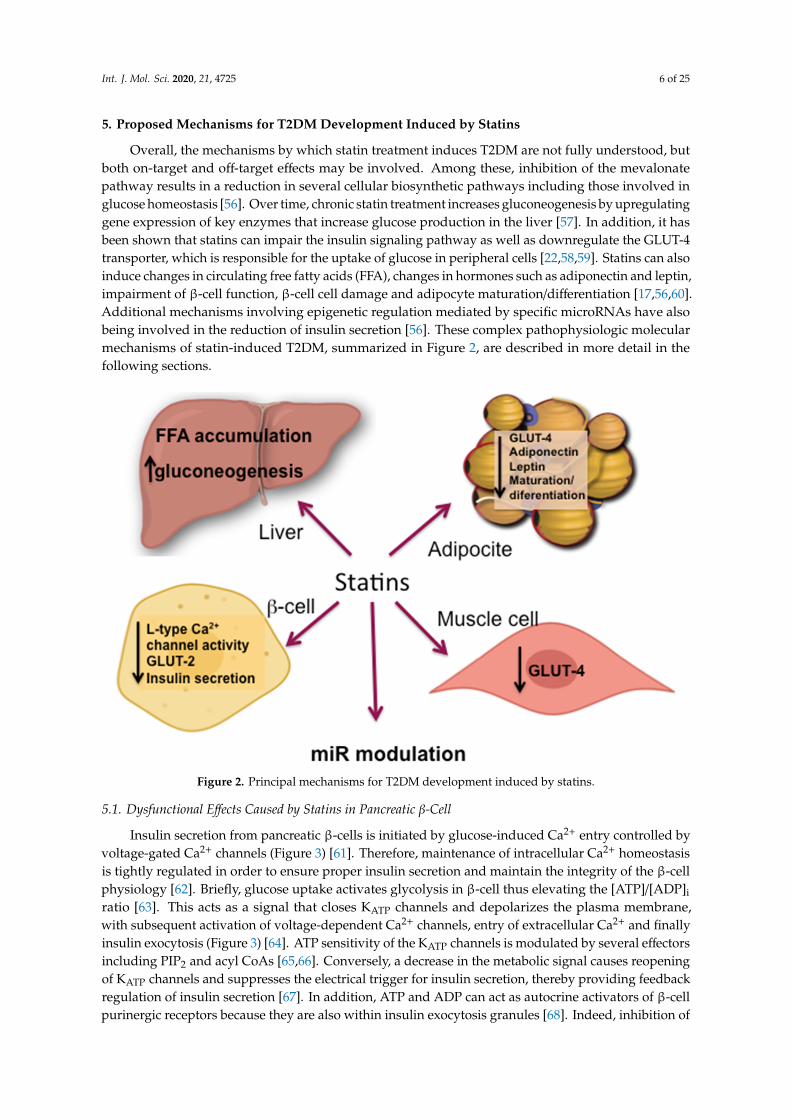
5. Proposed Mechanisms for T2DM Development Induced by Statins
Overall, the mechanisms by which statin treatment induces T2DM are not fully understood, butboth on-target and off-target effects may be involved. Among these, inhibition of the mevalonatepathway results in a reduction in several cellular biosynthetic pathways including those involved inglucose homeostasis [56]. Over time, chronic statin treatment increases gluconeogenesis by upregulatinggene expression of key enzymes that increase glucose production in the liver [57]. In addition, it hasbeen shown that statins can impair the insulin signaling pathway as well as downregulate the GLUT-4transporter, which is responsible for the uptake of glucose in peripheral cells [22,58,59]. Statins can alsoinduce changes in circulating free fatty acids (FFA), changes in hormones such as adiponectin and leptin,impairment of β-cell function, β-cell cell damage and adipocyte maturation/differentiation [17,56,60].Additional mechanisms involving epigenetic regulation mediated by specific microRNAs have alsobeing involved in the reduction of insulin secretion [56]. These complex pathophysiologic molecularmechanisms of statin-induced T2DM, summarized in Figure 2, are described in more detail in thefollowing sections.
Int. J. Mol. Sci. 2020, 21, x FOR PEER REVIEW 6 of 25
Overall, the mechanisms by which statin treatment induces T2DM are not fully understood, but both on-target and off-target effects may be involved. Among these, inhibition of the mevalonate pathway results in a reduction in several cellular biosynthetic pathways including those involved in glucose homeostasis [56]. Over time, chronic statin treatment increases gluconeogenesis by upregulating gene expression of key enzymes that increase glucose production in the liver [57]. In addition, it has been shown that statins can impair the insulin signaling pathway as well as downregulate the GLUT-4 transporter, which is responsible for the uptake of glucose in peripheral cells [22,58,59]. Statins can also induce changes in circulating free fatty acids (FFA), changes in hormones such as adiponectin and leptin, impairment of β-cell function, β-cell cell damage and adipocyte maturation/differentiation [17,56,60]. Additional mechanisms involving epigenetic regulation mediated by specific microRNAs have also being involved in the reduction of insulin secretion [56]. These complex pathophysiologic molecular mechanisms of statin-induced T2DM, summarized in Figure 2, are described in more detail in the following sections.
Figure 2. Principal mechanisms for T2DM development induced by statins.
5.1. Dysfunctional Effects Caused by Statins in Pancreatic β-cell
Insulin secretion from pancreatic β-cells is initiated by glucose-induced Ca2+ entry controlled by voltage-gated Ca2+ channels (Figure 3) [61]. Therefore, maintenance of intracellular Ca2+ homeostasis is tightly regulated in order to ensure proper insulin secretion and maintain the integrity of the β-cell physiology [62]. Briefly, glucose uptake activates glycolysis in β-cell thus elevating the [ATP]/[ADP]i ratio [63]. This acts as a signal that closes KATP channels and depolarizes the plasma membrane, with subsequent activation of voltage-dependent Ca2+ channels, entry of extracellular Ca2+ and finally insulin exocytosis (Figure 3) [64]. ATP sensitivity of the KATP channels is modulated by several effectors including PIP2 and acyl CoAs [65,66]. Conversely, a decrease in the metabolic signal causes reopening of KATP channels and suppresses the electrical trigger for insulin secretion, thereby providing feedback regulation of insulin secretion [67]. In addition, ATP and ADP can act as autocrine activators of β-cell purinergic receptors because they are also within insulin exocytosis granules [68]. Indeed, inhibition of both P2X and P2Y purinergic receptors causes a reduction in glucose-induced insulin secretion [69–72] (Figure 3).
Figure 2. Principal mechanisms for T2DM development induced by statins.
5.1. Dysfunctional Effects Caused by Statins in Pancreatic β-Cell
Insulin secretion from pancreatic β-cells is initiated by glucose-induced Ca2+ entry controlled byvoltage-gated Ca2+ channels (Figure 3) [61]. Therefore, maintenance of intracellular Ca2+ homeostasisis tightly regulated in order to ensure proper insulin secretion and maintain the integrity of the β-cellphysiology [62]. Briefly, glucose uptake activates glycolysis in β-cell thus elevating the [ATP]/[ADP]i
ratio [63]. This acts as a signal that closes KATP channels and depolarizes the plasma membrane,with subsequent activation of voltage-dependent Ca2+ channels, entry of extracellular Ca2+ and finallyinsulin exocytosis (Figure 3) [64]. ATP sensitivity of the KATP channels is modulated by several effectorsincluding PIP2 and acyl CoAs [65,66]. Conversely, a decrease in the metabolic signal causes reopeningof KATP channels and suppresses the electrical trigger for insulin secretion, thereby providing feedbackregulation of insulin secretion [67]. In addition, ATP and ADP can act as autocrine activators of β-cellpurinergic receptors because they are also within insulin exocytosis granules [68]. Indeed, inhibition of

Int. J. Mol. Sci. 2020, 21, 4725 7 of 25
both P2X and P2Y purinergic receptors causes a reduction in glucose-induced insulin secretion [69–72](Figure 3).
Int. J. Mol. Sci. 2020, 21, x FOR PEER REVIEW 7 of 25
Figure 3. Intracellular actions of statins in β-cells. Red lines indicate the mechanisms affected by statins.
To date, the relationship between statin-mediated inhibition of cholesterol synthesis and impaired L-type Ca2+ channel activity remains unclear. However, in vitro studies have indicated that simvastatin can directly inhibit L-type Ca2+ channels in rat pancreatic islet β-cells [41]. Specifically, because simvastatin was found to immediately inhibit channel activity, it has been suggested that there is a direct interaction between simvastatin and the channel. In contrast, pravastatin lacks L-type Ca2+ channels inhibition, possibly because of its lipophilicity [41]. Alternatively, other authors have suggested that the long-term cholesterol reduction caused by statins can lead to incorrect sorting of membrane lipid-raft bound proteins or conformational changes of the Ca2+ channel subunits [73]. More recently, it has been suggested that statins can reduce the membrane potential by inhibiting mitochondrial complex II activity, which causes oxidative stress [74]. These off-target effects of statins have been very recently corroborated by Curry et al. [75] in experiments showing that simvastatin impairs β-cell function by at least two mechanisms: (1) via direct inhibition of KATP channels in a mitochondria-independent manner and (2) via interference with mitochondrial respiration, thus decreasing cytosolic ATP levels and inhibiting metabolic upregulation of L-type Ca2+ channels [75].
As described before, insulin is secreted by β-cells in response to glucose uptake through GLUT receptors (primarily GLUT-1 to 4), with GLUT-2 being the predominant isoform in β-cells [76,77]. GLUT-2 represents a high-affinity and low-capacity glucose transporter [78]. It has been shown that treatment of β-cells with atorvastatin and pravastatin inhibited GLUT-2 expression in a
Figure 3. Intracellular actions of statins in β-cells. Red lines indicate the mechanisms affected by statins.
To date, the relationship between statin-mediated inhibition of cholesterol synthesis and impairedL-type Ca2+ channel activity remains unclear. However, in vitro studies have indicated that simvastatincan directly inhibit L-type Ca2+ channels in rat pancreatic islet β-cells [41]. Specifically, becausesimvastatin was found to immediately inhibit channel activity, it has been suggested that thereis a direct interaction between simvastatin and the channel. In contrast, pravastatin lacks L-typeCa2+ channels inhibition, possibly because of its lipophilicity [41]. Alternatively, other authors havesuggested that the long-term cholesterol reduction caused by statins can lead to incorrect sorting ofmembrane lipid-raft bound proteins or conformational changes of the Ca2+ channel subunits [73].More recently, it has been suggested that statins can reduce the membrane potential by inhibitingmitochondrial complex II activity, which causes oxidative stress [74]. These off-target effects of statinshave been very recently corroborated by Curry et al. [75] in experiments showing that simvastatinimpairs β-cell function by at least two mechanisms: (1) via direct inhibition of KATP channels ina mitochondria-independent manner and (2) via interference with mitochondrial respiration, thusdecreasing cytosolic ATP levels and inhibiting metabolic upregulation of L-type Ca2+ channels [75].
As described before, insulin is secreted by β-cells in response to glucose uptake through GLUTreceptors (primarily GLUT-1 to 4), with GLUT-2 being the predominant isoform in β-cells [76,77].GLUT-2 represents a high-affinity and low-capacity glucose transporter [78]. It has been shownthat treatment of β-cells with atorvastatin and pravastatin inhibited GLUT-2 expression in a

Int. J. Mol. Sci. 2020, 21, 4725 8 of 25
concentration-dependent manner [58]. However, rosuvastatin and pitavastatin showed a slightincrease in GLUT-2 expression [58]. In addition to this, it has also been observed in mouse pancreaticβ-cell line MIN6 cells that simvastatin treatment diminishes GLUT-2 mRNA and protein expression viaa dose-dependent reduction of ATP production [79]. Another mechanism through which statins mayinterfere with glucose metabolism is the statin-mediated LDLR upregulation that increases cholesteroluptake in the β-cell leading to reduced mRNA and protein expression of GLUT-2, consequently limitingglucose uptake [19,80].
The direct inhibition of the mevalonate pathway by statins reduces the intracellular concentration ofisoprenoids, the final products of the pathway. Isoprenoids are essential for G protein posttranslationalmodification, which is important for insulin granule exocytosis [17]. Interestingly, it has been shownthat the glucose-induced insulin secretion by lovastatin in normal rat islets is reduced by co-incubationwith mevalonate [81]. The adverse effects of statins are summarized in Figure 3.
5.2. Statin Induced IR
The binding of insulin to the insulin receptor (INSR) triggers insulin signaling with thephysiologic objective of normalizing high blood glucose levels [82]. Insulin binding induces structuralrearrangements in the INSR leading to auto-phosphorylation of tyrosine residues. The downstreamevents that follow INSR activation include recruitment of several adaptor proteins, facilitating asuitable binding site for insulin receptor substrates (IRSs) [82] that once phosphorylated, trigger severaldownstream signals [83]. Among them, IRS-1 is phosphorylated and activates different kinases suchas Akt, PKC, SIK2, S6K1, mTOR, ERK1/2 and ROCK1 [83,84]. IRS-1 activates PI3K, which in turn,catalyzes the conversion of PIP2 to PIP3, which activates Akt, among other targets [85]. Akt activationleads to glucose uptake by facilitating GLUT-4 translocation to the plasma membrane [86]. GLUT-4 isan insulin-dependent glucose transporter primarily expressed in adipose tissue, cardiomyocytes andskeletal muscle cells [87].
Akt also promotes glycogen synthesis by inhibiting glycogen synthase [88]. In addition, insulin alsotriggers several IRS-independent signaling pathways, among them those mediated by heterotrimericG protein and SOS-growth factor [89].
Several disturbances in insulin signal transduction mediated by statin treatment have beendescribed in different organs and tissues leading to a pathologic insulin resistance. This condition ischaracterized by a pathophysiologic failure to proper respond to normal circulatory levels of insulin ininsulin-sensitive cells, such as adipocytes, skeletal muscle cells and hepatocytes [90]. Below, we reviewsome proposed mechanisms through which statins interfere with the insulin response in each ofthese tissues.
5.2.1. Adipose Tissue
Recently, evidence that statin treatment impairs the insulin signal transduction process inadipocytes, including INSR, GLUT-4, Akt, some small GTP-binding proteins (G-proteins) and caveolaeintegrity has been demonstrated. Multiple studies have shown that atorvastatin and lovastatinreduce GLUT-4 expression at the plasma membrane in 3T3L1 adipocytes [91,92] and a similareffect has been described with atorvastatin in mouse-white adipose tissue, thus impairing glucosetolerance [22]. The statin-induced decrease in GLUT-4 translocation to the plasma membrane has beenattributed to inhibition of isoprenoid synthesis [22]. In fact, isoprenylation is essential for the correctfunctioning of several proteins involved in the GLUT-4 translocation process. As previously described,isoprenylation is impaired due to statin-induced inhibition of the mevalonate pathway. In oneillustrative example, it has been described that atorvastatin disrupts plasma membrane colocalizationof Rab-4 and RhoA through inhibition of geranylgeranyl pyrophosphate synthesis. Rab-4 and RhoA areisoprenoid-dependent proteins, which are involved in the insulin-induced translocation of GLUT-4, thustheir atorvastatin-mediated dysfunction may disturb overall insulin signaling [93]. RhoA modulates
Int. J. Mol. Sci. 2020, 21, 4725 9 of 25
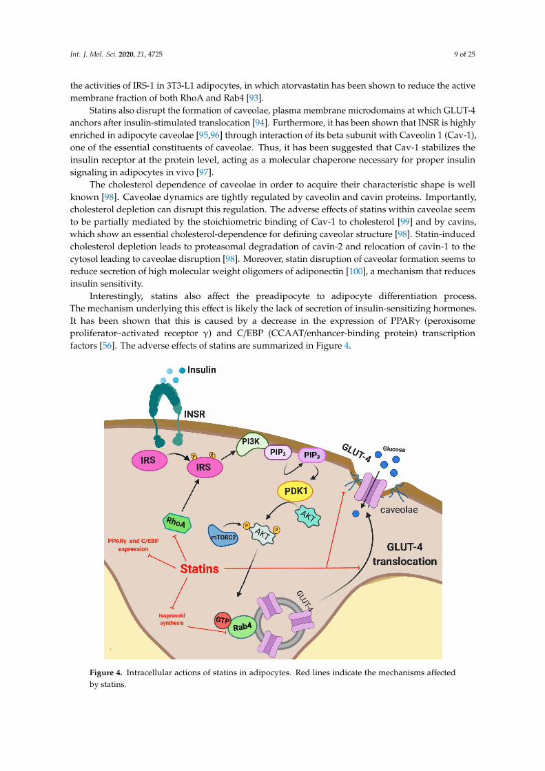
the activities of IRS-1 in 3T3-L1 adipocytes, in which atorvastatin has been shown to reduce the activemembrane fraction of both RhoA and Rab4 [93].
Statins also disrupt the formation of caveolae, plasma membrane microdomains at which GLUT-4anchors after insulin-stimulated translocation [94]. Furthermore, it has been shown that INSR is highlyenriched in adipocyte caveolae [95,96] through interaction of its beta subunit with Caveolin 1 (Cav-1),one of the essential constituents of caveolae. Thus, it has been suggested that Cav-1 stabilizes theinsulin receptor at the protein level, acting as a molecular chaperone necessary for proper insulinsignaling in adipocytes in vivo [97].
The cholesterol dependence of caveolae in order to acquire their characteristic shape is wellknown [98]. Caveolae dynamics are tightly regulated by caveolin and cavin proteins. Importantly,cholesterol depletion can disrupt this regulation. The adverse effects of statins within caveolae seemto be partially mediated by the stoichiometric binding of Cav-1 to cholesterol [99] and by cavins,which show an essential cholesterol-dependence for defining caveolar structure [98]. Statin-inducedcholesterol depletion leads to proteasomal degradation of cavin-2 and relocation of cavin-1 to thecytosol leading to caveolae disruption [98]. Moreover, statin disruption of caveolar formation seems toreduce secretion of high molecular weight oligomers of adiponectin [100], a mechanism that reducesinsulin sensitivity.
Interestingly, statins also affect the preadipocyte to adipocyte differentiation process.The mechanism underlying this effect is likely the lack of secretion of insulin-sensitizing hormones.It has been shown that this is caused by a decrease in the expression of PPARγ (peroxisomeproliferator–activated receptor γ) and C/EBP (CCAAT/enhancer-binding protein) transcriptionfactors [56]. The adverse effects of statins are summarized in Figure 4.
Int. J. Mol. Sci. 2020, 21, x FOR PEER REVIEW 9 of 25
which atorvastatin has been shown to reduce the active membrane fraction of both RhoA and Rab4 [93].
Statins also disrupt the formation of caveolae, plasma membrane microdomains at which GLUT-4 anchors after insulin-stimulated translocation [94]. Furthermore, it has been shown that INSR is highly enriched in adipocyte caveolae [95,96] through interaction of its beta subunit with Caveolin 1 (Cav-1), one of the essential constituents of caveolae. Thus, it has been suggested that Cav-1 stabilizes the insulin receptor at the protein level, acting as a molecular chaperone necessary for proper insulin signaling in adipocytes in vivo [97].
The cholesterol dependence of caveolae in order to acquire their characteristic shape is well known [98]. Caveolae dynamics are tightly regulated by caveolin and cavin proteins. Importantly, cholesterol depletion can disrupt this regulation. The adverse effects of statins within caveolae seem to be partially mediated by the stoichiometric binding of Cav-1 to cholesterol [99] and by cavins, which show an essential cholesterol-dependence for defining caveolar structure [98]. Statin-induced cholesterol depletion leads to proteasomal degradation of cavin-2 and relocation of cavin-1 to the cytosol leading to caveolae disruption [98]. Moreover, statin disruption of caveolar formation seems to reduce secretion of high molecular weight oligomers of adiponectin [100], a mechanism that reduces insulin sensitivity.
Interestingly, statins also affect the preadipocyte to adipocyte differentiation process. The mechanism underlying this effect is likely the lack of secretion of insulin-sensitizing hormones. It has been shown that this is caused by a decrease in the expression of PPARγ (peroxisome proliferator–activated receptor γ) and C/EBP (CCAAT/enhancer-binding protein) transcription factors [56]. The adverse effects of statins are summarized in Figure 4.
Figure 4. Intracellular actions of statins in adipocytes. Red lines indicate the mechanisms affected by statins.
5.2.2. Skeletal Muscle
Figure 4. Intracellular actions of statins in adipocytes. Red lines indicate the mechanisms affectedby statins.
Int. J. Mol. Sci. 2020, 21, 4725 10 of 25
5.2.2. Skeletal Muscle
Skeletal muscle is the major tissue consuming most of the glucose that enters circulation [101], andany impairment in glucose uptake by this tissue may result in T2DM development. GLUT-4 mediatesglucose transport into skeletal muscle cells, representing a key factor for blood sugar control [102].As indicated above, insulin binding to INSR causes Akt activation [103] and translocation of GLUT-4containing vesicles to the plasma membrane, thus facilitating the transport of glucose [104–106].Although the mechanism of statin induced T2DM is not completely understood, there are bothin vivo and in vitro studies that shed some light on this phenomenon in skeletal muscle. Some of themechanisms that have been previously described are statin-mediated inhibition of insulin stimulatedglucose uptake, impairment of intracellular signaling of the INSR and thereby of the Akt/mTORpathway, or an excess of FFA accumulation in skeletal muscle as a consequence of HMG-CoAreductase inhibition.
In support of a role for a statin-induced insulin resistance in skeletal muscle, a decreased GLUT-4expression has been found in L6 myotubes after simvastatin treatment [107]. Alternatively, it has beenmore recently shown that atorvastatin diminishes GLUT-4 translocation to the plasma membranewithout affecting total GLUT-4 protein expression in C2C12 myotubes [108]. Assessment of themechanism of simvastatin- or atorvastatin-associated impairment of glucose transport into myotubessuggests that impaired intracellular signaling of the INSR pathway also plays an important role. Indeed,Sanvee et al. [109] have shown that in C2C12 myotubes, simvastatin inhibits both INSR and mTORC2function leading to impaired Akt activation and decreased translocation of GLUT-4 and consequently,reduced glucose uptake into skeletal muscle [109]. This deficient GLUT-4 translocation is likely causedby impaired Akt-mediated phosphorylation of GSK3β. Additionally, they show that simvastatintreatment induces higher plasma glucose levels in mice despite increased insulin plasma concentrations,consistent with insulin resistance [109]. The sequence of events leading to diminished glucose uptakeinduced by simvastatin starts with impaired phosphorylation of INSR, specifically the β-chain, whichis considered to be essential for action of the receptor [101]. This results in deficient phosphorylation ofAkt, which needs to be phosphorylated at both Thr308 (through the insulin signaling pathway) andSer473 (by mTORC2) to become fully active [101]. Simvastatin treatment significantly impaired onlythe phosphorylation of Akt Ser473 due to an impaired phosphorylation of mTOR, one of the mTORC2constituents [109,110]. Since Akt requires both phosphorylations to be fully active, it is then unable toactivate glycogen synthase kinase 3β (GSK3β), which is involved in the translocation of GLUT-4 to theplasma membrane. Decreased GSK3β phosphorylation in the setting of simvastatin at least partiallyexplains impaired translocation of GLUT-4 to the plasma membrane.
Another adverse effect related to statin-induced T2DM and, similar to in adipocytes, is deficientprenylation of RabGTPases, which has been suggested to lead to impaired GLUT-4 translocation [111].Decreased intracellular cholesterol concentration is also considered a leading mechanism for impairedGLUT-4 translocation [108].
Alternatively, it has been suggested that simvastatin may cause insulin resistance through a novelfatty acid based mechanism independent of its cholesterol lowering effects. In their study, Kain etal. hypothesized that by blocking HMG CoA reductase, simvastatin may lead to accumulation ofacetyl CoA, a precursor of fatty acid synthesis that can promote an intracellular build-up of fatty acids.The resulting excess accumulation of FFA in skeletal muscle may inhibit glucose uptake by reducingGLUT translocation [112,113]. The adverse effects of statins in muscle cells are summarized in Figure 5.

Int. J. Mol. Sci. 2020, 21, 4725 11 of 25
Int. J. Mol. Sci. 2020, 21, x FOR PEER REVIEW 11 of 25
Figure 5. Intracellular actions of statins in muscle cells. White lines indicate the mechanisms affected by statins.
5.2.3. Liver
The liver plays a central role in glucose homeostasis and is exquisitely sensitive to insulin. In fact, insulin regulates many hepatic metabolic pathways ranging from the glucose output to lipid synthesis. Therefore, impairment of hepatic insulin sensitivity is rapidly reflected in glucose homeostasis and triglyceride levels. Emerging evidence has demonstrated that statin treatment is associated with worsening glycemic control in the liver [114]. Several mechanisms possibly involved with the effect of statins on glucose metabolism in the liver are summarized below.
Statin therapy is associated with a small increment in fasting blood glucose levels [115]. It has been shown that statins can stimulate endogenous glucose production by activation of phosphoenolpyruvate carboxykinase (PEPCK) and glucose-6-phosphatase (G6Pase) [116,117], the major rate-limiting gluconeogenic enzymes in human liver cells. The elevation of hepatic gluconeogenesis contributes to hyperglycemia, which is characteristic of insulin resistance and T2DM.
Regarding FFAs, it has been shown that an excess of FFA accumulation in liver cells can contribute to the development of T2DM [118,119]. Interestingly, atorvastatin and rosuvastatin treatment upregulates thyroid hormone-responsive spot 14 protein (THRSP) expression, which is a small protein predominantly expressed in lipid-producing tissues such as those found in the liver. THRSP has been implicated as a regulator of the lipogenic processes by controlling the expression of lipogenic genes such as fatty-acid synthase (FASN), ATP citrate lyase (ACLY) SREBP and ChREBP [120,121] or their activity [122]. The adverse effects of statins in the hepatocytes are summarized in Figure 6.
Figure 5. Intracellular actions of statins in muscle cells. White lines indicate the mechanisms affectedby statins.
5.2.3. Liver
The liver plays a central role in glucose homeostasis and is exquisitely sensitive to insulin. In fact,insulin regulates many hepatic metabolic pathways ranging from the glucose output to lipid synthesis.Therefore, impairment of hepatic insulin sensitivity is rapidly reflected in glucose homeostasis andtriglyceride levels. Emerging evidence has demonstrated that statin treatment is associated withworsening glycemic control in the liver [114]. Several mechanisms possibly involved with the effect ofstatins on glucose metabolism in the liver are summarized below.
Statin therapy is associated with a small increment in fasting blood glucose levels [115]. It has beenshown that statins can stimulate endogenous glucose production by activation of phosphoenolpyruvatecarboxykinase (PEPCK) and glucose-6-phosphatase (G6Pase) [116,117], the major rate-limitinggluconeogenic enzymes in human liver cells. The elevation of hepatic gluconeogenesis contributes tohyperglycemia, which is characteristic of insulin resistance and T2DM.
Regarding FFAs, it has been shown that an excess of FFA accumulation in liver cells cancontribute to the development of T2DM [118,119]. Interestingly, atorvastatin and rosuvastatin treatmentupregulates thyroid hormone-responsive spot 14 protein (THRSP) expression, which is a small proteinpredominantly expressed in lipid-producing tissues such as those found in the liver. THRSP has beenimplicated as a regulator of the lipogenic processes by controlling the expression of lipogenic genessuch as fatty-acid synthase (FASN), ATP citrate lyase (ACLY) SREBP and ChREBP [120,121] or theiractivity [122]. The adverse effects of statins in the hepatocytes are summarized in Figure 6.

Int. J. Mol. Sci. 2020, 21, 4725 12 of 25Int. J. Mol. Sci. 2020, 21, x FOR PEER REVIEW 12 of 25
Figure 6. Intracellular actions of statins in hepatocytes. White lines indicate the mechanisms affected by statins.
5.3. MicroRNAs and Impact of Statin Therapy on microRNA Expression Profile
MicroRNAs (miRs) are small (22 nucleotide) noncoding regulatory RNAs, which act as post-transcriptional regulators of gene expression [123,124]. miRs usually silence gene expression through mRNA degradation or sequestration of the target mRNA from translation machinery [125]. It has been shown that miRs are involved in many biological processes including insulin expression, skeletal muscle adaptation to elevated glucose, insulin sensitivity and glucose stimulated insulin secretion (GSIS) [126]. It has been shown that miRs likely mediate the pleiotropic effects of statins via modulation of lipid metabolism, enhancement of endothelial function, inhibition of inflammation, improvement of plaque stability and immune regulation. More specifically, miRs appear to regulate the fine-tuning of cellular phenotypes rather than serving as molecular on–off switches [127].
Statin therapy has been found to affect the expression of several miRs, which play a central role in the regulation of lipid and glucose metabolism [128] and that are associated with development of T2DM.
5.3.1. miR Modulation of Cholesterol and Lipid Homeostasis
miR-33a and miR-33b are encoded within the introns of the Srebp2 and Srebp1 genes, respectively, and modulate intracellular cholesterol and fatty acid homeostasis together with SREBP2 and SREBP1 [129–132]. Specifically, miR-33a targets genes involved in cholesterol export, inhibits ABCA1 and ABCG expression [130–132] and participates in the regulation of HDL levels in vivo. On the other hand, miR-33b modulates metabolic pathways related to of fatty acid metabolism [129,133]. Importantly, both miR-33a and miR-33b participate in the regulation of fatty acid metabolism and are involved in the regulation of lipid and glucose metabolism [129]. miR-33 also negatively affects IRS2 expression thereby affecting insulin signaling [129]. Collectively, both isoforms of miR-33 participate in the regulation of relevant pathways that impact the primary risk factors of insulin resistance.
It has been demonstrated that simvastatin and atorvastatin induce expression of miR-33a in the liver [134] thus suggesting a link between reduced insulin secretion and, ultimately, the development of statin-induced T2DM. miR-33a is an important regulator of ABCA1 and their expression levels are inversely proportional in β-cells [132,135]. miR-33a-mediated downregulation
Figure 6. Intracellular actions of statins in hepatocytes. White lines indicate the mechanisms affectedby statins.
5.3. MicroRNAs and Impact of Statin Therapy on microRNA Expression Profile
MicroRNAs (miRs) are small (22 nucleotide) noncoding regulatory RNAs, which act aspost-transcriptional regulators of gene expression [123,124]. miRs usually silence gene expressionthrough mRNA degradation or sequestration of the target mRNA from translation machinery [125].It has been shown that miRs are involved in many biological processes including insulin expression,skeletal muscle adaptation to elevated glucose, insulin sensitivity and glucose stimulated insulinsecretion (GSIS) [126]. It has been shown that miRs likely mediate the pleiotropic effects of statins viamodulation of lipid metabolism, enhancement of endothelial function, inhibition of inflammation,improvement of plaque stability and immune regulation. More specifically, miRs appear to regulatethe fine-tuning of cellular phenotypes rather than serving as molecular on–off switches [127].
Statin therapy has been found to affect the expression of several miRs, which play a central rolein the regulation of lipid and glucose metabolism [128] and that are associated with developmentof T2DM.
5.3.1. miR Modulation of Cholesterol and Lipid Homeostasis
miR-33a and miR-33b are encoded within the introns of the Srebp2 and Srebp1 genes, respectively,and modulate intracellular cholesterol and fatty acid homeostasis together with SREBP2 andSREBP1 [129–132]. Specifically, miR-33a targets genes involved in cholesterol export, inhibits ABCA1and ABCG expression [130–132] and participates in the regulation of HDL levels in vivo. On theother hand, miR-33b modulates metabolic pathways related to of fatty acid metabolism [129,133].Importantly, both miR-33a and miR-33b participate in the regulation of fatty acid metabolism and areinvolved in the regulation of lipid and glucose metabolism [129]. miR-33 also negatively affects IRS2expression thereby affecting insulin signaling [129]. Collectively, both isoforms of miR-33 participatein the regulation of relevant pathways that impact the primary risk factors of insulin resistance.
It has been demonstrated that simvastatin and atorvastatin induce expression of miR-33a in theliver [134] thus suggesting a link between reduced insulin secretion and, ultimately, the developmentof statin-induced T2DM. miR-33a is an important regulator of ABCA1 and their expression levels areinversely proportional in β-cells [132,135]. miR-33a-mediated downregulation of ABCA1 can also alter
Int. J. Mol. Sci. 2020, 21, 4725 13 of 25
islet cholesterol homeostasis and impair insulin secretion thus leading to β-cell dysfunction [136,137].However, additional studies are needed to further confirm the presence of a causal relationship betweenstatin treatment and miRs in T2DM. Several studies have shown that statin treatment can upregulatemiR-33b expression thus suggesting that statins could interfere fatty acid metabolism [138,139].
Recently, the miR-27 family (miR-27a and miR-27b) has emerged as a new key regulator ofcholesterol and lipid homeostasis [140–142]. Interestingly, the miR-27 family has been shown to beupregulated in a dose-dependent manner by simvastatin in HepG2 cells. Alvarez et al. demonstratedthat miR-27a directly decreases both LDLR RNA and protein levels by binding to the 3′UTR of the LDLRmRNA [143]. Moreover, miR-27a also decreases LDLR expression indirectly through upregulation ofPCSK9. They suggest that the potential binding site for miR-27a at position -1671 bp relative to thetranscription start site of PCSK9 may be responsible for the upregulation of PCSK9. In addition to thedirect and indirect downregulation of LDLR levels, miR-27a also indirectly affects LDLR efficiencythrough a mechanism in which miR-27a targets the 3′UTR sequence of two genes in the LDLRpathway: LRP6 and LDLRAP1 by downregulating their expression [143]. Both proteins are necessaryfor correct binding to clathrin and thus are essential for efficient endocytosis of the LDLR-LDL-Ccomplex [144–146]. Therefore, in addition to decreasing LDLR levels at the plasma membrane, miR-27amay also negatively affect LDLR efficiency. Deregulation of miR-27a has been reported in T2DM [147].Specifically, it has been shown to be upregulated in adipose tissue and in 3T3-L1 adipocytes exposed toincreased glucose concentration [147].
5.3.2. Modulation of Hepatic Glucose Production
A vast number of miRs have been described to modulate glucose homeostasis through variousmechanisms, leading to the question of whether some of them may potentially be involved in statins’diabetogenic effects. Specifically, it has been demonstrated that a direct effect of statins on hepatic glucoseproduction is mediated by upregulation of the miR-183/96/182 cluster by modulating the expressionof gluconeogenic enzymes [148]. It has been shown that incubation of hepatocytes with atorvastatin,simvastatin or pravastatin upregulates the expression the key gluconeogenic enzymes PEPCK andG6Pase [117,149]. The statin-mediated effects involve miR-183/96/182-mediated downregulation of thetranscription factor 7-like 2 (TCF7L2), which modulates hepatic and peripheral glucose metabolismand whole body glycemic control [148]. In regards to gluconeogenesis, TCF7L2 also reduces hepaticgluconeogenesis likely by decreasing the transcriptional activity of positive regulators of PEPCK andG6PC [150–154]. These results suggest that patients under long-term statin treatment would havepersistently elevated expression of the miR cluster and lead to sustained activation of the gluconeogenicpathway, ultimately contributing to T2DM.
5.3.3. Modulation of the Insulin Signaling Pathway
As mentioned above, the activation of INSR by insulin leads to structural rearrangements in thereceptor leading to autophosphorylation at tyrosine residues. Within the cell, phosphorylation levelsare tightly regulated by protein phosphatases, in this case protein tyrosine phosphatases (PTPAses).These PTPAses negatively modulate insulin signaling by removing phosphate groups from tyrosineresidues of the cytoplasmic domain of INSR. Specifically, protein tyrosine phosphatase non-receptortype 1 (PTPN1) has been predicted as an miR-146a target and the expression of PTPN1 is inverselycorrelated with miR-146a both in the skeletal muscle and in the liver of a T2DM rat model [155]. Of note,the role of miR-146a has been widely investigated in human T2DM pathogenesis and several studiesreport that it is downregulated in whole blood, plasma and some peripheral tissues [156]. Notably,it has been shown that simvastatin treatment also downregulates mir-146a expression after 6 monthsof therapy [157].
As mentioned above, IRSs link INSR activation to insulin metabolic effects through the intermediatemodulation of the PI3K/PDK1/Akt pathway. It has been described that expression levels of IRS1 aremodulated by miR-145 in hepatocytes [158] whereas in mice, upregulation of miR-145 in the liver leads

Int. J. Mol. Sci. 2020, 21, 4725 14 of 25
to insulin resistance [159]. Atorvastatin treatment has also been shown to differentially upregulatemiR-145 and modulates PI3K/Akt signaling pathway [160].
In hepatocytes, miR-33a and miR-33b have been reported to modulate fatty acid and cholesterolmetabolism as well as insulin signaling by targeting IRS2 [129,161]. In one study, miR-33boverexpression in the Huh7 human hepatocytes cell line resulted in reduced Akt and ERKphosphorylation secondary to IRS2 down-regulation [129].
Additional Akt-downstream kinases and phosphatases represent major regulators of insulinsignaling. Direct inactivation of AKT is mediated by protein phosphatase 2a (PP2A) [162]. PP2A activityhas been shown to be increased in primary rat hepatocytes in insulin resistance condition [163].Interestingly, insulin resistant Zucker Diabetic Fatty rats, PP2A mRNA is increased in liver, muscleand adipose tissue, thus suggesting a role for the phosphatase in deregulating insulin signaling inT2DM [163]. Importantly, PP2A expression is modulated by several miRs, among them miR-155 [164],whose expression has been found to be altered in T2DM. In addition, high dose rosuvastatin treatmenthas been shown to reduce the relative levels of serum miR-155 and therefore could lead to increasedexpression and activity of PP2A [165].
6. Differences in Diabetogenic Effects between Hydrophilic and Lipophilic Statins
As indicated in previous sections, lipophilic statins (atorvastatin, simvastatin, lovastatin,fluvastatin and pitavastatin) may be more diabetogenic than hydrophilic statins (pravastatin androsuvastatin) as they can more readily penetrate extrahepatic cell membranes such asβ-cells, adipocytesand skeletal muscle cells. Conversely, hydrophilic statins (e.g., pravastatin) are more hepatocytespecific and less likely to enter β-cells or adipocytes [29]. Indeed, a high hepato-selectivity translatesinto minimal interference with cholesterol metabolism in tissues other than the liver and consequentlyto a lesser diabetogenicity [29,30,56]. Several studies have shown that the detrimental effects of statinsare dose and potency dependent and primarily related to their lipophilicity [5,14,41,47,166].
While lipophilic statins have negative effects on pancreatic β-cell function, for hydrophilic statinssuch as pravastatin, neutral or improving effects have been observed [40,41,167]. As mentionedin Section 4, it has been reported that statins can inhibit glucose-induced cytosolic Ca2+ signalingand insulin secretion by blocking L-type Ca2+ channels in β-cells. These inhibitory potencies maybe particularly evident for the lipophilic rather than the hydrophilic statins [41,166,168]. Indeed,unlike hydrophilic statins, the lipophilic ones have a strong affinity for the cell membrane, and thereforehave easier access to the intracellular space [168]. In this context, statins may inhibit the endogenousmetabolic pathways described in Section 5.1 that are associated with glucose-stimulated insulinsecretion, including endogenous cholesterol synthesis [73,166] and Ca2+-dependent insulin responsesto glucose [168]. It has been shown that atorvastatin (lipophilic) but not pravastatin (hydrophilic)affects insulin release and mitochondrial metabolism due to the suppression of antioxidant defensesystem and induction of ROS production in pancreatic β-cell models [169].
As described in Sections 5.2.1–5.2.3, GLUT-4 mediates insulin-stimulated glucose uptake [86] in aprocess that requires fusion of the transporter with the plasma membranes facilitated by IRS-1 andseveral kinases [86,170,171]. The small GTP-binding proteins are also key players in this process [22,86]and they require isoprenylation by mevalonate products for their association with the cell membranes.The statin-mediated inhibition of the synthesis of the above products increases insulin resistancein parallel with the mevalonate synthesis inhibitory capacity [21,172]. Furthermore, several otherprocesses involved in the GLUT-4 signaling pathway may be inhibited by statins. These include IRS-1,insulin receptor β subunit, and Akt phosphorylation [22,166]. It has been suggested that these effectsare relevant only for lipophilic statins (e.g., atorvastatin and simvastatin), but not for hydrophilicstatins (e.g., pravastatin) [22,166]. The capacity of the former to enter adipocytes through passivediffusion can help explain this difference.
Int. J. Mol. Sci. 2020, 21, 4725 15 of 25
7. Conclusions
Taken together, the studies described in this review, ranging from clinical studies to in vivo andin vitro experimental results, confirm and reinforce the diabetogenic effect of statins. Although anumber of questions remain unanswered, the available evidence supports that statins do increase thechances of T2DM with some statins being more strongly related (e.g., simvastatin, rosuvastatin andatorvastatin) than others (e.g., pravastatin). Intense research is currently going on to elucidate themechanisms of statin induced T2DM at the molecular level. In light of the evidence from multipleobservational studies, it is important to emphasize that there is still a favorable risk–benefit ratio forstatin therapy, due to the large reduction in cardiovascular risk, despite the adverse effect of T2DMdevelopment. Overall, the risk of incident diabetes mellitus with statin therapy is present but largelyoutweighed by the actual cardiovascular benefits [16]. Thus, statins should be continued in patientsin whom these drugs are prescribed due to high or very high CVD risk, despite the risk of T2DMdevelopment until they achieve the target LDL-C levels. Before initiation of statin therapy the risk ofdiabetes should be assessed [8,16,173]. Statin-treated patients at high risk of developing diabetes shouldbe monitored for changes in blood glucose and HbA1c levels, and preventive lifestyle modificationshould be introduced. If diabetes develops, it should be managed according to the guidelines [16].Patients should be educated regarding the risk of incident diabetes mellitus with statins as with otherrisk–benefit of all therapies [174]. Lifestyle modification should be encouraged to lower cardiovascularrisk and that for developing T2DN [175] and national guidelines should be used to manage diabetesmellitus [176,177].
Several mechanisms through which statin treatment causes β-cell dysfunction and insulinresistance in peripheral tissues have been identified. Specifically, these the diabetogenic effects arerelated both to the dose and statin class. In addition, miRs are glucose homeostasis regulators throughthe specific modulation of insulin signaling components. Growing evidence indicates that statinmodulation of miRs expression may also be another mechanism through which statins increase therisk of T2DM. A multifactorial combination of these effects is what most likely contributes to thediabetogenic effects of statins described here. Clinically, these findings should encourage clinicians toconsider diabetes monitoring in patients receiving statin therapy in order to ensure early diagnosis andappropriate management. Ultimately, since the risk of statin-induced T2DM is still being characterized,and the efficacy of statins in preventing CVD is very well documented, statins remain a first linetreatment for prevention of CVD.
Funding: This work was supported by the Basque Government (Grupos Consolidados IT-1264-19). U.G.-G. wassupported by Fundación Biofísica Bizkaia. A.B.-V. was supported by Programa de especialización de PersonalInvestigador Doctor en la UPV/EHU (2019) 2019–2020. S.J. and A.L.-S. were supported by a grant PIF (2017–2018)and (2019–2020), Gobierno Vasco, respectively. A.L.-S. was partially supported by Fundación Biofísica Bizkaia.
Conflicts of Interest: The authors declare no conflict of interest.
Abbreviations
Cav-1 Caveolin 1CVD Cardiovascular diseaseFASN Fatty-acid synthaseFFA Free fatty acidsG-proteins Small GTP-binding proteinsG6Pase Glucose-6-phosphataseGLUT-4 Insulin-responsive glucose transporter 4GSIS Stimulated insulin secretionHMG-CoA 3-hydroxy-3-methyl-glutaryl coenzyme-AINSR Insulin receptorIRS Insulin receptor substrates

Int. J. Mol. Sci. 2020, 21, 4725 16 of 25
LDL-C LDL cholesterolLDLR LDL receptormiR MicroRNAPEPCK Phosphoenolpyruvate carboxykinasePP2A Protein phosphatase 2aPP2CA Protein phosphatase 2CAPTPAses Protein tyrosine phosphatasesPXR Pregnane X receptorRCT Randomized control trialsSGK2 Serum/glucocorticoid regulated kinase 2T2DM Type 2 diabetes mellitus
References
1. Baigent, C.; Keech, A.; Kearney, P.M.; Blackwell, L.; Buck, G.; Pollicino, C.; Kirby, A.; Sourjina, T.; Peto, R.;Collins, R.; et al. Efficacy and safety of cholesterol-lowering treatment: Prospective meta-analysis of datafrom 90,056 participants in 14 randomised trials of statins. Lancet 2005, 366, 1267–1278. [CrossRef] [PubMed]
2. Colhoun, H.M.; Betteridge, D.J.; Durrington, P.N.; Hitman, G.A.; Neil, H.A.W.; Livingstone, S.J.;Thomason, M.J.; Mackness, M.I.; Charlton-Menys, V.; Fuller, J.H. Primary prevention of cardiovasculardisease with atorvastatin in type 2 diabetes in the Collaborative Atorvastatin Diabetes Study (CARDS):Multicentre randomised placebo-controlled trial. Lancet 2004, 364, 685–696. [CrossRef]
3. Mihos, C.G.; Pineda, A.M.; Santana, O. Cardiovascular effects of statins, beyond lipid-lowering properties.Pharm. Res. 2014, 88, 12–19. [CrossRef] [PubMed]
4. Casula, M.; Mozzanica, F.; Scotti, L.; Tragni, E.; Pirillo, A.; Corrao, G.; Catapano, A.L. Statin use and riskof new-onset diabetes: A meta-analysis of observational studies. Nutr. Metab. Cardiovasc. Dis. 2017, 27,396–406. [CrossRef]
5. Cederberg, H.; Stancakova, A.; Yaluri, N.; Modi, S.; Kuusisto, J.; Laakso, M. Increased risk of diabetes withstatin treatment is associated with impaired insulin sensitivity and insulin secretion: A 6 year follow-upstudy of the METSIM cohort. Diabetologia 2015, 58, 1109–1117. [CrossRef]
6. Jones, M.; Tett, S.; Peeters, G.M.; Mishra, G.D.; Dobson, A. New-Onset Diabetes After Statin Exposure inElderly Women: The Australian Longitudinal Study on Women’s Health. Drugs Aging 2017, 34, 203–209.[CrossRef]
7. Lee, J.; Noh, Y.; Shin, S.; Lim, H.S.; Park, R.W.; Bae, S.K.; Oh, E.; Kim, G.J.; Kim, J.H.; Lee, S. Impact of statinson risk of new onset diabetes mellitus: A population-based cohort study using the Korean National HealthInsurance claims database. Ther. Clin. Risk Manag. 2016, 12, 1533–1543. [CrossRef]
8. Maki, K.C.; Diwadkar-Navsariwala, V.; Kramer, M.W. Statin use and risk for type 2 diabetes: What cliniciansshould know. Postgrad. Med. 2018, 130, 166–172. [CrossRef] [PubMed]
9. Crandall, J.P.; Mather, K.; Rajpathak, S.N.; Goldberg, R.B.; Watson, K.; Foo, S.; Ratner, R.; Barrett-Connor, E.;Temprosa, M. Statin use and risk of developing diabetes: Results from the Diabetes Prevention Program.BMJ Open Diabetes Res. Care 2017, 5, e000438. [CrossRef] [PubMed]
10. Ridker, P.M.; Pradhan, A.; MacFadyen, J.G.; Libby, P.; Glynn, R.J. Cardiovascular benefits and diabetes risksof statin therapy in primary prevention: An analysis from the JUPITER trial. Lancet 2012, 380, 565–571.[CrossRef]
11. Agarwala, A.; Kulkarni, S.; Maddox, T. The Association of Statin Therapy with Incident Diabetes: Evidence,Mechanisms, and Recommendations. Curr. Cardiol. Rep. 2018, 20, 50. [CrossRef] [PubMed]
12. Alberton, M.; Wu, P.; Druyts, E.; Briel, M.; Mills, E.J. Adverse events associated with individual statintreatments for cardiovascular disease: An indirect comparison meta-analysis. QJM 2012, 105, 145–157.[CrossRef]
13. Mills, E.J.; Wu, P.; Chong, G.; Ghement, I.; Singh, S.; Akl, E.A.; Eyawo, O.; Guyatt, G.; Berwanger, O.; Briel, M.Efficacy and safety of statin treatment for cardiovascular disease: A network meta-analysis of 170,255 patientsfrom 76 randomized trials. QJM 2011, 104, 109–124. [CrossRef] [PubMed]
Int. J. Mol. Sci. 2020, 21, 4725 17 of 25
14. Navarese, E.P.; Buffon, A.; Andreotti, F.; Kozinski, M.; Welton, N.; Fabiszak, T.; Caputo, S.; Grzesk, G.;Kubica, A.; Swiatkiewicz, I.; et al. Meta-analysis of impact of different types and doses of statins on new-onsetdiabetes mellitus. Am. J. Cardiol. 2013, 111, 1123–1130. [CrossRef] [PubMed]
15. Rajpathak, S.N.; Kumbhani, D.J.; Crandall, J.; Barzilai, N.; Alderman, M.; Ridker, P.M. Statin therapy and riskof developing type 2 diabetes: A meta-analysis. Diabetes Care 2009, 32, 1924–1929. [CrossRef] [PubMed]
16. Sattar, N.; Preiss, D.; Murray, H.M.; Welsh, P.; Buckley, B.M.; de Craen, A.J.M.; Seshasai, S.R.K.; McMurray, J.J.;Freeman, D.J.; Jukema, J.W.; et al. Statins and risk of incident diabetes: A collaborative meta-analysis ofrandomised statin trials. Lancet 2010, 375, 735–742. [CrossRef]
17. Betteridge, D.J.; Carmena, R. The diabetogenic action of statins-mechanisms and clinical implications.Nat. Rev. Endocrinol. 2016, 12, 99–110. [CrossRef]
18. Shetty, G.K.; Economides, P.A.; Horton, E.S.; Mantzoros, C.S.; Veves, A. Circulating adiponectin and resistinlevels in relation to metabolic factors, inflammatory markers, and vascular reactivity in diabetic patients andsubjects at risk for diabetes. Diabetes Care 2004, 27, 2450–2457. [CrossRef]
19. Kruit, J.K.; Brunham, L.R.; Verchere, C.B.; Hayden, M.R. HDL and LDL cholesterol significantly influencebeta-cell function in type 2 diabetes mellitus. Curr. Opin. Lipidol. 2010, 21, 178–185. [CrossRef]
20. Kruit, J.K.; Kremer, P.H.; Dai, L.; Tang, R.; Ruddle, P.; de Haan, W.; Brunham, L.R.; Verchere, C.B.; Hayden, M.R.Cholesterol efflux via ATP-binding cassette transporter A1 (ABCA1) and cholesterol uptake via the LDLreceptor influences cholesterol-induced impairment of beta cell function in mice. Diabetologia 2010, 53,1110–1119. [CrossRef]
21. Chamberlain, L.H. Inhibition of isoprenoid biosynthesis causes insulin resistance in 3T3-L1 adipocytes.FEBS Lett. 2001, 507, 357–361. [CrossRef]
22. Nakata, M.; Nagasaka, S.; Kusaka, I.; Matsuoka, H.; Ishibashi, S.; Yada, T. Effects of statins on the adipocytematuration and expression of glucose transporter 4 (SLC2A4): Implications in glycaemic control. Diabetologia2006, 49, 1881–1892. [CrossRef] [PubMed]
23. Paseban, M.; Butler, A.E.; Sahebkar, A. Mechanisms of statin-induced new-onset diabetes. J. Cell. Physiol.2019, 234, 12551–12561. [CrossRef] [PubMed]
24. Endo, A. A gift from nature: The birth of the statins. Nat. Med. 2008, 14, 1050–1052. [CrossRef]25. Fong, C.W. Statins in therapy: Understanding their hydrophilicity, lipophilicity, binding to 3-hydroxy-3-
methylglutaryl-CoA reductase, ability to cross the blood brain barrier and metabolic stability based onelectrostatic molecular orbital studies. Eur. J. Med. Chem. 2014, 85, 661–674. [CrossRef] [PubMed]
26. Endo, A. A historical perspective on the discovery of statins. Proc. Jpn. Acad. Ser. B 2010, 86, 484–493.[CrossRef] [PubMed]
27. Goldstein, J.L.; Brown, M.S. The low-density lipoprotein pathway and its relation to atherosclerosis. Annu.Rev. Biochem. 1977, 46, 897–930. [CrossRef] [PubMed]
28. Mason, R.P.; Walter, M.F.; Day, C.A.; Jacob, R.F. Intermolecular differences of 3-hydroxy-3-methylglutarylcoenzyme a reductase inhibitors contribute to distinct pharmacologic and pleiotropic actions. Am. J. Cardiol.2005, 96, 11–23. [CrossRef] [PubMed]
29. Schachter, M. Chemical, pharmacokinetic and pharmacodynamic properties of statins: An update. Fundam.Clin. Pharm. 2005, 19, 117–125. [CrossRef]
30. Shitara, Y.; Sugiyama, Y. Pharmacokinetic and pharmacodynamic alterations of 3-hydroxy-3-methylglutarylcoenzyme A (HMG-CoA) reductase inhibitors: Drug-drug interactions and interindividual differences intransporter and metabolic enzyme functions. Pharmacol. Ther. 2006, 112, 71–105. [CrossRef]
31. Davidson, M.H. Rosuvastatin: A highly efficacious statin for the treatment of dyslipidaemia. Expert Opin.Investig. Drugs 2002, 11, 125–141. [CrossRef] [PubMed]
32. Jialal, I.; Singh, G. Management of diabetic dyslipidemia: An update. World J. Diabetes 2019, 10, 280–290.[CrossRef]
33. American Diabetes Association. 15. Diabetes Care in the Hospital: Standards of Medical Care in Diabetes-2019.Diabetes Care 2019, 42, S173–S181. [CrossRef] [PubMed]
34. Jialal, I.; Chaudhuri, A. Targeting inflammation to reduce ASCVD in type 2 diabetes. J. Diabetes Complicat.2019, 33, 1–3. [CrossRef] [PubMed]
35. Koksal, M.; Eren, M.A.; Turan, M.N.; Sabuncu, T. The effects of atorvastatin and rosuvastatin on oxidativestress in diabetic patients. Eur. J. Intern. Med. 2011, 22, 249–253. [CrossRef] [PubMed]

Int. J. Mol. Sci. 2020, 21, 4725 18 of 25
36. Berthold, H.K.; Gouni-Berthold, I.; Bohm, M.; Krone, W.; Bestehorn, K.P. Patterns and predictors of statinprescription in patients with type 2 diabetes. Cardiovasc. Diabetol. 2009, 8, 25. [CrossRef]
37. Neil, H.A.; DeMicco, D.A.; Luo, D.; Betteridge, D.J.; Colhoun, H.M.; Durrington, P.N.; Livingstone, S.J.;Fuller, J.H.; Hitman, G.A.; Investigators, C.S. Analysis of efficacy and safety in patients aged 65–75 yearsat randomization: Collaborative Atorvastatin Diabetes Study (CARDS). Diabetes Care 2006, 29, 2378–2384.[CrossRef]
38. Cholesterol Treatment Trialists, C.; Kearney, P.M.; Blackwell, L.; Collins, R.; Keech, A.; Simes, J.; Peto, R.;Armitage, J.; Baigent, C. Efficacy of cholesterol-lowering therapy in 18,686 people with diabetes in14 randomised trials of statins: A meta-analysis. Lancet 2008, 371, 117–125. [CrossRef]
39. Collins, R.; Armitage, J.; Parish, S.; Sleigh, P.; Peto, R.; Heart Protection Study Collaborative, G. MRC/BHFHeart Protection Study of cholesterol-lowering with simvastatin in 5963 people with diabetes: A randomisedplacebo-controlled trial. Lancet 2003, 361, 2005–2016. [CrossRef]
40. Baker, W.L.; Talati, R.; White, C.M.; Coleman, C.I. Differing effect of statins on insulin sensitivity innon-diabetics: A systematic review and meta-analysis. Diabetes Res. Clin. Pract. 2010, 87, 98–107. [CrossRef]
41. Yada, T.; Nakata, M.; Shiraishi, T.; Kakei, M. Inhibition by simvastatin, but not pravastatin, of glucose-inducedcytosolic Ca2+ signalling and insulin secretion due to blockade of L-type Ca2+ channels in rat islet beta-cells.Br. J. Pharm. 1999, 126, 1205–1213. [CrossRef] [PubMed]
42. Carter, A.A.; Gomes, T.; Camacho, X.; Juurlink, D.N.; Shah, B.R.; Mamdani, M.M. Risk of incident diabetesamong patients treated with statins: Population based study. BMJ 2013, 346, f2610. [CrossRef] [PubMed]
43. Wang, S.; Cai, R.; Yuan, Y.; Varghese, Z.; Moorhead, J.; Ruan, X.Z. Association between reductions inlow-density lipoprotein cholesterol with statin therapy and the risk of new-onset diabetes: A meta-analysis.Sci. Rep. 2017, 7, 39982. [CrossRef] [PubMed]
44. Zaharan, N.L.; Williams, D.; Bennett, K. Statins and risk of treated incident diabetes in a primary carepopulation. Br. J. Clin. Pharm. 2013, 75, 1118–1124. [CrossRef]
45. Laakso, M.; Kuusisto, J.; Stancakova, A.; Kuulasmaa, T.; Pajukanta, P.; Lusis, A.J.; Collins, F.S.; Mohlke, K.L.;Boehnke, M. The Metabolic Syndrome in Men study: A resource for studies of metabolic and cardiovasculardiseases. J. Lipid Res. 2017, 58, 481–493. [CrossRef]
46. Ridker, P.M.; Danielson, E.; Fonseca, F.A.; Genest, J.; Gotto, A.M., Jr.; Kastelein, J.J.; Koenig, W.; Libby, P.;Lorenzatti, A.J.; MacFadyen, J.G.; et al. Rosuvastatin to prevent vascular events in men and women withelevated C-reactive protein. N. Engl. J. Med. 2008, 359, 2195–2207. [CrossRef]
47. Preiss, D.; Seshasai, S.R.; Welsh, P.; Murphy, S.A.; Ho, J.E.; Waters, D.D.; DeMicco, D.A.; Barter, P.; Cannon, C.P.;Sabatine, M.S.; et al. Risk of incident diabetes with intensive-dose compared with moderate-dose statintherapy: A meta-analysis. JAMA 2011, 305, 2556–2564. [CrossRef]
48. Waters, D.D.; Ho, J.E.; DeMicco, D.A.; Breazna, A.; Arsenault, B.J.; Wun, C.C.; Kastelein, J.J.; Colhoun, H.;Barter, P. Predictors of new-onset diabetes in patients treated with atorvastatin: Results from 3 largerandomized clinical trials. J. Am. Coll. Cardiol. 2011, 57, 1535–1545. [CrossRef]
49. Livingstone, S.J.; Looker, H.C.; Akbar, T.; Betteridge, D.J.; Durrington, P.N.; Hitman, G.A.; Neil, H.A.;Fuller, J.H.; Colhoun, H.M. Effect of atorvastatin on glycaemia progression in patients with diabetes:An analysis from the Collaborative Atorvastatin in Diabetes Trial (CARDS). Diabetologia 2016, 59, 299–306.[CrossRef]
50. Armitage, J.; Bowman, L.; Wallendszus, K.; Bulbulia, R.; Rahimi, K.; Haynes, R.; Parish, S.; Peto, R.; Collins, R.Intensive lowering of LDL cholesterol with 80 mg versus 20 mg simvastatin daily in 12,064 survivors ofmyocardial infarction: A double-blind randomised trial. Lancet 2010, 376, 1658–1669. [CrossRef] [PubMed]
51. Park, Z.H.; Juska, A.; Dyakov, D.; Patel, R.V. Statin-associated incident diabetes: A literature review.Consult. Pharm. 2014, 29, 317–334. [CrossRef]
52. Cai, R.; Yuan, Y.; Zhou, Y.; Xia, W.; Wang, P.; Sun, H.; Yang, Y.; Huang, R.; Wang, S. Lower intensified targetLDL-c level of statin therapy results in a higher risk of incident diabetes: A meta-analysis. PLoS ONE 2014,9, e104922. [CrossRef]
53. Thakker, D.; Nair, S.; Pagada, A.; Jamdade, V.; Malik, A. Statin use and the risk of developing diabetes:A network meta-analysis. Pharm. Drug Saf. 2016, 25, 1131–1149. [CrossRef] [PubMed]
54. Bellou, V.; Belbasis, L.; Tzoulaki, I.; Evangelou, E. Risk factors for type 2 diabetes mellitus: An exposure-wideumbrella review of meta-analyses. PLoS ONE 2018, 13, e0194127. [CrossRef]

Int. J. Mol. Sci. 2020, 21, 4725 19 of 25
55. Joseph, J.; Svartberg, J.; Njolstad, I.; Schirmer, H. Incidence of and risk factors for type-2 diabetes in a generalpopulation: The Tromso Study. Scand. J. Public Health 2010, 38, 768–775. [CrossRef]
56. Brault, M.; Ray, J.; Gomez, Y.H.; Mantzoros, C.S.; Daskalopoulou, S.S. Statin treatment and new-onsetdiabetes: A review of proposed mechanisms. Metabolism 2014, 63, 735–745. [CrossRef]
57. Wang, H.J.; Park, J.Y.; Kwon, O.; Choe, E.Y.; Kim, C.H.; Hur, K.Y.; Lee, M.S.; Yun, M.; Cha, B.S.; Kim, Y.B.; et al.Chronic HMGCR/HMG-CoA reductase inhibitor treatment contributes to dysglycemia by upregulatinghepatic gluconeogenesis through autophagy induction. Autophagy 2015, 11, 2089–2101. [CrossRef] [PubMed]
58. Zhao, W.; Zhao, S.P. Different effects of statins on induction of diabetes mellitus: An experimental study.Drug Des. Dev. Ther. 2015, 9, 6211–6223. [CrossRef] [PubMed]
59. Khan, T.; Hamilton, M.P.; Mundy, D.I.; Chua, S.C.; Scherer, P.E. Impact of simvastatin on adipose tissue:Pleiotropic effects in vivo. Endocrinology 2009, 150, 5262–5272. [CrossRef] [PubMed]
60. Mancini, G.B.; Baker, S.; Bergeron, J.; Fitchett, D.; Frohlich, J.; Genest, J.; Gupta, M.; Hegele, R.A.; Ng, D.;Pearson, G.J.; et al. Diagnosis, Prevention, and Management of Statin Adverse Effects and Intolerance:Canadian Consensus Working Group Update (2016). Can. J. Cardiol. 2016, 32, S35–S65. [CrossRef]
61. Braun, M.; Ramracheya, R.; Bengtsson, M.; Zhang, Q.; Karanauskaite, J.; Partridge, C.; Johnson, P.R.;Rorsman, P. Voltage-gated ion channels in human pancreatic beta-cells: Electrophysiological characterizationand role in insulin secretion. Diabetes 2008, 57, 1618–1628. [CrossRef] [PubMed]
62. Jahanshahi, P.; Wu, R.; Carter, J.D.; Nunemaker, C.S. Evidence of diminished glucose stimulation andendoplasmic reticulum function in nonoscillatory pancreatic islets. Endocrinology 2009, 150, 607–615.[CrossRef] [PubMed]
63. Rorsman, P.; Eliasson, L.; Renstrom, E.; Gromada, J.; Barg, S.; Gopel, S. The Cell Physiology of BiphasicInsulin Secretion. News Physiol. Sci. 2000, 15, 72–77. [CrossRef] [PubMed]
64. MacDonald, P.E.; Wheeler, M.B. Voltage-dependent K(+) channels in pancreatic beta cells: Role, regulationand potential as therapeutic targets. Diabetologia 2003, 46, 1046–1062. [CrossRef] [PubMed]
65. Fridlyand, L.E.; Tamarina, N.; Philipson, L.H. Bursting and calcium oscillations in pancreatic beta-cells:Specific pacemakers for specific mechanisms. Am. J. Physiol. Endocrinol. Metab. 2010, 299, E517–E532.[CrossRef]
66. Gilon, P.; Shepherd, R.M.; Henquin, J.C. Oscillations of secretion driven by oscillations of cytoplasmic Ca2+
as evidences in single pancreatic islets. J. Biol. Chem. 1993, 268, 22265–22268. [PubMed]67. Ashcroft, F.M.; Rorsman, P. K(ATP) channels and islet hormone secretion: New insights and controversies.
Nat. Rev. Endocrinol. 2013, 9, 660–669. [CrossRef]68. Bertrand, G.; Chapal, J.; Loubatieres-Mariani, M.M. Potentiating synergism between adenosine diphosphate
or triphosphate and acetylcholine on insulin secretion. Am. J. Physiol. 1986, 251, E416–E421. [CrossRef]69. Geisler, J.C.; Corbin, K.L.; Li, Q.; Feranchak, A.P.; Nunemaker, C.S.; Li, C. Vesicular nucleotide
transporter-mediated ATP release regulates insulin secretion. Endocrinology 2013, 154, 675–684. [CrossRef]70. Jacques-Silva, M.C.; Correa-Medina, M.; Cabrera, O.; Rodriguez-Diaz, R.; Makeeva, N.; Fachado, A.; Diez, J.;
Berman, D.M.; Kenyon, N.S.; Ricordi, C.; et al. ATP-gated P2X3 receptors constitute a positive autocrinesignal for insulin release in the human pancreatic beta cell. Proc. Natl. Acad. Sci. USA 2010, 107, 6465–6470.[CrossRef]
71. Khan, S.; Yan-Do, R.; Duong, E.; Wu, X.; Bautista, A.; Cheley, S.; MacDonald, P.E.; Braun, M. Autocrineactivation of P2Y1 receptors couples Ca (2+) influx to Ca (2+) release in human pancreatic beta cells.Diabetologia 2014, 57, 2535–2545. [CrossRef] [PubMed]
72. Wuttke, A.; Idevall-Hagren, O.; Tengholm, A. P2Y(1) receptor-dependent diacylglycerol signalingmicrodomains in beta cells promote insulin secretion. FASEB J. 2013, 27, 1610–1620. [CrossRef] [PubMed]
73. Xia, F.; Xie, L.; Mihic, A.; Gao, X.; Chen, Y.; Gaisano, H.Y.; Tsushima, R.G. Inhibition of cholesterol biosynthesisimpairs insulin secretion and voltage-gated calcium channel function in pancreatic beta-cells. Endocrinology2008, 149, 5136–5145. [CrossRef]
74. Sadighara, M.; Amirsheardost, Z.; Minaiyan, M.; Hajhashemi, V.; Naserzadeh, P.; Salimi, A.; Seydi, E.;Pourahmad, J. Toxicity of Atorvastatin on Pancreas Mitochondria: A Justification for Increased Risk ofDiabetes Mellitus. Basic Clin. Pharm. Toxicol. 2017, 120, 131–137. [CrossRef] [PubMed]
75. Curry, L.; Almukhtar, H.; Alahmed, J.; Roberts, R.; Smith, P.A. Simvastatin Inhibits L-Type Ca2+-ChannelActivity Through Impairment of Mitochondrial Function. Toxicol. Sci. 2019, 169, 543–552. [CrossRef][PubMed]

Int. J. Mol. Sci. 2020, 21, 4725 20 of 25
76. Gould, G.W.; Holman, G.D. The glucose transporter family: Structure, function and tissue-specific expression.Biochem. J. 1993, 295 Pt 2, 329–341. [CrossRef]
77. Thorens, B.; Sarkar, H.K.; Kaback, H.R.; Lodish, H.F. Cloning and functional expression in bacteria of a novelglucose transporter present in liver, intestine, kidney, and beta-pancreatic islet cells. Cell 1988, 55, 281–290.[CrossRef]
78. Baudry, A.; Leroux, L.; Jackerott, M.; Joshi, R.L. Genetic manipulation of insulin signaling, action andsecretion in mice. Insights into glucose homeostasis and pathogenesis of type 2 diabetes. EMBO Rep. 2002, 3,323–328. [CrossRef]
79. Zhou, J.; Li, W.; Xie, Q.; Hou, Y.; Zhan, S.; Yang, X.; Xu, X.; Cai, J.; Huang, Z. Effects of simvastatin on glucosemetabolism in mouse MIN6 cells. J. Diabetes Res. 2014, 2014, 376570. [CrossRef]
80. Roehrich, M.E.; Mooser, V.; Lenain, V.; Herz, J.; Nimpf, J.; Azhar, S.; Bideau, M.; Capponi, A.; Nicod, P.;Haefliger, J.A.; et al. Insulin-secreting beta-cell dysfunction induced by human lipoproteins. J. Biol. Chem.2003, 278, 18368–18375. [CrossRef]
81. Metz, S.A.; Rabaglia, M.E.; Stock, J.B.; Kowluru, A. Modulation of insulin secretion from normal rat islets byinhibitors of the post-translational modifications of GTP-binding proteins. Biochem. J. 1993, 295 Pt 1, 31–40.[CrossRef]
82. Lizcano, J.M.; Alessi, D.R. The insulin signalling pathway. Curr. Biol. 2002, 12, R236–R238. [CrossRef]83. Copps, K.D.; White, M.F. Regulation of insulin sensitivity by serine/threonine phosphorylation of insulin
receptor substrate proteins IRS1 and IRS2. Diabetologia 2012, 55, 2565–2582. [CrossRef]84. Kiselyov, V.V.; Versteyhe, S.; Gauguin, L.; De Meyts, P. Harmonic oscillator model of the insulin and IGF1
receptors’ allosteric binding and activation. Mol. Syst. Biol. 2009, 5, 243. [CrossRef] [PubMed]85. Ho, C.K.; Sriram, G.; Dipple, K.M. Insulin sensitivity predictions in individuals with obesity and type II
diabetes mellitus using mathematical model of the insulin signal transduction pathway. Mol. Genet. Metab.2016, 119, 288–292. [CrossRef] [PubMed]
86. Huang, S.; Czech, M.P. The GLUT4 glucose transporter. Cell Metab. 2007, 5, 237–252. [CrossRef]87. Moraes-Vieira, P.M.; Saghatelian, A.; Kahn, B.B. GLUT4 Expression in Adipocytes Regulates De Novo
Lipogenesis and Levels of a Novel Class of Lipids With Antidiabetic and Anti-inflammatory Effects. Diabetes2016, 65, 1808–1815. [CrossRef]
88. Koeppen, B.M.; Stanton, B.A. Berne & Levy Physiology; Elsevier Health Sciences: Amsterdam,The Netherlands, 2017.
89. Taniguchi, C.M.; Emanuelli, B.; Kahn, C.R. Critical nodes in signalling pathways: Insights into insulin action.Nat. Rev. Mol. Cell Biol. 2006, 7, 85–96. [CrossRef]
90. Samuel, V.T.; Shulman, G.I. The pathogenesis of insulin resistance: Integrating signaling pathways andsubstrate flux. J. Clin. Invest. 2016, 126, 12–22. [CrossRef]
91. Li, R.; Chen, L.Z.; Zhao, S.P.; Huang, X.S. Inflammation Activation Contributes to Adipokine Imbalance inPatients with Acute Coronary Syndrome. PLoS ONE 2016, 11, e0151916. [CrossRef]
92. Zhao, S.P.; Zhang, D.Q. Atorvastatin reduces interleukin-6 plasma concentration and adipocyte secretion ofhypercholesterolemic rabbits. Clin. Chim. Acta 2003, 336, 103–108. [CrossRef]
93. Takaguri, A.; Satoh, K.; Itagaki, M.; Tokumitsu, Y.; Ichihara, K. Effects of atorvastatin and pravastatin onsignal transduction related to glucose uptake in 3T3L1 adipocytes. J. Pharm. Sci. 2008, 107, 80–89. [CrossRef][PubMed]
94. Gustavsson, J.; Parpal, S.; Stralfors, P. Insulin-stimulated glucose uptake involves the transition of glucosetransporters to a caveolae-rich fraction within the plasma membrane: Implications for type II diabetes.Mol. Med. 1996, 2, 367–372. [CrossRef] [PubMed]
95. Gustavsson, J.; Parpal, S.; Karlsson, M.; Ramsing, C.; Thorn, H.; Borg, M.; Lindroth, M.; Peterson, K.H.;Magnusson, K.E.; Stralfors, P. Localization of the insulin receptor in caveolae of adipocyte plasma membrane.FASEB J. 1999, 13, 1961–1971. [CrossRef] [PubMed]
96. Sekimoto, J.; Kabayama, K.; Gohara, K.; Inokuchi, J. Dissociation of the insulin receptor from caveolae duringTNFalpha-induced insulin resistance and its recovery by D-PDMP. FEBS Lett. 2012, 586, 191–195. [CrossRef]
97. Cohen, A.W.; Razani, B.; Wang, X.B.; Combs, T.P.; Williams, T.M.; Scherer, P.E.; Lisanti, M.P.Caveolin-1-deficient mice show insulin resistance and defective insulin receptor protein expression inadipose tissue. Am. J. Physiol. Cell Physiol. 2003, 285, C222–C235. [CrossRef]

Int. J. Mol. Sci. 2020, 21, 4725 21 of 25
98. Breen, M.R.; Camps, M.; Carvalho-Simoes, F.; Zorzano, A.; Pilch, P.F. Cholesterol depletion in adipocytescauses caveolae collapse concomitant with proteosomal degradation of cavin-2 in a switch-like fashion.PLoS ONE 2012, 7, e34516. [CrossRef]
99. Murata, M.; Peranen, J.; Schreiner, R.; Wieland, F.; Kurzchalia, T.V.; Simons, K. VIP21/caveolin is acholesterol-binding protein. Proc. Natl. Acad. Sci. USA 1995, 92, 10339–10343. [CrossRef]
100. Krautbauer, S.; Neumeier, M.; Eisinger, K.; Hader, Y.; Dada, A.; Schmitz, G.; Aslanidis, C.; Buechler, C. LDLbut not HDL increases adiponectin release of primary human adipocytes. Exp. Mol. Pathol. 2013, 95, 325–329.[CrossRef]
101. Carnagarin, R.; Dharmarajan, A.M.; Dass, C.R. Molecular aspects of glucose homeostasis in skeletal muscle–Afocus on the molecular mechanisms of insulin resistance. Mol. Cell. Endocrinol. 2015, 417, 52–62. [CrossRef]
102. Bradley, H.; Shaw, C.S.; Worthington, P.L.; Shepherd, S.O.; Cocks, M.; Wagenmakers, A.J. Quantitativeimmunofluorescence microscopy of subcellular GLUT4 distribution in human skeletal muscle: Effects ofendurance and sprint interval training. Physiol. Rep. 2014, 2, e12085. [CrossRef] [PubMed]
103. Wang, Q.; Somwar, R.; Bilan, P.J.; Liu, Z.; Jin, J.; Woodgett, J.R.; Klip, A. Protein kinase B/Akt participates inGLUT4 translocation by insulin in L6 myoblasts. Mol. Cell. Biol. 1999, 19, 4008–4018. [CrossRef] [PubMed]
104. Funaki, M.; Randhawa, P.; Janmey, P.A. Separation of insulin signaling into distinct GLUT4 translocation andactivation steps. Mol. Cell. Biol. 2004, 24, 7567–7577. [CrossRef] [PubMed]
105. Govers, R.; Coster, A.C.; James, D.E. Insulin increases cell surface GLUT4 levels by dose dependentlydischarging GLUT4 into a cell surface recycling pathway. Mol. Cell. Biol. 2004, 24, 6456–6466. [CrossRef]
106. Sadler, J.B.; Bryant, N.J.; Gould, G.W.; Welburn, C.R. Posttranslational modifications of GLUT4 affect itssubcellular localization and translocation. Int. J. Mol. Sci. 2013, 14, 9963–9978. [CrossRef]
107. Yaluri, N.; Modi, S.; Kokkola, T. Simvastatin induces insulin resistance in L6 skeletal muscle myotubesby suppressing insulin signaling, GLUT4 expression and GSK-3beta phosphorylation. Biochem. Biophys.Res. Commun. 2016, 480, 194–200. [CrossRef]
108. Sun, B.; Zhong, Z.; Wang, F.; Xu, J.; Xu, F.; Kong, W.; Ling, Z.; Shu, N.; Li, Y.; Wu, T.; et al. Atorvastatinimpaired glucose metabolism in C2C12 cells partly via inhibiting cholesterol-dependent glucose transporter4 translocation. Biochem. Pharm. 2018, 150, 108–119. [CrossRef]
109. Sanvee, G.M.; Panajatovic, M.V.; Bouitbir, J.; Krahenbuhl, S. Mechanisms of insulin resistance by simvastatinin C2C12 myotubes and in mouse skeletal muscle. Biochem. Pharm. 2019, 164, 23–33. [CrossRef]
110. Bonifacio, A.; Sanvee, G.M.; Brecht, K.; Kratschmar, D.V.; Odermatt, A.; Bouitbir, J.; Krahenbuhl, S. IGF-1prevents simvastatin-induced myotoxicity in C2C12 myotubes. Arch. Toxicol. 2017, 91, 2223–2234. [CrossRef]
111. Li, W.; Liang, X.; Zeng, Z.; Yu, K.; Zhan, S.; Su, Q.; Yan, Y.; Mansai, H.; Qiao, W.; Yang, Q.; et al. Simvastatininhibits glucose uptake activity and GLUT4 translocation through suppression of the IR/IRS-1/Akt signalingin C2C12 myotubes. Biomed. Pharm. 2016, 83, 194–200. [CrossRef]
112. Kain, V.; Kapadia, B.; Misra, P.; Saxena, U. Simvastatin may induce insulin resistance through a novel fattyacid mediated cholesterol independent mechanism. Sci. Rep. 2015, 5, 13823. [CrossRef] [PubMed]
113. Roden, M. How free fatty acids inhibit glucose utilization in human skeletal muscle. News Physiol. Sci. 2004,19, 92–96. [CrossRef] [PubMed]
114. Ling, Z.; Shu, N.; Xu, P.; Wang, F.; Zhong, Z.; Sun, B.; Li, F.; Zhang, M.; Zhao, K.; Tang, X.; et al. Involvement ofpregnane X receptor in the impaired glucose utilization induced by atorvastatin in hepatocytes. Biochem. Pharm.2016, 100, 98–111. [CrossRef]
115. Piepoli, M.F.; Hoes, A.W.; Agewall, S.; Albus, C.; Brotons, C.; Catapano, A.L.; Cooney, M.T.; Corra, U.;Cosyns, B.; Deaton, C.; et al. 2016 European Guidelines on cardiovascular disease prevention in clinicalpractice: The Sixth Joint Task Force of the European Society of Cardiology and Other Societies onCardiovascular Disease Prevention in Clinical Practice (constituted by representatives of 10 societies and byinvited experts)Developed with the special contribution of the European Association for CardiovascularPrevention & Rehabilitation (EACPR). Eur. Heart J. 2016, 37, 2315–2381. [CrossRef] [PubMed]
116. Hatting, M.; Tavares, C.D.J.; Sharabi, K.; Rines, A.K.; Puigserver, P. Insulin regulation of gluconeogenesis.Ann. N. Y. Acad. Sci. 2018, 1411, 21–35. [CrossRef] [PubMed]
117. Gotoh, S.; Negishi, M. Statin-activated nuclear receptor PXR promotes SGK2 dephosphorylation by scaffoldingPP2C to induce hepatic gluconeogenesis. Sci. Rep. 2015, 5, 14076. [CrossRef]
118. Brookheart, R.T.; Michel, C.I.; Schaffer, J.E. As a matter of fat. Cell Metab. 2009, 10, 9–12. [CrossRef]

Int. J. Mol. Sci. 2020, 21, 4725 22 of 25
119. Wakil, S.J.; Abu-Elheiga, L.A. Fatty acid metabolism: Target for metabolic syndrome. J. Lipid Res. 2009, 50,S138–S143. [CrossRef]
120. LaFave, L.T.; Augustin, L.B.; Mariash, C.N. S14: Insights from knockout mice. Endocrinology 2006, 147,4044–4047. [CrossRef]
121. Zhu, Q.; Anderson, G.W.; Mucha, G.T.; Parks, E.J.; Metkowski, J.K.; Mariash, C.N. The Spot 14 protein isrequired for de novo lipid synthesis in the lactating mammary gland. Endocrinology 2005, 146, 3343–3350.[CrossRef]
122. Colbert, C.L.; Kim, C.W.; Moon, Y.A.; Henry, L.; Palnitkar, M.; McKean, W.B.; Fitzgerald, K.; Deisenhofer, J.;Horton, J.D.; Kwon, H.J. Crystal structure of Spot 14, a modulator of fatty acid synthesis. Proc. Natl. Acad.Sci. USA 2010, 107, 18820–18825. [CrossRef] [PubMed]
123. Bartel, D.P. MicroRNAs: Target recognition and regulatory functions. Cell 2009, 136, 215–233. [CrossRef][PubMed]
124. Friedman, R.C.; Farh, K.K.; Burge, C.B.; Bartel, D.P. Most mammalian mRNAs are conserved targets ofmicroRNAs. Genome Res. 2009, 19, 92–105. [CrossRef] [PubMed]
125. Filipowicz, W.; Bhattacharyya, S.N.; Sonenberg, N. Mechanisms of post-transcriptional regulation bymicroRNAs: Are the answers in sight? Nat. Rev. Genet. 2008, 9, 102–114. [CrossRef]
126. Williams, M.D.; Mitchell, G.M. MicroRNAs in insulin resistance and obesity. Exp. Diabetes Res. 2012, 2012,484696. [CrossRef]
127. Ambros, V. MicroRNA pathways in flies and worms: Growth, death, fat, stress, and timing. Cell 2003, 113,673–676. [CrossRef]
128. Fernandez-Hernando, C.; Ramirez, C.M.; Goedeke, L.; Suarez, Y. MicroRNAs in metabolic disease. Arterioscler.Thromb. Vasc. Biol. 2013, 33, 178–185. [CrossRef]
129. Davalos, A.; Goedeke, L.; Smibert, P.; Ramirez, C.M.; Warrier, N.P.; Andreo, U.; Cirera-Salinas, D.; Rayner, K.;Suresh, U.; Pastor-Pareja, J.C.; et al. miR-33a/b contribute to the regulation of fatty acid metabolism andinsulin signaling. Proc. Natl. Acad. Sci. USA 2011, 108, 9232–9237. [CrossRef]
130. Marquart, T.J.; Allen, R.M.; Ory, D.S.; Baldan, A. miR-33 links SREBP-2 induction to repression of steroltransporters. Proc. Natl. Acad. Sci. USA 2010, 107, 12228–12232. [CrossRef]
131. Najafi-Shoushtari, S.H.; Kristo, F.; Li, Y.; Shioda, T.; Cohen, D.E.; Gerszten, R.E.; Naar, A.M. MicroRNA-33 andthe SREBP host genes cooperate to control cholesterol homeostasis. Science 2010, 328, 1566–1569. [CrossRef]
132. Rayner, K.J.; Suarez, Y.; Davalos, A.; Parathath, S.; Fitzgerald, M.L.; Tamehiro, N.; Fisher, E.A.; Moore, K.J.;Fernandez-Hernando, C. MiR-33 contributes to the regulation of cholesterol homeostasis. Science 2010, 328,1570–1573. [CrossRef] [PubMed]
133. Gerin, I.; Clerbaux, L.A.; Haumont, O.; Lanthier, N.; Das, A.K.; Burant, C.F.; Leclercq, I.A.; MacDougald, O.A.;Bommer, G.T. Expression of miR-33 from an SREBP2 intron inhibits cholesterol export and fatty acid oxidation.J. Biol. Chem. 2010, 285, 33652–33661. [CrossRef] [PubMed]
134. Allen, R.M.; Marquart, T.J.; Albert, C.J.; Suchy, F.J.; Wang, D.Q.; Ananthanarayanan, M.; Ford, D.A.; Baldan, A.miR-33 controls the expression of biliary transporters, and mediates statin- and diet-induced hepatotoxicity.EMBO Mol. Med. 2012, 4, 882–895. [CrossRef] [PubMed]
135. Wijesekara, N.; Zhang, L.H.; Kang, M.H.; Abraham, T.; Bhattacharjee, A.; Warnock, G.L.; Verchere, C.B.;Hayden, M.R. miR-33a modulates ABCA1 expression, cholesterol accumulation, and insulin secretion inpancreatic islets. Diabetes 2012, 61, 653–658. [CrossRef]
136. Kruit, J.K.; Wijesekara, N.; Fox, J.E.; Dai, X.Q.; Brunham, L.R.; Searle, G.J.; Morgan, G.P.; Costin, A.J.; Tang, R.;Bhattacharjee, A.; et al. Islet cholesterol accumulation due to loss of ABCA1 leads to impaired exocytosis ofinsulin granules. Diabetes 2011, 60, 3186–3196. [CrossRef]
137. Lee, A.K.; Yeung-Yam-Wah, V.; Tse, F.W.; Tse, A. Cholesterol elevation impairs glucose-stimulated Ca(2+)signaling in mouse pancreatic beta-cells. Endocrinology 2011, 152, 3351–3361. [CrossRef]
138. Takwi, A.A.; Li, Y.; Becker Buscaglia, L.E.; Zhang, J.; Choudhury, S.; Park, A.K.; Liu, M.; Young, K.H.;Park, W.Y.; Martin, R.C.; et al. A statin-regulated microRNA represses human c-Myc expression and function.EMBO Mol. Med. 2012, 4, 896–909. [CrossRef]
139. Zhang, H.; Lamon, B.D.; Moran, G.; Sun, T.; Gotto, A.M., Jr.; Hajjar, D.P. Pitavastatin Differentially ModulatesMicroRNA-Associated Cholesterol Transport Proteins in Macrophages. PLoS ONE 2016, 11, e0159130.[CrossRef]

Int. J. Mol. Sci. 2020, 21, 4725 23 of 25
140. Zhang, M.; Wu, J.F.; Chen, W.J.; Tang, S.L.; Mo, Z.C.; Tang, Y.Y.; Li, Y.; Wang, J.L.; Liu, X.Y.; Peng, J.;et al. MicroRNA-27a/b regulates cellular cholesterol efflux, influx and esterification/hydrolysis in THP-1macrophages. Atherosclerosis 2014, 234, 54–64. [CrossRef]
141. Chen, W.J.; Yin, K.; Zhao, G.J.; Fu, Y.C.; Tang, C.K. The magic and mystery of microRNA-27 in atherosclerosis.Atherosclerosis 2012, 222, 314–323. [CrossRef]
142. Vickers, K.C.; Shoucri, B.M.; Levin, M.G.; Wu, H.; Pearson, D.S.; Osei-Hwedieh, D.; Collins, F.S.; Remaley, A.T.;Sethupathy, P. MicroRNA-27b is a regulatory hub in lipid metabolism and is altered in dyslipidemia.Hepatology 2013, 57, 533–542. [CrossRef] [PubMed]
143. Alvarez, M.L.; Khosroheidari, M.; Eddy, E.; Done, S.C. MicroRNA-27a decreases the level and efficiency ofthe LDL receptor and contributes to the dysregulation of cholesterol homeostasis. Atherosclerosis 2015, 242,595–604. [CrossRef] [PubMed]
144. Michaely, P.; Li, W.P.; Anderson, R.G.; Cohen, J.C.; Hobbs, H.H. The modular adaptor protein ARH isrequired for low density lipoprotein (LDL) binding and internalization but not for LDL receptor clusteringin coated pits. J. Biol. Chem. 2004, 279, 34023–34031. [CrossRef] [PubMed]
145. Ye, Z.J.; Go, G.W.; Singh, R.; Liu, W.; Keramati, A.R.; Mani, A. LRP6 protein regulates low density lipoprotein(LDL) receptor-mediated LDL uptake. J. Biol. Chem. 2012, 287, 1335–1344. [CrossRef]
146. He, G.; Gupta, S.; Yi, M.; Michaely, P.; Hobbs, H.H.; Cohen, J.C. ARH is a modular adaptor protein thatinteracts with the LDL receptor, clathrin, and AP-2. J. Biol. Chem. 2002, 277, 44044–44049. [CrossRef][PubMed]
147. Herrera, B.M.; Lockstone, H.E.; Taylor, J.M.; Ria, M.; Barrett, A.; Collins, S.; Kaisaki, P.; Argoud, K.;Fernandez, C.; Travers, M.E.; et al. Global microRNA expression profiles in insulin target tissues in aspontaneous rat model of type 2 diabetes. Diabetologia 2010, 53, 1099–1109. [CrossRef]
148. Marquart, T.J.; Allen, R.M.; Chen, M.R.; Dorn, G.W., II; Matkovich, S.J.; Baldan, A. Statins Stimulate HepaticGlucose Production via the miR-183/96/182 Cluster. BioRxiv 2019. [CrossRef]
149. Hakkola, J.; Rysa, J.; Hukkanen, J. Regulation of hepatic energy metabolism by the nuclear receptor PXR.Biochim. Biophys. Acta 2016, 1859, 1072–1082. [CrossRef]
150. Oh, K.J.; Park, J.; Kim, S.S.; Oh, H.; Choi, C.S.; Koo, S.H. TCF7L2 modulates glucose homeostasis by regulatingCREB- and FoxO1-dependent transcriptional pathway in the liver. PLoS Genet 2012, 8, e1002986. [CrossRef]
151. Neve, B.; Le Bacquer, O.; Caron, S.; Huyvaert, M.; Leloire, A.; Poulain-Godefroy, O.; Lecoeur, C.; Pattou, F.;Staels, B.; Froguel, P. Alternative human liver transcripts of TCF7L2 bind to the gluconeogenesis regulatorHNF4alpha at the protein level. Diabetologia 2014, 57, 785–796. [CrossRef]
152. Jin, T. Current Understanding on Role of the Wnt Signaling Pathway Effector TCF7L2 in Glucose Homeostasis.Endocr. Rev. 2016, 37, 254–277. [CrossRef] [PubMed]
153. Ip, W.; Shao, W.; Chiang, Y.T.; Jin, T. The Wnt signaling pathway effector TCF7L2 is upregulated by insulinand represses hepatic gluconeogenesis. Am. J. Physiol. Endocrinol. Metab. 2012, 303, E1166–E1176. [CrossRef][PubMed]
154. Norton, L.; Fourcaudot, M.; Abdul-Ghani, M.A.; Winnier, D.; Mehta, F.F.; Jenkinson, C.P.; Defronzo, R.A.Chromatin occupancy of transcription factor 7-like 2 (TCF7L2) and its role in hepatic glucose metabolism.Diabetologia 2011, 54, 3132–3142. [CrossRef] [PubMed]
155. Karolina, D.S.; Armugam, A.; Tavintharan, S.; Wong, M.T.; Lim, S.C.; Sum, C.F.; Jeyaseelan, K. MicroRNA144 impairs insulin signaling by inhibiting the expression of insulin receptor substrate 1 in type 2 diabetesmellitus. PLoS ONE 2011, 6, e22839. [CrossRef]
156. Alipoor, B.; Ghaedi, H.; Meshkani, R.; Torkamandi, S.; Saffari, S.; Iranpour, M.; Omrani, M.D. Association ofMiR-146a Expression and Type 2 Diabetes Mellitus: A Meta-Analysis. Int. J. Mol. Cell. Med. 2017, 6, 156–163.[CrossRef]
157. Yang, Y.M.; Seo, S.Y.; Kim, T.H.; Kim, S.G. Decrease of microRNA-122 causes hepatic insulin resistance byinducing protein tyrosine phosphatase 1B, which is reversed by licorice flavonoid. Hepatology 2012, 56,2209–2220. [CrossRef]
158. Wang, Y.; Hu, C.; Cheng, J.; Chen, B.; Ke, Q.; Lv, Z.; Wu, J.; Zhou, Y. MicroRNA-145 suppresses hepatocellularcarcinoma by targeting IRS1 and its downstream Akt signaling. Biochem. Biophys. Res. Commun. 2014, 446,1255–1260. [CrossRef]
159. Wen, F.; Yang, Y.; Jin, D.; Sun, J.; Yu, X.; Yang, Z. MiRNA-145 is involved in the development of resistin-inducedinsulin resistance in HepG2 cells. Biochem. Biophys. Res. Commun. 2014, 445, 517–523. [CrossRef]

Int. J. Mol. Sci. 2020, 21, 4725 24 of 25
160. Docrat, T.F.; Nagiah, S.; Krishnan, A.; Naidoo, D.B.; Chuturgoon, A.A. Atorvastatin induces MicroRNA-145expression in HEPG2 cells via regulation of the PI3K/AKT signalling pathway. Chem. Biol. Interact. 2018, 287,32–40. [CrossRef]
161. Tang, C.Y.; Man, X.F.; Guo, Y.; Tang, H.N.; Tang, J.; Zhou, C.L.; Tan, S.W.; Wang, M.; Zhou, H.D. IRS-2Partially Compensates for the Insulin Signal Defects in IRS-1(-/-) Mice Mediated by miR-33. Mol. Cells 2017,40, 123–132. [CrossRef]
162. Ugi, S.; Imamura, T.; Maegawa, H.; Egawa, K.; Yoshizaki, T.; Shi, K.; Obata, T.; Ebina, Y.; Kashiwagi, A.;Olefsky, J.M. Protein phosphatase 2A negatively regulates insulin’s metabolic signaling pathway by inhibitingAkt (protein kinase B) activity in 3T3-L1 adipocytes. Mol. Cell. Biol. 2004, 24, 8778–8789. [CrossRef] [PubMed]
163. Nigi, L.; Grieco, G.E.; Ventriglia, G.; Brusco, N.; Mancarella, F.; Formichi, C.; Dotta, F.; Sebastiani, G.MicroRNAs as Regulators of Insulin Signaling: Research Updates and Potential Therapeutic Perspectives inType 2 Diabetes. Int. J. Mol. Sci. 2018, 19, 3705. [CrossRef] [PubMed]
164. Lashine, Y.A.; Salah, S.; Aboelenein, H.R.; Abdelaziz, A.I. Correcting the expression of miRNA-155 repressesPP2Ac and enhances the release of IL-2 in PBMCs of juvenile SLE patients. Lupus 2015, 24, 240–247. [CrossRef][PubMed]
165. Xie, W.; Li, P.; Wang, Z.; Chen, J.; Lin, Z.; Liang, X.; Mo, Y. Rosuvastatin may reduce the incidenceof cardiovascular events in patients with acute coronary syndromes receiving percutaneous coronaryintervention by suppressing miR-155/SHIP-1 signaling pathway. Cardiovasc. Ther. 2014, 32, 276–282.[CrossRef]
166. Ishikawa, M.; Okajima, F.; Inoue, N.; Motomura, K.; Kato, T.; Takahashi, A.; Oikawa, S.; Yamada, N.;Shimano, H. Distinct effects of pravastatin, atorvastatin, and simvastatin on insulin secretion from a beta-cellline, MIN6 cells. J. Atheroscler. Thromb. 2006, 13, 329–335. [CrossRef]
167. Mita, T.; Watada, H.; Nakayama, S.; Abe, M.; Ogihara, T.; Shimizu, T.; Uchino, H.; Hirose, T.; Kawamori, R.Preferable effect of pravastatin compared to atorvastatin on beta cell function in Japanese early-state type 2diabetes with hypercholesterolemia. Endocr. J. 2007, 54, 441–447. [CrossRef]
168. Kostapanos, M.S.; Liamis, G.L.; Milionis, H.J.; Elisaf, M.S. Do statins beneficially or adversely affect glucosehomeostasis? Curr. Vasc. Pharm. 2010, 8, 612–631. [CrossRef]
169. Urbano, F.; Bugliani, M.; Filippello, A.; Scamporrino, A.; Di Mauro, S.; Di Pino, A.; Scicali, R.; Noto, D.;Rabuazzo, A.M.; Averna, M.; et al. Atorvastatin but Not Pravastatin Impairs Mitochondrial Function inHuman Pancreatic Islets and Rat beta-Cells. Direct Effect of Oxidative Stress. Sci. Rep. 2017, 7, 11863.[CrossRef]
170. Elmendorf, J.S.; Pessin, J.E. Insulin signaling regulating the trafficking and plasma membrane fusion ofGLUT4-containing intracellular vesicles. Exp. Cell Res. 1999, 253, 55–62. [CrossRef]
171. Khan, A.H.; Pessin, J.E. Insulin regulation of glucose uptake: A complex interplay of intracellular signallingpathways. Diabetologia 2002, 45, 1475–1483. [CrossRef]
172. Bliznakov, E.G. Diabetes and the role of isoprenoid biosynthesis. FEBS Lett. 2002, 525, 169–170. [CrossRef]173. Authors/Task Force Members; Ryden, L.; Grant, P.J.; Anker, S.D.; Berne, C.; Cosentino, F.; Danchin, N.;
Deaton, C.; Escaned, J.; Hammes, H.P.; et al. ESC Guidelines on diabetes, pre-diabetes, and cardiovasculardiseases developed in collaboration with the EASD: The Task Force on diabetes, pre-diabetes, andcardiovascular diseases of the European Society of Cardiology (ESC) and developed in collaborationwith the European Association for the Study of Diabetes (EASD). Eur. Heart J. 2013, 34, 3035–3087. [CrossRef]
174. Stone, N.J.; Robinson, J.G.; Lichtenstein, A.H.; Bairey Merz, C.N.; Blum, C.B.; Eckel, R.H.; Goldberg, A.C.;Gordon, D.; Levy, D.; Lloyd-Jones, D.M.; et al. 2013 ACC/AHA guideline on the treatment of bloodcholesterol to reduce atherosclerotic cardiovascular risk in adults: A report of the American College ofCardiology/American Heart Association Task Force on Practice Guidelines. Circulation 2014, 129, S1–S45.[CrossRef] [PubMed]
175. Eckel, R.H.; Jakicic, J.M.; Ard, J.D.; de Jesus, J.M.; Houston Miller, N.; Hubbard, V.S.; Lee, I.M.;Lichtenstein, A.H.; Loria, C.M.; Millen, B.E.; et al. 2013 AHA/ACC guideline on lifestyle management toreduce cardiovascular risk: A report of the American College of Cardiology/American Heart AssociationTask Force on Practice Guidelines. Circulation 2014, 129, S76–S99. [CrossRef] [PubMed]

Int. J. Mol. Sci. 2020, 21, 4725 25 of 25
176. American Diabetes Association. 7. Approaches to Glycemic Treatment. Diabetes Care 2016, 39 (Suppl. S1),S52–S59. [CrossRef]
177. Inzucchi, S.E.; Bergenstal, R.M.; Buse, J.B.; Diamant, M.; Ferrannini, E.; Nauck, M.; Peters, A.L.; Tsapas, A.;Wender, R.; Matthews, D.R. Management of hyperglycemia in type 2 diabetes, 2015: A patient-centeredapproach: Update to a position statement of the American Diabetes Association and the European Associationfor the Study of Diabetes. Diabetes Care 2015, 38, 140–149. [CrossRef]
© 2020 by the authors. Licensee MDPI, Basel, Switzerland. This article is an open accessarticle distributed under the terms and conditions of the Creative Commons Attribution(CC BY) license (http://creativecommons.org/licenses/by/4.0/).


![[PPT]Steroid Induced Diabetes - Healing, Teaching & … Induced... · Web viewACE/ADA Task Force on Inpatient Diabetes. Diabetes Care. 2006;29(8):1955-1962. Bolus insulin to keep](https://img.dokumen.tips/doc/110x75/5ab7ace77f8b9a684c8bcb29/pptsteroid-induced-diabetes-healing-teaching-inducedweb-viewaceada.jpg)