Embed Size (px)
Citation preview

Fujifilm Endoscopy System
State-of-the-art Electronic Video Endoscopy and Endoscopic Ultrasonography
Sp
ecifi
catio
ns a
re s
ubje
ct to
cha
nge
with
out n
otic
e. 1
0/1
2
www.fujifilm.eu
Heesenstr. 31, 40549 Düsseldorf, Germany Tel.: +49 211-50 89 0, Fax: +49 211-50 89 344 www.fujifilm.eu
FUJIFILM Europe GmbH

Enhancing the quality of life of people worldwide
Fujifilm is known as the world’s largest photographic and imaging company and is pioneering in diagnostic imag-
ing and information systems for health-care facilities. The current endoscopic equipment provides high-definition
video endo scopy and endoscopic ultrasound for gastroenterologists and pulmonologists. The actual range of
endoscopes and the EPX-4450HD processor technology come with FICE Dual Mode and DICOM on-board.
We will use leading-edge, proprietary technologies to provide top-quality products and services that contribute
to the advancement of culture, science, technology and industry, as well as improved health and environmen-
tal protection in society. Our overarching aim is to help enhance the quality of life of people worldwide.
Nowadays Fujifilm entities operate in over 50 group companies in Europe and employ more than 5,000
people engaged in R&D, manufacturing, sales, and service support.
Innovative solutions As one of the leading companies in the develop-
ment of endoscope technology, Fujifilm regularly
sets new benchmarks in the industry, for example
with devices for double balloon endoscopy and
transnasal endoscopy. However, the focus at
Fujifilm is very much on holistic patient care. Our
service portfolio therefore also includes competent
technical assistance, a comprehensive range of
hygiene products and individual consulting.
New opportunitiesWhether it is with the most advanced optical tech-
nology, state-of-the-art digital image processing or
new examination methods, Fujifilm is always creating
new opportunities in the world of endoscopy. In this
way, we are making a significant contribution to the
early detection of diseases and their successful
treatment.
Dedicated research, the continuous enhancement
of our technology, the highest quality demands and
close working relationships with international spe-
cialists set the global standard in Fujifilm endoscopy
and endosonography.
Index
4450HD system 04 – 05
500 series endoscopes 06 – 07
Video endoscopy system EPX-4450HD 08 – 09
FICE 10 – 11
Super CCD 590 series endoscopes 12 – 13
ColoAssist 14
CO2 insufflator/Water pump 15
2500 system 16 – 17
530 series upper gastrointestinal endoscopes 18 – 19
530 series duodenum endoscopes 20
530 series lower gastrointestinal endoscopes 21 – 22
FlushKnife 23
Endoscopic ultrasonography 24 – 27
Double balloon endoscopy 28 – 29
Technical specifications 30 – 31
2 3

Systems integration
DICOMTherapeuticEndoscopy
ESD EUS/EBUS
ColoAssist
Ultra-slim
TNEEndoscopy
500 series endoscopes & 4450HD system: a solution for improved
next generation endoscopy realized by fully digital technology
With advanced total solutions, Fujifilm is ready to fulfill a broad range of diagnostic and therapeutic endoscopic
requirements.
500 series endoscopes features leading-edge optical technologies to provide clear, bright endoscopic images
for easier and more accurate diagnosis. The ergonomically grip design ensures a smooth and comfortable
handling. The fully digital processor EPX-4450HD employs state-of-the-art digital signal processing. This system,
compatible with FICE, the image processing function to improve image visibility, takes the fullest advantage of
being completely digital. Fujifilm‘s endoscopy system is a total solution to support image input, processing and
sharing, surely contributing to more efficient endoscopy from now on with its excellent performance.
4 54450HD system 4450HD system

500SERIESSCOPE
500SERIESSCOPE
Circumference
- 10 mm
The newly developed grip fits gently into your hand, allowing full use of this high-performance endoscope.
Materials, processing and choice of parts have all been reviewed to reduce the grip weight for greater
maneuverability. The design is improved also to allow easier cleaning and disinfection. G-5 grip and
500 series endoscope in combination offer you added amenity in routine diagnosis.
Fits right. Moves agilely.
Light-weight grip for high operability
Designed lighter & slimmer20 % less in weight and 10 mm slimmer than that of our conventional
product. The angle operation knob is remodeled to accommodate
the fingers more firmly with better fit.
Multifunctionmagnification button
Freeze button
Remote control button
Suction valve
Air / water valve
Improved operabilityNew positioning of the functional switches,
air/water and suction valve minimize finger
travel and improve efficiency.
Water jet functionMain endoscopes for the lower gastroin-
testinal tract have a water jet nozzle in
addition to the forceps channel. The
water jet nozzle effectively removes
mucus on the surface being examined.
Light-weight connectorThe connectors incorporated in the 500 series endoscopes
are slim, light-weight, and easy to handle. Procedures are
easy when the endoscope has to be removed/attached for
cleaning and disinfection on every occasion of endoscopy.
Flexible portionIn upper and lower gastrointestinal endoscopy, the great
flexibility of the endoscope allows easy insertability and the
comfort of the examinee.
Improved cleaning and disinfectionCleanliness and safety focused on full defense against
contamination. Easily soiled air/water valve is removable
and autoclavable. A smoother, flatter surface assures all
areas receive optimal contact with cleaning and high-
performance disinfecting solutions.
6 7500 series endoscopes 500 series endoscopes

Fujifilm’s state-of-the-art technology for endoscopy system:
EPX-4450HD video processor
Giving you clear images with advanced imaging technology
Interface with excellent usability and safety
Clear and sharp image quality, advanced image processing features and interface allow for user-friendly operations and
efficient workflows. The high-end EPX-4450HD processor, from Fujifilm’s line-up of endoscopy systems, provides an optimal
environment for clinical examinations.
Anti-blur function: extracting the best still image from multiple imagesThe anti-blur function offers sharpest and clearest images for review and documentation in any occasion.
The operation screen is easy to use and supports the hospital workflowsBefore the examination, view the patient’s information
on screen for verification.
* You can also see the endoscope’s serial number and the direction of the forceps on the screen.* Cannot be displayed on some endoscopes.
The new screen layout on the monitor improves examination efficiencyPatient and imaging information are shown at the
bottom of the monitor. This user-friendly layout
supports an efficient endoscopy examination.
Achieving always optimal illuminated images with automatic control of the photometric modeThe automatic photometric mode optimally adjusts the
lighting in accordance with the positioning of the
endoscope, providing you with a well-balanced picture
from close-up to distant focusing.
* Available with the 500 series endoscopes
A sequence of images always kept in the background
* This diagram shows how the function works
Average photometric operation Peak photometric operation
Freezing the image during the examination
Automatic selection and display of the sharpest image
High-quality image provided by the HDTV and DVI outputsWhen the 500 series endoscopes, equipped with the high-resolution Super CCD, is used in combination with the full digital pro-
cessor EPX-4450HD, having two HDTV and DVI output terminals each, details of a lesion are displayed more clearly in high quality.
8 9Video endoscopy system EPX-4450HD Video endoscopy system EPX-4450HD

Tissue surface
Reconstructed image(FICE image)
Engine
Conventional imageWhite light
mn007mn004
FICE spectral image processing technology
widens the potential of endoscopic diagnosis
FICE – “Flexible spectral Imaging Color Enhancement“ – in the new
EPX-4450HD yields diagnostic results without any need for tissue
staining. The procedure digitally limits the wavelengths of the light and
displays it in up to ten different color combinations. The endoscope
switch allows physicians to switch between the conventional image
and the FICE image in a split second, ensuring an uninterrupted
examination with the eyes always concentrated on the monitor.
Change the FICE preset pattern with the endoscope switch in real-time*Use the endoscope button to select up to three wavelength patterns from presets. You can switch quickly, moving to the next
FICE image with a single push of a button which allows selection of the best pattern for the respective diagnosis.
White light FICE 0 FICE 1 FICE 8
Instant switching (switch patterns in rotation)
*Only when using the EPX-4450HD
Dual Mode
Dual Mode simultaneously display a FICE image and white light image on the same monitorBy having a dual view of a FICE image and white light image on the same monitor, you can collect more information for
examination and diagnosis.
White light FICE 1
FICE 1 FICE 8
EPX-4450HD integrates into the hospital network environment with DICOM interfaces
10 11FICE (Flexible spectral Imaging Color Enhancement) FICE (Flexible spectral Imaging Color Enhancement)

Light guide
Objective lens
Forceps channelAir/water nozzle
100°LEFT
100°RIGHT
210°UP
90°DOWN
Image area & Forceps entry position
Image area & Forceps entry position
Light guide
Objective lens
Forceps channelAir/water nozzle
100°LEFT
100°RIGHT
210°UP
90°DOWN
Image area & Forceps entry position
160°LEFT
160°RIGHT
180°UP
180°DOWN
Light guide
Objective lens
Forceps channelAir/water nozzle
Water jet nozzle
Image area & Forceps entry position
160°LEFT
160°RIGHT
180°UP
180°DOWN
Light guide
Objective lens
Forceps channelAir/water nozzle
Water jet nozzle
High-quality image endoscope with Super CCD
The Fujifilm Super CCD provides high-resolution image quality and supports the
detection of smallest lesions.
WM4 WI4 WL4
Viewing direction 0° (Forward)
Field of view 140°
Observation range 3-100 mm
Distal end diameter 12.8 mm
Flexible portion diameter 12.8 mm
Bending capabilityUP 180° / DOWN 180°
RIGHT 160° / LEFT 160°
Working length 1,330 mm 1,520 mm 1,690 mm
Total length 1,630 mm 1,820 mm 1,990 mm
Forceps channel diameter 3.8 mm
ZW3/M ZW3/L
Viewing direction 0° (Forward)
Field of view WD: 140° / TL: 55°
Observation range WD: 6-100 mm / TL: 2-3 mm
Distal end diameter 12.8 mm
Flexible portion diameter 12.8 mm
Bending capabilityUP 180° / DOWN 180°
RIGHT 160° / LEFT 160°
Working length 1,330 mm 1,690 mm
Total length 1,630 mm 1,990 mm
Forceps channel diameter 3.8 mm
Super CCD 590 series endoscope
For the Upper G.I. Tract – Optical Magnification
EG-590ZWEG-590ZW is a high-quality optical magnifying electronic endoscope for the upper
G.I. tract. The optical magnification enhances the images for easier and closer
observation. This endoscope has maximum optical magnification levels of up to
135 times when viewed on a 19 inch monitor and also an excellent field of view.
For the Upper G.I. Tract – Standard Type
EG-590WRThis endoscope is reasonably slim with a distal end of 9.6 mm, yet is equipped with
adequate functions necessary for routine examinations. This is a high-definition stan-
dard endoscope. The air/water nozzle is redesigned to constantly secure a clear field
of view, and its water filtering function is significantly improved.
For the Lower G.I. Tract – Optical Magnification
EC-590ZW3/M, EC-590ZW3/LThese optical magnifying endoscopes for the lower G.I. tract have a water jet function
which is effective for washing off mucus and securing a better field of view. These
endoscopes have a wide variety of functions such as a large 3.8 mm forceps channel,
optical magnifying function and water jet function.
For the Lower G.I. Tract – Standard Type
EC-590WM4, EC-590WI4, EC-590WL4These endoscopes for the lower G.I. tract routine examinations have an ultra-wide
140° field of view, a large 3.8 mm channel and also a water jet function which is
effective for washing off mucus.
Viewing direction 0° (Forward)
Field of view WD: 140° / TL: 55°
Observation range WD: 6 - 100 mm / TL: 2 - 3mm
Distal end diameter 10.8 mm
Flexible portion diameter 9.8 mm
Bending capability UP 210° / DOWN 90°RIGHT 100° / LEFT 100°
Working length 1,100 mm
Total length 1,400 mm
Forceps channel diameter 2.8 mm
Viewing direction 0° (Forward)
Field of view 140°
Observation range 6 - 100 mm
Distal end diameter 9.6 mm
Flexible portion diameter 9.3 mm
Bending capability UP 210° / DOWN 90°RIGHT 100° / LEFT 100°
Working length 1,100 mm
Total length 1,400 mm
Forceps channel diameter 2.8 mm
Stomach FICE 0
WIDE VIEWOPTICALMAGNIFYING
WIDE VIEW
WIDE VIEW
WIDE VIEW
WATER JET
ColoAssist
OPTICALMAGNIFYING
WATER JET
ColoAssist
12 13Super CCD 590 series endoscopes Super CCD 590 series endoscopes
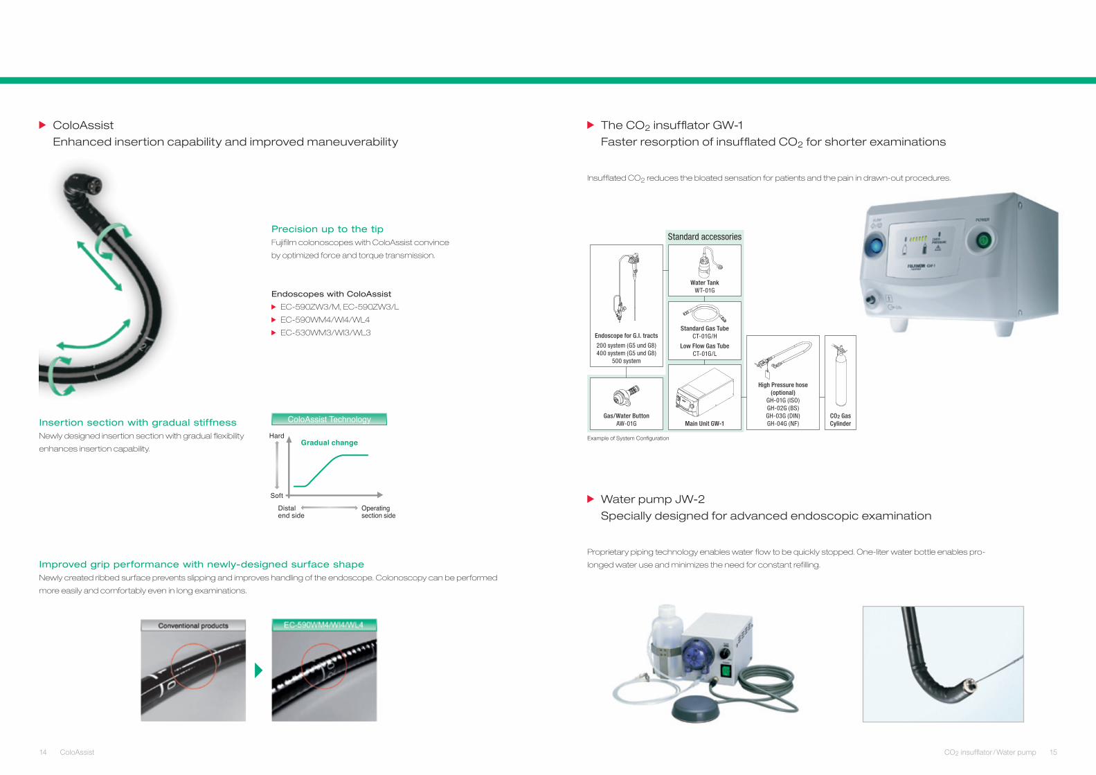
ColoAssist Technology Gas/Water ButtonAW-01G
CO2 Gas Cylinder
Standard accessories
Standard Gas TubeCT-01G/H
Low Flow Gas TubeCT-01G/L
Main Unit GW-1
High Pressure hose (optional)
GH-01G (ISO)GH-02G (BS)GH-03G (DIN)GH-04G (NF)
Endoscope for G.I. tracts
200 system (G5 und G8)400 system (G5 und G8)
500 system
Water TankWT-01G
Example of System Configuration
ColoAssist
Enhanced insertion capability and improved maneuverability
Insertion section with gradual stiffnessNewly designed insertion section with gradual flexibility
enhances insertion capability.
Endoscopes with ColoAssist
EC-590ZW3/M, EC-590ZW3/L
EC-590WM4/WI4/WL4
EC-530WM3/WI3/WL3
Improved grip performance with newly-designed surface shapeNewly created ribbed surface prevents slipping and improves handling of the endoscope. Colonoscopy can be performed
more easily and comfortably even in long examinations.
Precision up to the tipFujifilm colonoscopes with ColoAssist convince
by optimized force and torque transmission.
The CO2 insufflator GW-1
Faster resorption of insufflated CO2 for shorter examinations
Insufflated CO2 reduces the bloated sensation for patients and the pain in drawn-out procedures.
Water pump JW-2
Specially designed for advanced endoscopic examination
Proprietary piping technology enables water flow to be quickly stopped. One-liter water bottle enables pro-
longed water use and minimizes the need for constant refilling.
14 15ColoAssist CO2 insufflator / Water pump
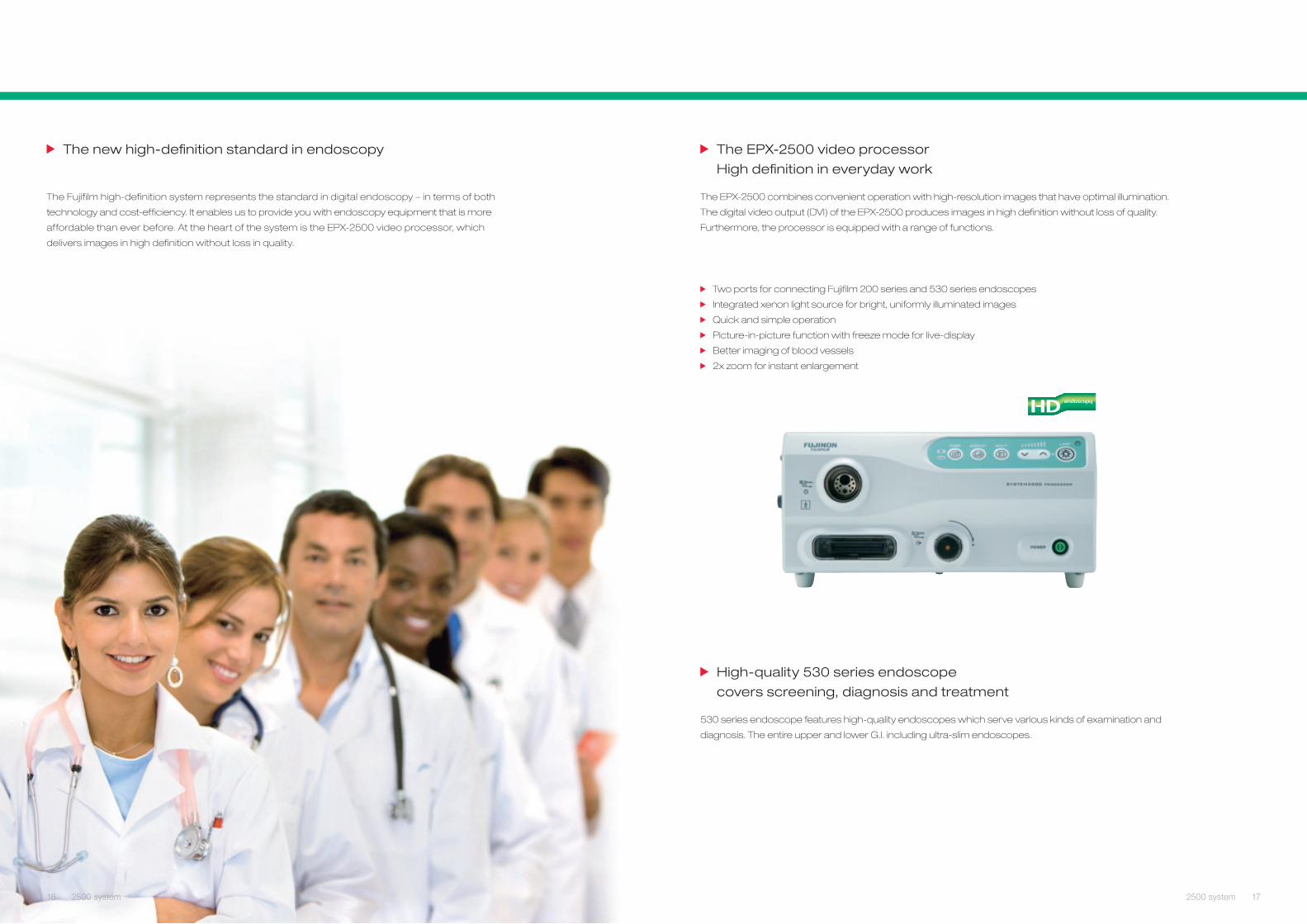
The new high-definition standard in endoscopy
The Fujifilm high-definition system represents the standard in digital endoscopy – in terms of both
technology and cost-efficiency. It enables us to provide you with endoscopy equipment that is more
affordable than ever before. At the heart of the system is the EPX-2500 video processor, which
delivers images in high definition without loss in quality.
The EPX-2500 video processor
High definition in everyday work
The EPX-2500 combines convenient operation with high-resolution images that have optimal illumination.
The digital video output (DVI) of the EPX-2500 produces images in high definition without loss of quality.
Furthermore, the processor is equipped with a range of functions.
Two ports for connecting Fujifilm 200 series and 530 series endoscopes
Integrated xenon light source for bright, uniformly illuminated images
Quick and simple operation
Picture-in-picture function with freeze mode for live-display
Better imaging of blood vessels
2x zoom for instant enlargement
High-quality 530 series endoscope
covers screening, diagnosis and treatment
530 series endoscope features high-quality endoscopes which serve various kinds of examination and
diagnosis. The entire upper and lower G.I. including ultra-slim endoscopes.
16 172500 system 2500 system

Air/water nozzle
210°UP
90°DOWN
100°LEFT
100°RIGHT
Light guide
Objective lens
Forceps channel
Image area & Forceps entry position
Light guide
Light guide
Objective lens
Forceps channel
Air/water nozzle
Water jet nozzle
Image area & Forceps entry position
100°LEFT
100°RIGHT
210°UP
90°DOWN
Objective lensForceps channel
Forceps channel
Light guide
Water jet nozzle
Light guide
Air/water nozzle
Image area & Forceps entry position
100°LEFT
100°RIGHT
210°UP
90°DOWN
Image area & Forceps entry position
Light guide
Objective lens
Forceps channelAir/water nozzle
210°UP
90°DOWN
100°LEFT
100°RIGHT
Image area & Forceps entry position
210°UP
120°DOWN
Light guide
Objective lens
Forceps channelAir/water nozzle
Image area & Forceps entry position
Light guide
Objective lens
Forceps channelAir/water nozzle
100°LEFT
100°RIGHT
210°UP
90°DOWN
Viewing direction 0° (Forward)
Field of view 120°
Observation range 3-100 mm
Distal end diameter 4.9 mm
Flexible portion diameter 5.1 mm
Bending capability UP 210° / DOWN 120°
Working length 1,100 mm
Total length 1,460 mm
Forceps channel diameter 2.0 mm
Viewing direction 0° (Forward)
Field of view 140°
Observation range 4-100 mm
Distal end diameter 9.4 mm
Flexible portion diameter 9.3 mm
Bending capabilityUP 210° / DOWN 90°RIGHT 100° / LEFT 100°
Working length 1,100 mm
Total length 1,400 mm
Forceps channel diameter 2.8 mm
Viewing direction 0° (Forward)
Field of view 140°
Observation range 4-100 mm
Distal end diameter 5.9 mm
Flexible portion diameter 5.9 mm
Bending capability UP 210° / DOWN 90°RIGHT 100° / LEFT 100°
Working length 1,100 mm
Total length 1,400 mm
Forceps channel diameter 2.0 mm
Viewing direction 0° (Forward)
Field of view 140°
Observation range 3-100 mm
Distal end diameter 10.8 mm
Flexible portion diameter 10.8 mm
Bending capabilityUP 210° / DOWN 90°RIGHT 100° / LEFT 100°
Working length 1,100 mm
Total length 1,400 mm
Forceps channel diameter 3.8 mm
Viewing direction 0° (Forward)
Field of view 140°
Observation range 3-100 mm
Distal end diameter 11.5 mm
Flexible portion diameter 11.5 mm
Bending capabilityUP 210° / DOWN 90°RIGHT 100° / LEFT 100°
Working length 1,090 mm
Total length 1,405 mm
Forceps channel diameter 3.8 mm / 2.8 mm
For the Upper G.I. Tract – Transnasal Type
EG-530NWThe ultra-slim gastroscope with a distal end diameter of 5.9 mm is made possible by
Fujifilm‘s proprietary microfabrication technology and offers a wide field of view with high-
resolution imaging similar to that obtainable with transoral gastroscopes. The flexible
gastroscope is ideal for transnasal insertion and provides the operator with highly visible
endoscopic images while reducing patient discomfort.
For the Upper G.I. Tract – Transnasal Type
EG-530NPEG-530NP slimmed down its endoscope to the utmost and realized a 4.9 mm distal
end (5.1 mm in the flexible portion), immensely improving the transnasal insertion
capability. This transnasal endoscope is also
equipped with dual light guides and a 2.0 mm
forceps channel.
For the Upper G.I. Tract – Standard Type
EG-530WRThe EG-530WR with a wide field of view of 140° provides exceptional visualization.
With the forceps channel of 2.8 mm, it is a standard endoscope producing high-
quality images, which is highly suited for
biopsies and treatment.
Viewing direction 0° (Forward)
Field of view 140°
Observation range 3-100 mm
Distal end diameter 8.5 mm
Flexible portion diameter 8.5 mm
Bending capability UP 210° / DOWN 90°RIGHT 100° / LEFT 100°
Working length 1,100 mm
Total length 1,400 mm
Forceps channel diameter 2.8 mm
For the Upper G.I. Tract – Slim Type
EG-530FPEG-530FP is a slim endoscope for the upper G.I. tract having a forceps channel of
2.8 mm diameter and a distal end of 8.5 mm. Observation capability has been
increased with a wide field of view of 140° and Fujifilm‘s Super CCD technology.
For the Upper G.I. Tract – Treatment Type
EG-530CTWith the forceps channel as wide as 3.8 mm, EG-530CT’s distal end is as slim as
10.8 mm in diameter. To support therapeutic
interventions, a water jet function is incorporated.
For the Upper G.I. Tract – Treatment Type
EG-530DEG-530D is an endoscope for treatment of the upper G.I. tract, having two forceps
channels, 3.8 mm and 2.8 mm, and a distal end as slim as 11.5 mm. Water jet function is
also incorporated for various treatment methods
during endoscopy.
WIDE VIEW
WIDE VIEW
WATER JET
WATER JET
BIG CHANNEL
DUAL CHANNEL
WIDE VIEW SLIM 8.5 mmULTRA-SLIM 5.9 mm
ULTRA-SLIM 4.9 mm
WIDE VIEW
TRANSNASAL
WIDE VIEW
TRANSNASAL
18 19530 series upper gastrointestinal endoscopes 530 series upper gastrointestinal endoscopes

Air/water nozzle(Internal)
Forceps channel
Light guide
Forceps elevator
Objective lens
Image area & Forceps entry position
90°LEFT
110°RIGHT
130°UP
90°DOWN
180°UP
180°DOWN
160°LEFT
160°RIGHT
Objective lensAir/water nozzle
EC-530LPEC-530MP
Forceps channel
Light guide
Image area & Forceps entry position
Image area & Forceps entry position
160°LEFT
160°RIGHT
180°UP
180°DOWN
Light guide
Objective lens
Forceps channelAir/water nozzle
Water jet nozzle
180°UP
180°DOWN
160°LEFT
160°RIGHT
Objective lensAir/water nozzle
Forceps channel
Light guide
Water jet nozzle
Image area & Forceps entry position
For the Duodenum
ED-530XT, ED-530XT8The structure of the distal end, bending portion and flexible portion is changed for
improved operability during examination
and treatment.
Viewing direction 98° (8° rearward)
Field of view 100°
Observation range 4-60 mm
Distal end diameter 13.1 mm
Flexible portion diameter 11.5 mm
Bending capabilityUP 130° / DOWN 90°RIGHT 110° / LEFT 90°
Working length 1,250 mm
Total length 1,550 mm
Forceps channel diameter 4.2 mm
Improved operabilityEasy to catch the papilla
The objective lens arrangement and bending per-
formance have been properly arranged to catch
the papilla easily from various endoscope positions.
Easy operability of the insertion portion
The stiffness of the insertion por-
tion has been impro ved for easier
stomach stretching and insertion
capability.
Improved insertion capability of ERCP
accessories into the papilla
Newly designed forceps elevator
has been applied to enhance
accessory control more precise-
ly and securely, facilitating easier
ERCP treatment.
Improved cleaning and disinfectionRemovable distal end cap*
The ED-530XT8 is equipped with a disposable distal end cap.
lt enables brushing all channels and helps to improve the
hygienic environment.*ED-530XT8 only
Covered tilt-up mechanism
A covered tilt-up mechanism of the
forceps elevator maintains the
elevator wire clean without any
additional cleaning procedure.
WM3 WI3 WL3
Viewing direction 0° (Forward)
Field of view 140°
Observation range 3-100 mm
Distal end diameter 12.8 mm
Flexible portion diameter 12.8 mm
Bending capabilityUP 180° / DOWN 180°
RIGHT 160° / LEFT 160°
Working length 1,330 mm 1,520 mm 1,690 mm
Total length 1,630 mm 1,820 mm 1,990 mm
Forceps channel diameter 3.8 mm
MT IT LT
Viewing direction 0° (Forward)
Field of view 140°
Observation range 3-100 mm
Distal end diameter 12.8 mm
Flexible portion diameter 12.8 mm
Bending capabilityUP 180° / DOWN 180°
RIGHT 160° / LEFT 160°
Working length 1,330 mm 1,520 mm 1,690 mm
Total length 1,630 mm 1,820 mm 1,990 mm
Forceps channel diameter 4.2 mm
MP LP
Viewing direction 0° (Forward)
Field of view 140°
Observation range 3-100 mm
Distal end diameter 11.0 mm
Flexible portion diameter 11.1 mm
Bending capabilityUP 180° / DOWN 180°
RIGHT 160° / LEFT 160°
Working length 1,330 mm 1,690 mm
Total length 1,630 mm 1,990 mm
Forceps channel diameter 3.2 mm
Excellent image qualityFujifilm‘s Super CCD, which has been exclusively developed for
the endoscope, is built-in, providing clear images.
For the Lower G.I. Tract – Standard Type
EC-530WM3, EC-530WI3, EC-530WL3With a wide field of view of 140°, these lower G.I. tract endoscopes have a greater
resolution. The newly ColoAssist design facilitates the insertion capability.
For the Lower G.I. Tract – Treatment Type
EC-530MT, EC-530IT, EC-530LTWith a large channel of 4.2 mm accommodating various treatment accessories,
these lower G.I. tract endoscopes are suited for examination and treatment, which
also have a rapid suction function.
For the Lower G.I. Tract – Slim Type
EC-530MP, EC-530LPThese are slim-type colonoscopes with the distal end of 11.0 mm. While these two
slimmed-down endoscopes have improved insertability, they retain a 3.2 mm forceps
channel to accommodate various treatment methods.
BIG CHANNEL
WIDE VIEW
WIDE VIEW
WATER JET
BIG CHANNEL
SLIM 11.0 mm
ColoAssist WIDE VIEW
WATER JET
20 21530 series duodenum endoscopes 530 series lower gastrointestinal endoscopes
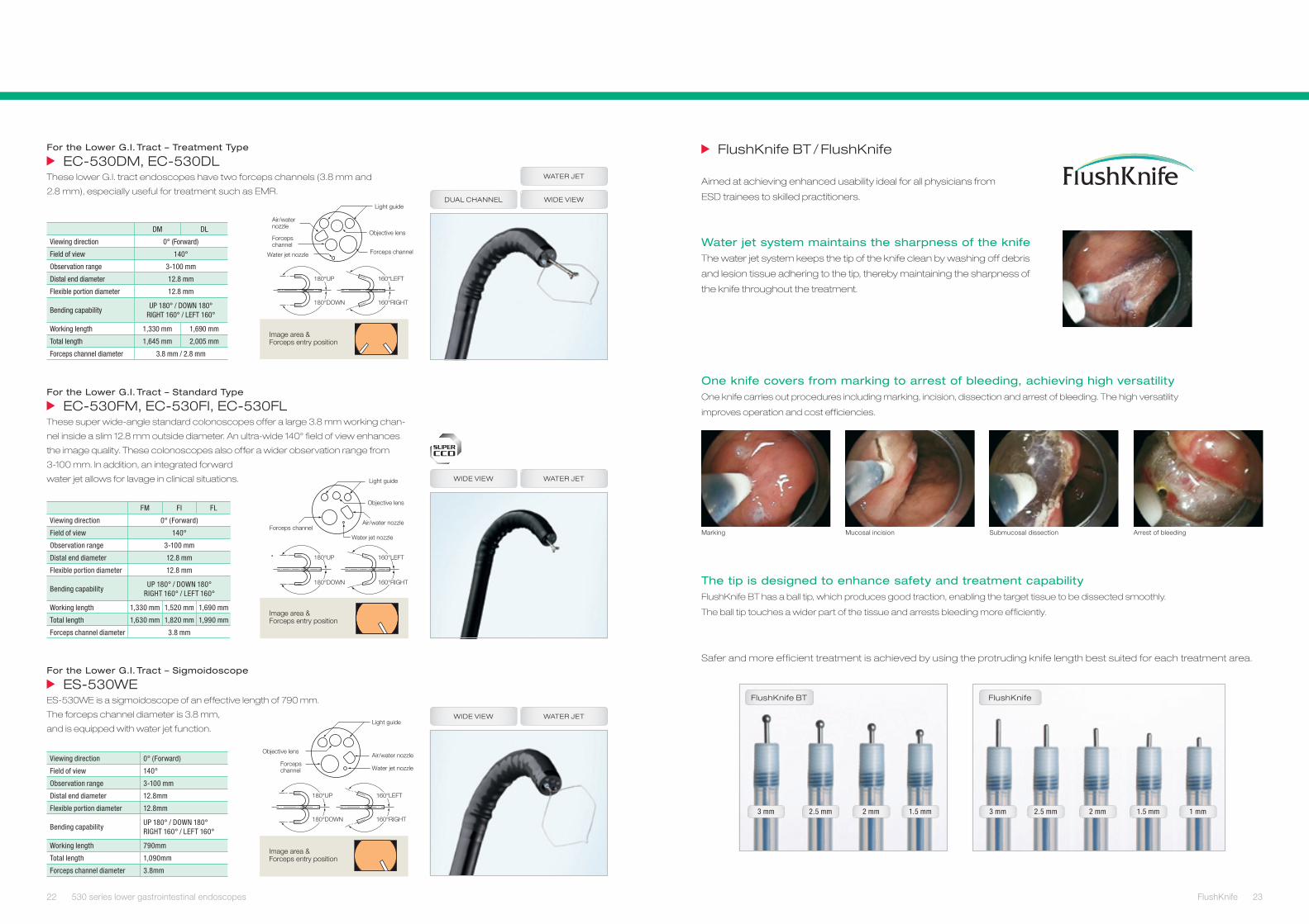
FlushKnife BT
3 mm 2.5 mm 2 mm 1.5 mm
FlushKnife
3 mm 2.5 mm 2 mm 1.5 mm 1 mm
180°UP
180°DOWN
160°LEFT
160°RIGHT
Air/water nozzle
Light guide
Objective lens
Forceps channelWater jet nozzle
Forceps channel
Image area & Forceps entry position
Image area & Forceps entry position
Air/water nozzle
Water jet nozzle
180°UP
180°DOWN
160°LEFT
160°RIGHT
Light guide
Objective lens
Forceps channel
180°UP
180°DOWN
160°LEFT
160°RIGHT
Objective lensAir/water nozzle
Forceps channel
Light guide
Water jet nozzle
Image area & Forceps entry position
DM DL
Viewing direction 0° (Forward)
Field of view 140°
Observation range 3-100 mm
Distal end diameter 12.8 mm
Flexible portion diameter 12.8 mm
Bending capabilityUP 180° / DOWN 180°
RIGHT 160° / LEFT 160°
Working length 1,330 mm 1,690 mm
Total length 1,645 mm 2,005 mm
Forceps channel diameter 3.8 mm / 2.8 mm
FM FI FL
Viewing direction 0° (Forward)
Field of view 140°
Observation range 3-100 mm
Distal end diameter 12.8 mm
Flexible portion diameter 12.8 mm
Bending capabilityUP 180° / DOWN 180°
RIGHT 160° / LEFT 160°
Working length 1,330 mm 1,520 mm 1,690 mm
Total length 1,630 mm 1,820 mm 1,990 mm
Forceps channel diameter 3.8 mm
Viewing direction 0° (Forward)
Field of view 140°
Observation range 3-100 mm
Distal end diameter 12.8mm
Flexible portion diameter 12.8mm
Bending capabilityUP 180° / DOWN 180°RIGHT 160° / LEFT 160°
Working length 790mm
Total length 1,090mm
Forceps channel diameter 3.8mm
For the Lower G.I. Tract – Treatment Type
EC-530DM, EC-530DLThese lower G.I. tract endoscopes have two forceps channels (3.8 mm and
2.8 mm), especially useful for treatment such as EMR.
For the Lower G.I. Tract – Standard Type
EC-530FM, EC-530FI, EC-530FLThese super wide-angle standard colonoscopes offer a large 3.8 mm working chan-
nel inside a slim 12.8 mm outside diameter. An ultra-wide 140° field of view enhances
the image quality. These colonoscopes also offer a wider observation range from
3-100 mm. In addition, an integrated forward
water jet allows for lavage in clinical situations.
For the Lower G.I. Tract – Sigmoidoscope
ES-530WEES-530WE is a sigmoidoscope of an effective length of 790 mm.
The forceps channel diameter is 3.8 mm,
and is equipped with water jet function.
FlushKnife BT / FlushKnife
Aimed at achieving enhanced usability ideal for all physicians from
ESD trainees to skilled practitioners.
Water jet system maintains the sharpness of the knifeThe water jet system keeps the tip of the knife clean by washing off debris
and lesion tissue adhering to the tip, thereby maintaining the sharpness of
the knife throughout the treatment.
Safer and more efficient treatment is achieved by using the protruding knife length best suited for each treatment area.
WIDE VIEW
WATER JET
WATER JETWIDE VIEW
WIDE VIEW
WATER JET
DUAL CHANNEL
Marking Mucosal incision Submucosal dissection Arrest of bleeding
The tip is designed to enhance safety and treatment capabilityFlushKnife BT has a ball tip, which produces good traction, enabling the target tissue to be dissected smoothly.
The ball tip touches a wider part of the tissue and arrests bleeding more efficiently.
One knife covers from marking to arrest of bleeding, achieving high versatilityOne knife carries out procedures including marking, incision, dissection and arrest of bleeding. The high versatility
improves operation and cost efficiencies.
22 23530 series lower gastrointestinal endoscopes FlushKnife

ZONE MethodConventional Method EUS Tower – all-in-one stack concept
Years of research and development to reduce patient discomfort and
improve operator efficiency during endoscope examinations led to the
development of Sonart, the integration of ultrasonographic diagnosis and
endoscopy systems.
For a more accurate diagnosis, advanced image processing technology
integrates improved endoscope maneuverability and insertion capability.
The compact, one-cart system supports various applications.
ZONE Sonography™ technology ensures high-quality imagesZONE Sonography™ technology defines conventional wisdom in
ultrasonography. The technology delivers wide ultrasound beams and
quickly acquires large amounts of echo data in sizeable zones. Split-
second data acquisition allows highly advanced image processing.
Sound Speed Correction technology improves image resolution Advanced image processing technology estimates the optimal speed of ultrasound travelling through the body
(sound speed) and constructs images.
1450 m/s ATS Phantom
What is Sound Speed Correction?
The resolution in the lateral dimension
deteriorates due to a difference in
sound speed. By correcting this and
carrying out optimization, the resolution
in the lateral dimension is improved.
Imaged at 1540 m/sbefore Sound Speed Correction
Imaged at 1450 m/safter Sound Speed Correction
C mode
The Color Doppler function obtains hemodynamic
information in disease areas and helps you locate the
observation site and vascular structures.
SU-8000 Scanning modes; C mode, Power Doppler,
Pulse wave, B mode, M mode
Frequency switching
A wide range of frequencies (5, 7.5, 10, and 12 MHz)
helps to delineate clear C mode images of the regions.
Display quality images in different modesTechnologies developed in the field of ultrasonographic diagnosis improve the quality of ultrasound images.
Images created from advanced image processing enable the use of appropriate modes for your setting.
CFM mode B mode
Flexible image display and switchingKeyboard operation facilitates
smooth examinations and allows
switching among an ultrasound
image, an endoscopic image,
and a picture-in-picture image
with patient‘s history images.
Picture-in-picture image Patient’s history image
Ultrasound image
Endoscopic image
24 25Endoscopic ultrasonography Endoscopic ultrasonography

Image area & Forceps entry position
180°UP
Light guide
Objective lens
Forceps channel
100°LEFT
100°RIGHT
Air/water nozzle
90°DOWN
Image area & Forceps entry position
Ultrasonic oscillator
Light guide
160°UP
160°DOWN
120°LEFT
120°RIGHT
Balloon water port
Air/water nozzle
Objective lensForceps elevator
Balloon slot
Ultrasonic endoscopes
Radial Scan Ultrasound Video Endoscope
EG-530UR2With a slim distal end of 11.4 mm and excellent bending capabilities, the EG-530UR2
allows physicians to perform endoscopic ultrasonography in a similar way to conven-
tional endoscopy.
Convex Scan Ultrasonic Video Endoscope
EG-530UT2With its forceps channel elevator function, the distal end of EG-530UT2 improves the
injection performance of the puncture needle. It also has a large channel which enables
various treatment accessories to be inserted. With excellent bending capabilities, the
EG-530UT2 provides greater flexibility in treatment.
Viewing direction 0° (Forward)
Field of view 140°
Observation range 3-100 mm
Distal end diameter 11.4 mm
Flexible portion diameter 11.5 mm
Bending capability UP 180° / DOWN 90°RIGHT 100° / LEFT 100°
Working length 1,250 mm
Total length 1,550 mm
Forceps channel diameter 2.2 mm
Scanning mode Color Doppler, Power Doppler,PW Doppler, B mode, M mode
Scanning method Electronic radial scan
Scanning angle 360°
Frequency 5 Mhz / 7.5 Mhz / 10 Mhz / 12 Mhz
Contact method Balloon method, degassed water congestion method, contacting method
Viewing direction Forward oblique 40°
Field of view 140°
Observation range 3-100 mm
Distal end diameter 13.9 mm
Flexible portion diameter 12.1 mm
Bending capability UP 160° / DOWN 160°RIGHT 120° / LEFT 120°
Working length 1,250 mm
Total length 1,550 mm
Forceps channel diameter 3.8 mm
Scanning modeColor Doppler, Power Doppler,PW Doppler, B mode, M mode, THI
Scanning method Electronic convex scan
Scanning angle110° (Combination with SU-7000)124° (Combination with SU-8000)
Frequency 5 Mhz / 7.5 Mhz / 10 Mhz / 12 Mhz
Contact methodBalloon method, degassed water congestion method, contacting method
Excellent insertion capabilityNewly designed structure of flexible portion improves
insertion capabilty. The tip with a small bending radius
allows better observation.
EG-530UR2, EG-530UT2EG-530UR2 and EG-530UT2 endoscopes combine Fujifilm’s high-quality endoscope features with the most advanced
ultrasound technology, to create an unsurpassed diagnostic and treatment system.
Consideration of the safety of fine needle aspirationDotted yellow guidelines are visualized on the monitor
to ensure the safety of paracentesis.
High-quality endoscopic imageEquipped with the Super CCD, this ultrasound endoscope offers bright, vivid, high-resolution image.
In pursuit of balloon operabilityAn air/water and suction button inflates water to the balloon and deflates water from the balloon.
EG-530UR2 EG-530UT2
26 27Endoscopic ultrasonography Endoscopic ultrasonography

TS-12140
TS-13140
BS-2
BS-1
Image area & Forceps entry position
Light guide
Objective lens
Forceps channelAir/water nozzle
160°LEFT
160°RIGHT
180°UP
180°DOWN
EN-450T5
Image area & Forceps entry position
Light guide
Objective lens
Forceps channelAir/water nozzle
160°LEFT
160°RIGHT
180°UP
180°DOWN
Image area & Forceps entry position
Light guideObjective lens
Forceps channel
Air/water nozzle
160°LEFT
160°RIGHT
180°UP
180°DOWN
Viewing direction 0° (Forward)
Field of view 120°
Observation range 5-100 mm
Distal end diameter 8.5 mm
Flexible portion diameter 8.5 mm
Bending capability UP 180° / DOWN 180°RIGHT 160° / LEFT 160°
Working length 2,000 mm
Total length 2,300 mm
Forceps channel diameter 2.2 mm
Viewing direction 0° (Forward)
Field of view 140°
Observation range 4-100 mm
Distal end diameter 9.4mm
Flexible portion diameter 9.3 mm
Bending capability UP 180° / DOWN 180°RIGHT 160° / LEFT 160°
Working length 2,000 mm
Total length 2,300 mm
Forceps channel diameter 2.8 mm
Viewing direction 0° (Forward)
Field of view 140°
Observation range 3-100 mm
Distal end diameter 9.4 mm
Flexible portion diameter 9.3 mm
Bending capability UP 180° / DOWN 180°RIGHT 160° / LEFT 160°
Working length 1,520 mm
Total length 1,820 mm
Forceps channel diameter 2.8 mm
Balloons and overtubes (consumable supplies) The exclusively developed specialized balloons and overtubes ensure
complete positioning of the endoscope in the small intestine. The distal end
of the endoscope can be smoothly inserted to reach the area of diagnosis.
Two balloons realize better insertability
into the depth of digestive tract
Balloon pump controller
PB-20The PB-20 balloon pump controller is de-
signed to simplify operation. Balloons can be
easily controlled via a hand-operated remote
control or foot switch – whichever is more
convenient for the physician.
The small intestine has long been the most difficult organ to access in
gastrointestinal endoscopy, therefore it has been known as “The Black
Box”. With new engineering innovation, Fujifilm‘s double balloon endo-
scopic system, designed for the small intestine, is equipped with exclusively
developed balloons, overtubes and balloon pump controller. Two balloons
improve the insertability of the endoscope into the small intestine.
Anterograde insertion Transanal insertion
Power AC 100 V 50 / 60 Hz 0.76 A
Power consumption (rated) 0.66 A
Set pressure accuracy ± 2 kPa
Set pressure of balloon 5.6 kPa
Maximum flow rate of pump 170 ml ± 50 ml / 10 sec
Dimensions (W x H x D) 350 × 130 × 420 mm
Weight 10 kg (Body), 0.4 kg (Remote switch)
Overtube model TS-12140 TS-13140 TS-13101
Outer diameter 12.2 mm 13.2 mm 13.2 mm
Total length 1,450 mm 1,450 mm 950 mm
Applicable endoscope
EN-450P5/20 EN-450T5 EC-450BI5
Balloon BS-1 BS-2
Outer diameter 25 mm 35 mm
Enteroscope – Standard Type
EN-450P5/20EN-450P5/20 is an endoscope for the small intestine examination. The relatively slim
overtubes (12.2 mm outer diameter) of the EN-450P5/20 allow for smooth insertion via
both the anterograde and transanal routes, depending on the position of lesion.
Enteroscope – Treatment Type
EN-450T5Treatment capacity has been greatly expanded with the EN-450T5, which is equipped
with a 2.8 mm forceps channel that allows the use of almost all general therapeutic
accessories and a variety of accessories such as APC probe, clip, diathermic coagu-
lator, and other therapeutic interventions.
Colonoscope – Standard Type
EC-450BI5Using balloons, the endoscope is stabilized in the intestinal tract, which leads to better
observation and treatment of lesions.
FOR TREATMENT
DOUBLE BALLOON
DOUBLE BALLOON
DOUBLE BALLOON
28 29Double balloon endoscopy Double balloon endoscopy

Power supply
AC120 V AC230 V
60 Hz 50 Hz
2.2 A 1.4 A
Current consumption (rated) 1.8 A 1.2 A
Applicable scopesEG-530U series scope
EB-530U series scope
Video output terminal
Video terminal (1 channel)
S video terminal (1 channel)
RGB PC terminal (1 channel)
RGB PC/TV terminal (1 channel)
DVI image input terminal (1 channel)
HD-SDI terminal (2 channels)
Audio output terminal RCA terminal (1 channel)
Video input terminal
DVI image input terminal (1 channel)
S video terminal (PROCESSOR) (1 channel)
S video terminal (SP702) (1 channel)
Control terminal
Remote terminal (2 channels)
Foot Switch terminal (1 channel)
Keyboard terminal (1 channel)
RS232C terminal (PROCESSOR) (1 channel)
RS232C terminal (SP702) (1 channel)
Network terminal (1 channel) Ethernet (100 BaseTX)
Image storageStorage CF memory card, networked shared folder (FTP, DICOM)
File format TIFF, JPEG
External dimensions (W x H x D) 375 x 215 x 445 mm (including protruding parts)
Weight 14 kg
Digital outputHD-SDI: HDTV 1080i (2ch)DVI (Digital Visual Interface): 1280 x 1024 pEthernet: 100 / 10 Base
Analog outputRGB: 1280 x 1024 pSDTV (120 V / NTSC, 230 V / PAL): RGB Y / C, Composite
Color adjustment Brightness, Red, Green, Blue, R-Hue, Chroma; 9 steps
Detail Hi, Lo; 9 steps
Contrast (gamma) 3 steps
Hyper-Sharpness Hi, Mid, Lo, Off
Color emphasis Hi, Mid, Lo, Off
FICEFlexible spectral imagingColor Enhancement 10 presets
Iris Average / Peak / Auto
Image storage CF Card
Power rating 120 V 60 Hz 0.8 A 230 V 50 Hz 0.5 A
Dimensions (W x H x D) 390 x 105 x 460 mm
Weight 9.5 kg
DICOM MWL, Store
Lamp rated valueMain Lamp: 300 W Xenon lamp LMP-002Emergency Lamp: 75 W Halogen lamp
Light control Automatic light control
Lamp cooling method Forced air cooling
Air supply pump High, Mid, Lo, Off
Light save On, Off
Transmitted illumination On, Off
Power rating 120 V 60 Hz 3.3 A 230 V 50 Hz 1.7 A
Dimensions (W x H x D) 390 x 155 x 450 mm
Weight 15 kg
XL-4450 Light source
EPX-4450HD EPX-2500
SU-8000
Video Processor Video Processor
Ultrasonic Processor
Digital output DVI (Digital Visual Interface): 1024 x 768 p
Analog outputRGB (2): SDTV (NTSC / PAL)Y/C (2): SDTV (NTSC / PAL)Composite: SDTV (NTSC / PAL)
Color adjustment Black, Red, Green, Blue, R-Hue, Chroma; 9 settings
Detail Hi, LO; 9 settings
Contrast (gamma) 9 settings
BLD Hi, Mid, Lo, Off
Picture in picture On, Off; Size: 1/4, 1/3
Auto gain control Off, +3 db, +6 db
Iris Average / Peak
Zoom Electric zoom: x1.0 – x2.0; 0.05 steps
Lamp rated valueMain lamp: 11.7 V 150 W Xenon lampEmergency lamp: 12 V 75 W Halogen lamp
Brightness control 9 settings
Lamp cooling method Forced air cooling
Air supply pump Hi, Low, Off
Power 120 V 60 Hz 2.7 A / 230 V 50 Hz 1.4 A
Dimensions (W x D x H) 375 x 495 x 190 mm (including projections)
Weight 17.0 kg
VP-4450 HD Processor
30 31Technical specifications Technical specifications