Embed Size (px)
Citation preview
C00h
w
TECHNOLOGY STATUS EVALUATION REPORT
opyright ª 2015 by the16-5107/$36.00ttp://dx.doi.org/10.1016
ww.giejournal.org
Endoscopic submucosal dissection
The American Society for Gastrointestinal Endoscopy(ASGE) Technology Committee provides reviews of exist-ing, new, or emerging endoscopic technologies thathave an impact on the practice of GI endoscopy.Evidence-based methodology is used, performing a MED-LINE literature search to identify pertinent clinical studieson the topic and a MAUDE (U.S. Food and Drug Adminis-tration Center for Devices and Radiological Health) data-base search to identify the reported adverse events of agiven technology. Both are supplemented by accessingthe “related articles” feature of PubMed and by scruti-nizing pertinent references cited by the identified studies.Controlled clinical trials are emphasized, but in manycases, data from randomized, controlled trials are lack-ing. In such cases, large case series, preliminary clinicalstudies, and expert opinions are used. Technical data aregathered from traditional and Web-based publications,proprietary publications, and informal communicationswith pertinent vendors. Technology Status EvaluationReports are drafted by 1 or 2 members of the ASGE Tech-nology Committee, reviewed and edited by the Committeeas a whole, and approved by the Governing Board ofthe ASGE. When financial guidance is indicated, themost recent coding data and list prices at the time of pub-lication are provided. For this review, the MEDLINE data-base was searched through April 2014 for relevant articlesby using the key words “endoscopic submucosal dissec-tion” and “ESD,” combined with other relevant termssuch as “gastric,” “esophageal,” “rectal,” “colonic,” and“adverse events,” among others. Technology Status Evalu-ation Reports are scientific reviews provided solely foreducational and informational purposes. TechnologyStatus Evaluation Reports are not rules and shouldnot be construed as establishing a legal standard ofcare or as encouraging, advocating, requiring, ordiscouraging any particular treatment or payment forsuch treatment.
BACKGROUND
Endoscopic submucosal dissection (ESD) is a well-established technique of endoscopic resection that allowsfor en bloc removal of GI epithelial lesions. ESD differs
American Society for Gastrointestinal Endoscopy.
/j.gie.2014.12.010
V
from EMR, the other type of endoscopic resection. Bothtechniques involve injection of a substance under the tar-geted lesion to act as a cushion. With EMR, the lesion isthen removed with a snare or suctioned into a cap andsnared. With ESD, the submucosa is instead dissected un-der the lesion with a specialized knife. This enablesremoval of larger and potentially deeper lesions with acurative intent than can be accomplished with EMR. ESDwas first described in 1988 as a technique to treat earlygastric neoplasia nonoperatively.1 Over the ensuing de-cades, procedural techniques and equipment for ESDhave evolved significantly, and applications for ESD tech-niques have expanded to locations throughout the GItract as well as to the treatment of deeper, nonepitheliallesions. The principles of ESD have also led to the develop-ment of procedures with a therapeutic intent other thanthe resection of neoplasia, including peroral endoscopicmyotomy for the treatment of achalasia.
TECHNOLOGY UNDER REVIEW
Proper patient and lesion selection for ESD are essen-tial. Endoscopic resection of neoplastic lesions shouldonly be undertaken when endoscopic and/or endosono-graphic evaluations predict a curative resection. However,one of the benefits of ESD is that the pathologist is pro-vided with an en bloc specimen, such that noncurative re-sections can be more easily detected and patients properlyreferred for further oncologic surgery.
ESD is accomplished in a sequential or stepwisemanner, and a variety of devices are available to assistthe endoscopist in performing each step. Typically, theESD steps during resection of a mucosal neoplastic lesionare as follows: (1) the perimeter of the lesion is markedwith cautery; (2) a lifting agent is injected into the submu-cosa around the perimeter of the lesion; (3) the mucosa isincised and then cut circumferentially around the lesionby using an electrosurgical knife; (4) the submucosabeneath the lesion is injected and then dissected in afree-hand manner by using an electrosurgical knife untilthe specimen has been completely resected; and (5) anyintraprocedural bleeding that occurs during the mucosalincision or submucosal dissection is managed by using awater jet for washing and hemostatic forceps or an electro-surgical knife by using coagulation current for vesselcoagulation.
Electrosurgical knives, discussed in the following, arethe main devices used in ESD that differentiate it from
olume 81, No. 6 : 2015 GASTROINTESTINAL ENDOSCOPY 1311
Endoscopic submucosal dissection
other types of endoscopic resection. The other tools used(eg, endoscope, electrosurgical unit [ESU], and other ancil-lary devices) are similar to those used for standard endos-copy. However, because of the complexity of theprocedure, special considerations in choosing these typesof equipment are also necessary.
Devices for ESDThe common attribute of all dedicated ESD devices is
their ability to perform submucosal dissection. However,some devices are also useful in earlier stages of the proce-dure, such as marking or initial mucosal incision. Theearliest dedicated ESD device simply added an insulatedball-like ceramic tip to an existing needle-knife to preventinadvertently deep dissection and thus potential perfora-tion.2 In addition to uncovered and covered (insulated-tip) needle-knife–like devices, a group of forceps-likedevices has now been developed. However, although awide variety of dedicated ESD devices are manufacturedworldwide, the number of ESD devices that are approvedby the U.S. Food and Drug Administration and availablein the United States is limited. Table 1 lists ESD devicesapproved by the U.S. Food and Drug Administration andtheir suitability for the different procedural steps in ESD.All ESD devices are designed for single use only. MostESD devices feature catheter outer diameters that arecompatible with a 2.8-mm endoscopic instrument channel.Some ESD devices are available in lengths compatiblewith use with a colonoscope.
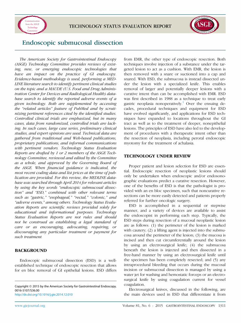
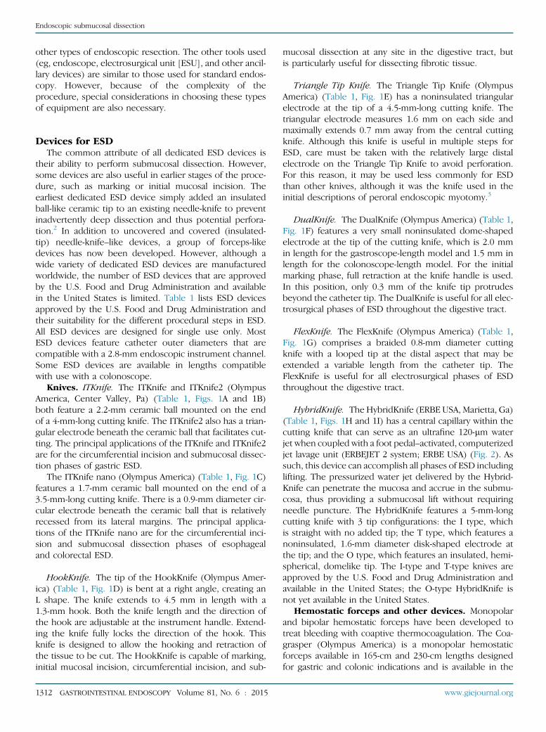
Knives. ITKnife. The ITKnife and ITKnife2 (OlympusAmerica, Center Valley, Pa) (Table 1, Figs. 1A and 1B)both feature a 2.2-mm ceramic ball mounted on the endof a 4-mm-long cutting knife. The ITKnife2 also has a trian-gular electrode beneath the ceramic ball that facilitates cut-ting. The principal applications of the ITKnife and ITKnife2are for the circumferential incision and submucosal dissec-tion phases of gastric ESD.
The ITKnife nano (Olympus America) (Table 1, Fig. 1C)features a 1.7-mm ceramic ball mounted on the end of a3.5-mm-long cutting knife. There is a 0.9-mm diameter cir-cular electrode beneath the ceramic ball that is relativelyrecessed from its lateral margins. The principal applica-tions of the ITKnife nano are for the circumferential inci-sion and submucosal dissection phases of esophagealand colorectal ESD.
HookKnife. The tip of the HookKnife (Olympus Amer-ica) (Table 1, Fig. 1D) is bent at a right angle, creating anL shape. The knife extends to 4.5 mm in length with a1.3-mm hook. Both the knife length and the direction ofthe hook are adjustable at the instrument handle. Extend-ing the knife fully locks the direction of the hook. Thisknife is designed to allow the hooking and retraction ofthe tissue to be cut. The HookKnife is capable of marking,initial mucosal incision, circumferential incision, and sub-
1312 GASTROINTESTINAL ENDOSCOPY Volume 81, No. 6 : 2015
mucosal dissection at any site in the digestive tract, butis particularly useful for dissecting fibrotic tissue.
Triangle Tip Knife. The Triangle Tip Knife (OlympusAmerica) (Table 1, Fig. 1E) has a noninsulated triangularelectrode at the tip of a 4.5-mm-long cutting knife. Thetriangular electrode measures 1.6 mm on each side andmaximally extends 0.7 mm away from the central cuttingknife. Although this knife is useful in multiple steps forESD, care must be taken with the relatively large distalelectrode on the Triangle Tip Knife to avoid perforation.For this reason, it may be used less commonly for ESDthan other knives, although it was the knife used in theinitial descriptions of peroral endoscopic myotomy.3
DualKnife. The DualKnife (Olympus America) (Table 1,Fig. 1F) features a very small noninsulated dome-shapedelectrode at the tip of the cutting knife, which is 2.0 mmin length for the gastroscope-length model and 1.5 mm inlength for the colonoscope-length model. For the initialmarking phase, full retraction at the knife handle is used.In this position, only 0.3 mm of the knife tip protrudesbeyond the catheter tip. The DualKnife is useful for all elec-trosurgical phases of ESD throughout the digestive tract.
FlexKnife. The FlexKnife (Olympus America) (Table 1,Fig. 1G) comprises a braided 0.8-mm diameter cuttingknife with a looped tip at the distal aspect that may beextended a variable length from the catheter tip. TheFlexKnife is useful for all electrosurgical phases of ESDthroughout the digestive tract.
HybridKnife. The HybridKnife (ERBE USA, Marietta, Ga)(Table 1, Figs. 1H and 1I) has a central capillary within thecutting knife that can serve as an ultrafine 120-mm waterjet when coupled with a foot pedal–activated, computerizedjet lavage unit (ERBEJET 2 system; ERBE USA) (Fig. 2). Assuch, this device can accomplish all phases of ESD includinglifting. The pressurized water jet delivered by the Hybrid-Knife can penetrate the mucosa and accrue in the submu-cosa, thus providing a submucosal lift without requiringneedle puncture. The HybridKnife features a 5-mm-longcutting knife with 3 tip configurations: the I type, whichis straight with no added tip; the T type, which features anoninsulated, 1.6-mm diameter disk-shaped electrode atthe tip; and the O type, which features an insulated, hemi-spherical, domelike tip. The I-type and T-type knives areapproved by the U.S. Food and Drug Administration andavailable in the United States; the O-type HybridKnife isnot yet available in the United States.
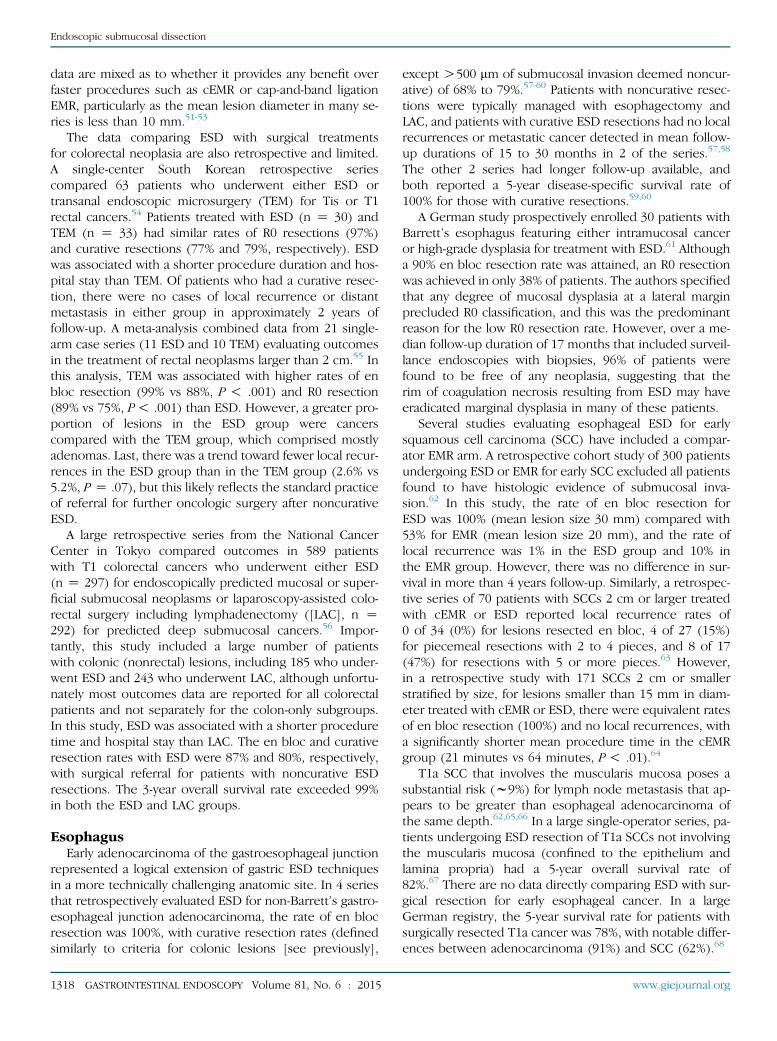
Hemostatic forceps and other devices. Monopolarand bipolar hemostatic forceps have been developed totreat bleeding with coaptive thermocoagulation. The Coa-grasper (Olympus America) is a monopolar hemostaticforceps available in 165-cm and 230-cm lengths designedfor gastric and colonic indications and is available in the
www.giejournal.org
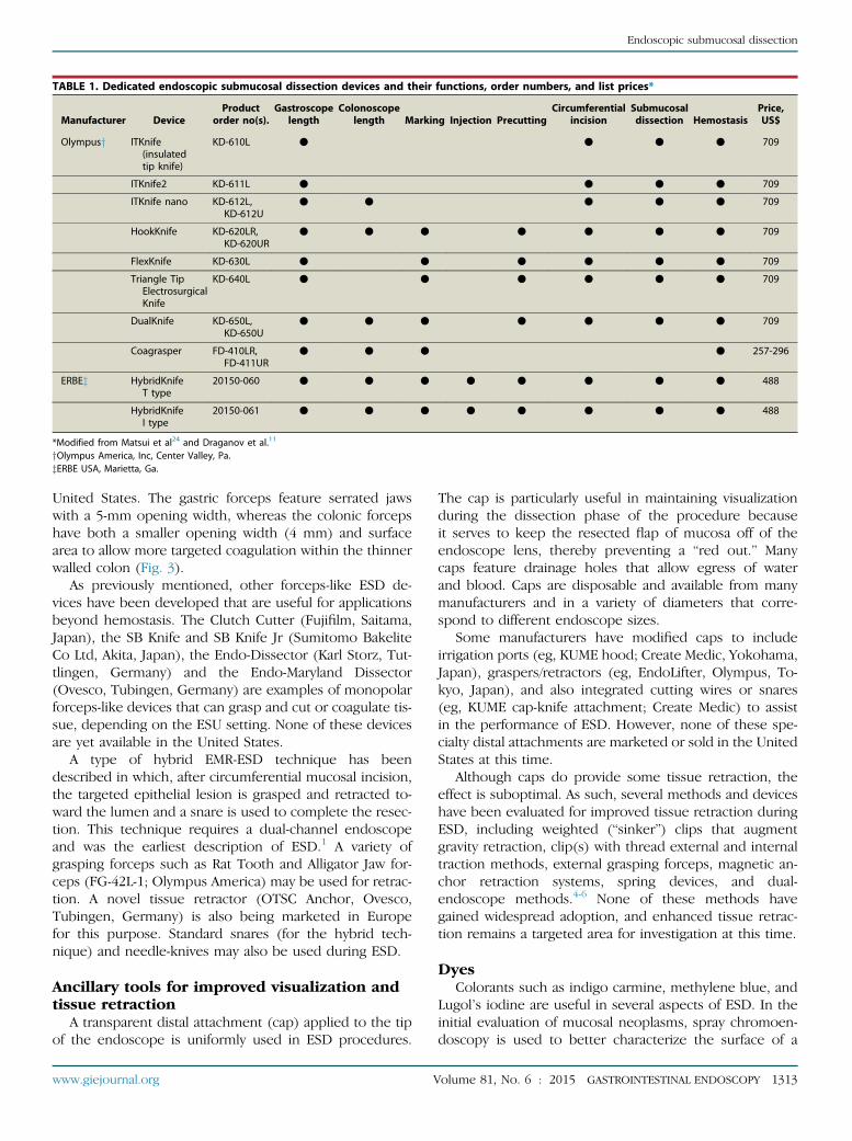
TABLE 1. Dedicated endoscopic submucosal dissection devices and their functions, order numbers, and list prices*
Manufacturer DeviceProduct
order no(s).Gastroscope
lengthColonoscope
length Marking Injection PrecuttingCircumferential
incisionSubmucosaldissection Hemostasis
Price,US$
Olympusy ITKnife(insulatedtip knife)
KD-610L C C C C 709
ITKnife2 KD-611L C C C C 709
ITKnife nano KD-612L,KD-612U
C C C C C 709
HookKnife KD-620LR,KD-620UR
C C C C C C C 709
FlexKnife KD-630L C C C C C C 709
Triangle TipElectrosurgicalKnife
KD-640L C C C C C C 709
DualKnife KD-650L,KD-650U
C C C C C C C 709
Coagrasper FD-410LR,FD-411UR
C C C C 257-296
ERBEz HybridKnifeT type
20150-060 C C C C C C C C 488
HybridKnifeI type
20150-061 C C C C C C C C 488
*Modified from Matsui et al24 and Draganov et al.11
yOlympus America, Inc, Center Valley, Pa.zERBE USA, Marietta, Ga.
Endoscopic submucosal dissection
United States. The gastric forceps feature serrated jawswith a 5-mm opening width, whereas the colonic forcepshave both a smaller opening width (4 mm) and surfacearea to allow more targeted coagulation within the thinnerwalled colon (Fig. 3).
As previously mentioned, other forceps-like ESD de-vices have been developed that are useful for applicationsbeyond hemostasis. The Clutch Cutter (Fujifilm, Saitama,Japan), the SB Knife and SB Knife Jr (Sumitomo BakeliteCo Ltd, Akita, Japan), the Endo-Dissector (Karl Storz, Tut-tlingen, Germany) and the Endo-Maryland Dissector(Ovesco, Tubingen, Germany) are examples of monopolarforceps-like devices that can grasp and cut or coagulate tis-sue, depending on the ESU setting. None of these devicesare yet available in the United States.
A type of hybrid EMR-ESD technique has beendescribed in which, after circumferential mucosal incision,the targeted epithelial lesion is grasped and retracted to-ward the lumen and a snare is used to complete the resec-tion. This technique requires a dual-channel endoscopeand was the earliest description of ESD.1 A variety ofgrasping forceps such as Rat Tooth and Alligator Jaw for-ceps (FG-42L-1; Olympus America) may be used for retrac-tion. A novel tissue retractor (OTSC Anchor, Ovesco,Tubingen, Germany) is also being marketed in Europefor this purpose. Standard snares (for the hybrid tech-nique) and needle-knives may also be used during ESD.
Ancillary tools for improved visualization andtissue retraction
A transparent distal attachment (cap) applied to the tipof the endoscope is uniformly used in ESD procedures.
www.giejournal.org V
The cap is particularly useful in maintaining visualizationduring the dissection phase of the procedure becauseit serves to keep the resected flap of mucosa off of theendoscope lens, thereby preventing a “red out.” Manycaps feature drainage holes that allow egress of waterand blood. Caps are disposable and available from manymanufacturers and in a variety of diameters that corre-spond to different endoscope sizes.
Some manufacturers have modified caps to includeirrigation ports (eg, KUME hood; Create Medic, Yokohama,Japan), graspers/retractors (eg, EndoLifter, Olympus, To-kyo, Japan), and also integrated cutting wires or snares(eg, KUME cap-knife attachment; Create Medic) to assistin the performance of ESD. However, none of these spe-cialty distal attachments are marketed or sold in the UnitedStates at this time.
Although caps do provide some tissue retraction, theeffect is suboptimal. As such, several methods and deviceshave been evaluated for improved tissue retraction duringESD, including weighted (“sinker”) clips that augmentgravity retraction, clip(s) with thread external and internaltraction methods, external grasping forceps, magnetic an-chor retraction systems, spring devices, and dual-endoscope methods.4-6 None of these methods havegained widespread adoption, and enhanced tissue retrac-tion remains a targeted area for investigation at this time.
DyesColorants such as indigo carmine, methylene blue, and
Lugol’s iodine are useful in several aspects of ESD. In theinitial evaluation of mucosal neoplasms, spray chromoen-doscopy is used to better characterize the surface of a
olume 81, No. 6 : 2015 GASTROINTESTINAL ENDOSCOPY 1313
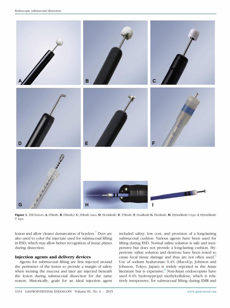
Figure 1. ESD knives. A, ITKnife. B, ITKnife2. C, ITKnife nano.D, HookKnife. E, TTKnife. F, DualKnife G, FlexKnife.H, HybridKnife I type. I, HybridKnifeT type.
Endoscopic submucosal dissection
lesion and allow clearer demarcation of borders.7 Dyes arealso used to color the injectate used for submucosal liftingin ESD, which may allow better recognition of tissue planesduring dissection.
Injection agents and delivery devicesAgents for submucosal lifting are first injected around
the perimeter of the lesion to provide a margin of safetywhen incising the mucosa and later are injected beneaththe lesion during submucosal dissection for the samereason. Historically, goals for an ideal injection agent
1314 GASTROINTESTINAL ENDOSCOPY Volume 81, No. 6 : 2015
included safety, low cost, and provision of a long-lastingsubmucosal cushion. Various agents have been used forlifting during ESD. Normal saline solution is safe and inex-pensive but does not provide a long-lasting cushion. Hy-pertonic saline solution and dextrose have been noted tocause local tissue damage and thus are not often used.8
Use of sodium hyaluronate 0.4% (MucoUp; Johnson andJohnson, Tokyo, Japan) is widely reported in the Asianliterature but is expensive.9 Non-Asian endoscopists haveused 0.4% hydroxypropyl methylcellulose, which is rela-tively inexpensive, for submucosal lifting during EMR and
www.giejournal.org
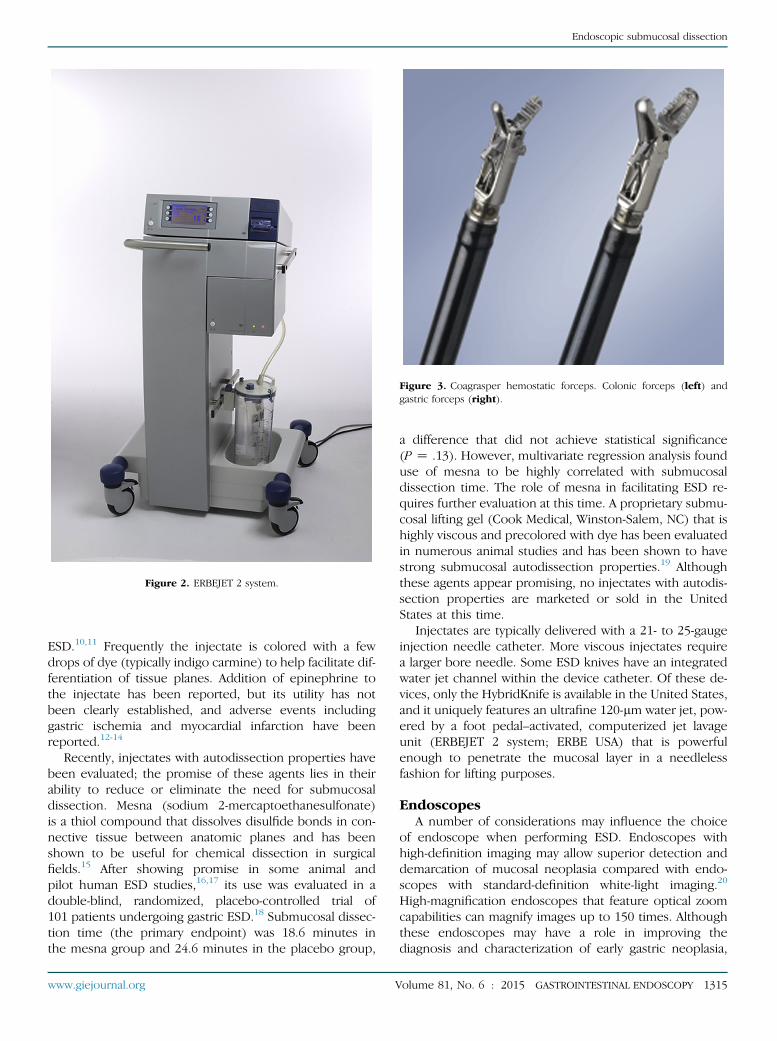
Figure 2. ERBEJET 2 system.
Figure 3. Coagrasper hemostatic forceps. Colonic forceps (left) andgastric forceps (right).
Endoscopic submucosal dissection
ESD.10,11 Frequently the injectate is colored with a fewdrops of dye (typically indigo carmine) to help facilitate dif-ferentiation of tissue planes. Addition of epinephrine tothe injectate has been reported, but its utility has notbeen clearly established, and adverse events includinggastric ischemia and myocardial infarction have beenreported.12-14
Recently, injectates with autodissection properties havebeen evaluated; the promise of these agents lies in theirability to reduce or eliminate the need for submucosaldissection. Mesna (sodium 2-mercaptoethanesulfonate)is a thiol compound that dissolves disulfide bonds in con-nective tissue between anatomic planes and has beenshown to be useful for chemical dissection in surgicalfields.15 After showing promise in some animal andpilot human ESD studies,16,17 its use was evaluated in adouble-blind, randomized, placebo-controlled trial of101 patients undergoing gastric ESD.18 Submucosal dissec-tion time (the primary endpoint) was 18.6 minutes inthe mesna group and 24.6 minutes in the placebo group,
www.giejournal.org V
a difference that did not achieve statistical significance(P Z .13). However, multivariate regression analysis founduse of mesna to be highly correlated with submucosaldissection time. The role of mesna in facilitating ESD re-quires further evaluation at this time. A proprietary submu-cosal lifting gel (Cook Medical, Winston-Salem, NC) that ishighly viscous and precolored with dye has been evaluatedin numerous animal studies and has been shown to havestrong submucosal autodissection properties.19 Althoughthese agents appear promising, no injectates with autodis-section properties are marketed or sold in the UnitedStates at this time.
Injectates are typically delivered with a 21- to 25-gaugeinjection needle catheter. More viscous injectates requirea larger bore needle. Some ESD knives have an integratedwater jet channel within the device catheter. Of these de-vices, only the HybridKnife is available in the United States,and it uniquely features an ultrafine 120-mm water jet, pow-ered by a foot pedal–activated, computerized jet lavageunit (ERBEJET 2 system; ERBE USA) that is powerfulenough to penetrate the mucosal layer in a needlelessfashion for lifting purposes.
EndoscopesA number of considerations may influence the choice
of endoscope when performing ESD. Endoscopes withhigh-definition imaging may allow superior detection anddemarcation of mucosal neoplasia compared with endo-scopes with standard-definition white-light imaging.20
High-magnification endoscopes that feature optical zoomcapabilities can magnify images up to 150 times. Althoughthese endoscopes may have a role in improving thediagnosis and characterization of early gastric neoplasia,
olume 81, No. 6 : 2015 GASTROINTESTINAL ENDOSCOPY 1315

TABLE 2. Reported settings for ERBE VIO300D for different stages of ESD24,25
ESD stage Device ESU setting
Marking Noninsulated tip ESD knife SOFT COAG, E5, 60-100 W
Precutting and circumferential incision Noninsulated tip ESD knife ENDOCUT I, E2-4, D1-3, I1-3
Submucosal dissection Any ESD knife* FORCED COAG, SWIFT COAG, DRY CUT, E2-3, 35-100 W
Hemostasis Hemostatic forceps SOFT COAG, E5, 60-100 W
ESD, endoscopic submucosal dissection; ESU, electrosurgical unit; E, effect; D, cut duration; I, cut interval.*Devices with a larger cutting surface (and thus decreased current density) may require a power setting at the upper end of the given range.
Endoscopic submucosal dissection
particularly when coupled with mucosal enhancementtechnologies (ie, narrow-band imaging),21 they offer noclear advantage during the performance of ESD. Becausebleeding commonly arises during ESD, an auxiliary waterchannel that may be used in conjunction with a peristalticflushing pump to produce a water-jet effect is a very usefulfeature for maintaining visualization and is available withmany endoscope models.22 Although ESD tools may bepassed through a 2.8-mm instrument channel, a largerdiameter “therapeutic” channel size will allow superior suc-tioning capabilities, particularly when an instrument is pre-sent in the channel. A high-definition therapeuticgastroscope with a single large (3.7 mm) instrument chan-nel (eg, GIF-1TH190; Olympus America) combines thedual advantages of superior optics and suctioning. Endo-scopes that feature 2 instrument channels allow dual-instrument use (eg, grasping forceps and an ESD knife),choice of channel with regard to optimal angle of approachfor dissection, or an open channel for suctioning if only1 instrument is being used. To overcome some of the lim-itations of flexibility inherent with standard endoscopeswhen approaching anatomically difficult lesions for ESD,a multibending endoscope has been developed (GIF-2TQ260M; Olympus) but is not marketed or sold in theUnited States.
Electrosurgical unitsESD devices apply high-frequency electrical current to
tissue by using either monopolar or bipolar circuits toachieve a desired effect. An ESU is required to powerthese devices. Several newer ESUs provide multiple fea-tures and functionality that facilitate safe and effectiveESD. Newer ESUs contain microprocessors that sensechanges in voltage due to increasing tissue impedanceduring electrosurgery and can responsively keep thevoltage constant to attain consistent and safe treatment ef-fects. Newer units also offer a wide array of electrosurgicalwaveforms that alter duty cycle and maximum peak voltageto produce a range of tissue effects. This flexibility is usefulduring ESD given the varied needs for marking, mucosalincision, submucosal dissection, and hemostasis as well asdifferent tissue characteristics in different patients (eg,fibrosis associated with a previously treated lesion). Finally,many ESUs are capable of delivering argon plasma coagula-tion, which may be useful for both marking and hemostasis.
1316 GASTROINTESTINAL ENDOSCOPY Volume 81, No. 6 : 2015
ESUs were thoroughly reviewed in a recent ASGE Technol-ogy Status Evaluation Report entitled “ElectrosurgicalGenerators.”23
Although multiple modern ESUs may be appropriatefor safe and effective ESD, specific ESU settings for thevarious stages of ESD in different anatomic locations inthe GI tract while using common ESD devices have beenmost robustly described for the ERBE VIO300D unit(ERBE USA).24,25 Some proprietary outputs of theVIO300D that are useful for ESD are briefly discussed inrelation to some basic relevant principles of electrosurgery.Specific settings for different phases of ESD are depictedin Table 2; although these settings have been reported,they are not meant to be inclusive of all useful ESD param-eters of the ERBE VIO300D unit.24,25
Peak voltage. The tissue effect of current behavesdifferently above and below a peak voltage (Vp) of 200 V.Above 200 V, a spark is generated, and an incision effectdue to cell bursting can be created even in “coagulation”modes when the current density is high due to a narrowcontact area. However, with a Vp of less than 200 V, onlydehydration and desiccation of the tissue occurs, withoutspark generation or cell bursting, thus providing a purecoagulation effect. The SOFT COAG mode of theVIO300D provides continuous current of less than 190Vp, and this setting is very useful for vessel coagulationwith hemostatic forceps (ie, Coagrasper) in the treatmentor prevention of bleeding during ESD (Table 2).24-26
Duty cycle. Duty cycle refers to the percentage of timethat the current is actually delivered. Continuously deliv-ered currents with Vp greater than 200 V are effectivelypure-cut currents. When the current is delivered in an in-terrupted manner, the tissue is allowed to cool duringthese interruptions, producing a greater coagulating effect.As an example, the FORCED COAG mode on the VIO300Dhas a duty cycle of 8%, whereas the DRY CUT mode has aduty cycle of 30%.23,25 FORCED COAG, SWIFT COAG, andDRY CUT are commonly used waveforms for the submuco-sal dissection phase of ESD (Table 2).24,25
ENDOCUT. ENDOCUT is a proprietary output modewith a 100% duty cycle that alternates a pure cutting cur-rent with the SOFT COAG mode. ENDOCUT also suppliesa higher power output to assist the successful initiationof a cut, then subsequently modifies the current in res-ponse to changing tissue impedance while providing the
www.giejournal.org

Endoscopic submucosal dissection
specified fractionation (cutting vs coagulation) of theoutput. Three parameters may be changed by the endo-scopist to alter the characteristics of the cut (speed of inci-sion: cut interval; width of incision: cut duration; andhemostatic effect: effect). ENDOCUT is frequently usedfor the precutting and circumferential incision phases ofESD (Table 2).23-25
Gas insufflationAlthough standard air insufflation has been safely used
for ESD procedures, luminal insufflation by using CO2
may hold some advantages. CO2 is absorbed across the in-testines 160 times more rapidly than nitrogen and 13 timesmore rapidly than oxygen, which are the principal gas com-ponents of air.27 As such, luminal distention with CO2
insufflation is less prolonged than with air and has beenassociated with less patient discomfort after longer endo-scopic procedures including colonoscopy28 and fewerpostprocedure admissions in a series of patients undergo-ing resection of large colonic lesions.29 The safety of CO2
insufflation during prolonged ESD procedures under mod-erate and deep sedation is well-established.30 Further, therapid reabsorption of CO2 may theoretically reduce thelikelihood of tension pneumoperitoneum developing inthe event of a perforation. CO2 was associated with areduced rate of radiographically detected pneumomedias-tinum compared with air insufflation after esophagealESD in a case-control study.31 Multiple CO2 regulatorsare available in the United States for use in endoscopicprocedures.32
EFFICACY AND COMPARISON WITHAVAILABLE TECHNOLOGIES
StomachIn large Asian series of patients with early gastric adeno-
carcinoma undergoing ESD, the rate of en bloc resectionranged from 86% to 97% and the rate of R0 (negativelateral and vertical margins) resection ranged from 88%to 93%.33-37 The rate of local recurrence generally approx-imates 1%, whereas 5-year overall survival ranges from96% to 100% and 5-year disease-specific survival rangesfrom 99% to 100%.29,31-33 Both immediate technical out-comes (eg, R0 resection) and the local recurrence rateare superior for lesions meeting Japanese Gastric CancerAssociation criteria38 (differentiated mucosal cancer, !2cm, without ulceration) than the expanded National Can-cer Center criteria,39 but there have been no differencesin mortality.33,35-39
Two meta-analyses evaluated ESD versus EMR forthe treatment of early gastric cancer.40,41 In these ana-lyses, ESD was associated with higher rates of en blocresection (92% vs 52%) and R0 resection (82%-92% vs42%-43%) than EMR, as well as a lower rate of local recur-rence (0.8% vs 5.0%-6.4%) than EMR. All-cause mortality at
www.giejournal.org V
mean follow-up durations of 36 to 43 months did not differbetween patients treated with ESD versus EMR for earlygastric cancer.
There are no data directly comparing modernsurgical resections and ESD for early gastric cancer. Itwas actually a retrospective review of 5265 Japanese pa-tients who had undergone gastrectomy and lymph nodedissection that was instrumental in determining the tumorfeatures that were associated with no nodal metastasisand thus appropriate for local (ie, endoscopic) treat-ment.42 A Japanese multicenter evaluation of laparoscopicgastrectomy (primarily distal gastrectomy) for earlygastric cancer reported 5-year disease-free survival ratesof 99.8% for stage T1a disease and 98.7% for stage T1bdisease.43
Colon and rectumESD in the colon has generally been used for laterally
spreading tumors larger than 2 cm in diameter. In a sys-tematic review of 22 colorectal ESD studies with morethan 2800 patients, the most common lesion site was therectum (44%) and the median of the mean tumor sizewas 32 mm.44 The histologic classification of resected le-sions predominantly included adenoma (median rate43%), intramucosal adenocarcinoma (44%), and submuco-sal adenocarcinoma (11%). In this review, the summary es-timate for an R0 resection rate was 88%. The 2010guidelines from the Japanese Society for Cancer of theColon and Rectum define R0 resections as curative whennone of the following are present: depth of submucosalinvasion greater than 1000 mm, lymphovascular invasion,poor differentiation, or higher grade (2 or 3) tumorbudding at the site of deepest invasion.45 However, ameta-analysis reported that the incidence of lymphaticmetastasis is 1.9% even when these criteria are satisfiedand also highlighted the limited quality and quantity ofthe source data.46
In 4 large retrospective series that compared conven-tional endoscopic resection (including lift polypectomyand cap-based EMR [cEMR]) with ESD for colorectal neo-plasms larger than 2 cm, the lesions in the ESD groupwere generally larger (29-37 mm vs 22-28 mm) and ESDwas associated with a higher rate of en bloc resection(84%-95% vs 33%-57%).47-50 Data regarding the curativeresection rate for ESD versus conventional polypectomy/EMR are mixed, with important caveats being that larger le-sions were being resected in the ESD groups in thesestudies and that lateral margins cannot be accurately as-sessed with piecemeal polypectomy/EMR.47,49 Over meanfollow-up durations ranging from 17 to 26 months, thelocal recurrence rate for conventional polypectomy/EMRranged from 12% to 26% compared with 0% to 2% forESD.48-50
With regard to nonepithelial colorectal neoplasia, ESDhas been evaluated for the endoscopic treatment of rectalcarcinoid tumors. Although ESD is effective in this setting,
olume 81, No. 6 : 2015 GASTROINTESTINAL ENDOSCOPY 1317
Endoscopic submucosal dissection
data are mixed as to whether it provides any benefit overfaster procedures such as cEMR or cap-and-band ligationEMR, particularly as the mean lesion diameter in many se-ries is less than 10 mm.51-53
The data comparing ESD with surgical treatmentsfor colorectal neoplasia are also retrospective and limited.A single-center South Korean retrospective seriescompared 63 patients who underwent either ESD ortransanal endoscopic microsurgery (TEM) for Tis or T1rectal cancers.54 Patients treated with ESD (n Z 30) andTEM (n Z 33) had similar rates of R0 resections (97%)and curative resections (77% and 79%, respectively). ESDwas associated with a shorter procedure duration and hos-pital stay than TEM. Of patients who had a curative resec-tion, there were no cases of local recurrence or distantmetastasis in either group in approximately 2 years offollow-up. A meta-analysis combined data from 21 single-arm case series (11 ESD and 10 TEM) evaluating outcomesin the treatment of rectal neoplasms larger than 2 cm.55 Inthis analysis, TEM was associated with higher rates of enbloc resection (99% vs 88%, P! .001) and R0 resection(89% vs 75%, P! .001) than ESD. However, a greater pro-portion of lesions in the ESD group were cancerscompared with the TEM group, which comprised mostlyadenomas. Last, there was a trend toward fewer local recur-rences in the ESD group than in the TEM group (2.6% vs5.2%, P Z .07), but this likely reflects the standard practiceof referral for further oncologic surgery after noncurativeESD.
A large retrospective series from the National CancerCenter in Tokyo compared outcomes in 589 patientswith T1 colorectal cancers who underwent either ESD(n Z 297) for endoscopically predicted mucosal or super-ficial submucosal neoplasms or laparoscopy-assisted colo-rectal surgery including lymphadenectomy ([LAC], n Z292) for predicted deep submucosal cancers.56 Impor-tantly, this study included a large number of patientswith colonic (nonrectal) lesions, including 185 who under-went ESD and 243 who underwent LAC, although unfortu-nately most outcomes data are reported for all colorectalpatients and not separately for the colon-only subgroups.In this study, ESD was associated with a shorter proceduretime and hospital stay than LAC. The en bloc and curativeresection rates with ESD were 87% and 80%, respectively,with surgical referral for patients with noncurative ESDresections. The 3-year overall survival rate exceeded 99%in both the ESD and LAC groups.
EsophagusEarly adenocarcinoma of the gastroesophageal junction
represented a logical extension of gastric ESD techniquesin a more technically challenging anatomic site. In 4 seriesthat retrospectively evaluated ESD for non-Barrett’s gastro-esophageal junction adenocarcinoma, the rate of en blocresection was 100%, with curative resection rates (definedsimilarly to criteria for colonic lesions [see previously],
1318 GASTROINTESTINAL ENDOSCOPY Volume 81, No. 6 : 2015
except O500 mm of submucosal invasion deemed noncur-ative) of 68% to 79%.57-60 Patients with noncurative resec-tions were typically managed with esophagectomy andLAC, and patients with curative ESD resections had no localrecurrences or metastatic cancer detected in mean follow-up durations of 15 to 30 months in 2 of the series.57,58
The other 2 series had longer follow-up available, andboth reported a 5-year disease-specific survival rate of100% for those with curative resections.59,60
A German study prospectively enrolled 30 patients withBarrett’s esophagus featuring either intramucosal canceror high-grade dysplasia for treatment with ESD.61 Althougha 90% en bloc resection rate was attained, an R0 resectionwas achieved in only 38% of patients. The authors specifiedthat any degree of mucosal dysplasia at a lateral marginprecluded R0 classification, and this was the predominantreason for the low R0 resection rate. However, over a me-dian follow-up duration of 17 months that included surveil-lance endoscopies with biopsies, 96% of patients werefound to be free of any neoplasia, suggesting that therim of coagulation necrosis resulting from ESD may haveeradicated marginal dysplasia in many of these patients.
Several studies evaluating esophageal ESD for earlysquamous cell carcinoma (SCC) have included a compar-ator EMR arm. A retrospective cohort study of 300 patientsundergoing ESD or EMR for early SCC excluded all patientsfound to have histologic evidence of submucosal inva-sion.62 In this study, the rate of en bloc resection forESD was 100% (mean lesion size 30 mm) compared with53% for EMR (mean lesion size 20 mm), and the rate oflocal recurrence was 1% in the ESD group and 10% inthe EMR group. However, there was no difference in sur-vival in more than 4 years follow-up. Similarly, a retrospec-tive series of 70 patients with SCCs 2 cm or larger treatedwith cEMR or ESD reported local recurrence rates of0 of 34 (0%) for lesions resected en bloc, 4 of 27 (15%)for piecemeal resections with 2 to 4 pieces, and 8 of 17(47%) for resections with 5 or more pieces.63 However,in a retrospective study with 171 SCCs 2 cm or smallerstratified by size, for lesions smaller than 15 mm in diam-eter treated with cEMR or ESD, there were equivalent ratesof en bloc resection (100%) and no local recurrences, witha significantly shorter mean procedure time in the cEMRgroup (21 minutes vs 64 minutes, P! .01).64
T1a SCC that involves the muscularis mucosa poses asubstantial risk (w9%) for lymph node metastasis that ap-pears to be greater than esophageal adenocarcinoma ofthe same depth.62,65,66 In a large single-operator series, pa-tients undergoing ESD resection of T1a SCCs not involvingthe muscularis mucosa (confined to the epithelium andlamina propria) had a 5-year overall survival rate of82%.67 There are no data directly comparing ESD with sur-gical resection for early esophageal cancer. In a largeGerman registry, the 5-year survival rate for patients withsurgically resected T1a cancer was 78%, with notable differ-ences between adenocarcinoma (91%) and SCC (62%).68
www.giejournal.org
Endoscopic submucosal dissection
DuodenumThe use of ESD in the duodenum for sessile adenomas,
early carcinomas, and carcinoid tumors has been des-cribed, but available data comprise only case reports andsmall case series.69-71
SAFETY
BleedingIntraprocedural bleeding is a common and expected
event during ESD. Typically, minor oozing from small ves-sels can be treated with coagulation current deliveredthrough the ESD knife, whereas more significant activebleeding is treated with hemostatic forceps. Efforts arealso made to identify larger nonbleeding submucosal ves-sels during the dissection for prophylactic coagulationwith hemostatic forceps.26 Although rare, severe intrapro-cedural bleeding that cannot be managed endoscopicallyhas been described. In a large South Korean series of1244 patients with early gastric cancer, 6 severe bleedingevents occurred that required urgent surgery (wedgeresection or laparoscopic gastrectomy).35
Delayed bleeding after ESD is more common in gastricESD than colorectal or esophageal sites. Although a meta-analysis of gastric ESD studies reported a 4.5% delayedbleeding rate,40 many individual studies from experiencedcenters have described higher rates, as high as 15.6%.34
Lesion size larger than 40 mm and resumption of oral an-tithrombotic therapy have been identified as risk factorsfor delayed bleeding after gastric ESD.72 Antisecretory ther-apy is routinely used to promote healing of ESD-related ul-cers, and a meta-analysis of 6 studies reported a reducedincidence of delayed bleeding after gastric ESD in patientstreated with a proton pump inhibitor compared with anH2 receptor antagonist (5.4% vs 10.5%; odds ratio 0.41;95% confidence interval, 0.20–0.85).73 Further, several ran-domized, controlled trials have demonstrated that thecombination of a mucosal protective agent and a protonpump inhibitor results in faster healing of gastric ESD ul-cers than a proton pump inhibitor alone, although animpact on delayed bleeding has not been shown.74-76 In1 series, 76% of delayed bleeds occurred within 24 hoursof ESD, whereas the remaining 24% occurred 2 to 15days after the procedure.77 Given this, the practice of per-forming a next-day “second look” endoscopy is common,but has not been clearly shown to improve outcomesincluding delayed bleeding.78 Delayed bleeding after non-gastric ESD is less common and has been reported in 0%to 5.2% of patients in series of esophageal ESD79 and 2%of patients in a meta-analysis of colorectal ESD.44
PerforationThe rate of perforation in meta-analyses of gastric ESD
is approximately 4.5%,40,41 and in a meta-analysis of colo-rectal ESD, it was 4.8%.44 In a review of esophageal ESD
www.giejournal.org V
adverse events, perforation rates of 0% to 10% are re-ported; a review of the data in these series suggests apooled perforation rate of 19 of 816 (2.3%).79 Fortunately,almost all perforations are recognized intraprocedurallyand are amenable to clip closure. A report on 117 consec-utive EMR/ESD gastric perforations between 1994 and 2004at a large Japanese cancer center described successful clipclosure and nonoperative management in 115 of 117 pa-tients (98%), with the remaining 2 patients needing urgentsurgery.80 Primary clip closure was used for defects 1 cmand smaller, and an “omental patch” method was usedfor larger defects, whereby the greater or lesser omentumis suctioned into the defect, and multiple clips are used tosecure the omentum to the gastric wall circumferentiallyaround the perforation. Patients in this series were initiallymanaged with nasogastric suction for 3 days, total paren-teral nutrition for 9 days, and a second-generation cephalo-sporin; a water-soluble contrast study was used to guidethe timing of return to oral intake. Later in the authors’experience, these timelines were shortened with nocompromise in outcomes. Use of other devices for suc-cessful closure of ESD-associated perforations includingover-the-scope clip(s) has also been reported.81
High rates of successful clip closure and conservativemanagement of colorectal ESD perforations have alsobeen reported. In a large series of 816 ESD resectionsof colorectal lesions, 16 perforations occurred (2%), 14 ofwhich were managed nonoperatively and 2 (0.2%) whichrequired urgent surgery.47 Although the majority of esoph-ageal perforations can also be managed with endoscopicclosure and conservative measures, in some patients life-threatening mediastinitis can develop, requiring urgentsurgery.62,79,82 In a small subset of patients, mediastinalemphysema will develop in the absence of a recognizedperforation, and conservative management also appearsto benefit these patients.62
A small subset of patients with perforations present ina delayed manner and have a less favorable clinical course.A Japanese group reported 6 delayed perforations in1159 patients (0.5%) after ESD for early gastric cancer.83
All presented 10 to 24 hours after the procedure with clin-ical signs and symptoms of peritonitis, and 5 of 6 requiredemergency surgery.
StricturePost-ESD esophageal stricture is generally defined as a
narrowing through which a standard gastroscope cannotbe advanced. Strictures develop in 12% to 17% of patientsafter esophageal ESD, with risk factors including thecircumference and length of the resection.62,84-86 ESD re-sections encompassing more than 75% of the circumfer-ence of the esophagus are at highest risk of stricturedevelopment.82 Due to the relatively high frequency ofthis adverse event, a number of strategies aimed at pre-venting and/or treating post-ESD esophageal stricturehave been used, including prophylactic serial dilation,
olume 81, No. 6 : 2015 GASTROINTESTINAL ENDOSCOPY 1319
Endoscopic submucosal dissection
intralesional steroid injection or topical steroid gel applica-tion, radial electroincision, and prophylactic placement offully covered self-expandable metal stents.79,87,88 Endo-scopic transplantation of tissue-engineered autologousoral mucosal epithelial cell sheets89 and resected gastricmucosa90 have also been reported for prophylaxis ofpost-ESD esophageal stricture, but remain experimental.Stricture development after gastric ESD is uncommonand anatomically limited to sites of relative luminal narrow-ing. In a review of 2011 gastric ESDs at a single Japanesecenter, strictures occurred in just 15 patients (0.7%) over-all, exclusively in resections involving the cardia (7/41,17%) or prepyloric antrum (8/115, 7%).91 Stricture devel-opment after colorectal ESD has not been reported.
EASE OF USE
Need for specialized trainingESD is a technically demanding procedure that requires
substantial training to achieve competence; inadequatetraining compromises both patient safety and technicaloutcomes. Two series reported on the outcomes of partic-ipants attending 2- to 3-day ESD courses that featuredhands-on stations in which live pigs were used.92,93 Inboth series, participant demographics indicated a meanof more than 10 years of clinical endoscopic experience,with some participants having limited previous experiencewith ESD. However, perforation rates of 22% to 63% wereobserved during gastric and esophageal ESD even withthese experienced endoscopists, suggesting that ESDposes significant risk when undertaken by an operatorinadequately trained in ESD.
Training modelsIn Japan, the training model is relatively established: af-
ter obtaining initial didactic training in ESD, learnersobserve experts for a variable number of procedures,then assist in a variable number of procedures beforefinally undertaking ESD on less technically challenging le-sions (generally in the distal stomach) under expert super-vision. In a survey of Japanese experts, observation of20 procedures and acting as an assistant in 5 procedureswere the most common responses for the minimumexperience needed before beginning ESD.94 However,this model is difficult to establish in Western countries,where both early gastric cancer and ESD experts are rare.As a result, the best training paradigm for Western learnersis likely to differ. Although animal models are suggestedbut optional for learners in Japan, they are essentiallymandatory for Western learners and represent the nextstep after initial didactic learning.
Ex vivo and in vivo porcine models for gastric ESD arewell studied and closely resemble human anatomy. Incontrast, the porcine colon is difficult to cleanse and isthinner walled and more mobile than the human colon;
1320 GASTROINTESTINAL ENDOSCOPY Volume 81, No. 6 : 2015
as such, the utility of the porcine colon model for ESDtraining is unclear. It has been suggested that 10 resectionsin an ex vivo porcine gastric model represent an adequateexperience to justify a transition to a live pig model, whichprovides a more realistic experience including the pres-ence of peristalsis, intraluminal secretions, and bleeding.95
In a study of 2 novice learners who each performed gastricESD on 60 lesions each in an ex vivo porcine model, thetotal resection time, en bloc resection rate, and perforationrate all improved for both endoscopists when the last 30resections were compared with the first 30 resections.96
The authors calculated that the cost to train an endoscopistwith 30 ex vivo gastric porcine procedures (assuming 6 le-sions per stomach) would be US$8410, given the cost ofthe simulator used, the gastric specimens, and the dispos-able devices used. In comparison, the authors calculatedthat the cost to train an endoscopist with 30 in vivo gastricporcine procedures (also assuming 6 lesions per stomach)would be about US$16,000.
Although observerships can be logistically challenging,some Western endoscopists have undertaken observer-ships in high-volume ESD centers in Asia, typically for 2to 5 weeks. One American endoscopist had performed 29resections in an ex vivo gastric porcine model beforeobserving 43 ESDs over 5 weeks at an expert center inJapan. The endoscopist’s next 9 resections in the gastricporcine model took 32.7 � 15.0 minutes to complete,which was significantly shorter than the mean duration ofhis last 9 resections before the observership (61.0 � 7.4 mi-nutes, P Z .001).97 As with other technically demandingprocedures, once the skill set for ESD is learned, it mustbe maintained over time by performing an ample volumeof cases and/or attending courses.
Lesion selection is important to maximize the chanceof a successful outcome in human patients early in theendoscopist’s ESD experience. Gastric antral lesions areeasily accessible, have a favorable wall thickness, andallow a stable endoscope position both forward-viewingand in retroflexion; for these reasons, smaller antrallesions are optimal for ESD learners. In 2 studies thateach evaluated the outcomes of the first 20 to 30 gastricESDs performed by Japanese trainees, risk factors fornonself-completion included size larger than 3 cm andlocation other than the antrum.98,99 Similarly, experts sug-gest that rectal ESD is anatomically favorable comparedwith colonic ESD and that smaller rectal lesions may be areasonable early target for Western endoscopists.100
Indeed, in some series of colorectal ESD performed bynovice/trainee endoscopists, all perforations occurred incolonic (nonrectal) cases.101,102
Learning curvesThere is no single “learning curve” for ESD, but rather
multiple learning curves that vary based on lesion charac-teristics (eg, anatomic site, size) and outcome of interest(eg, total procedure time, R0 resection rate, adverse event
www.giejournal.org

Endoscopic submucosal dissection
rate). Further, learners with abundant experience assistingin human procedures and/or performing resections in ani-mal models will enter at a higher point on most learningcurves and thus experience measurable improvements ata less rapid pace than more novice operators.98 Finally,as endoscopists become more comfortable with ESD,they tend to accept more challenging cases with regardto lesion size and location, and this may distort the upperend of a learning curve.103 With these caveats stated, manystudies reflect a breakpoint that occurs between 20 and50 human procedures, during which significant impro-vements across multiple outcomes can be demonstrated,irrespective of anatomic site.100,104-108 In centers withESD expertise, this level of experience may also corre-spond to graduating to performing unsupervised ESD.However, most ESD training studies incorporate fewerthan 50 procedures per endoscopist, and outcomes atthe conclusion of these studies for the most critical end-points (ie, R0 resection) still fall short of results achievedby providers at expert Japanese centers, indicating thatfurther improvement in operator skill occurs far out onthe learning curve.
Logistical issuesProcedure duration. Although high-volume Asian
centers have reported mean procedure times for gastricESD as short as 25 minutes,35 it should be recognizedthat ESD is a lengthy procedure for nonexperts. In aFrench multicenter survey, 188 ESD procedures (primarilygastric and rectal) from 16 centers were self-reported; themedian procedure duration was 105 minutes.109 In 2 Euro-pean series of 60 and 76 colorectal ESDs procedures per-formed early in these studies averaged more than 3hours in duration, whereas median procedure durationsnear the end of the studies varied from 70 to 136 mi-nutes.100,107 Finally, a relatively experienced German groupreported a median procedure duration of 74 minutes for29 gastric ESDs.110 As such, adequate time and resourcesmust be allotted before undertaking an ESD.
Sedation. Safe sedation for patients undergoing upperGI ESD has been described by using a range of levelsincluding moderate sedation (eg, by using midazolam),deep sedation (eg, by using propofol or dexmedetomi-dine), or general anesthesia.111-113 Retrospective data ongastric ESD in South Korea demonstrated higher en blocresection rates and shorter procedure durations inpatients cared for by an anesthesiologist.114 Given theduration of the procedure, need for fine-motor maneuvers,and potential for reflux and aspiration of secretions orblood, strong consideration should be given to using gen-eral anesthesia for upper GI lesions, particularly for endo-scopists who have limited experience in performing ESD.In contrast, moderate or deep sedation is generally suffi-cient for colorectal ESD; conscious sedation may facilitatechanges in patient position that beneficially use gravityfor countertraction on the lesion.100,107
www.giejournal.org V
FINANCIAL CONSIDERATIONS
ReimbursementDespite clinical benefits for patients, ESD remains
time-consuming and is not adequately reimbursed at thepresent time. There is no unique Current ProceduralTerminology (CPT) code for ESD. In 2014, new codes foresophagoscopy with EMR (43211) and EGD with EMR(43254) were introduced, and in 2015, there will be coun-terpart codes for colonoscopy. However, the several mo-dalities that are bundled in the EMR code (submucosalinjection, snare resection, biopsy if performed, control ofbleeding if performed) are not the key aspects of ESD,and CPT instructs that codes that are only approximateare not appropriate to report. The best 2 choices at presentwould be to report a snare polypectomy service (eg, 43251during EGD or 45385 during colonoscopy) and to reportan unlisted code to describe the remainder of thework or just to report an unlisted procedure code (43499for gastric, 45999 rectum, 44799 small intestine, 45399new code for unlisted procedure, colon) with supportingdocumentation to seek appropriate reimbursement. Inthis case, a cover letter submitted with the claim that ex-plains the nature of the procedure, equipment required(equipment invoice copies are helpful), estimatedpractice cost, and a comparison of physician work (time,intensity, risk) with other endoscopic services for whichthe payer has an established value should be included tothe payer. A center performing this procedure frequentlymight find it worthwhile to arrange a personal discussionbetween an endoscopist and the medical director of largerpayers to facilitate coverage and appropriate pricing.The dedicated ESD devices do add to the facility cost ofthe procedure largely without added reimbursement.
Device and equipment costsList prices for dedicated ESD devices are shown in
Table 1. The cost of a transparent distal attachment (cap)is approximately $30. The cost for several ESUs appro-priate for use during ESD is available in the ASGE Technol-ogy Status Evaluation report entitled “ElectrosurgicalGenerators.”23 The list price for the ERBEJET 2 system isUS$45,500.
AREAS FOR FUTURE RESEARCH
A key deficit that future studies should address is thelack of high-quality, randomized, controlled trial–leveldata comparing ESD with competing procedures such asEMR, TEM, and laparoscopic surgical resections. Studiesshould also address device-specific outcomes data. Al-though they are emerging, additional outcomes datafrom Western endoscopists would be useful. Human dataon newer autodissecting injectates are needed. If an agentcould be developed that markedly reduces the need for
olume 81, No. 6 : 2015 GASTROINTESTINAL ENDOSCOPY 1321
Endoscopic submucosal dissection
submucosal dissection, this would have the potential tosignificantly modify both the technical difficulty and therisk of ESD. Early pilot studies are ongoing to develop tech-niques that reduce the risks of postoperative adverseevents after ESD (eg, delayed bleeding, perforation)including endoscopic suturing of ESD defects115 and theuse of polyglycolic acid sheets and fibrin glue to “shield”defects.116 ESD techniques have been applied to the endo-scopic resection of neoplasia arising in the submucosa andmuscularis propria, including full-thickness resections.117
In some series, these deeper resections have been facili-tated by the variant technique of endoscopic submucosaltunnel dissection.118 These techniques remain in develop-ment and require further study.
SUMMARY
ESD is an established effective treatment modality forpremalignant and early-stage malignant lesions of thestomach, esophagus, and colorectum. Compared withEMR, ESD is generally associated with higher rates of enbloc, R0, and curative resections and a lower rate of localrecurrence. Oncologic outcomes with ESD compare favor-ably with competing surgical interventions, and ESD alsoserves as an excellent T-staging tool to identify noncurativeresections that will require further treatment. ESD is tech-nically demanding and has a higher rate of adverse eventsthan most endoscopic procedures including EMR. As such,sufficient training is critical to ensure safe conduct andhigh-quality resections. A standardized training model forWestern endoscopists has not been clearly established,but will be self-directed and include courses, animal modeltraining, and optimally an observership at an expert center.Numerous dedicated ESD devices are now available in theUnited States from different manufacturers. Although theuse of ESD in the United States is increasing, issues relatedto technical difficulty, limited training opportunities andmentors, risk of adverse events, long procedure duration,and suboptimal reimbursement may limit ESD adoptionin the United States to a modest number of academicreferral centers for the foreseeable future.
DISCLOSURE
Dr Hwang has received research support fromOlympus. Dr Abu Dayyeh has received research supportfrom Apollo Endoscopy, Aspire Bariatrics, and GI Dy-namics. Dr Konda has received honoraria from MaunaKea Technologies. All other authors disclosed no financialrelationships relevant to this article.
Abbreviations: cEMR, cap-based EMR; ESD, endoscopic submucosaldissection; ESU, electrosurgical unit; LAC, lymphadenectomy; SCC,squamous cell carcinoma; TEM, transanal endoscopic microsurgery;Vp, peak voltage.
1322 GASTROINTESTINAL ENDOSCOPY Volume 81, No. 6 : 2015
REFERENCES
1. Hirao M, Masuda K, Asanuma T, et al. Endoscopic resection of earlygastric cancer and other tumors with local injection of hypertonic sa-line-epinephrine. Gastrointest Endosc 1988;34:264-9.
2. Gotoda T, Kondo H, Ono H, et al. A new endoscopic mucosal resec-tion procedure using an insulation-tipped electrosurgical knife forrectal flat lesions: report of two cases. Gastrointest Endosc 1999;50:560-3.
3. Inoue H, Minami H, Kobayashi Y, et al. Peroral endoscopic myotomy(POEM) for esophageal achalasia. Endoscopy 2010;42:265-71.
4. Oyama T. Counter traction makes endoscopic submucosal dissectioneasier. Clin Endosc 2012;45:375-8.
5. Gotoda T, Oda I, Tamakawa K, et al. Prospective clinical trial ofmagnetic-anchor-guided endoscopic submucosal dissection forlarge early gastric cancer (with videos). Gastrointest Endosc2009;69:10-5.
6. Sakurazawa N, Kato S, Miyashita M, et al. An innovative technique forendoscopic submucosal dissection of early gastric cancer using a newspring device. Endoscopy 2009;41:929-33.
7. Mönkemüller K, Wilcox CM. Interventional chromoendoscopy. Gastro-intest Endosc 2013;78:346-50.
8. Fujishiro M, Yahagi N, Kashimura K, et al. Tissue damage of differentsubmucosal injection solutions for EMR. Gastrointest Endosc 2005;62:933-42.
9. Yamamoto H, Yahagi N, Oyama T, et al. Usefulness and safety of 0.4%sodium hyaluronate solution as a submucosal fluid “cushion” in endo-scopic resection for gastric neoplasms: a prospective multicenter trial.Gastrointest Endosc 2008;67:830-9.
10. Arantes V, Albuquerque W, Benfica E, et al. Submucosal injection of0.4% hydroxypropyl methylcellulose facilitates endoscopic mucosalresection of early gastrointestinal tumors. J Clin Gastroenterol2010;44:615-9.
11. Draganov PV, Gotoda T, Chavalitdhamrong D, et al. Techniques ofendoscopic submucosal dissection: application for the Western endo-scopist? Gastrointest Endosc 2013;78:677-88.
12. Lee SH, Chung IK, Kim SJ, et al. Comparison of postpolypectomybleeding between epinephrine and saline submucosal injectionfor large colon polyps by conventional polypectomy: a prospectiverandomized, multicenter study. World J Gastroenterol 2007;13:2973-7.
13. Probst A, Maerkl B, Bittinger M, et al. Gastric ischemia following endo-scopic submucosal dissection of early gastric cancer. Gastric Cancer2010;13:58-61.
14. Kim HH, Park MI, Park SJ, et al. Myocardial infarction thought to beprovoked by local epinephrine injection during endoscopic submu-cosal dissection. J Clin Med Res 2011;3:143-6.
15. Benassi L, Lopopolo G, Pazzoni F, et al. Chemically assisted dissectionof tissues: an interesting support in abdominal myomectomy. J AmColl Surg 2000;191:65-9.
16. Sumiyama K, Gostout CJ, Rajan E, et al. Chemically assisted endo-scopic mechanical submucosal dissection by using mesna. Gastroint-est Endosc 2008;67:534-8.
17. Sumiyama K, Tajiri H, Gostout CJ, et al. Chemically assisted submuco-sal injection facilitates endoscopic submucosal dissection of gastricneoplasms. Endoscopy 2010;42:627-32.
18. Sumiyama K, Toyoizumi H, Ohya TR, et al. A double-blind, block-randomized, placebo-controlled trial to identify the chemical assis-tance effect of mesna submucosal injection for gastric endoscopicsubmucosal dissection. Gastrointest Endosc 2014;79:756-64.
19. Khashab MA, Saxena P, Sharaiha RZ, et al. A novel submucosal gelpermits simple and efficient gastric endoscopic submucosal dissec-tion. Gastroenterology 2013;144:505-7.
20. Toyoizumi H, Kaise M, Arakawa H, et al. Ultrathin endoscopy versushigh-resolution endoscopy for diagnosing superficial gastricneoplasia: a prospective comparative study. Gastrointest Endosc2009;70:240-5.
www.giejournal.org
Endoscopic submucosal dissection
21. Ezoe Y, Muto M, Uedo N, et al. Magnifying narrowband imaging ismore accurate than conventional white-light imaging in diagnosisof gastric mucosal cancer. Gastroenterology 2011;141:2017-25.
22. Tatsumi K, Uedo N, Ishihara R, et al. A water-jet videoendoscope mayreduce operation time of endoscopic submucosal dissection for earlygastric cancer. Dig Dis Sci 2012;57:2122-9.
23. ASGE Technology Committee; Tokar JL, Barth BA, Banerjee S, et al.Electrosurgical generators. Gastrointest Endosc 2013;78:197-208.
24. Matsui N, Akahoshi K, Nakamura K, Ihara E, Kita H. Endoscopic submu-cosal dissection for removal of superficial gastrointestinal neoplasms:a technical review. World J Gastrointest Endosc 2012;4:123-36.
25. Morita Y. Electrocautery for ESD: settings of the electrical surgical unitVIO300D. Gastrointest Endosc Clin N Am 2014;24:183-9.
26. Yoshida N, Naito Y, Kugai M, et al. Efficient hemostatic method forendoscopic submucosal dissection of colorectal tumors. World J Gas-troenterol 2010;16:4180-6.
27. Saltzman HA, Siecker HO. Intestinal response to changing gaseousenvironments: normobaric and hyperbaric observations. Ann N YAcad Sci 1968;150:31-9.
28. Wu J, Hu B. The role of carbon dioxide insufflation in colonoscopy: asystematic review and meta-analysis. Endoscopy 2012;44:128-36.
29. Bassan MS, Holt B, Moss A, et al. Carbon dioxide insufflation reducesnumber of postprocedure admissions after endoscopic resection oflarge colonic lesions: a prospective cohort study. Gastrointest Endosc2013;77:90-5.
30. Takano A, Kobayashi M, Takeuchi M, et al. Capnographic monitoringduring endoscopic submucosal dissection with patients under deepsedation: a prospective, crossover trial of air and carbon dioxide in-sufflations. Digestion 2011;84:193-8.
31. Maeda Y, Hirasawa D, Fujita N, et al. A pilot study to assess medias-tinal emphysema after esophageal endoscopic submucosal dissec-tion with carbon dioxide insufflation. Endoscopy 2012;44:565-71.
32. ASGE Technology Committee; Maple JT, Banerjee S, Barth BA, et al.Methods of luminal distention for colonoscopy. Gastrointest Endosc2013;77:519-25.
33. Tanabe S, Ishido K, Higuchi K, et al. Long-term outcomes of endo-scopic submucosal dissection for early gastric cancer: a retrospectivecomparison with conventional endoscopic resection in a single cen-ter. Gastric Cancer 2014;27:130-6.
34. Chung IK, Lee JH, Lee SH, et al. Therapeutic outcomes in 1000 casesof endoscopic submucosal dissection for early gastric neoplasms:Korean ESD Study Group multicenter study. Gastrointest Endosc2009;69:1228-35.
35. Ahn JY, Jung HY, Choi KD, et al. Endoscopic and oncologic outcomesafter endoscopic resection for early gastric cancer: 1370 cases of ab-solute and extended indications. Gastrointest Endosc 2011;74:485-93.
36. Choi MK, Kim GH, Park DY, et al. Long-term outcomes of endoscopicsubmucosal dissection for early gastric cancer: a single-center expe-rience. Surg Endosc 2013;27:4250-8.
37. Goto O, Fujishiro M, Kodashima S, et al. Outcomes of endoscopicsubmucosal dissection for early gastric cancer with special referenceto validation for curability criteria. Endoscopy 2009;41:118-22.
38. Shimada Y. JGCA (The Japan Gastric Cancer Association). Gastric can-cer treatment guidelines. Jpn J Clin Oncol 2004;34:58.
39. Gotoda T, Iwasaki M, Kusano C, et al. Endoscopic resection of earlygastric cancer treated by guideline and expanded National CancerCentre criteria. Br J Surg 2010;97:868-71.
40. Park YM, Cho E, Kang HY, et al. The effectiveness and safety of endo-scopic submucosal dissection compared with endoscopic mucosalresection for early gastric cancer: a systematic review and metaanal-ysis. Surg Endosc 2011;25:2666-77.
41. Lian J, Chen S, Zhang Y, et al. A meta-analysis of endoscopic submu-cosal dissection and EMR for early gastric cancer. Gastrointest Endosc2012;76:763-70.
42. Gotoda T, Yanagisawa A, Sasako M, et al. Incidence of lymph nodemetastasis from early gastric cancer: estimation with a large numberof cases at two large centers. Gastric Cancer 2000;3:219-25.
www.giejournal.org V
43. Kitano S, Shiraishi N, Uyama I, et al; Japanese Laparoscopic SurgeryStudy Group. A multicenter study on oncologic outcome of laparo-scopic gastrectomy for early cancer in Japan. Ann Surg 2007;245:68-72.
44. Repici A, Hassan C, De Paula Pessoa D, et al. Efficacy and safety ofendoscopic submucosal dissection for colorectal neoplasia: a system-atic review. Endoscopy 2012;44:137-50.
45. Watanabe T, Itabashi M, Shimada Y, et al. Japanese Society for Cancerof the Colon and Rectum (JSCCR) guidelines 2010 for the treatmentof colorectal cancer. Int J Clin Oncol 2012;17:1-29.
46. Mou S, Soetikno R, Shimoda T, et al. Pathologic predictive factors forlymph node metastasis in submucosal invasive (T1) colorectal cancer:a systematic review and meta-analysis. Surg Endosc 2013;27:2692-703.
47. Nakajima T, Saito Y, Tanaka S, et al. Current status of endoscopicresection strategy for large, early colorectal neoplasia in Japan.Surg Endosc 2013;27:3262-70.
48. Saito Y, Fukuzawa M, Matsuda T, et al. Clinical outcome of endoscopicsubmucosal dissection versus endoscopic mucosal resection of largecolorectal tumors as determined by curative resection. Surg Endosc2010;24:343-52.
49. Lee EJ, Lee JB, Lee SH, et al. Endoscopic treatment of large colorectaltumors: comparison of endoscopic mucosal resection, endoscopicmucosal resection-precutting, and endoscopic submucosal dissec-tion. Surg Endosc 2012;26:2220-30.
50. Terasaki M, Tanaka S, Oka S, et al. Clinical outcomes of endoscopicsubmucosal dissection and endoscopic mucosal resection for laterallyspreading tumors larger than 20 mm. J Gastroenterol Hepatol2012;27:734-40.
51. Zhong DD, Shao LM, Cai JT. Endoscopic mucosal resection vs endo-scopic submucosal dissection for rectal carcinoid tumours: a system-atic review and meta-analysis. Colorectal Dis 2013;15:283-91.
52. Kim KM, Eo SJ, Shim SG, et al. Treatment outcomes according toendoscopic treatment modalities for rectal carcinoid tumors. ClinRes Hepatol Gastroenterol 2013;37:275-82.
53. Zhao ZF, Zhang N, Ma SR, et al. A comparative study on endoscopytreatment in rectal carcinoid tumors. Surg Laparosc Endosc PercutanTech 2012;22:260-3.
54. Park SU, Min YW, Shin JU, et al. Endoscopic submucosal dissection ortransanal endoscopic microsurgery for nonpolypoid rectal high gradedysplasia and submucosa-invading rectal cancer. Endoscopy 2012;44:1031-6.
55. Arezzo A, Passera R, Saito Y, et al. Systematic review and meta-analysis of endoscopic submucosal dissection versus transanal endo-scopic microsurgery for large noninvasive rectal lesions. Surg Endosc2014;28:427-38.
56. Kiriyama S, Saito Y, Yamamoto S, et al. Comparison of endoscopicsubmucosal dissection with laparoscopic-assisted colorectal surgeryfor early-stage colorectal cancer: a retrospective analysis. Endoscopy2012;44:1024-30.
57. Kakushima N, Yahagi N, Fujishiro M, et al. Efficacy and safety of endo-scopic submucosal dissection for tumors of the esophagogastricjunction. Endoscopy 2006;38:170-4.
58. Yoshinaga S, Gotoda T, Kusano C, et al. Clinical impact of endoscopicsubmucosal dissection for superficial adenocarcinoma located atthe esophagogastric junction. Gastrointest Endosc 2008;67:202-9.
59. Hirasawa K, Kokawa A, Oka H, et al. Superficial adenocarcinoma of theesophagogastric junction: long-term results of endoscopic submuco-sal dissection. Gastrointest Endosc 2010;72:960-6.
60. Yamada M, Oda I, Nonaka S, et al. Long-term outcome of endoscopicresection of superficial adenocarcinoma of the esophagogastric junc-tion. Endoscopy 2013;45:992-6.
61. Neuhaus H, Terheggen G, Rutz EM, et al. Endoscopic submucosaldissection plus radiofrequency ablation of neoplastic Barrett’s esoph-agus. Endoscopy 2012;44:1105-13.
62. Takahashi H, Arimura Y, Masao H, et al. Endoscopic submucosaldissection is superior to conventional endoscopic resection as a
olume 81, No. 6 : 2015 GASTROINTESTINAL ENDOSCOPY 1323
Endoscopic submucosal dissection
curative treatment for early squamous cell carcinoma of the esoph-agus (with video). Gastrointest Endosc 2010;72:255-64, 264.e1-2.
63. Ishihara R, Iishi H, Takeuchi Y, et al. Local recurrence of largesquamous-cell carcinoma of the esophagus after endoscopic resec-tion. Gastrointest Endosc 2008;67:799-804.
64. Ishihara R, Iishi H, Uedo N, et al. Comparison of EMR and endoscopicsubmucosal dissection for en bloc resection of early esophageal can-cers in Japan. Gastrointest Endosc 2008;68:1066-72.
65. Akutsu Y, Uesato M, Shuto K, et al. The overall prevalence of metas-tasis in T1 esophageal squamous cell carcinoma: a retrospective anal-ysis of 295 patients. Ann Surg 2013;257:1032-8.
66. Leers JM, DeMeester SR, Oezcelik A, et al. The prevalence of lymphnode metastases in patients with T1 esophageal adenocarcinomaa retrospective review of esophagectomy specimens. Ann Surg2011;253:271-8.
67. Toyonaga T, Man-i M, East JE, et al. 1,635 Endoscopic submucosaldissection cases in the esophagus, stomach, and colorectum: compli-cation rates and long-term outcomes. Surg Endosc 2013;27:1000-8.
68. Gertler R, Stein HJ, Langer R, et al. Long-term outcome of 2920 pa-tients with cancers of the esophagus and esophagogastric junction:evaluation of the New Union Internationale Contre le Cancer/Ameri-can Joint Cancer Committee staging system. Ann Surg 2011;253:689-98.
69. Kim GH, Kim JI, Jeon SW, et al. Endoscopic resection for duodenalcarcinoid tumors: a multicenter, retrospective study. J GastroenterolHepatol 2014;29:318-24.
70. Jung JH, Choi KD, Ahn JY, et al. Endoscopic submucosal dissection forsessile, nonampullary duodenal adenomas. Endoscopy 2013;45:133-5.
71. Takahashi T, Ando T, Kabeshima Y, et al. Borderline cases betweenbenignancy and malignancy of the duodenum diagnosed success-fully by endoscopic submucosal dissection. Scand J Gastroenterol2009;44:1377-83.
72. Koh R, Hirasawa K, Yahara S, et al. Antithrombotic drugs are risk fac-tors for delayed postoperative bleeding after endoscopic submucosaldissection for gastric neoplasms. Gastrointest Endosc 2013;78:476-83.
73. Yang Z, Wu Q, Liu Z, et al. Proton pump inhibitors versus histamine-2-receptor antagonists for the management of iatrogenic gastric ulcerafter endoscopic mucosal resection or endoscopic submucosal dissec-tion: a meta-analysis of randomized trials. Digestion 2011;84:315-20.
74. Asakuma Y, Kudo M, Matsui S, et al. Comparison of an ecabet sodiumand proton pump inhibitor (PPI) combination therapy with PPI alonein the treatment of endoscopic submucosal dissection (ESD)–inducedulcers in early gastric cancer: prospective randomized study. Hepato-gastroenterology 2009;56:1270-3.
75. Kato T, Araki H, Onogi F, et al. Clinical trial: rebamipide promotesgastric ulcer healing by proton pump inhibitor after endoscopic sub-mucosal dissection–a randomized controlled study. J Gastroenterol2010;45:285-90.
76. Shin WG, Kim SJ, Choi MH, et al. Can rebamipide and proton pumpinhibitor combination therapy promote the healing of endoscopicsubmucosal dissection-induced ulcers? A randomized, prospective,multicenter study. Gastrointest Endosc 2012;75:739-47.
77. Oda I, Gotoda T, Hamanaka H, et al. Endoscopic submucosal dissectionfor early gastric cancer: technical feasibility, operation time and com-plications from a large consecutive series. Dig Endosc 2005;17:54-8.
78. Ryu HY, Kim JW, Kim HS, et al. Second-look endoscopy is not associ-ated with better clinical outcomes after gastric endoscopic submuco-sal dissection: a prospective, randomized, clinical trial analyzed on anas-treated basis. Gastrointest Endosc 2013;78:285-94.
79. Isomoto H, Yamaguchi N, Minami H, et al. Management of compli-cations associated with endoscopic submucosal dissection/ endo-scopic mucosal resection for esophageal cancer. Dig Endosc2013;25(Suppl 1):29-38.
80. Minami S, Gotoda T, Ono H, et al. Complete endoscopic closure ofgastric perforation induced by endoscopic resection of early gastriccancer using endoclips can prevent surgery (with video). GastrointestEndosc 2006;63:596-601.
1324 GASTROINTESTINAL ENDOSCOPY Volume 81, No. 6 : 2015
81. Nishiyama N, Mori H, Kobara H, et al. Efficacy and safety of over-the-scope clip: including complications after endoscopic submucosaldissection. World J Gastroenterol 2013;19:2752-60.
82. Ono S, Fujishiro M, Niimi K, et al. Long-term outcomes of endoscopicsubmucosal dissection for superficial esophageal squamous cell neo-plasms. Gastrointest Endosc 2009;70:860-6.
83. Hanaoka N, Uedo N, Ishihara R, et al. Clinical features and outcomesof delayed perforation after endoscopic submucosal dissection forearly gastric cancer. Endoscopy 2010;42:1112-5.
84. Mizuta H, Nishimori I, Kuratani Y, et al. Predictive factors for esopha-geal stenosis after endoscopic submucosal dissection for superficialesophageal cancer. Dis Esophagus 2009;22:626-31.
85. Ono S, Fujishiro M, Niimi K, et al. Predictors of postoperative strictureafter esophageal endoscopic submucosal dissection for superficialsquamous cell neoplasms. Endoscopy 2009;41:661-5.
86. Kim JS, Kim BW, Shin IS. Efficacy and safety of endoscopic submuco-sal dissection for superficial squamous esophageal neoplasia: a meta-analysis. Dig Dis Sci 2014;59:1862-9.
87. Mori H, Rafiq K, Kobara H, et al. Steroid permeation into the artificialulcer by combined steroid gel application and balloon dilatation: pre-vention of esophageal stricture. J Gastroenterol Hepatol 2013;28:999-1003.
88. Wen J, Yang Y, Liu Q, et al. Preventing stricture formation by coveredesophageal stent placement after endoscopic submucosal dissectionfor early esophageal cancer. Dig Dis Sci 2014;59:658-63.
89. Ohki T, Yamato M, Ota M, et al. Prevention of esophageal strictureafter endoscopic submucosal dissection using tissue-engineeredcell sheets. Gastroenterology 2012;143:582-8.
90. Hochberger J, Koehler P, Wedi E, et al. Transplantation of mucosafrom stomach to esophagus to prevent stricture after circumferentialendoscopic submucosal dissection of early squamous cell. Gastroen-terology 2014;146:906-9.
91. Coda S, Oda I, Gotoda T, et al. Risk factors for cardiac and pyloric ste-nosis after endoscopic submucosal dissection, and efficacy of endo-scopic balloon dilation treatment. Endoscopy 2009;41:421-6.
92. Berr F, Ponchon T, Neureiter D, et al. Experimental endoscopic sub-mucosal dissection training in a porcine model: learning experienceof skilled Western endoscopists. Dig Endosc 2011;23:281-9.
93. Teoh AY, Chiu PW, Wong SK, et al. Difficulties and outcomes in start-ing endoscopic submucosal dissection. Surg Endosc 2010;24:1049-54.
94. Goda K, Fujishiro M, Hirasawa K, et al. How to teach and learn endo-scopic submucosal dissection for upper gastrointestinal neoplasm inJapan. Dig Endosc 2012;24(Suppl 1):136-42.
95. Parra-Blanco A, Gonzalez N, Arnau MR. Ex vivo and in vivo models forendoscopic submucosal dissection training. Clin Endosc 2012;45:350-7.
96. Kato M, Gromski M, Jung Y, et al. The learning curve for endoscopicsubmucosal dissection in an established experimental setting. SurgEndosc 2013;27:154-61.
97. Draganov PV, Chang M, Coman RM, et al. Role of observation of livecases done by Japanese experts in the acquisition of ESD skills by awestern endoscopist. World J Gastroenterol 2014;20:4675-80.
98. Tsuji Y, Ohata K, Sekiguchi M, et al. An effective training system forendoscopic submucosal dissection of gastric neoplasm. Endoscopy2011;43:1033-8.
99. Ono S, Kato M, Nakagawa M, et al. Outcomes and predictive factors of“not self-completion” in gastric endoscopic submucosal dissection fornovice operators. Surg Endosc 2013;27:3577-83.
100. Iacopini F, Bella A, Costamagna G, et al. Stepwise training in rectaland colonic endoscopic submucosal dissection with differentiatedlearning curves. Gastrointest Endosc 2012;76:1188-96.
101. Shiga H, Endo K, Kuroha M, et al. Endoscopic submucosal dissectionfor colorectal neoplasia during the clinical learning curve. SurgEndosc 2014;28:2120-8.
102. Hsu WH, Sun MS, Lo HW, et al. Clinical practice of endoscopic submu-cosal dissection for early colorectal neoplasms by a colonoscopist withlimited gastric experience. Gastroenterol Res Pract 2013;2013:262171.
www.giejournal.org
Endoscopic submucosal dissection
103. Kakushima N, Fujishiro M, Kodashima S, et al. A learning curve forendoscopic submucosal dissection of gastric epithelial neoplasms.Endoscopy 2006;38:991-5.
104. Yamamoto S, Uedo N, Ishihara R, et al. Endoscopic submucosal dissec-tion for early gastric cancer performed by supervised residents: assess-ment of feasibility and learning curve. Endoscopy 2009;41:923-8.
105. Oda I, Odagaki T, Suzuki H, et al. Learning curve for endoscopic sub-mucosal dissection of early gastric cancer based on trainee experi-ence. Dig Endosc 2012;24(Suppl 1):129-32.
106. Sakamoto T, Saito Y, Fukunaga S, et al. Learning curve associated withcolorectal endoscopic submucosal dissection for endoscopists expe-rienced in gastric endoscopic submucosal dissection. Dis ColonRectum 2011;54:1307-12.
107. Probst A, Golger D, Anthuber M, et al. Endoscopic submucosal dissec-tion in large sessile lesions of the rectosigmoid: learning curve in aEuropean center. Endoscopy 2012;44:660-7.
108. Hotta K, Oyama T, Shinohara T, et al. Learning curve for endoscopicsubmucosal dissection of large colorectal tumors. Dig Endosc2010;22:302-6.
109. Farhat S, Chaussade S, Ponchon T, et al. Endoscopic submucosaldissection in a European setting. A multi-institutional report of a tech-nique in development. Endoscopy 2011;43:664-70.
110. Schumacher B, Charton JP, Nordmann T, et al. Endoscopic submuco-sal dissection of early gastric neoplasia with a water jet-assisted knife:a Western, single-center experience. Gastrointest Endosc 2012;75:1166-74.
111. Kiriyama S, Gotoda T, Sano H, et al. Safe and effective sedation inendoscopic submucosal dissection for early gastric cancer: a random-ized comparison between propofol continuous infusion and intermit-tent midazolam injection. J Gastroenterol 2010;45:831-7.
112. Takimoto K, Ueda T, Shimamoto F, et al. Sedation with dexmedetomi-dine hydrochloride during endoscopic submucosal dissection ofgastric cancer. Dig Endosc 2011;23:176-81.
113. Dinis-Ribeiro M, Pimentel-Nunes P, Afonso M, et al. A European caseseries of endoscopic submucosal dissection for gastric superficial le-sions. Gastrointest Endosc 2009;69:350-5.
www.giejournal.org V
114. Park CH, Min JH, Yoo YC, et al. Sedation methods can determine per-formance of endoscopic submucosal dissection in patients withgastric neoplasia. Surg Endosc 2013;27:2760-7.
115. Kantsevoy SV, Bitner M, Mitrakov AA, et al. Endoscopic suturingclosure of large mucosal defects after endoscopic submucosal dissec-tion is technically feasible, fast, and eliminates the need for hospital-ization (with videos). Gastrointest Endosc 2014;79:503-7.
116. Tsuji Y, Ohata K, Gunji T, et al. Endoscopic tissue shielding methodwith polyglycolic acid sheets and fibrin glue to cover wounds aftercolorectal endoscopic submucosal dissection (with video). Gastroint-est Endosc 2014;79:151-5.
117. Abe N, Takeuchi H, Ooki A, et al. Recent developments in gastricendoscopic submucosal dissection: towards the era of endoscopicresection of layers deeper than the submucosa. Dig Endosc2013;25(Suppl 1):64-70.
118. Inoue H, Ikeda H, Hosoya T, et al. Submucosal endoscopic tumorresection for subepithelial tumors in the esophagus and cardia.Endoscopy 2012;44:225-30.
Prepared by:ASGE TECHNOLOGY COMMITTEEJohn T. Maple, DO, FASGEBarham K. Abu Dayyeh, MD, MPHShailendra S. Chauhan, MD, FASGEJoo Ha Hwang, MD, PhD, FASGESri Komanduri, MDMichael Manfredi, MDVani Konda, MDFaris M. Murad, MDUzma D. Siddiqui, MD, FASGESubhas Banerjee, MD, FASGE, Chair
This document is a product of the ASGE Technology Committee. Thisdocument was reviewed and approved by the Governing Board of theASGE.
olume 81, No. 6 : 2015 GASTROINTESTINAL ENDOSCOPY 1325


![Endoscopic submucosal dissection with a novel high …...ine, Na-alginate,Eleview(AriesPharmaceuticals,SanDiego,Ca-lifornia, United States) and mixtures of all those components [9,13]](https://img.dokumen.tips/doc/110x75/60fb40d53c76c06bd5143bf7/endoscopic-submucosal-dissection-with-a-novel-high-ine-na-alginateeleviewariespharmaceuticalssandiegoca-lifornia.jpg)