Embed Size (px)
Citation preview
Review began 07/16/2021 Review ended 07/23/2021 Published 07/29/2021
© Copyright 2021Alkhuraiji et al. This is an open accessarticle distributed under the terms of theCreative Commons Attribution LicenseCC-BY 4.0., which permits unrestricteduse, distribution, and reproduction in anymedium, provided the original author andsource are credited.
Spontaneous Spinal Epidural Hematoma in a 12-Year-Old ChildAbdullah S. Alkhuraiji , Osama A. Alrehaili , Ahmad A. Al Boukai
1. Department of Orthopaedic Surgery, King Saud University Medical City, Riyadh, SAU 2. Department of Radiology,King Saud University Medical City, Riyadh, SAU
Corresponding author: Abdullah S. Alkhuraiji , [email protected]
AbstractSpontaneous spinal epidural hematoma (SSEH) is uncommon, with an estimated incidence of one permillion per year in the general population. Since SSEH was first described, only 29 cases have been reportedin children. This condition is difficult to diagnose and needs immediate surgical intervention for hematomaevacuation and cord decompression to obtain optimal functional and neurological outcomes. Thepresentation in children might be atypical. We present a case that was managed surgically and yielded fullrecovery.
Categories: Radiology, Neurosurgery, OrthopedicsKeywords: cervical spine, epidural hematoma, children, spinal cord injury, trauma
IntroductionSpontaneous spinal epidural hematoma (SSEH) is a disorder characterized by accumulation of blood in theepidural space, which puts pressure and compression on the spinal cord. The first case was reported anddescribed in 1869 by Jackson [1]. The incidence has been estimated at one per million per year in the generalpopulation [2]. Only 29 cases have been reported in the pediatric population and the majority of the reportedcases spontaneously occurred [3-5]. These reports showed the importance of early detection and emergencyhematoma evacuation to achieve practical outcomes [3,6,7]. SSEH symptoms include an acute onset ofsevere neck pain which might be associated with mid back pain followed by symptoms of rapidly evolvingnerve root or spinal cord compression. Clinically, people affected had a wide range of presentation fromradiculopathy symptoms to quadriplegia depending on the haematoma size and site of cord compression.The presentation in children may vary or be atypical which is mainly affected by the age of the patient [8-11].SSEH's true etiology is unknown but several associations between SSEH and conditions such ascoagulopathy, arteriovenous malformation and use of anticoagulants have been reported [12,13]. In thiscase, we are presenting a late presentation case of spontaneous spinal epidural hematoma treated withsurgical intervention and resulting in complete recovery.
Case PresentationA 12-year-old female presented to our emergency with history of neck pain for seven days associated withgradual left side upper and lower limb weakness and numbness that affected her ability to walk. She deniedany history of trauma, fever, loss of weight, urinary and bowel symptoms.
At the start of her symptoms, she was managed in a small community hospital as a case of meningitis. Brainand cervical CT scans were unremarkable. An empirical antibiotic was commenced with no improvement. Asweakness progressed, patient was referred to the emergency department in our institute.
On presentation, the child was alert. She had an oral temperature of 37.3°C, 22/minute respiratory rate,85/minute pulse rate, and 114/56 mmHg blood pressure and 97% oxygen saturation on room air. Her motorexamination revealed intact motor power for right upper extremity (5/5), weakness of right lower extremity(3/5), and weakness of upper and lower left extremities (1/5). No sensory deficits were present. The patient’sdeep tendon reflexes were only appreciated in the right lower limb. Her rectal tone and peri-anal sensationwere intact. No other upper motor neuron lesion signs were detected.
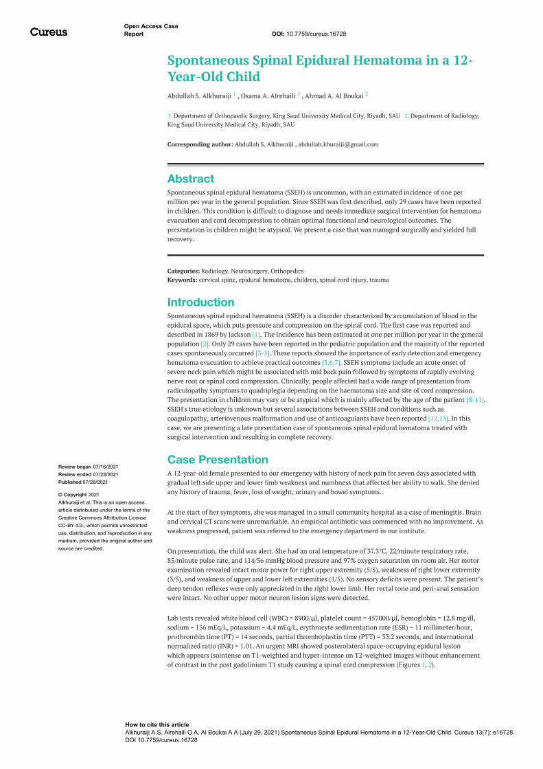
Lab tests revealed white blood cell (WBC) = 8900/μl, platelet count = 457000/μl, hemoglobin = 12.8 mg/dl,sodium = 136 mEq/L, potassium = 4.4 mEq/L, erythrocyte sedimentation rate (ESR) = 11 millimeter/hour,prothrombin time (PT) = 14 seconds, partial thromboplastin time (PTT) = 33.2 seconds, and internationalnormalized ratio (INR) = 1.01. An urgent MRI showed posterolateral space-occupying epidural lesionwhich appears isointense on T1-weighted and hyper-intense on T2-weighted images without enhancementof contrast in the post gadolinium T1 study causing a spinal cord compression (Figures 1, 2).
1 1 2
Open Access CaseReport DOI: 10.7759/cureus.16728
How to cite this articleAlkhuraiji A S, Alrehaili O A, Al Boukai A A (July 29, 2021) Spontaneous Spinal Epidural Hematoma in a 12-Year-Old Child. Cureus 13(7): e16728.DOI 10.7759/cureus.16728
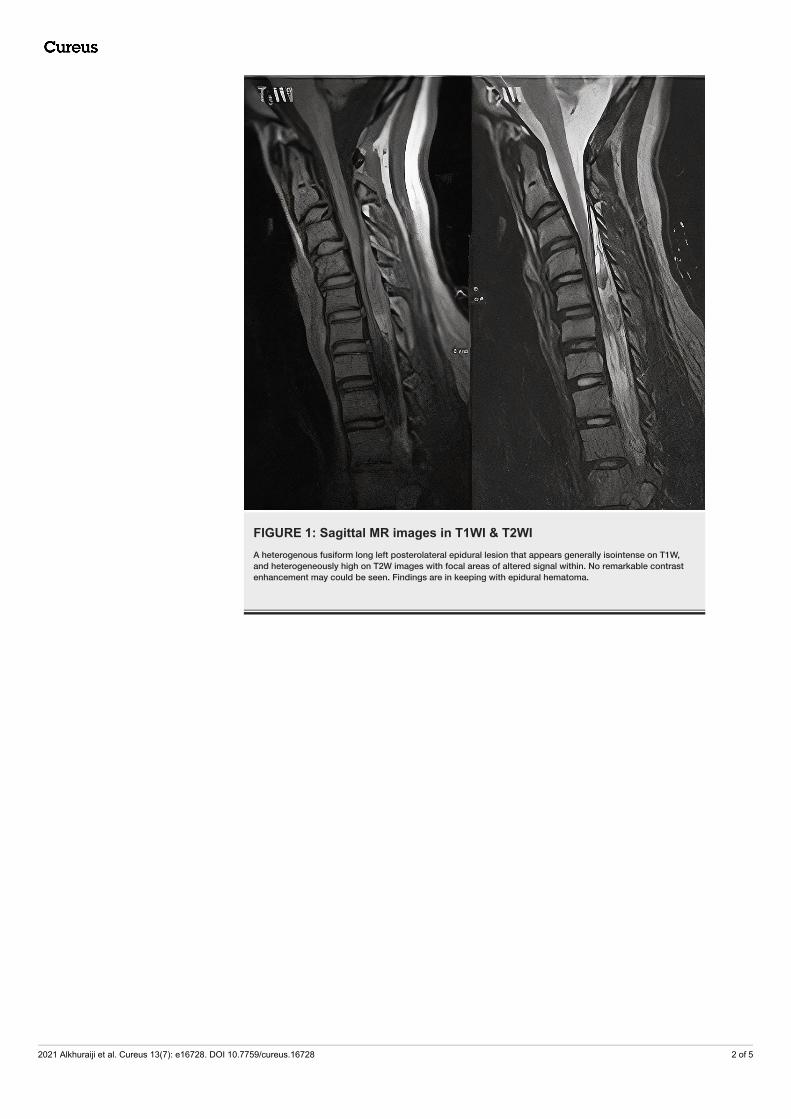
FIGURE 1: Sagittal MR images in T1WI & T2WIA heterogenous fusiform long left posterolateral epidural lesion that appears generally isointense on T1W,and heterogeneously high on T2W images with focal areas of altered signal within. No remarkable contrastenhancement may could be seen. Findings are in keeping with epidural hematoma.
2021 Alkhuraiji et al. Cureus 13(7): e16728. DOI 10.7759/cureus.16728 2 of 5
FIGURE 2: Axial MR images at same level in T2WI & T1 post contrastenhancementA heterogenous fusiform left posterolateral epidural lesion that appears heterogeneously high on T2Wimages with no remarkable contrast enhancement in the post gadolinium T1 study. Findings are in keepingwith epidural hematoma.
The patient was hospitalized in our institute, an emergent posterior cervical decompression and evacuationof hematoma from C4-6 along with instrumented fusion from C3-6 with bone grafting. Our aim was to startwith partial laminectomy but unfortunately it was not feasible to evacuate the hematoma due to blood clotsand this finding lead to do full laminectomy from C4-C6. There was no obvious pus or vascular malformationidentified. A tissue sample was taken for laboratory analysis and was sent for cultures and histopathology.Lateral mass screws were inserted from C3-6 bilaterally excluding C4 due to difficult entry. Homeostasis wasmaintained, bone graft applied over lateral mass and wound was closed over a drain. Intraoperative cultures
2021 Alkhuraiji et al. Cureus 13(7): e16728. DOI 10.7759/cureus.16728 3 of 5
did not grow any bacteria. The surgical pathology report revealed a histological feature consistent with anorganizing hematoma.
The patient’s initial post-operative recovery was slow, and neurological status remained the same until dayfive, then the patient started to improve. The patient was kept in our hospital for rehabilitation for twoweeks, upon discharge motor examination of left shoulder abduction and elbow flexion was 2/5 and forwrist/hand power was 4/5, motor examination of left lower extremity was 4/5, right upper and lowerextremity motor examination was 5/5. The patient’s latest follow-up three months after surgery showed fullneurological recovery.
DiscussionSSEH is an infrequent pathology in children, with only 29 cases having been reported. The majority of caseswere in the cervical spine (17 out of 29; 58.6%) [3-5]. Clinically, the presentation of epidural hematoma inthe pediatric age group varies significantly. Neck pain, abnormal neck position, focal or completeneurological weakness, numbness depending on level and location of blood accumulation, irritability orabnormal crying in infants has been reported [7,14]. The rate of symptom progression is typically fast,however slower progression of neurological symptoms has been seen. As a result of this ambiguouspresentation, a correct diagnosis of this etiology is difficult.
Appropriate laboratory tests including platelet count and coagulation panel are essential to rule out anycoagulation disorders or haemorrhagic diathesis. The cause of bleeding could not be identified. Although atthe start of symptoms our patient was diagnosed and treated as a case of meningitis which was ruled outafter final result of intra-operative culture and histology report, spontaneous spinal epidural hematomamimicking meningitis was reported in previous cases [10]. The spinal MRI with contrast is considered thebest available method to pick up such lesions [15,16]. The early MRI detection of hematoma offers a greatchance for early treatment, leading to optimal neurological recovery. The hematoma typically appearsisointense on T1-weighted and hyper-intense on T2-weighted MRI in the first 24 hours from onset ofsymptoms while it often appears hyper-intense on both T1- and T2-weighted images 24 hours later and mayappear hypo-intense on both T1- and T2-weighted images in chronic hematoma [15]. In our case thehematoma appeared isointense on T1-weighted and hyper-intense on T2-weighted although it wasperformed after seven days of onset of symptoms.
The mainstay of treatment is urgent surgical decompression and evacuation of accumulated blood fromneural structures [3,17]. Although the main factor determining the prognosis was the pre-operativeneurological status and not the onset of the symptoms, urgent surgical intervention and decompressionshould be considered in all symptomatic patients, especially those who present with incomplete cord injurygiven that these patients have higher chances of full recovery and resolution of neurological deficit [2].
A review of 29 pediatric cases reported by Pecha and Smeets showed complete resolution of neurologicaldeficits in 15 out of 29 (51.7%) patients, partial resolution in 13 (44.8%) patients, and death in one (3.4%)patient [3,5]. In the present report, the child showed partial recovery after five days from laminectomy andevacuation of hematoma and full recovery was reached after three months.
ConclusionsA high index of clinical suspicion followed by full investigation including an urgent MRI is crucial indiagnosing SSEH. Urgent surgical intervention is generally recommended as a first-line treatment followedby extensive rehabilitation program. This case was reported to be added to previously reported cases to giveattention to the existence of this unique condition and avoid missing patients with such presentation.
Additional InformationDisclosuresHuman subjects: Consent was obtained or waived by all participants in this study. Conflicts of interest: Incompliance with the ICMJE uniform disclosure form, all authors declare the following: Payment/servicesinfo: All authors have declared that no financial support was received from any organization for thesubmitted work. Financial relationships: All authors have declared that they have no financialrelationships at present or within the previous three years with any organizations that might have aninterest in the submitted work. Other relationships: All authors have declared that there are no otherrelationships or activities that could appear to have influenced the submitted work.
References1. Jackson R: Case of spinal apoplexy . Lancet. 1869, 94:5-6. 10.1016/s0140-6736(02)67624-x2. Bakker NA, Veeger NJ, Vergeer RA, Groen RJ: Prognosis after spinal cord and cauda compression in
spontaneous spinal epidural hematomas. Neurology. 2015, 84:1894-903. 10.1212/WNL.00000000000015453. Pecha M, Able A, Barber D, Willingham A: Outcome after spontaneous spinal epidural hematoma in
children: case report and review of the literature. Arch Phys Med Rehabil. 1998, 79:460-3. 10.1016/s0003-
2021 Alkhuraiji et al. Cureus 13(7): e16728. DOI 10.7759/cureus.16728 4 of 5

9993(98)90151-44. Epstein NE, Gilder M, Black K: Anterior thoracic extradural hematoma in a 5-year-old child . Pediatr
Neurosci. 1989, 15:48-52. 10.1159/0001204425. Smeets N, van den Akker M, Peters B, Vanderhasselt T, Jansen A: Spontaneous spinal epidural hematoma in
two toddlers: diagnostic pitfalls. Pediatr Dimens. 2016, 1:10.15761/pd.10001336. Alva N: Traumatic spinal epidural hematoma of a 10-month-old male: a clinical note . Pediatr Neurol. 2000,
23:88-9. 10.1016/s0887-8994(00)00151-X7. Kirwan R, Saigal G, Faingold R, O'Gorman A: Nontraumatic acute and subacute enhancing spinal epidural
hematoma mimicking a tumor in a child. Pediatr Radiol. 2004, 34:499-502. 10.1007/s00247-003-1129-98. Gopalkrishnan CV, Dhakoji A, Nair S: Spontaneous cervical epidural hematoma of idiopathic etiology: case
report and review of literature. J Spinal Cord Med. 2012, 35:113-7. 10.1179/2045772312Y.00000000019. Cakir E, Karaarslan G, Usul H, et al.: Clinical course of spontaneous spinal epidural haematoma mimicking
Guillain-Barré syndrome in a child: a case report and literature review. Dev Med Child Neurol. 2004, 46:838-42. 10.1017/s001216220400146x
10. Jumani DB, Littlewood R, Iyer A, et al.: Spontaneous spinal epidural haematoma mimicking meningitis in a2-year-old child--a case report and literature review. Childs Nerv Syst. 2013, 29:1795-8. 10.1007/s00381-013-2130-8
11. Patel H, Boaz J, Phillips J, Garg B: Spontaneous spinal epidural hematoma in children . Pediatr Neurol. 1998,19:302-7. 10.1016/s0887-8994(98)00059-9
12. Aycan A, Ozdemir S, Arslan H, Gonullu E, Bozkına C: Idiopathic thoracic spontaneous spinal epiduralhematoma. Case Rep Surg. 2016, 2016:1-4. 10.1155/2016/5430708
13. Taniguchi LU, Pahl FH, Lúcio JE, et al.: Complete motor recovery after acute paraparesis caused byspontaneous spinal epidural hematoma: case report. BMC Emerg Med. 2011, 11:10. 10.1186/1471-227X-11-10
14. Russman BS, Kazi KH: Spinal epidural hematoma and the Brown-Séquard syndrome . Neurology. 1971,21:1066-8. 10.1212/wnl.21.10.1066
15. Shima H, Yasuda M, Nomura M, et al.: A spinal epidural hematoma with symptoms mimicking cerebralstroke. Nagoya J Med Sci. 2012, 74:207-10.
16. Holtås S, Heiling M, Lönntoft M: Spontaneous spinal epidural hematoma: findings at MR imaging andclinical correlation. Radiology. 1996, 199:409-13. 10.1148/radiology.199.2.8668786
17. Groen RJ: Non-operative treatment of spontaneous spinal epidural hematomas: a review of the literatureand a comparison with operative cases. Acta Neurochir (Wien). 2004, 146:103-10. 10.1007/s00701-003-0160-9
2021 Alkhuraiji et al. Cureus 13(7): e16728. DOI 10.7759/cureus.16728 5 of 5
![A Traumatic Cervical Epidural Hematoma that Showed Rapid · Cervical spinal epidural hematoma is rare, and most cases are caused by spontaneous bleeding [1]. Traumatic cervical spinal](https://img.dokumen.tips/doc/110x75/5d1b365088c993dc468c7296/a-traumatic-cervical-epidural-hematoma-that-showed-rapid-cervical-spinal-epidural.jpg)