Embed Size (px)
Citation preview

Abstract Spinal epidural abscess (SEA) was first de-scribed in the medical literature in 1761 and representsa severe, generally pyogenic infection of the epiduralspace requiring emergent neurosurgical intervention toavoid permanent neurologic deficits. Spinal epiduralabscess comprises 0.2 to 2 cases per 10,000 hospitaladmissions. This review intends to offer detailed evalu-ation and a comprehensive meta-analysis of the interna-tional literature on SEA between 1954 and 1997, espe-cially of patients who developed it following anestheticprocedures in the spinal canal. In this period, 915 casesof SEA were published. This review is the most com-prehensive literature analysis on SEA to date. Mostcases of SEA occur in patients aged 30 to 60 years, butthe youngest patient was only 10 days old and the old-est was 87. The ratio of men to women was 1:0.56. Themost common risk factor was diabetes mellitus, fol-lowed by trauma, intravenous drug abuse, and alcohol-ism. Epidural anesthesia or analgesia had been per-formed in 5.5% of the patients with SEA. Skin abscess-es and furuncles were the most common source of in-fection. Of the patients, 71% had back pain as the ini-tial symptom and 66% had fever. The second stage ofradicular irritation is followed by the third stage, withbeginning neurological deficit including muscle weak-ness and sphincter incontinence as well as sensory defi-
cits. Paralysis (the fourth stage) affected only 34% ofthe patients. The average leukocyte count was15,700/µl (range 1,500–42,000/µl), and the averageerythrocyte sedimentation rate was 77 mm in the firsthour (range 2–50 mm). Spinal epidural abscess is pri-marily a bacterial infection, and the gram-positive Sta-phylococcus aureus is its most common causativeagent. This is true also for patients who develop SEAfollowing spinal anesthetics. Magnetic resonance imag-ing (MRI) displays the greatest diagnostic accuracy andis the method of first choice in the diagnostic process.Myelography, commonly used previously to diagnoseSEA, is no longer recommended. Lumbar puncture todetermine cerebrospinal fluid protein concentrations isnot needed for diagnosis and entails the risk of spread-ing bacteria into the subarachnoid space with conse-quent meningitis; therefore, it should not be performed.The therapeutic method of choice is laminectomy com-bined with antibiotics. Conservative treatment alone isjustifiable only for specific indications. Laminotomy isa therapeutic alternative for children. The mortality ofSEA dropped from 34% in the period of 1954–1960 to15% in 1991–1997. At the beginning of the twentiethcentury, almost all patients with SEA died. Parallel toimprovements in the mortality rate, today more patientsexperience complete recovery from SEA. The progno-sis of patients who develop SEA following epidural an-esthesia or analgesia is not better than that of patientswith noniatrogenic SEA, and the mortality rate is alsocomparable. The essential problem of SEA lies in thenecessity of early diagnosis, because only timely treat-ment is able to avoid or reduce permanent neurologicdeficits.
“The problem with spinal epidural abscesses is nottreatment, but early diagnosis – before massive neuro-logical symptoms occur” (Strohecker and Grobovschek1986).
Keywords Epidural abscess · Meta-analysis · Epidemiology · Outcome
Inaugural dissertation, University of Ulm 2000, supported by theUniversity Clinic of Anesthesiology and the Neurosurgical Clinicof the German Military Hospital of Ulm
E. ReihsausLudwigsburg-Bietigheim General Hospital, Department of Anesthesiology, Riedstrasse 12, 74321 Bietigheim-Bissingen, Germany
H. WaldbaurNeurosurgical Clinic, German Military Hospital, Oberer Eselsberg 40, 89081 Ulm, Germany
W. SeelingUniversity Clinic of Anesthesiology, Section of Pain Therapy, University of Ulm, Steinhövelstrasse 9, 89075 Ulm, Germany
Neurosurg Rev (2000) 232:175–204 © Springer-Verlag 2000
R E V I E W
E. Reihsaus · H. Waldbaur · W. Seeling
Spinal epidural abscess: a meta-analysis of 915 patients
Received: 18 July 2000 / Accepted: 14 September 2000
första tecknen på cancer i urinblåsan

Introduction
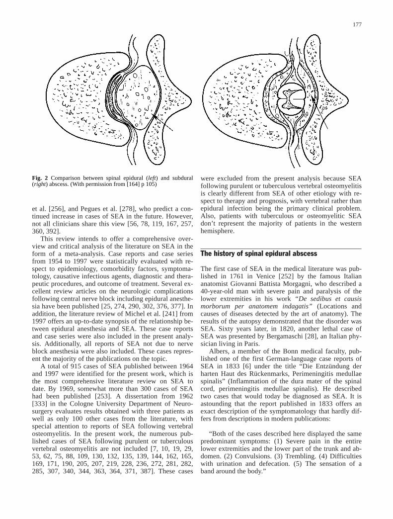
Spinal epidural abscess (SEA) represents a neurosurgicalemergency requiring immediate action. It usually presentsas a suppurative process localized between the spinal duramater and the vertebral periosteum within the spinal epi-dural space (Fig. 1).
According to Strohecker and Grobovschek [361], “theproblem with spinal epidural abscesses is not treatment,but early diagnosis – before massive neurological symp-toms occur.” Almost all reviews and case series on spinalepidural abscess emphasize this point [8, 16, 31, 67, 80,82, 98, 154, 158, 164, 179, 189, 195, 201, 213, 214, 230,232, 235, 262, 264, 283, 297, 300, 306, 309, 314, 316,330, 350, 366, 378, 401]. Immediate therapy is necessaryto prevent compression of the spinal cord and cauda equ-ina, with the corresponding neurological lesions (Fig. 2).Early surgical decompression and abscess drainage sup-ported by broad antibiotic coverage can lead to completerecovery.
Intramedullary [20, 238] and subdural spinal abscess-es [54, 111, 255, 275, 279, 367] occur only rarely. Incontrast, recent studies seem to document an increasingincidence of purulent infections in the spinal epiduralspace [67, 73, 154, 195, 314]. In fact, the authors of astudy on the MRI characteristics of cervical SEA from
1994 commented: “In our experience, this disorder ismore common than previously reported” [112]. Althoughthe prevalence may be increasing, the incidence of 0.2 totwo cases per 10,000 hospital admissions [16, 154], orless than one case per million residents [164], is stillquite low. It may present with back pain as the initialsymptom (see “Symptoms and laboratory findings”);however, back pain also occurs due to a large number ofother disorders. In Great Britain, the yearly incidence ofback pain is estimated to be 28,000 cases per million res-idents. This helps explain the problem of SEA as de-scribed by Strohecker and Grobovschek [361] and oth-ers. “Early diagnosis” [361] is only possible if SEA isconsidered in the differential diagnosis of back pain,which in turn is dependent on familiarity with the mani-festations of SEA.
The advanced age of the general population may beresponsible for the greater prevalence of SEA, since ag-ing is associated with an increase in risk factors (see“Pathogenesis and risk factors”). Abuse of intravenousdrugs, especially in the United States, also contributes tothe higher prevalence of SEA, since bacterial contamina-tion of syringes, needles, and other equipment can leadto SEA by hematogenous spread. More immunocompro-mised individuals in the last two decades have also con-tributed to the growing incidence of SEA.
The use of regional spinal and epidural anesthesia –even in patients with comorbidity factors such as diabe-tes mellitus – may be one reason for the observed preva-lence of SEA according to Du Pen et al. [87], Ngan Kee
176
Fig. 1 Cross-section through the fourth cervical vertebra. Menin-ges and epidural space. (With permission from [209] p 108)
et al. [256], and Pegues et al. [278], who predict a con-tinued increase in cases of SEA in the future. However,not all clinicians share this view [56, 78, 119, 167, 257,360, 392].
This review intends to offer a comprehensive over-view and critical analysis of the literature on SEA in theform of a meta-analysis. Case reports and case seriesfrom 1954 to 1997 were statistically evaluated with re-spect to epidemiology, comorbidity factors, symptoma-tology, causative infectious agents, diagnostic and thera-peutic procedures, and outcome of treatment. Several ex-cellent review articles on the neurologic complicationsfollowing central nerve block including epidural anesthe-sia have been published [25, 274, 290, 302, 376, 377]. Inaddition, the literature review of Michel et al. [241] from1997 offers an up-to-date synopsis of the relationship be-tween epidural anesthesia and SEA. These case reportsand case series were also included in the present analy-sis. Additionally, all reports of SEA not due to nerveblock anesthesia were also included. These cases repres-ent the majority of the publications on the topic.
A total of 915 cases of SEA published between 1964and 1997 were identified for the present work, which isthe most comprehensive literature review on SEA todate. By 1969, somewhat more than 300 cases of SEAhad been published [253]. A dissertation from 1962[333] in the Cologne University Department of Neuro-surgery evaluates results obtained with three patients aswell as only 100 other cases from the literature, withspecial attention to reports of SEA following vertebralosteomyelitis. In the present work, the numerous pub-lished cases of SEA following purulent or tuberculousvertebral osteomyelitis are not included [7, 10, 19, 29,53, 62, 75, 88, 109, 130, 132, 135, 139, 144, 162, 165,169, 171, 190, 205, 207, 219, 228, 236, 272, 281, 282,285, 307, 340, 344, 363, 364, 371, 387]. These cases
were excluded from the present analysis because SEAfollowing purulent or tuberculous vertebral osteomyelitisis clearly different from SEA of other etiology with re-spect to therapy and prognosis, with vertebral rather thanepidural infection being the primary clinical problem.Also, patients with tuberculous or osteomyelitic SEAdon’t represent the majority of patients in the westernhemisphere.
The history of spinal epidural abscess
The first case of SEA in the medical literature was pub-lished in 1761 in Venice [252] by the famous Italiananatomist Giovanni Battista Morgagni, who described a40-year-old man with severe pain and paralysis of thelower extremities in his work “De sedibus et causismorborum per anatomem indagatis” (Locations andcauses of diseases detected by the art of anatomy). Theresults of the autopsy demonstrated that the disorder wasSEA. Sixty years later, in 1820, another lethal case ofSEA was presented by Bergamaschi [28], an Italian phy-sician living in Paris.
Albers, a member of the Bonn medical faculty, pub-lished one of the first German-language case reports ofSEA in 1833 [6] under the title “Die Entzündung derharten Haut des Rückenmarks, Perimeningitis medullaespinalis” (Inflammation of the dura mater of the spinalcord, perimeningitis medullae spinalis). He describedtwo cases that would today be diagnosed as SEA. It isastounding that the report published in 1833 offers anexact description of the symptomatology that hardly dif-fers from descriptions in modern publications:
“Both of the cases described here displayed the samepredominant symptoms: (1) Severe pain in the entirelower extremities and the lower part of the trunk and ab-domen. (2) Convulsions. (3) Trembling. (4) Difficultieswith urination and defecation. (5) The sensation of aband around the body.”
177
Fig. 2 Comparison between spinal epidural (left) and subdural(right) abscess. (With permission from [164] p 105)

The “convulsions” and “trembling” described by Albers[6] are likely to represent the radiculopathic stage of SEA(see “Clinical and laboratory findings”).
In 1877, Lewitzky [210] of Warsaw and, in 1879,Spencer [353] of Bristol published other cases of SEAwith lethal clinical courses.
Until the 1930s, there was a lack of consistency in theterminology used to describe SEA, which was describedas peripachymeningitis spinalis [210], pachymeningitis(spinalis) externa (purulenta) [72, 113, 150, 243, 329,331, 353], perimeningitis [159, 175], extradural spinalsuppuration [268], (acute) purulent peripachymeningitis[145, 251], perimeningitis purulenta [153], extraduralabscess [355], epimeningitis spinalis [42], and extrathe-cal abscess [368]. As late as 1928, Bensheim [27], a phy-sician at the Internal Medicine Department of the Heidel-berg University, then under the direction of professorFreiherr von Weizsäcker, presented a case of SEA usingthe terminology peripachymeningitis spinalis externa pu-rulenta. In 1931, Pollak [286] used the term perimeningi-tis, although the autopsy report presented in his articleclearly documented that the leptomeninges were“smooth and free of exudates.” Mixter [247] was the firstto use the concept of epidural (intra)spinal abscess.
Irrespective of the terminology preferred by the dif-ferent authors, it is remarkable how consistent their de-scriptions of the disease manifestations are. The connec-tion between distant sources of infection and the occur-rence of SEA (see “Pathogenesis and risk factors”) wasalready recognized in the early days of the new scienceof bacteriology [247, 268, 329].
Furuncles were not the only cause of SEA to be iden-tified. Nonne [258] viewed “otogenic suppuration” ascausal for the development of SEA, and in 1921 Hinz[153] described the first recognized connection betweenthe development of SEA and the postpartal period.
Despite the progress in knowledge of the etiology andsymptoms of this disorder, the lack of treatment possibil-ities led to a more or less nihilistic attitude towards thetherapy until the beginning of the twentieth century. Ac-cordingly, the medical literature on SEA in the nine-teenth century consists almost completely of autopsy re-ports [6, 210, 353]. Unfortunately, until the 1930s mostpatients with SEA died [27, 42, 113, 145, 150 153, 243,247, 251, 268, 286, 329, 355]. In 1926, Nonne [258]wrote: “The prognosis of spinal cord abscesses is ex-tremely poor.” However, he was mistaken to claim thatno case had been cured up to that date. The Americanauthors Taylor and Kennedy [368] reported one success-fully treated case of SEA, and Dandy [72] describedthree other therapeutic successes in 1926. The method oftreatment was operative laminectomy (see “Treatment”).
The first operative intervention, laminectomy, was re-ported in 1892 by the French physician Delorme (quotedin Ref. 331). The laminectomy was performed from theseventh to the 11th thoracic vertebrae and revealed a“spongy, not especially adherent mass on the dura.” Thepatient did not survive the intervention. The first surgeonto report a successful laminectomy was Barth [21] in
1901. The 21-year-old patient had developed post-trau-matic SEA following a knife wound around theeighth/ninth thoracic vertebrae. The knife wound hadpunctured the dura and led to an epidural abscess thatwas drained operatively. However, there were postopera-tive neurological deficits.
However, it was in the 1920s that laminectomy wasfirst intensively discussed and utilized as a therapeuticoption for SEA. Taylor and Kennedy [368] and Dandy[72] in the USA were the first to use this method suc-cessfully. Since then, laminectomy has been standardtreatment (see “Treatment”). Following the above men-tioned articles, there were numerous communications onthe operative treatment aspects of SEA in the medical lit-erature from the 1930s to the 1950s [8, 15, 26, 34, 47,48, 51, 64, 68, 69, 83, 90, 120, 131, 136, 151, 197, 218,246, 248, 284, 289, 295, 296, 301, 310, 349, 356, 396].
Initial enthusiasm about the possibility of successfullytreating this disease, which had so long been associatedwith an extremely poor prognosis, is reflected in the ti-tles of the articles published in this period:
– 1932: Metastatic epidural abscess of the spinal cord.Recovery after operation [69].
– 1932: Epidural abscess complicated by staphylococ-cal meningitis. Report of a case with complete recov-ery following operation [310].
– 1934: Metastatic spinal epidural abscess. Report of acase with recovery following operation [349].
– 1940: Acute metastatic spinal epidural abscess. Re-port of two cases with recovery following laminecto-my [301].
– 1941: Emergency laminectomy for acute epidural ab-scess of the spinal canal. Report of four cases with re-covery in three [90].
– 1944: Acute epidural abscess of the spinal canal.Complete recovery following emergency laminecto-my and penicillin [83].
However, case studies continued to be published inwhich patients were described who died despite treat-ment with laminectomy [9, 26, 131, 136, 289, 313], es-pecially in the early years of use of this method. None-theless, the prognosis of the disease had been significant-ly improved. Starting from the early 1930s, early surgi-cal intervention has been emphasized as a prerequisitefor successful treatment of SEA [51, 349, 396].
Grant [129] analyzed 88 cases of SEA published be-fore 1937 and calculated a mortality rate of 48%. In con-trast, the same author described a mortality rate abouthalf as much, or 26%, for cases occurring after 1937. Inaddition to laminectomy, the use of antibiotics and sul-fonamides for the treatment of patients with SEA after1941 is likely to be responsible for the drop in mortality.Donathan [83] from Knoxville, USA described one caseof SEA in 1944 and Heusner [151] from Boston de-scribed 12 who received medical treatment in addition tolaminectomy. Sulfadiazine and penicillin were the antibi-otics used in that period. According to Donathan [83],
178

penicillin is “of prime importance and should be startedimmediately.”
Whereas Donathan [83] had described a 12-year-oldgirl with SEA in 1935, Gasul and Jaffe [120] describedcases of SEA in children aged 3, 8, and 12 years. In allcases, laminectomy was performed. The 3-year-old boywas the only patient who died in the postoperative peri-od. This publication is the first to deal specifically withthe problem of SEA in children, but the discussionsabout SEA in pediatric age groups do not differ fromthose on SEA in adults.
Usually, gram-positive cocci and especially Staphylo-coccus aureus represent the etiologic agent in cases ofSEA (see “Causative organisms”). Therefore, the firstdescription of SEA due to Actinomyces sp., irregularlyformed, non-spore-forming gram-positive rods, is espe-cially interesting from the point of view of medical his-tory. The case was published in 1940 by Krumdieck andStevenson [197] in New York. The 54-year-old patientwas first treated by radiation for suspected Hodgkin’sdisease but developed paraplegia and fever during thecourse of his hospital stay. The diagnosis of actinomy-cotic SEA was made by autopsy.
The first documented case of iatrogenic SEA due tolumbar puncture was described in 1945 by the Americanauthors Rangell and Glassman [295]. Viscous pus wasdrained from between the third and fourth lumbar verte-brae following laminectomy in this 28-year-old patient.The patient recovered following postoperative treatmentwith sulfadiazine but had residual neurological deficits.
Epidemiology
Spinal epidural abscess occurs primarily in individualsover 30 years of age. However, published case serieswith more than ten patients report differing average agesfor affected patients. The lowest age was reported byDei-Anang et al. [80], who found a mean age of only 34years in a group of 15 patients. Maslen et al. [231] re-ported a mean of 67 years of age in a group of 28 pa-tients with SEA, which represents the highest averageage in all published case series [16, 31, 73, 74, 80, 81,98, 141, 154, 172, 177, 189, 201, 212, 213, 231, 235,253, 283, 297, 300, 306, 316, 370, 391, 401]. However,Dei-Anang et al. [80] included in their patient group fourchildren aged 2 to 6 as well as five patients from 19 to38. The range of ages reported by Dei-Anang et al. was 2 to 74 years, while the patient group investigated byMaslen et al. [231] had an age range of 45 to 87 years.Ravicovitch and Spallone [297] of the Botkin Hospital inMoscow reported a significantly lower average age (30.1years) for their patient group than Dei-Anang et al. Noneof the patients reported by Ravicovitch and Spallone wasyounger than 15 years.
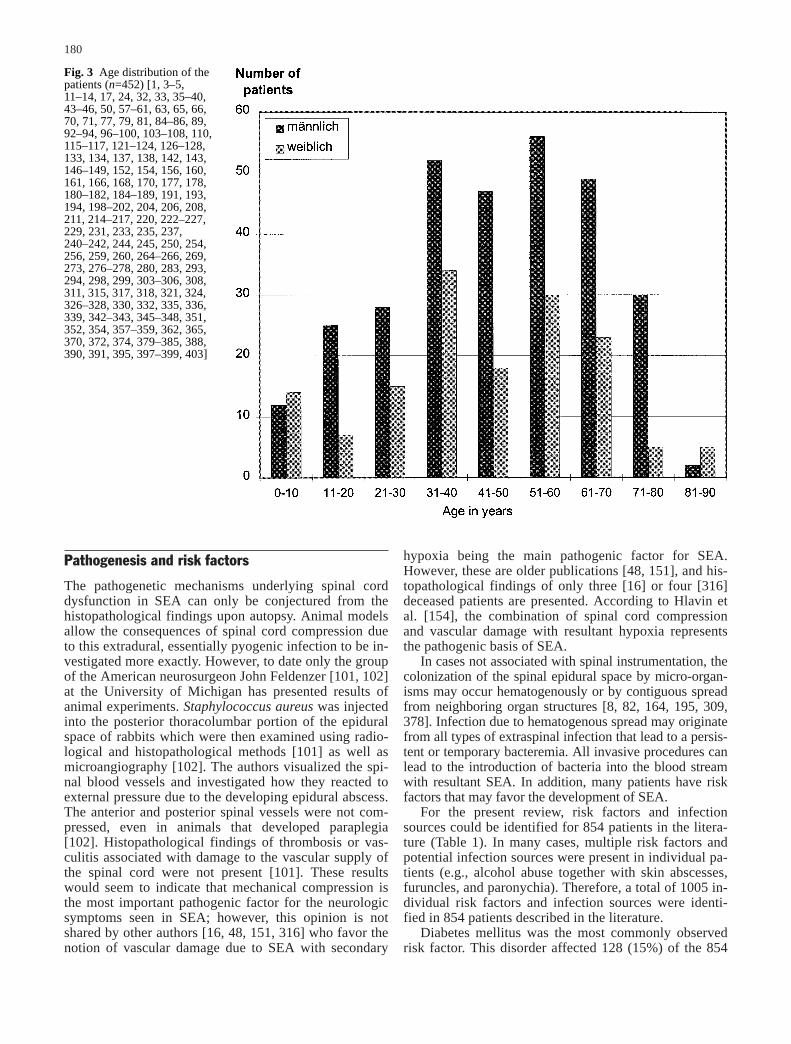
In this review, articles published between 1954 and1997 concerning a total of 915 patients with SEA werestatistically analyzed [1, 3–5, 11–14, 16, 17, 24, 31–33,35–40, 43–46, 50, 55, 57–61, 63, 65, 66, 70, 71, 73, 74,
77, 79–81, 84–86, 89, 92–94, 96–100, 103–108, 110,115–117, 121–124, 126–128, 133, 134, 137, 138,141–143, 146–149, 152, 154, 156, 158, 160, 161, 166,168, 170, 172, 176–178, 180–182, 184–189, 191, 193,194, 198–202, 204, 206, 208, 211–217, 220, 222–227,229, 231–233, 235, 237, 240–242, 244, 245, 250, 253,254, 256, 259, 260, 264–266, 269–271, 273, 276–278,280, 283, 293, 294, 297–300, 303–306, 308, 311,315–318, 321, 324, 326–328, 330, 332, 335, 336, 339,341–343, 345–348, 351, 352, 354, 357–359, 362, 365,370, 372, 373, 379–385, 388, 390, 391, 395, 397–399,401, 403]. Some publications did not include detailed in-formation on patient age [16, 31, 55, 73, 74, 80, 141,158, 172, 176, 206, 212, 213, 232, 253, 297, 300, 316,401]. Additionally, some only indicate the average ageand the range of ages, without indicating the exact age ofindividual patients [16, 31, 73, 74, 80, 141, 158, 172,177, 212, 213, 232, 253, 297, 300, 316, 401]. Therefore,it was possible to calculate the age distribution for 452patients with SEA (Fig. 3). In contrast to the commonconception that SEA mainly affects individuals olderthan 50 [195, 314], there was a broader distribution ofage groups among patients affected by SEA, especiallyamong males (Fig. 3). The age distribution does not sup-port the view that there is a peak exclusively in the sixthand seventh decades of life [230].
Of a total of 301 men with SEA, 204 (68%) were be-tween 31 and 70 years old. An age predilection for menbetween 51 and 70 was not observed. From a total of 151women with SEA, 105 (70%) were between 31 and 70years, again without an obvious predilection for any giv-en decade. The youngest patient with SEA was only 10days old [133] and the oldest was 87 [231, 235].
Ruiz et al. [314] deny the existence of a gender pref-erence for SEA in their review article published in 1995,whereas Youmans [402] wrote in a textbook contributionin 1985 that men develop SEA more frequently. Kraussand McCormick [195] do not pronounce a definite opin-ion on this issue. In the present work, essentially all pub-lications on SEA appearing after 1954 were included,and the evaluation of the correspondingly large numberof cases allows statistically sound analysis.
All large case series observed a preference for themale gender in the development of SEA [31, 73, 74, 80,81, 98, 141, 154, 172, 177, 189, 201, 212, 213, 231, 235,253, 283, 297, 300, 316, 370, 391, 401]. Only one articleon a series of 39 patients with SEA [16] reports amale:female ratio of 0.5:1. The 915 cases from the litera-ture in the present work included 520 men and 289 wom-en. For 106 patients, no gender information was avail-able in the original publications [55, 158, 172, 297, 401].Therefore, the male:female ratio is 1:0.56, which corre-sponds to a definite prevalence for males. The reasonsfor this cannot be clearly derived from the literature, butit may be assumed that different risk factors are partiallyresponsible (see “Pathogenesis and risk factors”). Alco-hol abuse, use of intravenous drugs, and trauma affectmen more often than women and have been observed tobe associated with SEA.
179
Pathogenesis and risk factors
The pathogenetic mechanisms underlying spinal corddysfunction in SEA can only be conjectured from thehistopathological findings upon autopsy. Animal modelsallow the consequences of spinal cord compression dueto this extradural, essentially pyogenic infection to be in-vestigated more exactly. However, to date only the groupof the American neurosurgeon John Feldenzer [101, 102]at the University of Michigan has presented results ofanimal experiments. Staphylococcus aureus was injectedinto the posterior thoracolumbar portion of the epiduralspace of rabbits which were then examined using radio-logical and histopathological methods [101] as well asmicroangiography [102]. The authors visualized the spi-nal blood vessels and investigated how they reacted toexternal pressure due to the developing epidural abscess.The anterior and posterior spinal vessels were not com-pressed, even in animals that developed paraplegia[102]. Histopathological findings of thrombosis or vas-culitis associated with damage to the vascular supply ofthe spinal cord were not present [101]. These resultswould seem to indicate that mechanical compression isthe most important pathogenic factor for the neurologicsymptoms seen in SEA; however, this opinion is notshared by other authors [16, 48, 151, 316] who favor thenotion of vascular damage due to SEA with secondary
hypoxia being the main pathogenic factor for SEA.However, these are older publications [48, 151], and his-topathological findings of only three [16] or four [316]deceased patients are presented. According to Hlavin etal. [154], the combination of spinal cord compressionand vascular damage with resultant hypoxia representsthe pathogenic basis of SEA.
In cases not associated with spinal instrumentation, thecolonization of the spinal epidural space by micro-organ-isms may occur hematogenously or by contiguous spreadfrom neighboring organ structures [8, 82, 164, 195, 309,378]. Infection due to hematogenous spread may originatefrom all types of extraspinal infection that lead to a persis-tent or temporary bacteremia. All invasive procedures canlead to the introduction of bacteria into the blood streamwith resultant SEA. In addition, many patients have riskfactors that may favor the development of SEA.
For the present review, risk factors and infectionsources could be identified for 854 patients in the litera-ture (Table 1). In many cases, multiple risk factors andpotential infection sources were present in individual pa-tients (e.g., alcohol abuse together with skin abscesses,furuncles, and paronychia). Therefore, a total of 1005 in-dividual risk factors and infection sources were identi-fied in 854 patients described in the literature.
Diabetes mellitus was the most commonly observedrisk factor. This disorder affected 128 (15%) of the 854
180
Fig. 3 Age distribution of thepatients (n=452) [1, 3–5,11–14, 17, 24, 32, 33, 35–40,43–46, 50, 57–61, 63, 65, 66,70, 71, 77, 79, 81, 84–86, 89,92–94, 96–100, 103–108, 110,115–117, 121–124, 126–128,133, 134, 137, 138, 142, 143,146–149, 152, 154, 156, 160,161, 166, 168, 170, 177, 178,180–182, 184–189, 191, 193,194, 198–202, 204, 206, 208,211, 214–217, 220, 222–227,229, 231, 233, 235, 237,240–242, 244, 245, 250, 254,256, 259, 260, 264–266, 269,273, 276–278, 280, 283, 293,294, 298, 299, 303–306, 308,311, 315, 317, 318, 321, 324,326–328, 330, 332, 335, 336,339, 342–343, 345–348, 351,352, 354, 357–359, 362, 365,370, 372, 374, 379–385, 388,390, 391, 395, 397–399, 403]
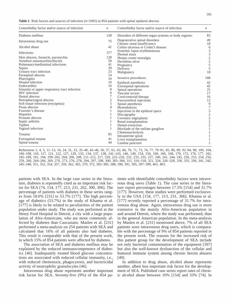
patients with SEA. In the large case series in the litera-ture, diabetes is repeatedly cited as an important risk fac-tor for SEA [74, 154, 177, 213, 231, 262, 300, 306]. Thepercentage of patients with diabetes in these series rang-es from 18.0% [231] to 53.7% [177]. The high percent-age of diabetics (53.7%) in the study of Khanna et al.[177] is likely to be related to peculiarities of the patientpopulation under study. The study was performed at theHenry Ford Hospital in Detroit, a city with a large popu-lation of Afro-Americans, who are more commonly af-fected by diabetes than Caucasians. Maslen et al. [231]performed a meta-analysis on 254 patients with SEA andcalculated that 16% of all patients also had diabetes.This result is comparable with that of the present work,in which 15% of 854 patients were affected by diabetes.
The association of SEA and diabetes mellitus may beexplained by the reduced immunocompetence of diabet-ics [46]. Inadequately treated blood glucose concentra-tions are associated with reduced cellular immunity, i.e.,with reduced chemotaxis, phagocytosis, and bactericidalactivity of neutrophilic granulocytes [46].
Intravenous drug abuse represents another importantrisk factor for SEA. Seventy-five (9%) of the 854 pa-
tients with identifiable comorbidity factors were intrave-nous drug users (Table 1). The case series in the litera-ture report percentages between 17.5% [154] and 31.7%[177]. However, these studies were performed exclusive-ly in the USA [154, 177, 213, 231, 306]. Khanna et al.[177] recently reported a percentage of 31.7% for intra-venous drug abuse. Again, intravenous drug use is moreextensive in the mainly Afro-American population inand around Detroit, where the study was performed, thanin the general American population. In the meta-analysisby Maslen et al. [231] mentioned above, 7% of all SEApatients were intravenous drug users, which is compara-ble with the percentage of 9% of 854 patients reported inthe present work. The reasons for the increased risk ofthis patient group for the development of SEA includenot only bacterial contamination of the equipment [397]but also the well-known dysfunction of the cellular andhumoral immune system among chronic heroin abusers[49].
In addition to drug abuse, alcohol abuse representsanother, albeit less important risk factor for the develop-ment of SEA. Published case series report rates of chron-ic alcohol abuse between 10% [154] and 33% [74]. In
181
Table 1 Risk factors and sources of infection (n=1095) in 854 patients with spinal epidural abscess
Comorbidity factor and/or source of infection n Comorbidity factor and/or source of infection n
Diabetes mellitus 128
Intravenous drug use 75
Alcohol abuse 41
Infections: 377Skin abscess, furuncle, paronychia 128Vertebral osteomyelitis/discitis 59Pulmonary/mediastinal infections 41Sepsis 39Urinary tract infection 23Paraspinal abscess 14Pharyngitis 10Wound infection 10Endocarditis 10Sinusitis or upper respiratory tract infection 8HIV infection 9Dental abscess 7Retropharyngeal abscess 5Soft tissue infection (erysipelas) 5Psoas abscess 4Fournier’s disease 1Hepatitis 1Prostate abscess 1Septic arthritis 1Typhus 1Vaginal infection 1
Trauma: 85Extraspinal trauma 41Spinal trauma 44
Disorders of different organ systems or body regions: 83Degenerative spinal disorders 49Chronic renal insufficiency 18Colitis ulcerosa or Crohn’s disease 6Systemic lupus erythematosus 3Dermal sinus 3Herpes zoster neuralgia 2Decubitus ulcer 2Pregnancy 5Delivery 5Malignancy 18
Invasive procedures: 188Epidural anesthesia 42Extraspinal operations 42Spinal operations 25Vascular access 17Corticosteroid therapy 16Paravertebral injections 12Spinal anesthesia 9Hemodialysis 7Injections in the epidural space 5Discography 2Coronary angioplasty 2Renal transplantation 2Dental extraction 2Blockade of the stellate ganglion 1Chemonucleolysis 1Intrauterine spiral 1Liver transplantation 1Lumbar puncture 1
References 1, 4, 5, 11–13, 16, 24, 31, 33, 35–40, 43–46, 50, 57, 61, 63, 66, 70, 71, 73, 74, 77, 79–81, 85, 86, 89, 92–94, 98, 100, 104,106–108, 110, 117, 121, 122, 127, 128, 133, 134, 137, 138, 141–143, 146, 149, 154, 156, 160, 166, 168, 170, 172, 176, 177, 182,185–189, 191, 194, 199–202, 204, 206, 208, 211–215, 217, 220, 222–226, 232, 233, 235, 237, 240, 241, 244, 245, 250, 253, 254, 256,259, 260, 264–266, 269, 270, 273, 276, 278, 294, 297, 298, 300, 305–306, 311, 316–318, 321, 324, 326–328, 330, 335, 336, 341, 342,345–348, 351, 352, 354, 357, 359, 362, 365, 370, 372, 383–385, 388, 390, 391, 395, 397–399, 401, 403.

the present work, 41 (5%) of 854 patients described inthe international medical literature were alcoholics.Among the 254 patients from different publications ana-lyzed by Maslen et al. [231], 4% were alcoholics.
In many cases, alcoholics consume diets deficient inprotein, which is thought to compromise the immunesystem [46]. It remains speculative whether other factorssuch as inadequate personal hygiene or other sociologi-cal characteristics among alcoholics may also be associ-ated with the observed increased coincidence of SEAand chronic alcohol abuse.
On the other hand, there is a definite connection be-tween sources of infection near to and distant from thevertebral canal. Skin abscesses, furuncles, and parony-chia are often found to be SEA’s source of infection. He-matogenous spread into the epidural space is the impor-tant pathogenic factor. Patients with SEA who report aprevious infectious condition of the skin made up a high-er proportion of all patients in older case series than inthose from the 1990s. In 1954, Hulme and Dott [158] de-scribed cutaneous and subcutaneous infections in 33% oftheir patients and Yang [401] found 44% in his case se-ries from 1982. However, the latter study was performedin Tientsin, China. In more recent American studies, theincidence of cutaneous infections was calculated to be 7% [231], 12.2% [177], 15.4% [154], and 20% [300];the present meta-analysis found an incidence of cutane-ous infections of 15% (128 of 854 patients) (Table 1).Maslen et al. [231] observed a rate of 25% for dermaland soft-tissue infections among the 254 literature casesthey analyzed. Sources of infection near the vertebral ca-nal include paraspinal abscess (14 of 854 cases), retro-pharyngeal abscess (five of 854), and psoas abscess (fourof 854) (Table 1). In these cases, the colonization of thespinal epidural space occurred by contiguous spread [46,61, 71, 133, 168, 188, 294, 321, 372].
Taken together, infectious processes were identifiedin 377 (44%) of the 854 cases identified in the literatureand thus represent the most common comorbidity factor.In most cases, the infection led to seeding of the epiduralspace by hematogenous spread.
Trauma represents another important group of riskfactors (Table 1). In the patient group presented in thiswork, 85 of 854 patients (10%) had suffered extraspinalor spinal trauma preceding development of SEA. In thelarger case series of the literature, trauma was reported in25% [158] to 34.7% [306] of patients with SEA. Traumacan favor hematogenous spread of micro-organisms bypenetration of anatomic barriers, but spinal trauma is es-pecially important, since it may create a site of entry formicro-organisms into the epidural space [16, 151, 158,172]. Spinal hematomas associated with severe blunttrauma to the back represent an important pathogeneticfactor that can favor the development of SEA and couldalso explain the potential development of SEA followingepidural blood patch [221] (see below).
In the present meta-analysis, 49 (6%) of 854 patientswith SEA were identified who also had degenerative ver-tebral disease as a comorbidity factor (Table 1). Degen-
erative changes of the intervertebral disks could act assites of reduced resistance against hematogenous spread,similarly to hematomas due to spinal trauma, and there-by favor the development of SEA.
Patients with chronic renal insufficiency and especial-ly those who require dialysis often show reduced immu-nocompetence. Eighteen (2%) of the 854 patients fromthe literature discussed in the present work had chronicrenal insufficiency (Table 1) [154, 187, 212, 213, 264,306, 343, 351].
Of 854 patients with SEA, six had Crohn’s disease orcolitis ulcerosa (Table 1) [4, 110, 146, 254, 300, 383].Both immunosuppressive treatment and the tendency toformation of fistulas represent factors that could favorthe development of SEA. One patient with Crohn’s dis-ease developed SEA due to a fistula that led into thelumbosacral canal [110], and another 42-year-old withCrohn’s disease developed SEA due to an enteroepiduralfistula following proctocolectomy and creation of anilioanal anastomosis [254].
Cancer patients may also display reduced immuno-competence. Eighteen (2%) of the 854 patients with SEAalso had cancer [12, 37, 73, 80, 147, 154, 177, 201, 211,300, 304, 326, 395]. Some of these patients were also re-ceiving chemotherapy [395]. Because of the simulta-neous presence of several different comorbidity factorsin older patients, it is not always possible to determinethe exact contribution of cancer to the development ofSEA in these cases.
Pregnancy and delivery represent other risk factors.Ten of 854 patients in the literature developed SEA dur-ing pregnancy or the postpartal period [16, 70, 122, 160,176, 182, 185, 224]. Postpartal impairment of immunedefense may be related to the development of SEA inthese patients [224].
Invasive procedures led to SEA in 188 (22%) of the854 patients in the literature (Table 1). Spinal and extra-spinal operations, which may create a contiguous port ofentry for micro-organisms into the epidural space or leadto hematogenous seeding, and especially anesthesiologi-cal procedures may favor the development of SEA. Al-though spinal anesthesia [76, 174] and lumbar puncturein children with bacteremia [369] can lead to the develop-ment of bacterial meningitis, most authors agree that therisk of infection following nerve block anesthesia nearthe spinal cord should not be overestimated [78, 119, 157,257, 290, 302, 337, 360, 376, 377]. Spinal epidural ab-scess is generally thought to be a “very rare, severe com-plication” [302] of central nerve blocks. The fact that 42(5%) of the 854 cases of SEA were associated with epi-dural anesthesia must be seen in relation to the quite com-mon use of this form of regional anesthesia. Among505,000 epidural anesthetics performed between 1982and 1986 for obstetric indications, only a single case ofSEA was observed [337]. Perioperative bacteremia is re-garded as a relative but not absolute contraindication forregional epidural or spinal anesthesia [25].
Michel et al. [241] identified 40 patients with SEA fol-lowing epidural anesthesia or analgesia in articles pub-
182

lished between 1947 and 1996; they also quoted the workof Du Pen et al. [87], who had described 15 other cases ofinfections in the epidural space following epidural anes-thetic procedures. Kindler et al. [179] published an evalu-ation of the literature from 1974 to 1996 with 42 patientswith SEA following epidural anesthesia or analgesia.There was a remarkably high percentage of 33% follow-ing thoracic catheterization compared to 45% followinglumbar catheterization, even though thoracic catheters areused much less often than lumbar catheters. The authorsspeculate that this is related to the generally more diffi-cult insertion of thoracic catheters and their longer dura-tion of use. The information in the literature on the inci-dence of SEA following anesthesiological procedures isnot reliable because of the rarity of this complication. Theincidence of SEA due to short-duration catheterizations isabout 1:100,000 [25], whereas that of SEA with tunneledperidural catheters is about 4% [87].
Du Pen et al. [87] report on 350 patients with a totalof 32,354 catheter days and calculate a risk of 1 per 1702catheter days. In all, there is a low incidence of infec-tious complications following anesthesiological proce-dures in the epidural and spinal spaces [25]. In this meta-analysis of 854 patients, there were 42 cases of SEA fol-lowing catheter epidural anesthesia, nine following spi-nal anesthesia, and five following “single shot” injec-tions into the epidural space (Table 1) [24, 36, 39, 45,57, 70, 79, 86, 104, 106, 117, 122, 128, 142, 152, 186,213, 215, 225, 233, 241, 256, 259, 260, 265, 278, 304,318, 335, 342, 351, 352, 362, 365, 382, 385].
Williams et al. [394] report on a case of epidural he-matoma following repeated steroid injections that had tobe surgically decompressed. Hemorrhages into the epi-dural space following epidural anesthesia were demon-strated postmortem [400] and could represent a site ofreduced resistance to colonization by micro-organisms,thus favoring the development of SEA. Occasionally theformation of fistulas has also been observed after cathe-terization of the epidural space [334, 386].
Vascular access may also represent a port of entry forinfectious agents. Seventeen (2%) of the 854 patientswith SEA in the literature contracted the infection in thisway (Table 1) [12, 74, 154, 187, 201, 214, 264, 343].
The other invasive procedures performed on patientswho then developed SEA are listed in Table 1.
Of the 738 patients in this meta-analysis for whomthe localization of SEA could be identified from descrip-tions in the literature, 19% (140) had cervical SEA and7% (53) had cervicothoracic SEA (Table 2). AlthoughLasker [204] indicates a lower frequency of 12% for cer-vical SEA, Friedman reported a frequency of 32% only 7years later. In the majority of the cases, cervical epiduralabscesses originated as a consequence of vertebral bodyosteomyelitis or discitis affecting vertebrae C4–C7 in ananterior position [314].
The most common location for SEA is the thoracicepidural space. Of the 738 patients, 35% (255) had tho-racic epidural abscesses and 30% (223) had lumbar orlumbosacral abscesses (Table 2). This localization corre-
sponds to the results of most large case series in the liter-ature [16, 138, 177, 235, 253]. The preferential thoracicor lumbar localization of SEA is likely related to thegreater extension of the epidural space in the thoraco-lumbar segment of the vertebral column and to the well-developed extradural venous plexus in this region [366].
Clinical and laboratory findings
In 1948, Corradini et al. [68] concisely defined themeaning of the symptoms of SEA: “Furthermore, we be-lieve that the syndrome is so characteristic that its recog-nition depends only upon acquaintance with the symp-tomatology.” Even today, this statement is still correct.
Also in 1948, Heusner [151] published his classic de-scription of the various stages of SEA that continues tobe referred to in the literature up to the present. Follow-ing the initial stage of severe back pain associated withfever and local tenderness in the area of the spinal column, the second stage is dominated by signs of spinalirritation. These manifestations include Lasègue’s, Kernig’s, and Lhermitte’s signs, Brudzinski’s reflex, andneck stiffness [241, 309]. Additionally, back pain can ra-diate into the arms or legs, depending on the craniocau-dal location of the abscess. In Heusner’s third stage[151], initial neurologic deficits are observed such asweakness of the voluntary musculature or fecal or uri-nary incontinence. Sensory deficits may also occur. Inthe fourth stage, muscle weakness progresses to paraly-sis. The duration of the individual stages varies. Impor-tantly, the transition to the terminal stage with paralysiscan occur very quickly [158]. In addition to neurologicaldeficits [151], patients with SEA may also develop fever
183
Table 2 Craniocaudal localization of spinal epidural abscess in738 patients
Localization Number (%*)
Cervical 140 (19)Cervicothoracic 53 (7)Thoracic 255 (35)Thoracolumbar 54 (7)Lumbar 133 (18)Lumbosacral 90 (12)Sacral 3 (–)Cervicothoracolumbar 8 (1)Thoracolumbosacral 2 (–)Total 738 (100)
*Rounded off.References 1, 3–5, 11–14, 16, 17, 31–33, 35–40, 43, 44, 50, 55,57–61, 63, 65, 66, 70, 74, 77, 79, 81, 84, 85, 89, 92–94, 96,98–100, 103–108, 110, 115–117, 121, 122, 124, 127, 128, 133,134, 137, 138, 141–143, 146–149, 152, 154, 156, 160, 161, 166,168, 170, 172, 177–178, 180–182, 185–188, 191, 193, 194,198–201, 204, 208, 211, 212, 214–217, 222–227, 229, 232, 233,235, 237, 240, 241, 244, 245, 250, 254, 256, 259, 264–266,269–271, 273, 276, 277, 280, 283, 293, 297, 299, 300, 303, 305,306, 308, 311, 315, 316, 318, 321, 324, 326–328, 332, 336, 339,341–343, 345–348, 351, 352, 354, 357–359, 362, 365, 372, 374,379, 381–384, 390, 391, 395, 397–399, 401, 403.

in the initial stage due to the inflammation associatedwith the abscess [82, 164, 230, 309, 378]. This is accom-panied by an accelerated erythrocyte sedimentation rate(ESR) and leukocytosis.
The combination of severe pain near the spine withfever is characteristic [68] and should always be regard-ed as a warning sign of possible SEA. The early recogni-tion of these symptoms before the occurrence of neuro-logic deficits is exactly the “problem of spinal epiduralabscesses” described by Strohecker and Grobovchek[361].
For 871 of the 915 patients with SEA identified in theliterature, information about the symptoms of the diseasewere available from case reports or case series (Table 3).
Back pain was the most common symptom, occurringin 71% of the patients with SEA. Fever was present in66%. Accordingly, two thirds of the SEA patients couldbe diagnosed in an early stage if this disorder is consid-ered in the differential diagnosis, and neurologic deficitscould be avoided with timely therapeutic intervention.
Most of the large case series in the literature with atleast 18 patients describe a somewhat higher incidence ofback pain with SEA. All 18 patients of Yang [401], all 29of Del Curling et al. [81], and all 39 of Baker et al. [16]displayed spine-related back pain due to SEA. Eightypercent of the SEA patients described by Nussbaum et al.[262], 89% of those of Khanna et al. [177], 89% of thoseof Liem et al. [213], and 92% of those of Maslen et al. [231] also reported back pain. Darouiche et al. [74]
found 72% of their patient group to have back pain, andRigamonti et al. [306] found 74%; these are the only twolarge case studies with rates of back pain comparable to that calculated from the present meta-analysis. Thecase studies describe groups of patients treated by onephysician or group of physicians, while the present meta-analysis summarizes published data from numerous au-thors, so it is possible that back pain was not noted insome articles. Even if one author performs a retrospectivechart analysis, clinical data are not always complete. Forinstance, Hancock [141] reports that the clinical data onthe symptoms experienced by 49 SEA patients in hisanalysis were sufficiently documented in only 24 cases.
Fever developed in 66% of the patients with SEA inthe present meta-analysis (Table 3). Even though in-creased body temperature is an essential symptom of thissevere inflammation of the epidural space, publishedcase series document a quite variable incidence of feverin these patients. During the course of disease, 29% of the patients of Liem et al. [213], 39.1% of those ofRigamonti et al. [306], 45% of those of Nussbaum et al.[262], 52% of those of Maslen et al. [231], 61% of thoseof Khanna et al. [177], and 62% of those of Del Curlinget al. [81] as well as all patients described by Baker et al.[16] developed increased body temperature. This broadrange may be related to differing definitions of fever inthe different studies. In the present meta-analysis, the ad-jective “feverish” in the case reports was regarded assufficient to include a patient in the group with fever.
In the initial stage of back pain, 17% of the 871 pa-tients from the international medical literature had localtenderness of the spine and its surroundings (Table 3).This finding is not always consistently documented inthe literature. Most case series do not analyze the inci-dence of local tenderness of the spine using statisticalmethods. The only exceptions to this are the articles ofBaker et al. [16] and Maslen et al. [231]. Whereas theformer document the occurrence of local tenderness inall examined patients with SEA, Maslen et al. [231]found this manifestation to be present in only 31% oftheir patients and 24% of the 207 patients they analyzedfrom the international literature.
Irritability was seen in eight of 871 patients (Table 3)and is a typical symptom of SEA in small children [3,32, 96, 125, 228, 271, 315, 380]. Aicardi and Lepintre[3] describe a 3-month-old infant with SEA and con-clude that conspicuous crying and obvious pain on beingtouched may be the only signs of SEA in this age group.
Spinal irritability and signs of initial or progressiveneurological deficits occurred in only a fifth to a third ofthe 871 patients reported in the international literaturefrom 1954 to 1997 (Table 3). Perhaps many patientswere diagnosed in early stages. Comparable indicationson the incidence of signs of spinal irritation are generallynot found in the large case series. Radiculopathic com-plaints were present in 12.2% of the patients describedby Khanna et al. [177], as the presenting symptom in19% of those described by Maslen et al. [231], and in atotal of 47% of patients described by Darouiche et al.
184
Table 3 Symptoms and signs of spinal epidural abscess in 871 pa-tients
Clinical findings n patients (%)
Fever 571 (66)
Initial pain symptom:Back pain 620 (71)Neck pain 29 (3)Headache 25 (3)Local tenderness 151 (17)Irritability 8 (1)Spinal irritation 174 (20)
Beginning neurologic deficit:Muscle weakness 225 (26)Incontinence 210 (24)Sensory deficit 115 (13)
Advanced neurologic deficit:Paraparesis/paraplegia 268 (31)Tetraparesis/tetraplegia 29 (3)
References 1, 3–5, 11–14, 16, 17, 24, 31–33, 35–40, 43–46, 50,55, 57–61, 63, 65, 66, 71, 74, 77, 79–81, 84, 85, 89, 92–94, 96, 97,99, 100, 103–108, 110, 115–117, 121–124, 126–128, 133, 134,137, 138, 141–143, 146–149, 152, 154, 156, 160, 161, 166, 168,170, 172, 176–178, 180–182, 185, 187–189, 191, 193, 194,198–202, 204, 206, 208, 211, 213–217, 220, 222–227, 229,231–233, 235, 237, 240–242, 244, 245, 250, 253, 254, 256, 259,260, 264–266, 269–271, 273, 276, 277, 280, 293, 294, 297–300,303–306, 308, 311, 315–318, 321, 324, 326–328, 330, 332,335–336, 339, 341–343, 345–348, 351, 352, 354, 357–359, 362,365, 370, 372, 374, 379–385, 388, 390, 391, 397–399, 401, 403.

[74]. In this meta-analysis of 871 patients, 20% showedsigns of spinal irritation (Table 3) including radicularcomplaints.
Weakness of the voluntary musculature as well as uri-nary or fecal incontinence were seen in 26% and 24% ofthe cases included in this meta-analysis, respectively(Table 3). There were sensory deficits in 13%.
In case series of the literature, 27% to 35% of the pa-tients had weakness of the voluntary musculature [74,231]. Sphincter dysfunction was present in 30.4% to38% [74, 213, 262, 306]. The incidence of sensory defi-cits is reported to be between 23% and 43.4% [74, 213,231, 262, 306].
The results of this meta-analysis concerning the per-centual prevalence of signs of the second and third stag-es of SEA are somewhat lower than those of individualcase series. This could be due to poorer documentationof these manifestations on the part of some or many ofthe case reports that were included in this meta-analysis,since individual case reports often emphasize one specif-ic aspect of SEA such as an unusual concurrent disease,a rare infectious agent, or special radiological tech-
niques. Systematic descriptions of signs and symptomssimilar to those of large case series is generally not a fea-ture of individual case reports. However, these case re-ports were included in the evaluation for the presentwork.
In 268 (31%) of the 871 patients from the literature,paraparesis or paraplegia developed, and 29 (3%) devel-oped tetraparesis or tetraplegia (Table 3). Published caseseries seldom differentiate between paraparesis/paraple-gia and tetraparesis/tetraplegia or even between paresisand paralysis. Instead, the findings are generally sub-sumed under the heading of paralysis. Of the patients ofDarouiche et al., 21% [74] developed paralysis, and 32%of the patients of Maslen et al. [231] had it upon admis-sion to the hospital. Twenty-nine (3%) of the 871 pa-tients from the literature developed tetraparesis or tetra-plegia. Most of these cases were due to cervical or cervi-cothoracic SEA [38, 44, 59, 99, 100, 116, 117, 121, 143,199, 223, 311, 326, 345, 359, 382, 395].
The manifestations of the different stages of SEA areaccompanied by various laboratory findings that reflectthe severe inflammation, including leukocytosis and an
185
Fig. 4 Leukocytes/µl in 218patients with SEA [3, 5, 11, 13,14, 17, 31, 32, 40, 44, 46, 50,55, 58, 60, 61, 63, 77, 79, 84,93, 94, 96, 97, 100, 103, 105,108, 110, 117, 122, 123,126–128, 133, 134, 143,147–149, 152, 160, 161, 166,168, 172, 176, 178, 180, 181,185–189, 191, 193–194, 198,202, 204, 208, 211, 214, 215,225, 227, 229, 232, 233, 240,242, 244, 245, 250, 256, 259,264, 266, 269, 271, 273, 276,293, 299, 303, 304, 306, 308,315, 317, 321, 324, 326, 328,332, 335, 336, 339, 341, 342,348, 352, 354, 357, 362, 372,374, 379–381, 383–385, 390,399, 403]

accelerated erythrocyte sedimentation rate (ESR). Forthe 218 patients from the international literature whoseleukocyte counts were described in case reports, the av-erage was 15,700 leukocytes/µl and the count rangedfrom 1,500 [189] to 42,000 [14] leukocytes/µl (Fig. 4).
The patient with the lowest leukocyte count (1,500]was a 42-year-old alcohol- and drug-dependent man witha positive tuberculin test [189]. Whether this patient wasHIV-positive cannot be determined from the article,which describes a patient group in New York treatedfrom 1984 to 1987. The patient with the highest leuko-cyte count (42,000) was a 10-year-old African child fromBrazzaville [14]. Hlavin et al. [54] report an average leu-kocyte count of 13,400/µl, with a range of 4,000 to33,000/µl. The leukocyte count of the seven patients de-scribed by Mattle et al. [232] was between 10,000/µl and24,600/µl with an average of 14,100/µl. In the presentwork, 171 (78%) of the 218 patients from the literaturehad a leukocyte count above 10,000/µl. For 68% of thepatients of Maslen et al. [231] and 76% of the patients ofDel Curling et al. [81], a leukocyte count was shown
above 10,000/µl. In the 88 patients identified in the liter-ature by Maslen et al. [231], there was leukocytosis (i.e.,above 10,000/µl) in 77%.
The ESR was indicated for only 117 of the 915 pa-tients with SEA identified in the literature (Fig. 5). Theaverage ESR was 77 mm in the first hour, with a rangeof 2 to 150 mm. One hundred ten (94%) of the patientsshowed an ESR above 20 mm (Fig. 5). A high ESR wasconsistently found in most patients described in the liter-ature. All 40 patients examined by Hlavin et al. [154]showed an ESR above 30 mm in the first hour, and all 28patients described by Maslen et al. [231] had more than25 mm.
In summary, the evaluation of 915 patients with SEAdescribed in the literature allows the conclusion that thecombination of back pain and abnormal inflammationparameters (fever, leukocytosis, high ESR) is character-istic and should always make the clinician include SEAin the differential diagnosis.
However, many patients are misdiagnosed initially.Table 4 summarizes the diagnoses for which patients
186
Fig. 5 Erythrocyte sedimenta-tion rate (ESR) in 117 patientswith SEA [5, 11, 14, 17, 31, 40,44, 46, 50, 60, 63, 84, 93, 99,103, 105, 108, 110, 117, 121,123, 127, 128, 142, 149, 160,161, 180, 184, 186, 187, 194,198, 208, 211, 214–216, 220,229, 232, 237, 240, 253, 259,265, 293, 299, 306, 308, 315,317, 321, 324, 326, 332, 336,342, 347, 374, 380, 383, 384,399, 403]

with SEA in the literature were initially treated. This in-formation was available for only 159 of 915 patients.The most common mistaken diagnosis was meningitis,followed by intervertebral disk prolapse.
Causative organisms
Spinal epidural abscess is primarily a bacterial infection.The few reported cases of fungal infections were seenmainly in immunocompromised patients, and individualcases of SEA due to parasites have been reported fromcertain geographic regions.
At the beginning of the twentieth century, Staphylo-coccus was described as the principle etiologic agent ofSEA [159, 247, 268], and this has remained true up tothe present day. Staphylococcus aureus was present in551 (73%) of 753 patients from the international litera-ture for whom the causative agent of SEA was identified(Table 5). Skin abscesses and furuncles represent com-mon risk factors for SEA (see “Pathogenesis and riskfactors”), and are mainly due to Staphylococcus. Staphy-lococcus was isolated in 80% of the 42 SEA patients ofRavicovitch and Spallone [297], and infections of theskin were again the primary source of infection. In otherlarge case series, the incidence of staphylococcal infec-tion was 59.2% [231], 61% [177], 62% [74, 154, 213],64% [300], 67.5% [262], 69.6% [306], 71% [235], and93% [316]. The Canadian group of Russell et al. [316]published a case series in 1979 describing 30 patientswith SEA, 93% of whom had staphylococcal infection.The study included patients treated in Toronto starting in 1946. All other case series reporting an incidence ofStaphylococcus as the causative agent from 59.2% to69.6% were published in the mid-1990s and describe pa-tients treated in 1979 or later [262, 306]. With the excep-tion of an Australian [235] and a Canadian [300] study,all studies were performed in the USA. The smaller pro-portion of Staphylococcus aureus among patients withSEA in the more recent publications may reflect a partialchange of etiologic agents [74]. However, the ubiquity ofStaphylococcus aureus continues to make it the most im-portant etiologic agent of SEA.
Gram-negative bacteria may be more common incases of SEA in drug users, according to the experienceof Kaufman et al. [172]. The use of intravenous drugshas become much more common in industrial nations inthe last several decades, especially in the USA. Since in-travenous drug use represents a comorbidity factor forthe development of SEA, this would result in an in-creased incidence of gram-negative bacteria in this pa-tient group with a corresponding reduction in the inci-dence of Staphylococcus aureus. Koppel et al. [189] ofthe Metropolitan Hospital in New York report on a groupof 18 drug-dependent patients with SEA. In contrast tothe results of Kaufman et al. [172], an increased inci-dence of gram-negative bacteria was not observed inthese patients. Hlavin et al. [154] reach a similar conclu-sion. The lower proportion of Staphylococcus aureus inthe above mentioned publications from the 1990s onSEA patients from the USA could reflect a worldwideshift in bacteria in this age of antibiotics and isn’t neces-sarily due to a predilection of certain subgroups of pa-tients with SEA for other infectious agents.
187
Table 4 Initial diagnosis (n=159) in patients with spinal epiduralabscess
Diagnosis n
Cerebral nervous system disorders:Meningitis 25Cerebral ischemia 2
Vertebral/spinal disorders:Intervertebral disk prolapse 30Vertebral osteomyelitis 9Lumbar ischialgia 9Spinal tumor or metastases 8Poliomyelitis 4Paraparesis 4Guillain-Barré syndrome 3Idiopathic back pain 3Discitis 2Polyneuritis 2Transverse myelitis 2Arachnoiditis 1Syndrome of the anterior spinal artery 1Brown-Séquard’s syndrome 1Herpes zoster 1Infectious vertebral spondylitis 1Partial cauda equina syndrome 1Intraspinal space-occupying lesion 1Landry’s paralysis 1Spinal hematoma 1Spinal tuberculosis 1Idiopathic spinal lesion 1Vertebral compression fracture 1Cervical myeloradiculopathy 1Cervical spondylosis 1
Infectious diseases except spinal, vertebral, and cerebral infections:Urinary tract infection 9Sepsis 8Endocarditis 3Paravertebral abscess 2Dentogenic abscess 1Pelvic inflammatory disease 1Pneumonia 1Prostatitis 1Retropharyngeal abscess 1Septic arthritis 1Virus infection syndrome 1
Other disorders:Hysteria 4Urolithiasis 2Rheumatoid arthritis 2Appendicitis 1Burkitt’s lymphoma 1Diabetic coma 1Mastoiditis 1Myocardial infarction 1Sacroileitis 1
References 4, 13, 16, 33, 35, 36, 38, 50, 57, 58, 66, 70, 74, 80, 92,96, 100, 110, 116, 117, 137, 143, 148, 154, 156, 170, 176, 178,180, 184, 186, 187, 193, 204, 206, 215, 227, 232, 237, 244, 245,250, 253, 256, 264, 265, 271, 293, 318, 326, 336, 342, 346, 348,362, 374, 379–381, 384, 388, 403.

188
Table 5 Infectious agents in830 patients with SEA Infectious agent Number
Bacteria 753
Gram-positive cocci. Micrococcaceae:Staphylococcus aureus 551Coagulase-neg. staphylococci: 35Staphylococcus epidermidis 9Staphylococcus mitis 1Staphylococcus sp. 25
Gram-positive bacteria, catalase-negative. Streptococcaceae:Streptococcus pneumoniae 12Streptococcus viridans 11Streptococcus pyogenes 7Streptococcus agalactiae 4Streptococcus milleri 4Streptococcus sanguis 2Streptococcus bovis 1Streptococcus constellatus 1Streptococcus sp. 14Peptostreptococcus sp. 1Enterococcus sp. 1
Facultatively anaerobic, gram-negative rods. Enterobacteriaceae:Escherichia coli 21Proteus mirabilis 3Proteus sp. 1Enterobacter cloacae 2Enterobacter sp. 2Enterobacter typhi 1Salmonella typhimurium 1Salmonella sp. 2Klebsiella sp. 2Citrobacter diversus 1Serratia liquefaciens 1
Facultatively anaerobic, gram-negative rods. Pasteurellaceae: haemophilus paraphrophilus 1
Anaerobic, gram-negative, straight, curved, and helical rods. bacteriodaceae:Bacteroides melanogenicus, 2Bacteroides bovis 1Bacteroides necrophorum 1Bacteroides urealyticus 1
Aerobic, gram-negative, flagellated rods. Pseudomonaceae: 14, 1pseudomonas aeruginosa, pseudomonas spec.
Gram-negative cocci and rods. Neisseriaceae: neisseria sp., acinetobacter 1, 1
Genera/species without family classification: brucella melitensis, brucella spec. 1, 1
Spore-forming gram-positive rods: Bacillaceae. Clostridium perfringens 1
Irregularly formed, non-spore-forming rods: actinomyces bovis, actinomyces sp. 1, 2
Regularly formed, non-spore-forming, gram-positive rods: propionibacterium sp. 2
Mycobacteria: Mycobacterium tuberculosis 9
Pleomorphic, gram-positive rods: Nocardia asteroides 2
Mixed bacterial infections 27
Fungi: 13Aspergillus fumigatus 9Aspergillus sp. 1Sporotrichium schenkii 1Torulopsis glabrata 2
Parasites: 3Dracunculus midinensis, echinococcus granulosus 2, 1
No growth or agent not specified 61
Taxonomic classification ofbacteria was done according toKayser et al. [173].References 1, 3–5, 11–14, 16,17, 24, 31, 32, 35, 37–40, 43,44, 50, 55, 58, 59, 61, 63, 66,70, 74, 77, 79–81, 84, 85, 89,93, 94, 96–100, 103–105, 107,108, 115–117, 121–124,126–129, 133, 134, 138,141–143, 146–149, 152, 154,156, 161, 166, 168, 170, 172,176–178, 180–182, 185–189,191, 193, 194, 202, 204, 208,211–217, 222–227, 229,231–233, 235, 240, 242, 244,245, 253, 254, 256, 259, 260,264–266, 269–271, 276–278,280, 293, 294, 298–300,303–306, 308, 311, 315–318,321, 324, 326–328, 330, 332,335, 336, 339, 341, 345–347,351, 352, 354, 358, 359, 362,365, 374, 379–384, 390, 391,397–399, 401.

Among gram-negative bacteria, 21 (3%) of the 753patients from the literature with bacterial SEA had infec-tions due to Escherichia coli and 14 (2%) had infectionsdue to Pseudomonas aeruginosa (Table 5). Other gram-negative bacteria were identified in individual cases.Spinal epidural abscess due to Escherichia coli andPseudomonas aeruginosa was described in case seriesthat did not undertake differentiated analysis of infectionsources or risk factors [16, 74, 81, 98, 154, 172, 177,189, 213, 235, 300, 391]. There are only five publishedcase reports that allow such an analysis; SEA due toEscherichia coli was identified in two diabetic patients,one of whom was also alcoholic and underwent severalendoscopic investigations because of pancreatitis [148,327] and the other three case reports described Pseudo-monas aeruginosa as the infectious agent [24, 278, 351].These three patients all underwent spinal procedures. Inone case, an epidural catheter was left in place for 5 days[278] and in another for 6 weeks [351]. The third patientreceived five single shots for a nonoperative analgesicindication [24].
Among the 915 patients with SEA analyzed in thepresent meta-analysis, 26 were identified who had re-ceived epidural anesthesia by means of an indwellingcatheter and in whom the etiologic agent was determined[35, 36, 39, 70, 79, 104, 152, 233, 241, 259, 260, 265,278, 304, 318, 335, 342, 351, 352, 362, 365]. There were 18 patients with Staphylococcus aureus, four withStaphylococcus epidermidis, two with Pseudomonas ae-ruginosa, and one each with coagulase-negative Staphy-lococci and Pyocyaneus species. Among the 915 pa-tients, only nine received spinal or peridural anesthesiaby single-shot injections [24, 45, 57, 94, 128, 186, 256,317, 382]. In one case, trigger point infiltration had beenperformed in the paraspinal cervical region at five differ-ent trigger points [94]. In eight patients, Staphylococcusaureus was identified and in one a Pseudomonas species.
Therefore, Staphylococci were the most common etio-logic agents in the 31 patients with epidural anesthesia oranalgesia for whom an infectious agent was identified,being found in 26 of them (81%). Holt et al. [155] foundmainly coagulase-negative Staphylococci in the 78 pa-tients they investigated by cultures of epidural cathetertips, and in only 35% did they find Staphylococcus aureus. They report on two patients with SEA; in oneStaphylococcus aureus was identified and in the otherPseudomonas aeruginosa.
Infection due to epidural catheters can occur via thecatheter lumen or the insertion canal [119]. However,bacterial colonization of the catheter tip does not neces-sarily result in SEA, especially since catheter tips arecolonized by bacteria in about 25% of cases, accordingto Ungemach et al. [375]. In contrast, Bauer et al. [23]identified bacterial contamination in only 5.7% of epi-dural catheters, in one case with an aerobic organism(Staphylococcus epidermidis) and in three others withanaerobic bacteria (Propionibacterium species). The au-thors investigated women who received epidural anes-thesia during labor and do not specify the average time
the catheters remained in place. The average time in thestudy of Ungemach et al. [375] was 32 hours; this studyclassifies results according to specialty (orthopedics, sur-gery, urology), number of subsequent injections, and siteof insertion. If the catheter was inserted at L2/L3 orabove, the rate of contamination was only half that if thecatheter was inserted at L3/L4 or below. However, bacte-rial colonization of the catheter tip does not correlatewith bacterial colonization of the epidural space [18].
According to Sato et al. [325], bacterial colonizationof the skin represents a potential source of infection.They found that a 10% polyvidone iodine solution is in-ferior to 0.5% chlorhexidine solution with respect to itsbactericidal effect. However, even after application ofthese disinfectants, bacteria remain on the skin. In thestudy of Abouleish et al. [2], the application of polyvi-done-iodine solution for 1 minute was sufficient to pre-vent infection of the epidural space. Zenz et al. [405]used suture fixation to secure and maintain indwellingcatheters for morphine analgesia of carcinoma patients;bathing these patients in a solution containing polyvi-done-iodine may help greatly as infection prophylaxis.According to a Japanese study, positive bacterial culturesfrom the skin of the back are more common in summerthan in winter [320], which may be due to increasedsweating in summer. Therefore, the authors recommendmore frequent disinfection of the skin in summer beforethe insertion of epidural catheters. Continuous infusionsand avoidance of frequent manipulations of the epiduralcatheter in order to give intermittent bolus injections,and strict aseptic technique for catheter insertion canalso reduce the risk of infection [362]. It is also interest-ing that local anesthetics, morphine, and related opioidsmay possess a certain antimicrobial effect that varies ac-cording to types of local anesthetic and infectious agent,temperature, and pH of the solution [79].
Two comprehensive and recent studies that investigatethe risk of infection for children following epidural anes-thesia conclude that the risk is very small for short-term(2- or 3-day) catheterization performed with strictly steriletechnique [192, 360]. In nine children aged 10 years orless who had developed spontaneous SEA, seven cases ofinfection with Staphylococcus aureus [14, 17, 133, 271,336, 380, 384] and one case of infection with Streptococ-cus pneumoniae [96] were identified. In another childwith SEA, unspecified Staphylococci were identified [32].
Among a total of 830 patients with SEA from the in-ternational literature in whom the infectious agent wasidentified, there were 13 cases of fungal infections, nineof which were due to Aspergillus fumigatus (Table 5)[85, 126, 149, 177, 339, 341, 381]. Three of these pa-tients had acute myeloid leukemia [85], one was HIV-positive, and three others were receiving corticosteroids[149, 341, 381].
In three patients, parasites were identified as the caus-ative organism for SEA (Table 5). Two Nigerian patientshad a 70-cm long worm that was operatively removedfrom the epidural space. In both cases, the worm wasidentified as Dracunculus medinensis, which is endemic
189

in Africa [178], causing dracunculiasis (Medina or Guineaworm infection). Another case involved a cyst of Echino-coccus granulosus located in the epidural space at Th11 ina 44-year-old man from New York [172]. The cyst wasalso operatively removed.
Diagnosis
Radiological investigations have been the mainstay ofdiagnosis for patients with SEA. However, the dictum ofSchlossberg and Shulman from 1997 [330] on the centralaspect of the diagnosis of SEA still remains valid: “Themost important step in diagnosing spinal epidural ab-scess is consideration of the entity.”
Before the era of computed tomography (CT) andmagnetic resonance imaging (MRI), the only imagingmodalities of use in diagnosis were conventional radio-graphs and myelography, and these are the only investi-gations mentioned in the literature up to 1980 as diag-nostic methods for patients with SEA (Table 6).
However, conventional radiography of the vertebralcolumn is not necessarily reliable for SEA [8, 82, 164,230, 234, 267, 314, 350, 378]. Only after arrosion orsclerotic changes of bony structures, which may occurafter SEA has been present for some time, do conven-tional radiographic investigations of the spine offer valu-able diagnostic information. However, radiologically vis-ible changes are to be expected only after several weeksof chronic SEA [8, 378]. Only nine (23%) of 39 patientswith SEA evaluated by Baker et al. [17] and 11 (37%) ofthe 30 evaluated by Russell et al. [316] demonstratedspinal radiograph findings compatible with an inflamma-tory spinal process. Such a low sensitivity is not suffi-cient for diagnostic purposes.
Myelography has long been considered the method ofchoice for the diagnosis of SEA. It reveals evidence of SEAreliably because there is contrast medium blockage aboveor below the abscess [8, 82, 164, 230, 350, 378, 402].
In all three periods evaluated for this meta-analysis ofthe international literature, myelography was the mostcommonly used diagnostic method (Table 6). However,MRI is beginning to replace myelography as the method ofchoice; for instance, MRI alone was used in the diagnosisof a patient with SEA published by this center [241]. Al-though myelography provides reliable results, it is an inva-sive procedure and may lead to the spread of micro-organ-isms into the subarachnoidal space [164, 300, 350, 402].
The introduction of CT into routine clinical use at thebeginning of the 1980s replaced myelography as the di-agnostic method of first choice for patients with SEA[41, 52, 181, 211, 270, 354, 391]. Computed tomograph-ic imaging enables axial tomographs of the spine withhigh resolution and reproducibility. In contrast to myel-ography, CT is noninvasive. However, delineation of thespinal cord from the epidural space can be difficult withCT [350]. Of nine patients with SEA, the diagnosis byCT could not be made in six; repeat CT could only dem-onstrate SEA in one of these six patients [73]. Therefore,the authors judged the value of CT in the diagnosis ofSEA to be limited. On the other hand, it may enable ear-ly diagnosis [52], which is not usually possible with con-ventional radiographs or sonography. In addition, CT isadvantageous for planning the operative procedure andperforming percutaneous needle biopsies of the epiduralspace or of bony structures involved in osteomyelitis inorder to isolate the causative micro-organism [46, 52].
The combination of CT with myelography (myelo-CT) is also possible. Among the patients represented inthis meta-analysis, myelo-CT was used more often thanCT alone (Table 6). It enables high diagnostic accuracyand exact judgments concerning the paraspinal space[154, 164, 350]. In one of the patients reported by O’Sul-livan et al., only the use of myelo-CT enabled a defini-tive diagnosis, which had not been possible with CTalone [270]. The diagnostic value of CT in patients withSEA can also be increased by the application of intrave-nous contrast medium [354, 391].
190
Table 6 Radiologic and isoto-pic examinations (n=1142) inpatients with spinal epiduralabscess
Examination technique 1954–1980 1981–1990 1991–1997
n % n % n %
Spinal radiograph 53 34 142 31 90 17Myelography 104 66 104 23 112 21Computed tomography – – 33 7 56 11Myelography and computed tomography – – 89 20 96 18MRI – – 38 8 90 17Gadolinium-enhanced MRI – – – – 57 11Isotopic examinations* – – 50 11 28 5
Total 157 100 456 100 529 100
*Includes radioisotope bone scan, 111indium scan, 67gallium, and 99mtechnetium scan.References 1, 3–5, 11–14, 16, 24, 32, 35–40, 43, 46, 50, 57, 61, 63, 65, 66, 71, 74, 77, 79, 84, 85, 89,92, 94, 96, 97, 99, 100, 103, 108, 110, 115, 117, 121, 124, 126, 128, 133, 134, 137, 138, 142, 143,146, 149, 152, 154, 156, 161, 166, 168, 170, 172, 176–178, 180–182, 184–189, 191, 193, 194,198–202, 204, 206, 208, 211, 213–217, 220, 222–227, 229, 232, 233, 235, 240–242, 245, 250, 254,256, 259, 260, 264–266, 269–271, 273, 276, 277, 280, 293, 294, 299, 300, 303–306, 308, 311,315–318, 321, 326–328, 330, 332, 335, 336, 339, 341–343, 345, 346, 348, 351, 352, 354, 357–359,362, 365, 372, 374, 379–385, 388, 390, 391, 395, 397–399, 401, 403.

In contrast to CT, MRI is able to form multiplanartomographic images with high contrast among soft-tissuestructures and without bone artifacts. In addition, imagingof the spinal canal can be varied with respect to the inten-sities of its components by varying the excitation parame-ters [249, 287]. The lack of radiation represents anotheradvantage of MRI over CT. In this meta-analysis, the fre-quency of use of MRI had increased more than that ofany other diagnostic tool when comparing the periods1981–1990 and 1991–1997 (Table 6). Hlavin et al. [154]report a sensitivity of 91% in MRI for the definitive diag-nosis of SEA. This is comparable with the 92% sensitivi-ty with myelo-CT, which these authors evaluated in par-allel. Beyond that, MRI allows spinal tumors, hemato-mas, transverse myelitis, spinal cord infarction, or inter-vertebral disk prolapse to be differentiated from SEA
[154, 203]. In one of the two patients described bySchmutzhard et al. [332], CT demonstrated an isodensespace-occupying lesion in the spinal canal but did not al-low the delineation of the lesion as an abscess, scar, orgranulomatous process. Subsequently, MRI was used toidentify the process as SEA.
The literature on SEA since 1991 has contained re-ports of the use of gadolinium as a contrast medium forMRI [196, 200, 225, 261, 319, 322, 370]. In the presentmeta-analysis, MRI examinations with gadolinium wereperformed in 57 (11%) of the 529 radiological or nucle-ar-medicine examinations reported in the SEA literaturefrom 1991 to 1997. This rate is comparable to that of CT(Table 6). The use of gadolinium in MRI allows betterdelineation of SEA from contiguous structures [319].Nuclear-medicine imaging is being used less and less of-ten in the diagnosis of SEA (Table 6).
In the youngest reported patient with SEA, a 10-day-old newborn, MRI and sonography were employed tomake the diagnosis [133]. Epidurography has not yetbeen used for the diagnosis of SEA [338, 373].
Figure 6 displays the distribution of CSF proteinconcentrations in patients with SEA. The average CSF protein concentration in the 130 patients for whom in-
191
Fig. 6 CSF protein concentration in 117 patients with spinal epi-dural abscess. Thirteen other patients demonstrated protein con-centrations of 1500 mg/dl to 5100 mg/dl [3, 14, 17, 24, 31, 35, 36,38, 50, 59, 60, 66, 77, 80, 84, 89, 99, 100, 103, 104, 108, 117, 121,134, 152, 166, 172, 178, 180, 187–189, 198, 204, 206, 208,214–216, 232, 244, 253, 266, 271, 293, 299, 315, 317, 318, 328,330, 332, 336, 341, 342, 345, 348, 379, 381, 383, 384, 390]

formation was available in the original publicationswas 538 mg/dl (normal range: 15–45 mg/dl) (referenc-es: see legend to Fig. 6). The reported concentrationsranged from 17 mg/dl [383] to 5100 mg/dl [80]. Mostauthors regard lumbar puncture as an unnecessary ex-amination that does not contribute to exact diagnosis incases of suspected SEA. Because of the danger of in-ducing bacterial meningitis by spreading the bacteriainto the subarachnoidal space, many authors recom-mend that lumbar puncture should be avoided [8, 82,154, 195, 300].
In a review from 1996 about spinal infections, Martinand Yuan [230] categorically reject lumbar puncture:“Lumbar puncture to obtain the cerebrospinal fluid(CSF) for analysis is mentioned to be condemned. Thisprocedure risks spreading the infection to the intrathecalcompartment”.
In summary, MRI, especially in combination with ga-dolinium, now represents the method of first choice forthe diagnosis of SEA. It has made other diagnostic pro-cedures essentially superfluous [99, 225, 370].
Treatment
In 1986, Strohecker and Grobovschek [361] summarizedthe principles of treatment for SEA: “Although conser-vative treatment modalities have occasionally been re-ported to be successful ... , in our opinion, an operationis the method of choice, according to the surgical motto‘ubi pus, ibi evacua’.” This is also the conclusion of theoverwhelming majority of the articles in the literature [8,46, 67, 82, 164, 195, 230, 241, 309, 378, 390, 402]. Sur-gical intervention is usually performed with dorsal ac-cess as a laminectomy or removal of the spinous processand vertebral arch (lamina) including the posterior longi-tudinal ligament in order to drain the abscess [140, 239].The surgical intervention can be combined with suction-irrigation drainage [118] and is generally performed withpre- and postoperative administration of antibiotics.
Information on treatment details was available for639 of the 915 patients with SEA from the internationalliterature (Table 7). In 567 patients (89%), surgical andconservative treatment were combined as describedabove. The most commonly used operative procedurewas laminectomy (339 patients). For 197 patients, thesurgical procedure was not specified in the correspond-ing publications, but it may be assumed that laminecto-my was used in most cases. Intraoperative sonography isrecommended by many authors to judge the extent of theabscess and to plan the laminectomy [101, 222, 288,291]. Intraoperative sonography is especially useful inthe surveillance of surgical decompression of anteriorlylocated extensions of the abscess [103, 222, 228].
Anterior decompression is necessary for SEA in theanterior segment of the epidural space; this operation isusually combined with anterior corpectomy [100, 124,133, 177, 213]. This technique was used in only 14 of atotal of 639 patients (Table 7). Surgery is performed
from a ventral approach in the cervical region and fromeither an anterior transthoracic or dorsolateral approachfor thoracic abscesses; the anterior transthoracic ap-proach is associated with the risk of infection of thepleural cavity or mediastinum [64].
Percutaneous abscess drainage is a further option thatwas used in nine of the 639 patients [71, 235, 306, 336,365, 384]. One of them was a 9-month-old child withSEA [384]. The authors utilized fluoroscopically guidedpercutaneous drainage because the abscess was easily de-lineated by MRI in the dorsolumbar portion of the epidur-al space. In addition, the authors did not want to exposethe child to increased risk of long-term complications fol-lowing lumbar laminectomy. At the age of 15 months, thepatient displayed no neurological deficits. Walter et al.[384] view nonlumbar or anterior localization as a rela-tive contraindication to percutaneous drainage. Schweichand Hurt [336] successfully performed percutaneous ab-scess drainage on 3-year-old and 11-year-old children,both of whom recovered completely.
Laminotomy was performed on seven of the 639 pa-tients from the literature for whom details of the thera-peutic approach were available [108, 170, 193, 380].Five of the seven patients were children aged between 2months and 11 years [108, 380]. In contrast to laminecto-my, laminotomy represents an attempt to close the poste-rior covering of the spinal canal following the surgicalintervention. Following temporary removal of the spi-nous processes and vertebral laminae, the spinous pro-cesses are reconnected to the roots of the vertebral archof the corresponding vertebrae, once abscess drainagehas been performed (Fig. 7) [239, 292].
Restoration of the anatomical integrity of the spine isespecially advantageous for children operated upon forSEA. Kyphosis, anterior subluxation, and spinal instabil-ity are potential complications of multisegment laminec-
192
Table 7 Therapeutic approach (n=639) in patients with spinal epi-dural abscess
Treatment N patients
Solely conservative treatment 72
Combined surgical and conservative treatment 567Laminectomy/hemilaminectomy 339Anterior decompression 14Percutaneous abscess drainage 9Laminotomy 7Spondylodesis 1Unspecified operation 197
References 1, 3–5, 11–14, 16, 17, 24, 32, 33, 35–40, 43–46, 50,55, 57–61, 63, 65, 66, 70, 71, 74, 77, 79, 81, 84, 85, 89, 92–94,96–100, 103–108, 110, 115–117, 121–124, 126, 128, 133, 134,137, 138, 142, 143, 147–149, 154, 156, 160, 161, 166, 168, 170,172, 176–178, 180–182, 185–188, 191, 193, 194, 198, 199, 202,204, 206, 208, 214–217, 220, 222–224, 226, 227, 229–233, 235,237, 240, 241, 244–245, 250, 253, 256, 259, 260, 265, 266, 270,271, 273, 276, 277, 293, 294, 297–299, 303–305, 308, 311,315–318, 324, 327, 328, 332, 335, 336, 339, 341–343, 345, 346,348, 352, 354, 357–359, 362, 365, 370, 372, 374, 379–385, 388,397, 398, 401, 403.

tomy in the pediatric age group [108, 158, 292]. Eventhough two reports on laminotomy in adults have beenpublished [170, 193], laminectomy is the procedure offirst choice for the treatment of SEA in adults and shouldbe performed emergently following diagnosis [46, 164,241, 309, 361, 378].
Only 72 (11%) of 639 patients with SEA analyzed inthis work received exclusively antibiotic treatment with-out operative drainage of the abscess (Table 7). In addi-tion to several case reports, three case series concerningthe conservative treatment of SEA have been published,although only three to six patients were analyzed in eachreport [142, 211, 226]. The authors share the opinion thatconservative therapy of SEA is only indicated when pa-tients do not yet have severe neurologic symptoms.Computed tomography or MRI must be available at alltimes in order to perform emergent radiological examin-ations in case the patient’s clinical condition worsenssuddenly [142]. Close follow-up is absolutely necessary.Early identification of the causative infectious agent isan absolute prerequisite for sole conservative treatment[142, 226, 263] to allow specific antibiotic treatment.For patients who have been completely paralyzed for 3days or more, Leys et al. [211] recommend just conser-vative treatment, because surgical decompression is notlikely to produce significant benefits such as recovery ofneurological function after this duration of complete pa-ralysis. However, this opinion is not shared by all clini-
cians. On the other hand, treatment with antibiotics aloneof patients with other severe medical problems may beindicated [142, 211]. Those with large abscesses extend-ing from the cervical to the lumbar segments may also becandidates for conservative treatment [211]. However,such patients have also been successfully treated surgi-cally. For instance, Donowitz et al. [84] successfullytreated a 13-year-old girl with cervicothoracolumbarSEA by laminectomy. The patient recovered completely.The admittedly cautious recommendation of Hanigan etal. [142] to treat children conservatively if an operationwould require multisegment laminectomy is debatable.Not only the above mentioned case [84] but also the op-tion of laminotomy represent arguments against this rec-ommendation. In summary, purely conservative treat-ment is an option for selected patients but not indicatedfor the majority of cases.
The antibiotics used for purely conservative or com-bined conservative and surgical treatment should fulfillthe criteria of Leys et al. [211]: (1) efficacy againstStaphylococcus aureus, the most common cause of SEA,(2) low toxicity to enable treatment over several weeks,and (3) the ability to penetrate bony tissues, as also isnecessary in treating spondylodiscitis.
Over the period of 1954 to 1997, a large number ofdifferent antibiotics were used (Table 8) which reflectthe preferences of each center and the development ofantibiotics in the last four decades.
Initial treatment with a combination of antibiotics is in-dicated while waiting for definitive identification of thecausative micro-organism. In 1991, Ingham et al. [164]recommended the combination of flucloxacillin, ampicil-lin, gentamicin, and metronidazole. Mampalam et al. [226]
193
Fig. 7 Laminotomy: the continuity of the vertebral arch is re-paired with osteoplastic technique. (With permission from [292] p 558)

recommend the combination of a third-generation cephalo-sporin with antibiotics with activity against Staphylococcussuch as vancomycin or nafcillin. In the case reported byour department, in which Staphylococcus epidermidis wasisolated, vancomycin and ofloxacin were administered[241].
The duration of antibiotic administration is up to 12weeks but usually 4 to 6 weeks. Antibiotics should beinitially given intravenously but may be continued withoral formulations [164, 195, 309, 323, 378, 402].
A standard antibiotic treatment regimen cannot be de-rived from published results. The choice of antibiotics isbased on the preferences of the clinician and the institu-tion, resistance testing, and the country of treatment.
Outcome
In 1926, Dandy [72] published one of the first compre-hensive reviews on SEA. Of the 32 patients summarizedin his report, 26 died, corresponding to a mortality rateof 81%. In the series published by Nussbaum et al. [262]in 1992, just two of 40 patients died (5%). Laminectomyand antibiotic therapy have greatly improved the progno-sis of this severe pyogenic infection.
For the 915 cases of SEA evaluated in the present re-view, information as to treatment results was availablefor 599 patients. Death occurred in 94 (16%) of these pa-tients. If results are classified according to periods, acontinual reduction in mortality from 34% to 16% can beseen from 1954 to 1980 (Table 9). Since 1980, the mor-tality rate has remained constant and was 15% from1991 to 1997 (Table 9). The large case series from 1954to 1996 with at least 18 patients with SEA report mortal-ity rates from 0% to 36%:
– 0%: no deaths in 21 patients; year of publication:1991 [235]
– 5%: two deaths in 43 patients; year of publication:1992 [74]
– 7%: two deaths in 29 patients; year of publication:1990 [81]
– 13%: three deaths in 23 patients; year of publication:1994 [306]
– 13%: four deaths in 30 patients; year of publication:1979 [316]
– 14%: five deaths in 35 patients; year of publication:1987 [73]
– 14%: three deaths in 21 patients; year of publication:1994 [213]
– 18%: seven deaths in 39 patients; year of publication:1975 [16]
– 20%: eight deaths in 41 patients; year of publication:1996 [177]
– 20%: five deaths in 25 patients; year of publication:1992 [300]
– 22%: four deaths in 18 patients; year of publication:1982 [401]
– 23%: nine deaths in 39 patients; year of publication:1990 [154]
– 36%: nine deaths in 25 patients; year of publication:1954 [158]
For all patients, the average mortality of the three peri-ods of 1971–1980, 1981–1990, and 1991–1997 was con-sistently between 13% and 16% (Table 9). Despite mod-ern diagnostic methods (see “Diagnosis”), there has beenno significant change in the mortality of SEA patients inthe last 25 years. This may be related to the rarity of thedisorder, which is apparently still taken into consider-ation all too infrequently [195].
In parallel to the overall reduction in mortality since1954, there has been an increase in the number of pa-tients who completely recover from SEA (Table 9).However, the proportion has not changed significantlysince 1971 and lies between 41% and 47% (Table 9). A
194
Table 8 Antibiotics used for treatment of patients with spinal epidural abscess according to period
Period Antibiotics
1954–1980 Achromycin, amikacin, azlocillin, cefazolin, ceftriaxone, cephalexin, cephalothin, chloramphenicol, chlormycetin, cloxacillin, dicloxacillin, erythromycin, gentamicin, kanamycin, methicillin, nafcillin, oxacillin, penicillin, polybactrim, polymyxin B, procaine-penicillin, streptomycin, sulfadiazine, sulfisoxazole, tetracycline
1981–1990 Amikacin, ampicillin, amoxicillin-clavulanic acid, benzylpenicillin, cefataxime, cefotiam, cefuroxime, cephalexin, chloramphenicol, clindamycin, cloxacillin, dicloxacillin, erythromycin, flucloxacillin, fosfomycin, fusidinic acid, gentamicin, lincomycin, metronidazole, minocycline, nafcillin, natilmicin, oxacillin, penicillin G, pristinamycin, rifampicin, streptomycin, tetracycline, trimethoprim-sulfmethoxazole, tobramycin, vancomycin
1991–1997 Amikacin, amoxicillin, amoxicillin-clavulanic acid, ampicillin, arbekacin, benzyl penicillin, cefalexin, cefazolin, cefmenoxim, cefotaxime, cefpirom, ceftazidime, ceftriaxone, cefuroxime, cephalothin, cephmetazole, cephradine, chloramphenicol, ciprofloxacin, clindamycin, cloxacillin, dicloxacillin, doxycycline, erythromycin, flomexef-sodium, flucloxacillin, fosfomycin, fusidinic acid, gentamicin, imipenem, metronidazole, mezlocillin, minocycline, nafcillin, natilmycin, ofloxacin, oxacillin, penicillin G, piperacillin, rifampicin, streptomycin, sulfbactam, tobramycin, tosufloxacin, trimethoprim-sulfmethoxazole, vancomycin
References 1, 3, 5, 12–14, 17, 32, 35–40, 44, 59–61, 65, 66, 77,79, 84, 85, 93, 96–100, 105, 108, 110, 117, 121–123, 126–128,133, 134, 143, 146–149, 152, 154, 160, 161, 166, 170, 178, 180,182, 185–187, 193, 198, 201, 204, 211, 214–216, 224, 227–229,
233, 240, 241, 244, 245, 250, 254, 259, 260, 264–266, 270, 271,273, 276, 277, 280, 293, 294, 299, 303, 304, 308, 315, 324, 326,328, 332, 335, 336, 339, 341, 342, 345, 352, 354, 357, 358, 362,365, 372, 374, 381, 383–385, 388, 390, 397–399, 401, 403.
total of 160 of 599 patients (27%) retained permanentneurologic deficits (except paresis and paralysis) in theperiod between 1954 and 1997, and 87 had either paresisor paralysis (15%). There has been no obvious change inthese proportions over the entire period (Table 9). Tacco-ni et al. [366] published a case series on ten patients withSEA in 1996 and emphasized the reduction in mortalitycoupled with unchanged problems of morbidity. Howev-er, the present meta-analysis demonstrates a clear in-crease in the proportion of patients with complete recov-ery (see above), with a parallel decrease in mortality. Itis therefore conceivable that a shift has occurred betweenpatient groups with different outcomes. Patients whomight have died in earlier decades may today survivewith residual paralysis, those that might have developedparalysis might today have other, milder neurologic defi-cits, and those who earlier would have recovered withneurologic deficits may today experience complete re-covery. This speculation seems probable but of coursecannot be proven. Proof would entail controlled, pro-spective case studies in which the complete therapeuticrepertoire is not administered to one group in order tocompare it with another group with all therapeutic op-tions. This is not possible because the severity of the dis-order makes it ethically impossible to withhold therapeu-tic options to the patients afflicted. Moreover, its rarity
would not allow sufficient numbers of cases to be evalu-ated.
Among the 559 patients of the international literaturefor whom information on the outcome was available,there were 29 with SEA following epidural anesthesia oranalgesia (Table 10) [33, 35, 36, 45, 57, 79, 98, 104, 106,122, 128, 142, 152, 186, 225, 233, 241, 259, 304, 317,318, 335, 352, 362, 365, 382, 385, 386]. Patients inwhom instrumental intervention of the spinal canal hadbeen performed (i.e., mostly catheterization) had a mor-tality rate of 14%. The mortality of patients with sponta-neous SEA was slightly higher at 16% (Table 10). Com-plete recovery was experienced by 43% of patients withspontaneous SEA and 38% of patients with SEA follow-ing anesthesiological interventions in the epidural space.The percentage of patients with mild neurological deficitswas 26%, clearly higher than the 21% with such deficitsdue to SEA following anesthesiologic interventions in theepidural space. Paresis or paralysis developed in 27% ofSEA patients following anesthesiologic interventions,which was significantly higher than in patients with spon-taneous SEA (15%). All in all, there has been no im-provement in outcome for patients with SEA followinganesthetic or analgesic procedures in the epidural space.A possible explanation for this observation could be thatsigns of the infection in the epidural space are confused
195
Table 9 Outcome for 599 patients with SEA
Outcome 1954–1960 1961–1970 1971–1980 1981–1990 1991–1997
n % n % n % n % n %
Complete recovery 8 28 8 31 63 46 89 47 90 41Neurological deficits (without paresis) 6 21 7 27 27 20 57 30 63 29Paresis/paralysis 5 17 5 19 24 18 19 10 34 16Death 10 34 6 23 22 16 24 13 32 15
Total 29 100 26 100 136 100 189 100 219 100
References 1, 3–5, 11–13, 16, 17, 32, 33, 35–40, 44, 45, 50,57–61, 65, 66, 71, 77, 79–81, 84, 92–94, 96–100, 103–108, 110,116, 117, 121–124, 126–128, 133–134, 137, 138, 142, 143, 146,149, 152, 154, 156, 158, 160, 161, 166, 170, 172, 176–178,180–182, 185–188, 191, 193, 194, 198–202, 204, 206, 208, 211,
214–217, 222–227, 232, 233, 235, 237, 240–242, 244, 245, 250,253, 256, 259, 260, 264, 266, 269, 271, 276, 277, 280, 283, 293,294, 298–300, 303–306, 308, 311, 315–318, 321, 326–328, 332,335, 336, 339, 341–343, 345, 346, 348, 352, 354, 357–359, 362,365, 370, 372, 374, 381–385, 390, 395, 397–399, 403.
Table 10 Outcome for 589 patients with spontaneous SEAor following anesthesiologicprocedures in the epiduralspace (epiduralanesthesia/analgesia – single shot, catheterinsertion, and epidural steroidapplication)
Outcome n (%) of patients n (%) of patients with SEA following with spontaneous SEA anesthetic procedures in the epidural space
Complete recovery 240 (43) 11 (38)Neurologic deficits 146 (26) 6 (21)
except paresis/paralysisParesis/paralysis 84 (15) 8 (27)Death 90 (16) 4 (14)
Total 560 (100) 29 (100)
References 1, 3–5, 11–13, 16, 17, 32, 33, 35–40, 44, 45, 50, 57–61, 65, 66, 71, 77, 79–81, 84, 92, 93,96–100, 103–108, 110, 116, 117, 121–124, 126–128, 133, 134, 137, 138, 142, 143, 146, 149, 152,154, 156, 158, 160, 161, 166, 170, 172, 176–178, 180–182, 185–188, 191, 193, 194, 198–202, 204,206, 208, 211, 214–217, 222–227, 232, 233, 235, 237, 240–242, 244, 245, 250, 253, 256, 259, 260,264, 266, 269, 271, 276, 277, 280, 283, 293, 294, 298–300, 303–306, 308, 311, 315–318, 321,326–328, 332, 335, 336, 339, 342, 343, 345, 346, 348, 352, 354, 357–359, 362, 365, 370, 372, 374,381–385, 390, 395, 397–399, 403.

with consequences of the operation or the disorder forwhich the operation was performed. This could in turnlead to delays in the diagnosis of SEA. On the other hand,the index of suspicion for a pyogenic infection of the epi-dural space should be higher in these patients, especiallyif back pain and fever develop. Kindler et al. [179] founda similar outcome in their retrospective study of 42 casesof SEA following insertion of peridural catheters in pa-tients treated between 1974 and 1996; 45% of patients re-covered completely and 48% had residual neurologicaldeficits (including paresis or paralysis).
The case published by this department was unusual inthat the infection developed under antibiotic therapy[241]. Since the patient developed only a mild neurologi-cal deficit, surgical decompression in combination withfurther antibiotic therapy led to complete recovery. Itmay be assumed that the index of suspicion for SEA ishigher with patients who have received nerve block pro-cedures near the spine or other procedures in the spinalcanal than for patients developing spontaneous SEA.This confirms once again the statement of Stroheckerand Grobovchek [361]: “The problem with spinal epidu-ral abscesses is not treatment, but early diagnosis – be-fore massive neurological symptoms occur.”
It is often emphasized in textbooks and review arti-cles that early diagnosis and timely therapeutic interven-tion in the form of surgical decompression lead to betteroutcomes for patients with SEA [8, 67, 82, 156, 164,230, 231, 309, 378, 402]. More severe preoperative neu-rologic deficits are generally associated with a worseprognosis, which is confirmed by all the large case seriesof the literature [16, 73, 81, 154, 213, 235, 300, 306,316, 401].
Additionally, the duration of neurologic abnormalitieshas an influence on the outcome. Heusner [151] noted in1948 that SEA patients without paralysis preoperativelyor whose paralysis had developed less than 36 hours be-fore the operation had better prognoses with respect tosurvival and recovery of function. In contrast, no pa-tients with paralysis developing 48 hours or more beforesurgical decompression showed recovery of neurologicfunction. All of the deaths reported in the work ofHeusner [151] occurred in this patient group. These re-sults from more than three decades ago were confirmedby Yang [401] in a case series on 18 patients with SEA.Russell et al. [316] also reported a lack of recovery ofneurologic function in five of a total of 30 patients.These five patients had demonstrated paraplegia beforesurgery. Information on the duration of paraplegia is notavailable in that publication. The duration of 36 hours,after which, according to Heusner [151], patients withparalysis due to SEA generally do not recover neurologicfunction, was recently confirmed by Rigamonti et al.[306]. The extent of spinal cord compression is thoughtto represent the decisive pathophysiological factor foroutcome and prognosis [177]. A rehabilitation programis desirable for SEA patients [389].
In summary, analysis of 915 patients from the inter-national literature on SEA allows the following conclu-
sion: a high index of suspicion for the early symptomsand signs of SEA (spinal back pain, local tenderness,meningitic signs, and parameters of acute inflamma-tion), emergent diagnostic measures (MRI), and treat-ment (laminectomy, antibiotics) before onset of neuro-logic deficits can prevent these deficits from becomingpermanent.
References
1. Abdel-Magid RA, Kotb HIM (1990) Epidural abscess afterspinal anesthesia: favorable outcome. Neurosurgery 27:310–311
2. Abouleish E, Orig T, Amortegui AJ (1980) Bacteriologic com-parison between epidural and caudal techniques. Anesthesiolo-gy 53:511–514
3. Aicardi J, Lepintre J (1967) Spinal epidural abscess in a 1-month-old child. Am J Dis Child 114:665–667
4. Aitken RJ, Wright JP, Bok A, Elliot MS (1986) Crohn’s des-ease precipitating a spinal extradural abscess and paraplegia.Br J Surg 73:1004–1005
5. Akai S, Ohno T, Kamura S, Kadoya S, Kadoya H, Matsuzaki 0(1990) Epidural abscess following septic arthritis in a rheuma-toid patient. Spine 15:603–605
6. Albers E (1833) Die Entzündung der harten Haut des Rücken-marks, perimeningitis medullae spinalis. J Chir Augenheilk19:347–369
7. Albright L, Toczek S, Brenner VJ, Ommaya AK (1974) Osteo-myelitis and epidural abscess caused by Arachnia propionica.Case report. J Neurosurg 40:115–119
8. Allen Jr MB, Beveridge WD (1985) Spinal epidural and sub-dural abscesses. In: Wilkins RH, Rengachary SS (eds): Neuro-surgery. McGraw-Hill, New York, pp 1972–1975
9. Allen SS, Kahn EA (1932) Acute pyogenic infection of thespinal epidural space. J Am Med Ass 98:875–878
10. Altrocchi PH (1963) Acute spinal epidural abscess vs. acutetransverse myelopathy. Arch Neurol 9:17–25
11. Angtuaco EJC, McConnell JR, Chadduck WM, Flanigan S(1987) MR imaging of spinal epidural sepsis. Am J Roentgenol149:1249–1253
12. Ashpole RD, Jenkins A (1995) Cervical extradural abscesscomplicating discitis and associated disc prolapse, secondaryto a long line infection. J Neurol Neurosurg Psychiatr 58:510–511
13. Atkinson L, Deambrosis W, Sheehy J, Teoh G (1978) A spinalepidural abscess complicating an intrauterine contraceptivedevice. Aust N Z J Obstetr Gynecol 18:272–273
14. Bader B, Labrune P, Zerah M, Lebras P, Wood C, Devictor 0,Huault G (1986) Traitement médical d’une épidurite à staphyl-ocoques. Arch Fr Pédiatr 43:335–336
15. Bailey M, Love JG (1941) Epidural abscess of the spinal cord:report of two cases. Proc Mayo Clin 16:24–28
16. Baker AS, Ojemann RG, Swartz MN, Richardson EP (1975)Spinal epidural abscess. New Engl J Med 293:463–468
17. Baker CJ (1971) Primary spinal epidural abscess. Am J DisChild 121:337–339
18. Barreto RS (1962) Bacteriological culture of indwelling epidu-ral catheters. Anesthesiology 23:643–646
19. Bartal AD, Schiffer J, Heilbronn YD, Yahel M (1972) Anteriorinterbody fusion for cervical osteomyelitis. Reversal of quad-riplegia after evacuation of epidural spinal abscess. J NeurolNeurosurg Psychiatr 35:133–136
20. Bartels RHMA, Gonera EG, van der Spek JAN, ThijssenHOM, Mullaart RA, Gabreëls FJM (1995) Intramedullary spi-nal cord abscess. A case report. Spine 20:1199–1204
21. Barth H (1901) Operative Behandlung der eitrigen Meningitis.Dtsch Med Wochenschr I:136–138
22. Batson 0V (1940) The function of the vertebral veins and theirrole in the spread of metastases. Ann Surg 112:138–149
196

23. Bauer M, Knippenberger H, Kubli F, Fournier D, Radacic A,Mehringer R (1979) Die bakterielle Kontamination von Epidural-Kathetern mit aeroben und anaeroben Keimen in der Geburts-hilfe. Geburtsh Frauenheilk 39:1054–1056
24. Beaudoin MG, Klein L (1984) Epidural abscess followingmultiple spinal anaesthetics. Anaesth Intens Care 12:163–164
25. Beland B, Prien T, van Aken H (1997) Rückenmarknahe Regionalanästhesien bei Bakteriämie. Anaesthesist 46:536–547
26. Bellerose A, Amyot R (1932) Metastatic epidural abscess ofthe spinal marrow. Can Med Ass J 27:629–631
27. Bensheim H (1928) Über Peripachymeningitis spinalis externapurulenta. Ztschr Ges Neurol Psychiatr 110:777–782)
28. Bergamaschi G (1820) Sulla mielitide stenica e sul tetano, loroidentia. Thesis. G. Torri, Parvia
29. Bergman I, Wald ER, Meyer JD, Painter MJ (1983) Epiduralabscess and vertebral osteomyelitis following serial lumbarpunctures. Pediatrics 72:476–480
30. Bertino RE, Porter BA, Stimac GK, Tepper SJ (1988) Imagingspinal osteomyelitis and epidural abscess with short TI inver-sion recovery (STIR). Am J Neuroradiol 9:563–564
31. Bischof W, Nittner K (1965) Zur Pathogenese, Klinik undBehandlung des spinalen Epiduralabszesses. Zentralbl Neuchir26:193–210
32. Bock SA, Sickler D, Chhabra OP (1976) Spinal epidural ab-scess in a five-week-old infant. A problem in diagnosis. ClinPediatr 15:286–287
33. Boden 0 (1954) Letale Komplikation einer extraduralen Spin-alanaesthesie. Anaesthesist 3:127–128
34. Boharas S, Koskoff YD (1941) The early diagnosis of acutespinal epidural abscess. J Am Med Ass 117:1085–1088
35. Bollensen E, Menck S, Buzanoski J, Prange HW (1993) Iatro-genic epidural spinal abscess. Clin Invest 71:780–786
36. Bollensen E, Prange HW (1991) Epiduraler spinaler Abszeßals letale Komplikation einer Periduralanaesthesie. Reg Anae-sth 14:101–103
37. Bonomo RA, Strauss M, Blinkhorn R, Salata RA (1996) Toru-lopsis (Candida) glabrata: a new pathogen found in spinalepidural abscess. Clin lnfect Dis 22:588–589
38. Boongird P, Vimolchalao M, Khantanaphar S, Bunyaratavej S(1974) Acute spinal epidural abscess: a report of two cases,one with autopsy findings. J Med Ass Thailand 57:564–570
39. Borum SE, McLeskey CH, Williamson JB, Harris FS, KnightAB (1995) Spinal epidural abscess. Anesthesiology 82:1523–1526
40. Bouchez B, Arnott G, Delfosse JM (1985) Acute spinal epidu-ral abscess. J Neurol 231:343–344
41. Brant-Zawadzki M, Burke VD, Jeffrey RB (1983) CT in theevaluation of spine infection. Spine 8:358–364
42. Braun H (1922) Über Epimeningitis spinalis. Zentralbl Chir35:1374–1376
43. Brian Jr JE, Westerman GR, Chadduck WM (1984) Septiccomplications of chemonucleolysis. Neurosurgery 15:730–734
44. Brisseau JM, Derriennic M, Fritz A, Leveiller D, Courtieu AL,Barrier JH (1988) Septicaemia due to Actinobacillus actino-mycetemcomitans with endocarditis and spinal epidural ab-scess. J Infection 17:131–134
45. Bromage PR (1993) Spinal extradural abscess: pursuit of vigi-lance. Br J Anaesth 70:471–473
46. Broner FA, Garland DE, Zigler JE (1996) Spinal infections inthe immunocompromised host. Orthop CIin North Am 27:37–46
47. Browder J, Meyers R (1937) Infections of the spinal epiduralspace: an aspect of vertebral osteomyelitis. Am J Surg 37:4–26
48. Browder J, Meyers R (1941) Pyogenic infections of the spinalepidural space. A consideration of the anatomic and physio-logic pathology. Surgery 10:296–308
49. Brown SM, Stimmel B, Taub RN, Kochwa S, Rosenfield RE(1974) lmmunologic dysfunction in heroin addicts. Arch In-tern Med 134:1001–1006
50. Bullock R (1981) Unusual presentation of pyogenic spinal epi-dural abscess. A case report. South Afr Med J 59:723–724
51. Bunch GH, Madden E (1933) Acute extradural abscess withcompression of the cord. Am J Surg 20:763–771
52. Burke DR, Brant-Zawadzki M (1985) CT of pyogenic spineinfection. Neuroradiology 27:131–137
53. Buruma 0JS, Craane H, Kunst MW (1979) Vertebral osteomy-elitis and epidural abscess due to mucormycosis. A case re-port. Clin Neurol Neurosurg 81:39–44
54. Butler EG, Dohrmann PJ, Stark RJ (1988) Spinal subdural ab-scess. CIin Exp Neurol 25:67–70
55. Byers K, Axelrod P, Michael S, Rosen S (1995) lnfectionscomplicating tunneled intraspinal catheter systems used totreat chronic pain. Clin lnfect Dis 21:403–408
56. Carson D, Wildsmith JAW (1995) The risk of extradural ab-scess. Br J Anaesth 75:520–521
57. Chan S-T, Leung S (1989) Spinal epidural abscess followingsteroid injection for sciatica. Spine 14:106–108
58. Chang C-Z, Huang T-Y, Howng S-L (1997) Spinal epiduralabscess – a case report. Kaoshiung J Med Sci 13:457–461
59. Chappel R, Verhelst JA, Nagler JM, Dom L, Appel B, Herre-gods P (1986) Epidural abscess causing tetraparesis: case re-port. Paraplegia 24:364–369
60. Chee YC, Poh SC (1983) Aspergillus epidural abscess in a pa-tient with obstructive airway disease. Postgrad Med J 59:43–45
61. Chen JY, Chen W-J, Huang T-J, Shih C-H (1992) Spinal epi-dural abscess complicating cervical spine fracture with hypo-pharyngeal perforation. A case report. Spine 17:971–974
62. Cheng T-C, Tsai T-C, Lin G-J (1995) Successful medical treat-ment for staphylococcal vertebral osteomyelitis complicatedby spinal epidural abscess, psoas abscess and meningitis: acase report. Kaohsiung J Med Sci 113:295–299
63. Clark R, Carlisle JT, Valainis GT (1989) Streptococcus pneu-moniae endocarditis presenting as an epidural abscess. Rev In-fect Dis 11:338–340
64. Cohen I (1938) Epidural spinal infections. Ann Surg 108:992–1000
65. Colle I, Peeters P, Le Roy I, Diltoer M, D’Haens J (1996) Epi-dural abscess: case report and review of the literature. ActaCIin Belg 51:412–416
66. Cooper K (1984) Spinal epidural abscess: a case report. DelMed J 56:343–348
67. Corboy JR, Price RW (1993) Myelitis and toxic, inflammato-ry, and infectious disorders. Curr Opin Neurol Neurosurg6:564–570
68. Corradini EW, Turney MF, Browder EJ (1948) Spinal epiduralinfection. N Y State J Med 48:2367–2370
69. Craig WMK, Doyle JB (1932) Metastatic epidural abscess ofthe spinal cord. Recovery after operation. Ann Surg 95:58–66
70. Crawford JS (1975) Pathology in the extradural space. Br JAnaesth 47:412–415
71. Cwikiel W (1991) Percutaneous drainage of abscess in psoascompartment and epudural space. Case report and review ofthe literature. Acta Radiol 32:159–161
72. Dandy WE (1926) Abscesses and inflammatory tumors in thespinal epidural space (so-called pachymeningitis externa).Arch Surg 13:477–494
73. Danner RL, Hartman BJ (1987) Update of spinal epidural ab-scess: 35 cases and review of the literature. Rev lnfect Dis9:265–274
74. Darouiche RO, Hamill RJ, Greenberg SB, Weathers SW,Musher DM (1992) Bacterial spinal epidural abscess. Reviewof 43 cases and literature survey. Medicine 71:369–385
75. David CV, Balasubramaniam P (1981) Acute osteomyelitisof the spine with paraplegia. Aust N Z J Surg 51:544–545
76. Davidson IM (1947) Pseudomonas pyocyanea meningitis fol-lowing spinal analgesia. Lancet I:653–654
77. Davidson RI, Zajac JJ (1978) Spinal epidural empyema withBrown-Séquard syndrome. Surg Neurol 9:195–197
78. Davies JM, Thistlewood JM, Rolbin SH, Douglas MJ (1988)Infections and the parturient: anaesthetic considerations. CanJ Anaesth 35:270–277
197

79. Dawson P, Rosenfeld JV, Murphy MA, Hellyar AG (1991)Epidural abscess associated with postoperative epidural anal-gesia. Anaesth Intens Care 19:569–572
80. Dei-Anang K, Hase U, Schürmann K (1990) Epidural spinalabscesses. Neurosurg Rev 13:285–288
81. Del Curling O, Gower DJ, McWhorter JM (1990) Changingconcepts in spinal epidural abscess: a report of 29 cases.Neurosurgery 27:185–192
82. DiNubile MJ (1990) Spinal epidural abscess. In: SchlossbergD (ed) Infections of the nervous system. Springer, BerlinHeidelberg New York Tokyo, pp 171–178
83. Donathan ER (1944) Acute epidural abscess of the spinal ca-nal. Complete recovery following emergency laminectomyand penicillin. J Am Med Ass 126:956–957
84. Donowitz LG, Cole WQ, Lohr JA (1983) Acute spinal epidu-ral abscess presenting as hip pain. Pediatr lnfect Dis 2:44–45
85. Dubbeld P, van Oostenbrugge RJ, Twijnstra A, Schouten HC(1996) Spinal epidural abscess due to Aspergillus infectionof the vertebrae: report of 3 cases. Neth J Med 48:18–23
86. Ducroq JL, Tchaoussoff J, Zarour J, Le Gars D, Dorde T, Boulard M, Ossart M (1983) Complication iatrogène infectieusede l’analgésie péridurale en continu. Agressologie 24:179–180
87. Du Pen SL, Peterson DG, Williams A, Bogosian AJ (1990)Infection during chronic epidural catheterization: diagnosisand treatment. Ansthesiology 73:905–909
88. Durity F, Thompson GB (1968) Localized cervical extraduralabscess. Case report. J Neurosurg 28:387–390
89. Dús V (1960) Spinal peripachymeningitis (epidural abscess).J Neurosurg 17:973–983
90. Echols DH (1941) Emergency laminectomy for acute epidur-al abscess of the spinal canal. Report of four cases with re-covery in three. Surgery 10:287–295
91. Editorial (1988) Frequency of neurosurgical disorders in theUK. Br J Neurosurg 2:281–283
92. Egashira T, Nose T, Maki Y (1984) A case of spinal epiduralabscess in a renal transplant recipient. No Shinkei Geka 12:739–744
93. Elian D, Hassin D, Tomer A, Bank H, Eisenstein Z (1984)Spinal epidural abscess: an unusual complication of bacterialendocarditis. Infection 12:258–259
94. Elias M (1994) Cervical epidural abscess following triggerpoint injection. J Pain Sympton Manage 9:71–72
95. Emmanuel ER (1994) Post-sacral extradural catheter abscessin a child. Br J Anaesth 73:548–549
96. Enberg RN, Kaplan RJ (1974) Spinal epidural abscess inchildren. Early diagnosis and immediate surgical drainage isessential to forestall paralysis. CIin Pediatr 13:247–253
97. Epstein S, Holden M, Feldshuh J, Singer JM (1963) Unusualcase of spinal cord compression: nocardiosis. N Y State JMed 63:3422–3427
98. Ericsson M, Algers G, Schliamser SE (1990) Spinal epiduralabscesses in adults: review and report of iatrogenic cases.Scand J lnfect Dis 22:249–257
99. Erntell M, Holtas S, Norlin K, Dahlquist E, Nilsson-Ehle I(1988) Magnetic resonance imaging in the diagnosis of spi-nal epidural abscess. Scand J Infect Dis 20:323–327
100. Esposito DP, Gulick TA, Sullivan HG, Allen MB Jr (1984)Acute anterior spinal epidural abscess. South Med J77:1171–1172
101. Feldenzer JA, McKeever PE, Schaberg DR, Campbell JA,Hoff JT (1987) Experimental spinal epidural abscess: a patho-physiological model in the rabbit. Neurosurgery 20:859–867
102. Feldenzer JA, McKeever PE, Schaberg DR, Campbell JA,Hoff JT (1988) The pathogenesis of spinal epidural abscess:microangiographic studies in an experimental model. J Neu-rosurg 69:110–114
103. Feldenzer JA, Waters DC, Knake JE, Hoff JT (1986) Anteri-or cervical epidural abscess: the use of intraoperative spinalsonography. Surg Neurol 25:105–108
104. Ferguson JF, Kirsch WM (1974) Epidural empyema follow-ing thoracic extradural block. Case report. J Neurosurg 41:762–764
105. Ferree BA, Stambough JL, Greiner AL (1989) Spinal epidur-al abscess. A case report and literature review. Orthop Rev18:75–80
106. Fine PG, Hare BD, Zahniser JC (1988) Epidural abscess fol-lowing epidural catherization in a chronic pain patient: a di-agnostic dilemma. Anesthesiology 69:422–424
107. Firsching R, Frowein RA, Nittner K (1985) Acute spinal epi-dural empyema. Observations from seven cases. Acta Neuro-chir 74:68–71
108. Fischer EG, Greene CS, Winston KR (1981) Spinal epiduralabscess in children. Neurosurgery 9:257–260
109. Forsythe M, Rothman RH (1978) New concepts in the diagnosis and treatment of infections of the cervical spine.Orthop Clin North Am 9:1039–1051
110. Frank B, Dörr F, Penkert G, Vogel E, Tidow G (1991) Epidu-raler spinaler Abszeß mit Kaudasymptomatik als Komplikationeines Morbus Crohn. Dtsch Med Wochenschr 116:1313–1316
111. Fraser RAR, Ratzan K, Wolpert SM, Weinstein L (1973) Spi-nal subdural empyema. Arch Neurol 28:235–238
112. Friedman DP, Hills JR (1994) Cervical epidural spinal infec-tion: MR imaging characteristics. Am J Roentgenol 163:699–704
113. Fuchs L (1920) Kasuistischer Beitrag zur Pathologie derRückenmarkshäute (Pachymeningitis externa). Dtsch ZtschrNervenheilkde 66:231–242
114. Fuente Aguado J, Arzuaga Torre JA, Yusta Izquierdo A,Garcıa Andrade L, Martínez López de Letona L (1992)Absceso epidural espinal. Experienca de ocho anos (Spinalepidural abscess. 8 years’ experience). Rev Clın Esp 191:264–266
115. Fujimoto N, Nabatame H, Nakamura K, Konishi T, KamiyaY (1991) Spinal epidural abscess as the cause of torticollis –diagnosis by magnetic resonance imaging. Clin Neurol31:49–53
116. Fujiwara S, Morioka T, lshibashi H, Takaki T, Fukui M(1994) Acute purulent discitis with epidural abscess of thecervical spine in an adult. Case report. Neurol Med Chir34:382–384
117. Fukui T, Ichikawa H, Kawate N, Nozawa T, Sugita K (1992)Acute spinal epidural abscess and spinal leptomeningitis: re-port of 2 cases with comparative neuroradiological and au-topsy study. Eur Neurol 32:328–333
118. Garrido E, Rosenwasser RH (1983) Experience with the suc-tion-irrigation technique in the management of spinal epidur-al infection. Neurosurgery 12:678–679
119. Gastmeier K, Gastmeier P (1991) Zur lnfektionsgefahr beiPatienten mit Langzeitperiduralkathetern. Anaesthesiol Re-animat 16:267–270
120. Gasul BM, Jaffe RH (1935) Acute epidural spinal abscess – aclinical entity. Arch Pediatr 52:361–390
121. Gelber BR, Pierson EW, Birkmann LW (1981) Spinal epidu-ral abscess. Nebraska Med J 66:10–14
122. Gelfand MS, Bakhtian BJ, Simmons BP (1991) Spinal sepsisdue to Streptococcus milleri: two cases and review. Rev In-fect Dis 13:559–563
123. Ghosh K, Duncan R, Kennedy PGE (1988) Acute spinal epi-dural abscess caused by Streptococcus milleri. J lnfection16:303–312
124. Gilliland K (1989) Epidural abscesses of the spine: casecomparisons. J Neurosci Nurs 21:185–189
125. Glazer PA, Hu SS (1996) Pediatric spinal infections. OrthopClin North Am 27:111–123
126. Go BM, Ziring DJ, Kountz DS (1993) Spinal epidural ab-scess due to Aspergillus sp in a patient with acquired immu-nodeficiency syndrome. South Med J 86:957–960
127. Godeau B, Brun-Buisson C, Brugières P, Roucoules J,Schaeffer A (1993) Complete resolution of spinal epiduralabscess with short medical treatment alone. Eur J Med2:510–511
128. Goucke CR, Graziotti P (1990) Extradural abscess followinglocal anaesthetic and steroid injection for chronic low backpain. Br J Anaesth 65:427–429
198

129. Grant FC (1945) Epidural spinal abscess. J Am Med Ass128:509–512
130. Griffiths HED, Jones DM (1971) Pyogenic infection of thespine. A review of twenty-eight cases. J Bone Joint Surg 53B:383–391
131. Grossman MO, Kesert BH, Voris HC (1944) Chronic spinalepidural granuloma. Report of two cases. Ann Surg 119:897–902
131. Gruß P, Friedrich B, Mertens HG, Bockhorn J (1976) Puru-lent osteomyelitis of the cervical spine with epidural abscess.Operative treatment by means of dorsal and ventral ap-proach. Clin Neurol Neurosurg 79:57–61
133. Gudinchet F, Chapuis L, Berger D (1991) Diagnosis of ante-rior cervical spinal epidural abscess by US and MRI in anewborn. Pediatr Radiol 21:515–517
134. Guerrero IC, Slap GB, MacGregor RR, Lawner P, Ruggeri S,Gennarelli T (1978) Anaerobic spinal epidural abscess. Casereport. J Neurosurg 48:465 469
135. Gutmanis PA, Rorabeck CH (1979) Pyogenic infections ofthe adult spine. Can J Surg 22:162–166
136. Guttmann E, Singer L (1931) Der Epiduralabszeß oder diePachymeningitis spinalis externa purulenta. Arch Klin Chir166:183–191
137. Hainline B, Tuszynski MH, Posner JB (1992) Ataxia in epi-dural spinal cord compression. Neurology 42:2193–2195
138. Hakin RN, Burt AA, Cook JB (1979) Acute spinal epiduralabscess. Paraplegia 17:330–336
139. Hamada J, Sato K, Seto H, Ushio Y (1991) Epidural tubercu-loma of the spine: case report. Neurosurgery 28:161–163
140. Hamer J, Dosch C (1978) Neurochirurgische Operationen.Springer, Berlin Heidelberg New York
141. Hancock DO (1973) A study of 49 patients with acute spinalextradural abscess. Paraplegia 10:285–288
142. Hanigan WC, Asner NG, Elwood PW (1990) Magnetic reso-nance imaging and the nonoperative treatment of spinal epi-dural abscess. Surg Neurol 34:408–413
143. Harris LF, Haws FP, Triplett JN Jr, Maccubbin DA (1987)Subdural empyema and epidural abscess: recent experiencein a community hospital. South Med J 80:1254–1258
144. Harston PK (1992) Spinal epidural abscess as a complicationof duodenolumbar fistula. A case report. Spine 17:593–596
145. Hassin GB (1925) Circumscribed suppurative (nontubercu-lous) peripachymeningitis. Histopathologic study of a case.Arch Neurol Psychiatr 20:110–129
146. Hefter H, Piontek M, Aulich A (1991) Bacterial meningitisand dorsal spinal epidural abscess caused by Crohn’s disease.Neurology 41:606–608
147. Henderson JM, Coonrod JD (1990) Spinal epidural abscess:an unusual complication of a duodenal ulcer. J Clin Gastro-enterol 12:672–674
148. Hendlisz A, Rypens F, Gay F, Vangossum A, Cremer M(1995) Spinal epidural abscess after endoscopic treatment forchronic pancreatitis. Pancreas 10:204–215
149. Hendrix WC, Arruda LK, Platts-Mills TAE, Haworth CS,Jabour R, Ward GW Jr (1992) Aspergillus epidural abscessand cord compression in a patient with aspergilloma and em-pyema. Survival and response to high dose systemic ampho-tericin therapy. Am Rev Resp Dis 145:1483–1486
150. Henneberg NN (1921) Pachymeningitis externa purulenta nachNackenabsceß. Zentralbl Ges Neurol Psychiatr 25:96–97
151. Heusner AP (1948) Nontuberculous spinal epidural infec-tions. New EngI J Med 239:845–854
152. Higuchi T, Imagawa A, Murahashi M, Hara H, Wakayama Y(1996) Spinal epidural abscess associated with epidural anes-thesia: gadolinium-enhanced magnetic resonance imagingand its usefulness in diagnosis and treatment. Intern Med35:902–904
153. Hinz F (1921) Ueber einen Fall von Perimeningitis puru-lenta. Dtsch Med Wochenschr 47:1229
154. Hlavin ML, Kaminsky HJ, Ross JS, Ganz E (1990) Spinalepidural abscess: a ten-year perspective. Neurosurgery 27:177–184
155. Holt HM, Andersen SS, Andersen 0, Gahrn-Hansen B,Siboni K (1995) Infections following epidural catheteriza-tion. J Hosp lnfect 30:253–260
156. Hoppe B (1996) Spinal epidural abscess: the nurse’s role inearly detection and intervention. Heart Lung 25:463–466
157. Horlocker TT, McGregor DG, Matsushige DK, SchroederDR, Besse JA, Perioperative Outcome Group (1997) A retro-spective review of 4767 consecutive spinal anesthetics: cen-tral nervous system complications. Anesth Analg 84:578–584
158. Hulme A, Dott NM (1954) Spinal epidural abscess. Br Med JI:64–68
159. Hunt J (1904) Acute infectious osteomyelitis of the spine andacute suppurative perimeningitis. Med Rec 65:641–650
160. Hunter JC, Ryan MD, Taylor TKF, Pennington JC (1977)Spinal epidural abscess in pregnancy. Aust N Z J Surg 47:672–674
161. Hutchinson FD (1955) Non-tuberculous spinal epidural ab-scess. Can Med Ass J 72:208–210
162. Hutton PW (1956) Acute osteomyelitis of cervical spine withepidural abscess. Br Med J I:153–154
163. Iannettoni MD, Whyte RI, Orringer MB (1995) Catastrophiccomplications of the cervical esophagogastric anastomosis. JThorac Cardiovasc Surg 110:1493–1501
164. Ingham HR, Sisson PR, Mendelow AD, Kalbag RM (1991)Pyogenic neurosurgical infections. Edward Arnold, London
165. Jabban R, Pierce JF (1977) Spinal cord compression due topseudomonas in a heroin addict. Case report. Neurology 27:1034–1037
166. Jacobsen FS, Sullivan B (1994) Spinal epidural abscesses inchildren. Orthopedics 17:1131–1138
167. Jakobsen KB, Christensen M-K, Carlsson PS (1995) Extra-dural anaesthesia for repeated surgical treatment in the pres-ence of infection. Br J Anaesth 75:536–540
168. Jamison MH, Stanworth P, Maclennan I (1984) An usual rec-tal fistula: extradural abscess discharging per rectum. Br JSurg 71:651–652
169. Johnston JDH, Ashbell TS, Rosomoff HL (1962) Isolated in-traspinal extradural tuberculosis. New Engl J Med 266:703–705
170. Junila J, Niinimäki T, Tervonen 0 (1997) Epidural abscessafter lumbar discography. A case report. Spine 22:2191–2193
171. Kannangara DW, Tanaka T, Thadepalli H (1981) Spinal epidur-al abscess due to Actinomyces israelii. Neurology 31:202–204
172. Kaufman DM, Kaplan JG, Litman N (1980) lnfectious agentsin spinal epidural abscesses. Neurology 30:844–850
173. Kayser FH, Bienz KA, Eckert J, Zinkernagel RM (1998) Me-dizinische Mikrobioiogie. 9. Aufl. Georg Thieme, Stuttgart
174. Keefer CS (1950) Bacterial meningitis following lumbarpuncture and spinal anesthesia. Am Pract 1:679–682
175. Keienburg F (1924) Über akute eitrige Perimeningitis. MedKlin 19:640–642
176. Keon-Cohen BT (1968) Epidural abscess simulating disc he-mia. J Bone Joint Surg 50B:128–130
177. Khanna RK, Malik GM, Rock JP, Rosenblum ML (1996)Spinal epidural abscess: evaluation of factors influencingoutcome. Neurosurgery 39:958–964
178. Khwaja MS, Dossetor JFB, Lawrie JH (1975) Extraduralguinea-worm abscess. J Neurosurg 43:627–630
179. Kindler CH, Seeberger MD, Staender SE (1998) Epidural ab-scess complicating epidural anesthesia and analgesia. An analy-sis of the literature. Acta Anaesthesiol Scand 42:614–620
180. King M (1994) Spinal epidural abscess: an elusive diagnosis.South Med J 87:288–289
181. Kirzner H, Oh YK, Lee SH (1988) Intraspinal air: a CT find-ing of epidural abscess. Am J Roentgenol 151:1217–1218
182. Kitching AJ, Rice ASC (1993) Extradural abscess in thepostpartum period. Br J Anaesth 70:703–704
183. Klein M, Ahn C-S, Drum DE, Tow DE (1994) Gallium-67scintigraphy as an aid in the detection of spinal epidural ab-scess. Clin Nucl Med 19:761–762
199

184. Klinger M, Druschky KF, Mokrusch T (1987) Spinaler epi-duraler Abszeß als Ursache eines progredienten Querschnitt-syndroms. In: Bock WJ, Schirmer M (eds) Differential-diagnosen in der Neurochirurgie. Urban and Schwarzenberg,Munich, pp 170–172
185. Klygis LM, Reisberg BE (1991) Spinal epidural abscesscaused by group G streptococci. Am J Med 91:89–90
186. Knight JW, Cordingley JJ, Palazzo MGA (1997) Epidural ab-scess following epidural steroid and local anaesthetic injec-tion. Anaesthesia 52:576–585
187. Kolmos HJ (1979) Spinal epidural abscess in patients onmaintenance haemodialysis (a presentation of two cases). IntUrol Nephrol 11:249–253
188. Koopmann CF Jr, Miller RW, Couthard SW (1984) Retro-pharyngeal abscess associated with progressive quadriplegia,an epidural abscess, renal failure, and jaundice. OtolaryngolHead Neck Surg 92:114–118
189. Koppel BS, Tuchman AJ, Mangiardi JR, Daras M, Weitzner I(1988) Epidural spinal infections in intravenous drug abus-ers. Arch Neurol 45:1331–1337
190. Korbin WM (1958) Spinal epidural abscess. Review of theliterature and report of case. Bull Los Angeles Neurol Soc23:21–26
191. Korovessis P, Sidiropoulos P, Piperos G, Karagiannis A(1993) Spinal epidural abscess complicated closed vertebralfracture. A case report and review of literature. Spine 18:671–674
192. Kost-Byerly S, Tobin JR, Greenberg RS, Billett C, ZahurakM, Yaster M (1998) Bacterial colonization and infection rateof continuous epidural catheters in children. Anesth Analg86:712–716
193. Kotilainen E, Sonninen P, Kotilainen P (1996) Spinal epiduralabscess: an unusual cause of sciatica. Eur Spine J 5:201–203
194. Kotlarek F, Schütz E (1981) Spinaler Epiduralabszeß beieinem 11 Jahre alten Jungen. Monatsschr Kinderheilkde 129:48–50
195. Krauss WE, McCormick PC (1992) Infections of the duralspaces. Neurosurg Clin North Am 3:421–433
196. Kricun R, Shoemaker EI, Chovanes GI, Stephens HW (1992)Epidural abscess of the cervical spine: MR findings in fivecases. Am J Roentgenol 158:1145–1149
197. Krumdieck N, Stevenson L (1940) Spinal epidural abscessassociated with actinomycosis. Arch Pathol 30:1223–1226
198. Kuiters RRF, Douma G, Hekster REM (1987) Spinal epidur-al abscess. Report of two cases. Clin Neurol Neurosurg 89:255–260
199. Kuwata T, Nishiguchi T, Kinoshita Y, Kubo K, Komai N(1990) Cervical spinal epidural abscess: case report. NoShinkei Geka 18:285–288
200. Lang IM, Hughes DG, Jenkins JPR, St Clair Forbes W,McKenna F (1995) MR imaging appearances of cervical epi-dural abscess. Clin Radiol 50:466–471
201. Lange M. Tiecks F, Schielke E, Yousry T, Haberl R, Oeckler R(1993) Diagnosis and results of different treatment regimes inpatients with spinal abscesses. Acta Neurochir 125:105–114
202. Larkin EB, Scott SD (1994) Metastatic paraspinal abscessand paraplegia secondary to dental extraction. Br Dent J177:340–342
203. Larsson E-M, Holtås S, Cronqvist S (1988) Emergency mag-netic resonance examination of patients with spinal cordsymptoms. Acta Radiol 29:69–75
204. Lasker BR, Harter DH (1987) Cervical epidural abscess.Neurology 37:1747–1753
205. Latronico N, Tansini A, Gualandi GF, Bonetti M, Scalzini A,Candiani A, Chiesa A (1993) Successful nonoperative treat-ment of tuberculous spinal epidural abscess: the role of mag-netic resonance imaging. Eur Neurol 33:177–180
206. Law WB (1969) Acute spinal epidural abscess. Aust N Z JSurg 38:354–357
207. Lee HJ, Bach JR, White RE (1992) Spinal epidural abscesscomplicating vertebral osteomyelitis: an insidious cause ofdeteriorating spinal cord function. J Am Paraplegia Soc 15:19–21
208. Leela Naidu PS, Dinakar I, Sitharma W, Rao SY, SathyavathiS, Fakruddin L (1979) Spinal epidural abscess of Salmonellaetiology – case report. Indian J Pathol Microbiol 22:367–370
209. Leonhardt H, Tillmann B, Töndury G, Zilles K (1987) Ana-tomie des Menschen. Lehrbuch und Atlas. Bd. 3: Nerven-system, Sinnesorgane. Georg Thieme, Stuttgart
210. Lewitzky P (1877) Ein Fall von Peripachymeningitis spina-lis. Berlin Klin Wochenschr 14:227–230
211. Leys D, Lesoin F, Viaud C, Pasquier F, Rousseaux M, JominM, Petit H (1985) Decreased morbidity from acute bacterialspinal epidural abscesses using computed tomography and non-surgical treatment in selected patients. Ann Neurol 17:350–355
212. Leys D, Lesoin F, Destee A, Jomin M, Christiaens JL, WarotP (1984) Les épidurites aigues à germes banales. Vingt-troisobservations. Presse Méd 13:597–599
213. Liem LK, Rigamonti D, Wolf AL, Robinson WL, EdwardsCC, DiPatri A (1994) Thoracic epidural abscess. J Spinal Dis7:449–454
214. Lindner A, Warmuth-Metz M, Becker G, Toyka KV (1997)Iatrogenic spinal epidural abscesses: early diagnosis essentialfor good outcome. Eur J Med Res 2:201–205
215. Liu K-N, Wu J-M, Fairholm D (1981) Acute spinal epiduralabscess. Report of 4 cases. Taiwan I Hsueh Hui Tsa Chi (J Formosan Med Ass) 80:971–976
216. Loar CR, Connelly TR (1976) Acute spinal epidural abscess.West Virg Med J 72:56–57
217. Loarie DJ, Fairley HB (1978) Epidural abscess followingspinal anesthesia. Anesth Analg 57:351–353
218. MacDonald NS (1928) A case of intraspinal extradural ab-scess. J Am Med Ass 90:1114–1115
219. Madsen JR, Rosenberg AE (1992) Case records of the Mas-sachusetts General Hospital: weekly clinico-pathological ex-ercises. New Engl J Med 16:1070–1076
220. Mahendru V, Bacon DR, Lema MJ (1994) Multiple epiduralabscesses and spinal anesthesia in a diabetic patient. CaseReport. Reg Anesth 19:66–68
221. Maier C (1997) Aufklärung bei epiduralem Blutpatch. An-aesthesist 46:255–258
222. Mak KH, Au KK, Fung KY, Chan YW (1996) Spinal epiduralabscess: a report of nine cases and the use of intra-operativeultrasonography. Aust N Z Surg 66:287–290
223. Makiuchi T, Kondo T, Yamakawa K, Shinoura N, YatsushiroK, Ichi S, Yoshioka M (1993) Stellate ganglion blocks as thesuspected route of infection in a case of cervical epidural ab-scess. No Shinkei Geka 21:805–808
224. Male CG, Martin R (1973) Puerperal spinal epidural abscess.Lancet I:608–609
225. Mamourian AC, Dickman CA, Drayer BP, Sonntag VKH(1993) Spinal epidural abscess: three cases following spinalepidural injection demonstrated with magnetic resonance im-aging. Anesthesiology 78:204–207
226. Mampalam TJ, Rosegay H, Andrews BT, Rosenblum ML,Pitts LH (1989) Nonoperative treatment of spinal epidural in-fections. J Neuosurg 71:208–210
227. Mansharami RK, Singh SD, Ranawat CS, Jain AC, ThakarSV (1965) Spinal epidural abscess. Case reports with reviewof literature. Ind J Med Sci 19:136–141
228. Marks WA, Bodensteiner JB (1987) Anterior cervicaI epidu-ral abscess with pneumococcus in an infant. J Child Neurol2:25–29
229. Martin MJ, Lee PYC (1995) Streptoccoccus mitis causingepidural abscess. Postgrad Med J 71:251
230. Martin RJ, Yuan HA (1996) Neurosurgical care of spinal epi-dural, subdural, and intramedullary abscesses and arachnoidi-tis. Orthop Clin North Am 27:125–136
231. Maslen DR, Jones SR, Crislip MA, Bracis R, Dworkin RJ,Flemming JE (1933) Spinal epidural abscess. Optimizing pa-tient care. Arch Intern Med 153:1713–1721
232. Mattle H, Jaspert A, Forsting M, Sieb JP, Hanny P, Ebeling V(1986) Der akute spinale Epiduralabszeß. Dtsch MedWochenschr 111:1642–1646
233. McDonogh AJ, Cranney BS (1984) Delayed presentation ofan epidural abscess. Anaesth Intens Care 12:364–365
200

234. McGahan JR, Dublin AB (1985) Evaluation of spinal infectionsby plain radiographs, computed tomography, intrathecal metri-zamide, and CT-guided biopsy. Diagn Imag Clin Med 54:11–20
235. McGee-Collett M, Johnston IA (1991) Spinal epidural ab-scess: presentation and treatment. A report of 21 cases. MedJ Aust 155:14–17
236. McKnight P, Friedman J (1992) Torticollis due to cervicalepidural abscess and osteomyelitis. Neurology 42:696–697
237. Meinecke F-W (1964) Epiduraler Absceß am Rückenmarkals Unfallfolge. Monatsschr Unfallheilkde 67:403–405
238. Menezes AH, Graf CJ, Perret GE (1977) Spinal cord abscess:a review. Surg Neurol 8:461–467
239. Merrem G, Goldhahn W-E (1981) Neurochirurgische Ope-rationen. 2. Aufl. Springer, Berlin Heidelberg New York
240. Messer HD, Lenchner GS, Brust JCM, Resor S (1977) Lum-bar spinal abscess managed conservatively. J Neurosurg46:825–829
241. Michel H, Steffen P, Weichel T, Seeling W (1997) Epiduritisnach Langzeitschmerztherapie mit einem Epiduralkatheter –eine Literaturübersicht mit einem aktuellen Fallbericht. An-aesthesiol Reanimat 22:69–79
242. Miller WH, Hesch JA (1962) Nontuberculous spinal epiduralabscess. Report of a case in a 5-week-old infant. Am J DisChild 104:269–275
243. MilIs CK, Spiller WG (1902) Case of external spinal pachy-meningitis, implicating the entire ventral surface of the spinaldura. Brain 25:318–325
244. Mincks JR, Pulaski EJ (1956) Acute lumbar epidural abscessin a thirty months old child; complete recovery followingsurgery and antibiotic therapy. Antibiot Med Clin Ther 3:202–206
245. Mitohell DIG, Crandon IW (1994) Spinal epidural abscess.Diagnosis and management. West Ind Med J 43:104–106
246. Mitchell WRD (1938) Acute epidural abscess. Br Med JI:1149–1151
247. Mixter WJ (1916) A case of epidural intraspinal abscess ofpyogenic origin. Boston Med Surg J 175:864–865
248. Mixter WJ, Smithwick RH (1932) Acute intraspinal epiduralabscess. New Engl J Med 207:126–131
249. Modic MT, Masaryk T, Paushter D (1986) Magnetic resonanceimaging of the spine. Radiol Clin North Am 24:229–245
250. Mooney RP, Hockberger RS (1987) Spinal epidural abscess:a rapidly progressive disease. Ann Emerg Med 16:1168–1170
251. Morawitz P (1919) Über akute eitrige Perimeningitis (Peri-pachymeningitis). Ein charakteristisches Krankheitsbild beiStaphylokokkenerkrankungen. Dtsch Arch Klin Med 128:294–316
252. Morgagni G (1967) De sedibus et causis morborum (Venedig1761). Ausgewählt, übertragen, eingelassen und mit Erkennt-nissen versehen von Markwart Michler. Huber, Bern
253. Müller A (1969) Der spinale epidurale Abszeß. SchweizArch Neurol Neurochir Psychiatr 104:247–273
254. Murr MM, Metcalf AM (1993) Spinal epidural abscess com-plicating an ileal J-pouch-anal anastomosis. Report of a case.Dis Colon Rectum 36:293–294
255. Negrin J Jr, Clark RA Jr (1952) Pyogenic subdural abscess ofthe spinal meninges. Report of two cases. J Neurosurg 9:95–100
256. Ngan Kee WD, Jones MR, Thomas P, Worth RJ (1992) Ex-tradural abscess complicating extradural anaesthesia for cae-sarean section. Br J Anaesth 69:647–652
257. Nickels JH, Poulos JG, Chaouki K (1989) Risks of infectionfrom short-term epidural catheter use. Reg Anesth 14:88–89
258. Nonne M (1926) Otogener Rückenmarksabsceß. Z Hals- Nasen- und Ohrenheilkde 13:574–579
259. Nordström O, Sandin R (1993) Delayed presentation of an extradural abscess in a patient with alcohol abuse. Br JAnaesth 70:368–369
260. North JB, Brophy BP (1979) Epidural abscess: a hazard ofspinal epidural anaesthesia. Aust N Z J Surg 49:484–485
261. Numaguchi Y, Rigamonti D, Rothman MI, Sato S, Mihara F,Sadato N (1993) Spinal epidural abscess: evaluation with ga-dolinium-enhanced MR imaging. Radiographics 13:545–559
262. Nussbaum ES, Rigamonti D, Standiford H, Numaguchi Y,Wolf AL, Robinson WL (1992) Spinal epidural abscess: a re-port of 40 cases and review. Surg Neurol 38:225–231
263. Obana WG, Rosenblum ML (1992) Nonoperative treatmentof neurosurgical infections. Neurosurg Clin North Am 3:359–373
264. Obrador GT, Levenson DJ (1996) Spinal epidural abscess inhemodialysis patients; report of three cases and review of theliterature. Am J Kidney Dis 27:75–83
265. Oepen G, Thron A, Thoden U (1978) Pyocyaneusspondylitismit Epiduralabszeß und Kompression. Nervenarzt 49:609–612
266. Ohtsuka K, Tsunoda M, Nakagawa Y, Tashiro K (1979) Spi-nal epidural abscess – report of two cases. No Shinkei Geka7:499–504
267. Olcott EW, Dillon WP (1993) Plain film clues to the diagno-sis of spinal epidural neoplasm and infection. Neuroradiolo-gy 35:288–292
268. Oppenheim EA (1910) Ueber einen Fall von extraduralerSpinaleiterung. Berlin Klin Wochenschr 47:1412–1414
269. Oriscello RG, Fineman S, Vigario JC, Robertello ME (1994)Epidural abscess as a complication of coronary angioplasty: arare but dangerous complication. Cath Cardiovasc Diagn33:36–38
270. O’Sullivan R, McKenzie A, Hennessy 0 (1988) Value of CTscanning in assessing location and extent of epidural andparaspinal inflammatory conditions. Aust Radiol 32:203–206
271. Palmer JJ, KeIly WA (1972) Epidural abscess in a 3-week-old infant: a case report. Pediatrics 50:817–820
272. Pareyson D, Savoiardo M, D’Incerti L, Sghirlanzoni A(1995) Spinal epidural abscess complicating tuberculousspondylitis. Ital J Neurol Sci 16:321–325
273. Park CW, Shin YS, Shin MJ, Koh SH, Chang KU, Ahn YB,Chang YS, Bang BK (1997) Pyoderma gangraenosum andspinal epidural abscess after subcutaneous administration ofrecombinant human erythropoeitin. Nephrol Dial Transplant12:1506–1508
274. Parnass SM, Schmidt KJ (1990) Adverse effects of spinaland epidural anaesthesia. Drug Safety 5:179–194
275. Patronas NJ, Marx WJ, Duda EE (1979) Radiographic pre-sentation of spinal abscess in the subdural space. Am JRoentgenol 132:138–139
276. Pawlak A, Learner J, Kurtz A, Blackburn GW (1984) Spinalepidural abscess caused by streptococcus MG intermediuswith complicating leptomeningitis: report of case. J AmOsteopath Ass 83:589–592
277. Paz IF, Alvarez FJ, Roda IM, Frutos R, IsIa A (1994) Spinalepidural abscess caused by Brucella: case report. J NeurosurgSci 38:245–249
278. Pegues DA, Carr DB, Hopkins CC (1994) Infectious compli-cations associated with temporary epidural catheters. Clinlnfect Dis 19:970–972
279. Penkert G, Fliedner E (1982) Spinales subdurales Empyemnach Stromverletzung. Unfallheilkunde 85:473–477
280. Pérez-Calvo J, Matamala C, Sanjoaquin I, Rodriguez-Benavente A, Ruiz-Laiglesia F, Bueno-Gomez J (1994) Epidural abscess due to acute Brucella melitensis infection.Arch Intern Med 154:1410–1411
281. Petersen IM, Awad I, Ahmad M, Bay J, McHenry MC (1983)Nocardia osteomyelitis and epidural abscess in the nonim-munosuppressed host. Cleve Clin Quart 50:453–459
282. Petty BG, Burrow CR, Robinson RA, Bulkley GB (1985)Haemophilus aphrophilus meningitis followed by vertebralosteomyelitis and suppurative psoas abscess. Am J Med78:159–162
283. Phillips GE, Jefferson A (1979) Acute spinal epidural abscess.Observations from fourteen cases. Postgrad Med J 55:712–715
284. Pincoffs MC (1949) Epidural spinal abscess with paraplegia.A report of three cases. Int Clin 3:49–69
285. Pirofski L-A, Casadevall A (1990) Mixed staphylococcal andcryptococcal epidural abscess in a patient with AIDS. RevInfect Dis 12: 964–965
286. Pollak E (1931) Zur Frage der Perimeningitis. Arb NeurolInst Univ Wien 33:297–314
201

287. Post MJD, Bowen BC, Sze G (1991) Magnetic resonance imag-ing of spinal infection. Rheum Dis Clin North Am 17:773–794
288. Post MJD, Quencer RM, Montalvo BM, Katz BH, EismontFJ, Green BA (1988) Spinal infection: evaluation with MRimaging and intraoperative US. Radiology 169:765–771
289. Puckett HL, Harris EM (1933) Acute epidural abscess of thespinal cord. US Nav Med Bull 31:29–301
290. Pujol S, Torrielli R (1996) Accidents neurologiques aprèsanesthésie péridurale en milieu obstétrical. Cah Anesthesiol44:341–345
291. Quencer RM, Montalvo BM, Green BA, Eismont FJ (1984) In-traoperative spinal sonography of soft-tissue masses of the spi-nal cord and spinal canal. Am J Nucl Med Radiol 5:507–515
292. Raimondi AJ, Gutierrez FA, Di Rocco C (1976) Laminotomyand total reconstruction of the posterior spinal arch for spinalcanal surgery in childhood. J Neurosurg 45:555–560
293. Ramani A, Raja A, Kundaje GN, Subramania NKR, ShubhaK (1986) Acute spinal epidural abscess. J Ass Phys Ind34:522–523
294. Rana PVS, Raghunath D, Parakkal U, Biswas S, Prakash S(1985) Spinal epidural abscess due to Salmonella Group Cmonophasic 1.5. J Neurosurg 62:942–943
295. Rangell L, Glassman F (1945) Acute spinal epidural abscessas a complication of lumbar puncture. J Nerv Ment Dis102:8–18
296. Rankin RM, Flothow PG (1946) Pyogenic infection of the spi-nal epidural space. West J Surg Obstetr Gynecol 54:320–323
297. Ravicovitch MA, Spallone A (1982) Spinal epidural abscess-es. Surgical and parasurgical management. Eur Neurol 21:347–354
298. Rea GL, McGregor JM, Miller CA, Miner ME (1992) Surgi-cal treatment of the spontaneous spinal epidural abscess.Surg Neurol 37:274–279
299. Reddy DR, Murthy DK, Waghray VN, Krishna RV (1972) Acase of chronic spinal epidural abscess. J Ass Phys Ind20:803–804
300. Redekop GJ, Del Maestro RF (1992) Diagnosis and manage-ment of spinal epidural abscess. Can J Neurol Sci19:180–187
301. Reeves DL (1940) Acute metastatic spinal epidural abscess.Report of two cases with recovery following laminectomy.Arch Surg 41:994–1003
302. Renck H (1995) Neurological complications of central nerveblocks. Acta Anaesthesiol Scand 39:859–868
303. Renjhen RC, Nangia VK, Joshi V, Khare NP (1977) Spinalepidural abscess. A case report. Neurol India 25:193–194
304. Reynolds PC, Hahn MB (1991) Early diagnosis of a spinalepidural abscess. Reg Anesth 16:57–58
305. Richmond BK, Schmidt JH III (1994) Seventeen level lami-nectomy for extensive spinal epidural abscess: case reportand review. West Virg Med J 90:468–471
306. Rigamonti D, Liem L, Wolf AL, Fiancada MS, NumaguchiY, Hsu FP, Nussbaum ES (1994) Epidural abscess in the cer-vical spine. Mt Sinai J Med 61:357–362
307. Roberts WA (1988) Pyogenic vertebral osteomyelitis of alumbar facet joint with associated epidural abscess. Spine 13:948–952
308. Rockney R, Ryan R, Knuckey N (1989) Spinal epidural ab-scess. An infectious emergency. Case report and review. ClinPediatr 28:332–334
309. Roos KL (1988) Epidural abscess: a review. Indiana Med81:1015–1019
310. Rosamond E (1932) Epidural abscess complicated by staphy-lococcus meningitis. Report of a case with complete recoveryfollowing operation. J Pediatr 1:230–232
311. Rossitch E Jr, Maggio MI, Andersson EE (1991) Spinal epi-dural abscess as a complication of Fournier’s gangrene. JNeurosurg Sci 35:225–227
312. Rubin G, Michowiz SD, Ashakenasi A, Tadmor R, Rapp-aport ZH (1993) Spinal epidural abscess in the pediatric agegroup: case report and review of the literature. Pediatr InfectDis J 12:1007–1011
313. Rucks WW, Nicholson BH (1932) Acute epidural abscess ofthe spinal canal of probable metastatic origin. Am J DisChild 43:569–662
314. Ruiz A, Post MJD, Ganz WI (1995) Inflammatory and infec-tious processes of the cervical spine. Neuroimag Clin NorthAm 5:401–425
315. Rushworth RG, Martin PB (1958) Acute spinal epidural ab-scess: a case in an infant with recovery. Arch Dis Child 33:261–264
316. Russell NA, Vaughan R, Morley TP (1979) Spinal epiduralinfection. Can J Neurol Sci 6:325–328
317. Rustin MHA, Flynn MD, Commes EN (1983) Acute sacralepidural abscess following local anaesthetic injection. Post-grad Med J 59:399–400
318. Saady A (1976) Epidural abscess complicating thoracic epi-dural analgesia. Anesthesiology 44:244–246
319. Sadato N, Numaguchi Y, Rigamonti D, Kodama T, Nuss-baum E, Sato S, Rothman M (1994) Spinal epidural abscesswith gadolinium-enhanced MRI: serial follow-up studies andclinical correlations. Neuroradiology 36:44–48
320. Sakuragi T, Higa K, Dan K, Okubo M (1990) Skin disinfec-tants for nerve blocks and their long-lasting antimicrobial ef-fects. Masui 39:328–334
321. Sanchez J, Jiménez-Escrig A, Saldaña C, Santoni C, de And-res R, Varrela de Seijas E (1992) Cervical epidural abscess:approaches to diagnosis. J Neurosurg Sci 36:121–125
322. Sandhu FS, Dillon WP (1992) Spinal epidural abscess: eval-uation with contrast-enhanced MR imaging. Am J Roent-genol 158:405–411
323. Sapico FL (1996) Microbiology and antimicrobial therapy ofspinal infections. Orthop Clin North Am 27:9–13
324. Sarrell WG, LaFia DJ (1954) Acute lumbar epidural abscess.Report of a case. New Engl J Med 250:318–320
325. Sato S, Sakuragi T, Dan K (1996) Human skin flora as a po-tential source of epidural abscess. Anesthesiology 85:1276–1282
326. Sawada M, Iwamura M, Hirata T, Sakai N (1996) Cervicaldiscitis associated with spinal epidural abscess caused bymethicillin-resistent Staphylococcus aureus. Case report.Neurol Med Chir 36:40–44
327. Sawamura Y, Kinomoto H, Mabuchi S, Iwasaki Y, Itoh T,Abe H, Tsuru M (1983) A case of spinal epidural abscess ex-tending over twenty-one vertebral levels from cervicalthrough lumbar spines. No Shinkei Geka 11:299–303
328. Scerpella EG, Wu S, Oefinger PE (1994) Case report of spi-nal epidural abscess caused by Haemophilus paraphrophilus.J Clin Microbiol 32:563–564
329. Schick K (1909) Pachymeningitis spinalis externa purulentaals Metastase nach Diplokokkenbronchitis. Wien KlinWochenschr 22:1185–1188
330. Schlossberg D, Shulman JA (1977) Spinal epidural abscess.South Med J 70:669–673
331. Schmalz A (1925) Über akute Pachymeningitis spinalis ex-terna. Virchow Arch Pathol Anat 257:521–560
332. Schmutzhard E, Aichner F, Dierckx RA, Gerstenbrand F,Willeit J (1986) New perspectives in acute spinal epiduralabscess illustrated by two cases. Acta Neurochir 80:105–108
333. Schnell U (1962) Zur Pathogenese und Klinik der spinalenEpiduralabscesse. Klinische Studie unter besonderer Be-rücksichtigung der Wirbelosteomyelitis. Dissertation, Uni-versity of Cologne
334. Schregel W, Hartmann K, Schmitz C, Baumgärtner D, CunitzG (1992) Entzündliche Fistel nach Periduralkatheter. Anaes-thesist 41:346–347
335. Schröter J, Djamba D, Hoffmann V, Bach A, Motsch J (1997)Epidural abscess after combined spinal-epidural block. Can JAnaesth 44:300–304
336. Schweich PJ, Hurt TL (1992) Spinal epidural abscess in chil-dren: two illustrative cases. Pediatr Emerg Care 8:84–87
337. Scott DB, Hibbard BM (1990) Serious non-fatal complica-tions associated with extradural block in obstetric practice.Br J Anaesth 64:537–541
202

338. Seeling W, Tomczak R, Merk J, Mrakovcic N (1995) Ver-gleich von konventionellen und CT-Epidurographien nachKontrastmittelinjektion über thorakale Epiduralkatheter. Anaesthesist 44:24–36
339. Seres JL, Ono H, Benner EJ (1972) Aspergillosis presentingas a spinal cord compression. Case report. J Neurosurg 36:221–224
340. Seward DNL (1976) Some problems in diagnosis and man-agement of spinal tuberculosis. Med J Aust 1:822–826
341. Sheth NK, Varkey B, Wagner DK (1985) Spinal cord Asper-gillus invasion – complication of an aspergilloma. Am J ClinPathol 84: 763–769
342. Shintani S, Tanaka H, Irifune A, Mitoh Y, Udono H, KanedaA, Shiigai T (1993) Iatrogenic acute spinal epidural abscesswith septic mengitis: MR findings. Clin Neurol Neurosurg94:253–255
343. Shirakawa H, Nakagawa H, Itoh H, Hara H, Asano G (1988)Spinal epidural abscess in a hemodialyzed patient. Case re-port. Neurol Med Chir 28:828–832
344. Shope TR, Garrett AL, Waecker NJ Jr (1994) Mycobacteri-um bovis spinal epidural abscess in a 6-year-old boy withleukemia. Pediatrics 93:835–837
345. Siao TC P, McCabe P, Yagnik P (1989) Nocardial spinal epi-dural abscess. Neurology 39:996
346. Siao TC P, Yagnik P (1988) Spinal epidural abscess. J EmergMed 6:391–396
347. Sirang H (1977) Chronischer, epiduraler intraspinaler Abszeßnach Lumbalpunktion. Neurochirurgia 20:173–177
348. Slade WR, Lonano F (1990) Acute spinal epidural abscess. J Natl Med Assoc 82:713–716
349. Slaughter RF, Fremont-Smith F, Munro D (1934) Metastaticspinal epidural abscess. Report of a case with recovery fol-lowing operation. J Am Med Assoc 102:1468–1470
350. Smith AS, Blaser SI (1991) Infectious and inflammatory pro-cesses of the spine. Radiol Clin North Am 29:809–827
351. Sollmann W-P, Gaab MR, Panning B (1987) Lumbales epi-durales Hämatom und spinaler Abszeß nach Periduralanaes-thesie. Reg Anaesth 10:121–124
352. Sowter MC, Burgess NA, Woodsford PV, Lewis MH (1992)Delayed presentation of an extradural abscess complicatingthoracic extradural analgesia. Br J Anaesth 68:103–105
353. Spencer WH (1879) Case of idiopathic inflammation of thespinal dura mater (Pachymeningitis spinalis externa). LancetI:836–837
354. Spiegelmann R, Findler G, Faibel M, Ram Z, Shacked I,Sahar A (1991) Postoperative spinal epidural empyema.Clinical and computed tomography features. Spine 16:1146–1179
355. Spiller WG, Wright VWM (1921) Extradural abscess of themidthoracic region of the spinal canal secondary to a boil inthe neck. Arch Neurol Psychiatr 5:107–108
356. Stammers FAR (1938) Spinal epidural suppuration, with spe-cial reference to osteomyelitis of the vertebrae. Br J Surg26:366–374
357. Stephanides GC, Gibson RM (1984) Paraplegia caused byspinal epidural abscess. Postgrad Med J 64:603–605
358. Stewart IC, Fort MJ, Heading RC, Mendelow AD, Harris P(1981) Acute spinal epidural abscess – a cause of meningism.Scott Med J 26:348–349
359. Still JM, Abramson R, Law EJ (1995) Development of an epidural abscess following staphylococcal septicemia inan acutely burned patient: case report. J Trauma 38:958–959
360. Strafford MA, Wilder RT, Berde CB (1995) The risk of in-fection from epidural analgesia in children: a review of 1620cases. Anesth Analg 80:234–238
361. Strohecker J, Grobovschek M (1986) Der spinale Epidural-abszeß: eine interdisziplinäre Notfallsituation. Zentralbl Neu-rochir 47:120–124
362. Strong WE (1991) Epidural abscess associated with epiduralcatherization: a rare event? Report of two cases with marked-ly delayed presentation. Anesthesiology 74:943–946
363. Struyf N, Moens E, Convens C, Verhelst J, Appel B, MahlerC (1994) Tuberculous spinal epidural abscess: case reportand MR imaging. Eur J Radiol 18:36–37
364. Sugimoto M, Tamaki N, Nagashima T, Kokunai T, Tomita H,Sakagami Y (1993) Cervical spinal epidural abscess causedby methicillin-resistant staphylococcus aureus (MRSA).Spine 20:10–21
365. Tabo E, Ehkuma Y, Kimura S, Nagaro T, Arai T (1994) Suc-cessful percutaneous drainage of epidural abscess with epi-dural needle and catheter. Anesthesiology 80:1393–1395
366. Tacconi L, Johnston FG, Symon L (1986) Spinal epidural ab-scess – review of 10 cases. Acta Neurochir 138:520–523
367. Takenaka K, Kobayash H, Niikawa S, Hattori T, Ohkuma A,Nokura H, Sakai N, Yamada H, Sasaoka I (1989) Spinal sub-dural abscess – report of a case and a review of the literatureof 43 cases. No To Shinkei 41: 331–336
368. Taylor AS, Kennedy F (1923) A case of extrathecal abscessof the spinal cord. Arch Neurol Psychiatr 9:652–653
369. Teele DW, Dashefsky B, Rakusan T, Klein JO (1981) Menin-gitis after lumbal puncture in children with bacteremia. NewEngl J Med 305:1079–1081
370. Teman AJ (1992) Spinal epidural abscess. Early detectionwith gadolineum magnetic resonance imaging. Arch Neurol49:743–746
371. Terada Y, Matsunobe S, Nemoto T, Tsuda T, Tonomura S(1992) Contents of chest wall cold abscess flowing into theepidural space in miliary tuberculosis. Chest 101:590–591
372. Tolnay M, Bregenzer T, Heilbronner R, Stock KW, DalquenP (1996) Zervikale prävertebrale Phlegmone mit spinalemEpiduralabszess als Komplikation des Diabetes mellitus.Schweiz Rundschau Med 85:197–202
373. Tomczak R, Seeling W, Rieber A, Sokiranski R, Rilinger N,Brambs H-J (1996) Die Epidurographie: Vergleich mit CT-,Spiral-CT- und MR-Epidurographie. Fortschr Röntgenstr165:123–129
374. Tyson GW, Grant A, Strachan WE (1979) Spinal epidural ab-scess presenting as acute abdomen in a child. Br J Surg66:3–4
375. Ungemach J, Jaminet M, Striebel J-P, Tolksdorf W (1980)Bakteriologische Studien bei Katheter-Periduralanästhesien.In: Weis KH, Cunitz G (eds) Anaesthesiologie und Inten-sivmedizin. Vol 130. Springer, Berlin Heidelberg New York,pp 362–365
376. Usubiaga JE (1975) Neurological complications followingepidural anesthesia. Int Anaesthesiol Clin 13:1–157
377. Vandermeulen E, Gogarten W, van Aken H (1997) Risikenund Komplikationsmöglichkeiten der Periduralanästhesie.Anaesthesist 46 [Suppl 3]:179–186
378. Verner EF, Musher DM (1985) Spinal epidural abscess. MedClin North Am 69:375–384
379. Vilke GM, Honingford EA (1996) Cervical spine epiduralabscess in a patient with no predisposing risk factors. AnnEmerg Med 27:777–780
380. de Villiers JC, de Clüver PF (1978) Spinal epidural abscessin children. South Afr J Surg 16:149–155
381. Wagner DK, Varkey B, Sheth NK, Da Mert GJ (1985) Epidu-ral abscess, vertebral destruction, and paraplegia caused byextending infection from an aspergilloma. Am J Med 78:518–522
382. Waldman SD (1991) Cervical epidural abscess after cervicalepidural nerve block with steroids. Anesth Analg 72:713–718
383. WaIIace JR, Luchi M (1995) Crohn’s disease complicated bymeningitis and spinal epidural abscess. Clin Infect Dis 20:1428–1429
384. Walter RS, King JC Jr, Manley J, Rigamonti D (1991) Spinalepidural abscess in infancy: successful percutaneous drainagein a nine-month-old and review of the literature. Pediatr In-fect Dis J 19:860–864
385. Wang JS, Fellows DG, Vakharia S, Rosenbaum AE, ThomasPS (1996) Epidural abscess – early magnetic resonance im-aging detection and conservative therapy. Anesth Analg82:1069–1071
203

386. Wanscher M, Riishede L, Krogh B (1985) Fistula formationfollowing epidural catheter. A case report. Acta AnaesthesiolScand 29:552–553
387. Watts JW, Mixter WJ (1931) Spinal epidural granuloma.New Engl J Med 204:1336–1344
388. Wayne DA, Muizelaar JP (1989) Acute lumbosacral epiduralabscess after percutaneous transluminal angioplasty. Am JMed 87:478
389. Weingarden SI, Swarczinski C (1991) Non-granulomatousspinal epidural abscess: a rehabilitation perspective. Para-plegia 29:628–631
390. Wheeler D, Keiser P, Rigamonti D, Keay S (1992) Medicalmanagement of spinal epidural abscesses: case report and re-view. Clin Infect Dis 15:22–27
391. Whelan MA, Schonfeld S, Post JD, Svigals P, Meisler W,Weingarten K, Kricheff II (1985) Computed tomography ofnontuberculous spinal infection. J Comp Ass Tomogr 9:280–287
392. Wildsmith JAW (1993) Extradural abscess after dentral neu-ral block. Br J Anaesth 70:387–388
393. Wilensky AO (1929) Osteomyelitis of the vertebrae. AnnSurg 89:561–570
394. Williams KN, Jackowski A, Evans PJD (1990) Epidural hae-matoma requiring surgical decompression following repeatedcervical epidural steroid injections for chronic pain. Pain42:197–199
395. Williams JW, Powell T (1990) Epidural abscess of the cervi-cal spine: case report and literature review. Br J Radiol 63:576–578
396. Wilson G, Kammer AG (1934) Abscess of the spinal epiduralarea. Med Clin North Am 18:287–295
397. van Winter JT, Nielsen SNJ, Jr PL (1991) Epidural abscessassociated with intravenous drug abuse in a pregnant patient.Mayo Clin Proc 66:1036–1039
398. Wszolek ZK, McCashland TM, Witte RJ, Brandenberg GA,Steg RE (1996) Spinal epidural abscess in a liver transplantrecipient. Transplant Proc 28:2978–2979
399. Wu L-L, Chen S-T, Tang L-M (1994) Nonsurgical treatmentof spinal epidural abscess: report of a case. Taiwan I HsuehHui Tsa Chi (J Formos Med Ass) 93:253–255
400. Wulf H, Striepling E (1990) Postmortem findings after epi-dural anaesthesia. Anaesthesia 45:357–361
401. Yang S-Y (1982) Spinal epidural abscess. N Z Med J95:302–304
402. Youmans JR (1985) Infections of the spine and spinal cord.In: Youmans JR (ed) Neurological surgery. Part XI: Infec-tion. Saunders, Philadelphia, pp 3270–3304
403. Yu L, Emans JB (1988) Epidural abscess associated withspondylosis. A case report. J Bone Joint Surg 70A:444–447
404. Zamora-Chávez A, Soto-Mancilla JL, de la O-Cavazos ME,Berrones Espericueta D, Paredes-Diaz E (1990) Epidural spi-nal abscess. Diagnosis of a case using nuclear magnetic reso-nance. Bol Med Hosp Infant Mex 47:589–592
405. Zenz M, Piepenbrock S, Hüsch M, Schappler-Scheele B,Neuhaus R (1981) Erfahrungen mit längerliegenden Peridu-ralkathetern – peridurale Morphin-Analgesie bei Karzinom-patienten. Reg Anaesth 4:26–28
204