Embed Size (px)
Citation preview
79 International Journal of Scientific Study | May 2015 | Vol 3 | Issue 2
Spectrum of Breast Neoplasms in Females: A 10 Years Histopathological Review in a Tertiary Care HospitalVani Dayanand1, HB Shashidhar1, M Sandhya2, NS Ashwini2, M Bharathi31Associate Professor, Department of Pathology, Mysore Medical College and Research Institute, Mysore, Karnataka, India, 2Post-graduate Student, Department of Pathology, Mysore Medical College and Research Institute, Mysore, Karnataka, India, 3Professor and Head, Department of Pathology, Mysore Medical College and Research Institute, Mysore, Karnataka, India
diseases (BBDs) constitute the majority accounting for 90% of breast lesions worldwide. The BBDs spectrum range from developmental abnormalities, inflammatory, epithelial, fibro-epithelial, stromal proliferations and neoplastic tumors. Most of these lesions are seen in women of reproductive age and are associated with hormonal influences. However, studies have reported an increasing trend of these lesions in children and adolescents.4 Though benign breast neoplasms are more common and completely curable, they are overshadowed by the magnitude of the problems of malignant tumors of the breast.5 Breast carcinoma ranks first among the malignant tumors affecting females in many pails of the world.6 Breast cancer is a public health problem worldwide; therefore, it is critical that efforts in prevention and early diagnosis of breast cancer are implemented everywhere. One of the main problems concerning breast cancer relates to the lack of patients awareness about the disease. Breast awareness is a part of general body awareness. Learning how your breasts feel at
INTRODUCTION
Breast, an anatomical site which is constantly under the varying influence of sex hormones is one of the frequent sites of neoplasms in females.1 Breast diseases are showing a rising trend worldwide. There is a wide variation in the spectrum of breast diseases and the epidemiology of breast cancer in various countries or ethnic groups.2 Currently, the female breast is one of the most commonly biopsied tissues because of the myriad of diseases and lesions that arise from it.3 Among these lesions, benign breast
Original Article
AbstractIntroduction: Currently, the female breast is one of the most commonly biopsied tissues today because of the myriads of diseases and lesions that arise from it. These lesions constitute a source of morbidity and mortality among women globally.
Objective: To highlight the prevalence, spectrum and anatomopathological patterns of breast neoplasms in a tertiary care hospital.
Materials and Methods: A 10 years retrospective descriptive analysis of all histopathologically diagnosed breast neoplasms in women. In this study, the records of all the breast specimens received in the Department of Pathology, Mysore Medical College and Research Institute, Mysore, Karnataka were considered.
Results: Among 1285 cases of neoplastic lesions, 851 (66.2%) were benign and 434 (33.8%) cases were malignant. The majority of cases were fibroadenoma, i.e., 531 (41.3%). Invasive carcinoma of no special type was the most common malignant neoplasm, i.e., 371 (85.5%) cases, followed by invasive lobular carcinoma, which constituted 22 (5.1%) cases.
Conclusion: This study highlights the importance of histopathological examination in breast lumps not only in establishing the final diagnosis, but also in predicting the prognosis of breast neoplasms. These findings underscore the need for urgent public health intervention.
Key words: Benign, Breast, Malignant, Neoplasms
Access this article online
www.ijss-sn.com
Month of Submission : 03-2015 Month of Peer Review : 04-2015 Month of Acceptance : 04-2015 Month of Publishing : 05-2015
Corresponding Author: Dr. Vani Dayanand, Department of Pathology, Mysore Medical College and Research Institute, Mysore, Karnataka, India. Phone: +91-9986028077. E-mail: [email protected]
DOI: 10.17354/ijss/2015/218

80International Journal of Scientific Study | May 2015 | Vol 3 | Issue 2
Dayanand, et al.: The Spectrum of Breast Neoplasms in Females
different times will help you to know what is normal for you. The introduction of mammographic screening has led to an increased detection of breast cancers, at early stages. Fine-needle aspiration cytology is part of the triple assessment for the diagnosis of breast lesions. It is an established, highly accurate method for diagnosing breast cancer and has given rise to a reduction in the number of excision biopsies for BBDs.7 While most reports indicate that breast lumps are predominantly benign and mostly non-proliferative epithelial lesions, there has, however, been increasing recognition of the risk implications of the various forms of premalignant lesions. Researchers widely believe that cancer risk is increased in patients with atypical ductal and atypical lobular hyperplasia. The natural history of breast cancer continues to baffle both the surgeons and pathologists. However, there is no uniform pattern of study regarding the incidence and biology of the breast tumors in different parts of India. It is, therefore, pertinent for pathologists, oncologists, and radiologists not only to recognize and distinguish BBD from breast cancer but also to have in-depth knowledge of the pattern of occurrence of these disorders in their geographical locale.8 We are presenting the findings of a retrospective analysis of various breast diseases in females from 2004 to 2013, which was carried out at the Mysore Medical College and Research Institute, Mysore, Karnataka.
ObjectivesTo h igh l i gh t the p reva l ence , spec t r um and anatomopathological patterns of breast neoplasms in a tertiary care hospital.
MATERIALS AND METHODS
A retrospective descriptive analysis of all histopathologically diagnosed breast neoplasms in women over a period of 10 years was done. Request forms were scrutinized for clinical data. In this study the records of all the breast specimens including mastectomy, lobectomy, and other open breast biopsy specimens received in the Department of Pathology, Mysore Medical College and Research Institute, Mysore, Karnataka were considered. A total of 1285 specimens were analyzed over a period of 10 years, i.e., from 2004 to 2013. Paraffin-embedded sections were stained with the routine hematoxylin and eosin method. Special stains and immunohistochemistry were performed wherever required.
RESULTS
During the period described, a total of 1285 breast specimens were received. Out of these there were 425 (33.07%) mastectomy and 860 (66.92%) lumpectomy
specimens. Among 1285 cases of neoplastic lesions, 851 (66.2%) were benign, and 434 (33.8%) cases were malignant (Graph 1).
In this study, the majority of cases were fibroadenoma, i.e., 531 (41.3%) (Figure 1). Other entities encountered include 138 (16.2%) cases of fibrocystic change, 65 (7.6%)
Figure 1: Fibroadenoma, ×10 H and E
Figure 2: Benign phyllodes, ×10 H and E
BREAST NEOPLASMS
BENIGN
MALIGNANT
Graph 1: Benign versus malignant breast neoplasms
81 International Journal of Scientific Study | May 2015 | Vol 3 | Issue 2
Dayanand, et al.: The Spectrum of Breast Neoplasms in Females
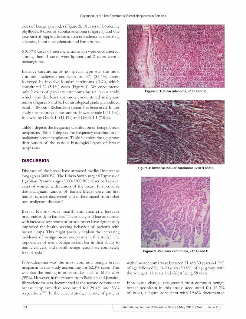
cases of benign phyllodes (Figure 2), 10 cases of borderline phyllodes, 8 cases of tubular adenoma (Figure 3) and one case each of nipple adenoma, apocrine adenoma, sclerosing adenosis, blunt duct adenosis and hamartoma.
6 (0.7%) cases of mesenchymal origin were encountered, among them 4 cases were lipoma and 2 cases were a hemangioma.
Invasive carcinoma of no special type was the most common malignant neoplasm i.e., 371 (85.5%) cases, followed by invasive lobular carcinoma (ILC), which constituted 22 (5.1%) cases (Figure 4). We encountered only 2 cases of papillary carcinoma breast in our study, which was the least common encountered malignant tumor (Figures 5 and 6). For histological grading, modified Scraff - Bloom - Richardson system has been used. In this study, the majority of the tumors showed Grade I (51.1%), followed by Grade II (41.1%) and Grade III (7.8%).
Table 1 depicts the frequency distribution of benign breast neoplasms. Table 2 depicts the frequency distribution of malignant breast neoplasms. Table 3 depicts the age group distribution of the various histological types of breast neoplasms.
DISCUSSION
Diseases of the breast have attracted medical interest as long ago as 3000 BC. The Edwin Smith surgical Papyrus of Egyptian Pyramids age (3000-2500 BC) described several cases of women with tumors of the breast. It is probable that malignant tumors of female breast were the first human cancers discovered and differentiated from other non-malignant diseases.9
Breast lesions pose health and cosmetic hazards predominantly in females. The anxiety and fear associated with increased awareness of breast cancer have significantly improved the health seeking behavior of patients with breast lumps. This might partially explain the increasing incidence of benign breast neoplasms in this study.8 The importance of many benign lesions lies in their ability to mimic cancers, and not all benign lesions are completely free of risks.5
Fibroadenoma was the most common benign breast neoplasm in this study accounting for 62.3% cases. This was also the finding in other studies such as Malik et al. (59%). However, in the reports from Pakistan and Jamaica, fibroadenoma was documented as the second commonest breast neoplasm that accounted for 29.4% and 33% respectively.10,11 In the current study, majority of patients
with fibroadenoma were between 21 and 30 years (41.9%) of age followed by 11-20 years (30.5%) of age group, with the youngest 11 years and oldest being 58 years.
Fibrocystic change, the second most common benign breast neoplasm in this study, accounted for 16.2% of cases, a figure consistent with 19.6% documented
Figure 3: Tubular adenoma, ×10 H and E
Figure 4: Invasive lobular carcinoma, ×10 H and E
Figure 5: Papillary carcinoma, ×10 H and E

82International Journal of Scientific Study | May 2015 | Vol 3 | Issue 2
Dayanand, et al.: The Spectrum of Breast Neoplasms in Females
in Nigeria.8 Fibrocystic change seems relatively more common in Pakistanis as Memmon reported a high frequency of 66.3% and observed a changing trend of benign tumors from fibroadenoma to fibrocystic change.11 This trend was however not observed in this study as fibroadenoma remained the commonest benign breast neoplasm throughout the 10 years study period. The
fibrocystic change consists of a spectrum of morphologic changes comprising cysts, adenosis, epithelial hyperplasia, and fibrosis. The age range of patients with fibrocystic change in our study was 15-60 years with a mean age of 30 years, which is in consonance with 30, 32, 33 and 37 years documented in Nigeria, Ibadan, Kano and Ife respectively.12-14 In contrast to fibrocystic change which showed a relatively high prevalence up to the fifth decade, a sharp decline in the occurrence of fibroadenoma was observed after the third decade.
One case each of eccrine acrospiroma, chondroid syringoma and dermatofibrosarcoma protuberans was encountered.
In our study, only one case of atypical ductal hyperplasia was reported. It has 4-5 fold risk of progression to invasive breast carcinoma.
Carcinoma of the breast ranked second in this study. The vast majority of the cases (371 out of 434) were invasive ductal carcinoma. There were only 6 cases of ductal carcinoma in-situ. In addition, 22 cases of ILC, 21 cases of medullary carcinoma, 6 cases of metaplastic carcinoma (Figure 7), 5 cases of malignant phyllodes,
Figure 6: Papillary carcinoma, ×10 IHC P63
Table 1: Benign breast neoplasms (851/1285)Type of lesion Number of patients PercentageApocrine adenoma 1 0.1Blunt duct adenosis 1 0.1Fibroadenoma 531 62.4Fibrocystic change 138 16.2Hamartoma 1 0.1Intraductal papilloma 8 0.9Hemangioma 2 0.2Lipoma 4 0.5Nipple adenoma 1 0.1Sclerosingadenosis 1 0.1Tubular adenoma 8 0.9Usual ductal hyperplasia 63 7.4Phyllodes - benign 65 7.6Borderline 10 1.2Chondroidsyringoma 1 0.1Eccrine spiroma 1 0.1DFSP 1 0.1ADH 1 0.1ADH: Atypical Ductal Hyperplasia, DFSP: Dermatofibrosarcoma protuberans
Table 2: Malignant breast neoplasms (434/1285)Type of lesion Number of cases PercentageDuctal carcinoma in‑situ 6 1.4Invasive ductal carcinoma 371 85.5ILC 22 5.1Medullary carcinoma 21 4.8Metaplastic carcinoma 6 1.4Mucinous carcinoma 3 0.7Papillary carcinoma 2 0.5Phyllodes-malignant 5 1.5Non Hodgkin lymphoma 4 0.9Pleomorphic MFH 1 0.2Carcinosarcoma 1 0.2Squamous cell carcinoma 1 0.2Undifferentiated carcinoma 2 0.5MFH: Malignant fibrous histiocytomas, ILC: Invasive lobular carcinoma
Table 3: Age distribution in the breast neoplasms‑ no. (percentage %)Age in years→ 10-20 21-30 31-40 41-50 51-60 61-70fibroadenoma 162 (30.5) 223 (41.9) 121 (22.7) 22 (4.1) 3 (0.5)fibrocystic change 35 (25.3) 60 (43.4) 35 (25.3) 7 (5.1) 1 (0.7)Usual Epithelial hyperplasia 20 (31.7) 20 (31.7) 18 (28.6) 5 (7.9)DCIS 0 1 (16.7) 2 (33.3) 2 (33.3) 0 1 (16.7)Invasive carcinoma 0 32 (7.5) 114 (26.8) 130 (30.6) 91 (21.4) 58 (13.6)Benign phyllodes 10 (15.3) 17 (26.2) 18 (27.7) 15 (23.1) 5 (7.7)Borderline phyllodes 0 1 (10) 5 (50) 3 (30) 1 (10)Malignant phyllodes 2 (40) 0 1 (20) 1 (20) 1 (20)Lymphoma 0 1 (25) 0 1 (25) 2 (50)Ductal carcinoma in‑situ
83 International Journal of Scientific Study | May 2015 | Vol 3 | Issue 2
Dayanand, et al.: The Spectrum of Breast Neoplasms in Females
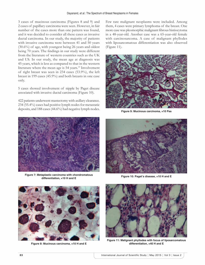
3 cases of mucinous carcinoma (Figures 8 and 9) and 2 cases of papillary carcinoma were seen. However, in fair number of the cases more than one pattern was found, and it was decided to consider all these cases as invasive ductal carcinoma. In our study, the majority of patients with invasive carcinoma were between 41 and 50 years (30.6%) of age, with youngest being 26 years and oldest being 70 years. The findings in our study were different from the literature of western countries such as the UK and US. In our study, the mean age at diagnosis was 45 years, which is less as compared to that in the western literature where the mean age is 54 years.15 Involvement of right breast was seen in 234 cases (53.9%), the left breast in 199 cases (45.9%) and both breasts in one case only.
5 cases showed involvement of nipple by Paget disease associated with invasive ductal carcinoma (Figure 10).
422 patients underwent mastectomy with axillary clearance. 234 (55.4%) cases had positive lymph nodes for metastatic deposits, and 188 cases (44.6%) had negative lymph nodes.
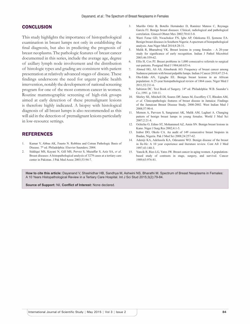
Few rare malignant neoplasms were included. Among them, 4 cases were primary lymphoma of the breast. One more case was pleomorphic malignant fibrous histiocytoma in a 48-year-old. Another case was a 65-year-old female with carcinosarcoma. A case of malignant phyllodes with liposarcomatous differentiation was also observed (Figure 11).
Figure 7: Metaplastic carcinoma with chondromatous differentiation, ×10 H and E
Figure 8: Mucinous carcinoma, ×10 H and E
Figure 10: Paget’s disease, ×10 H and E
Figure 9: Mucinous carcinoma, ×10 Pas
Figure 11: Malignant phyllodes with focus of liposarcomatous differentiation, ×40 H and E
84International Journal of Scientific Study | May 2015 | Vol 3 | Issue 2
Dayanand, et al.: The Spectrum of Breast Neoplasms in Females
CONCLUSION
This study highlights the importance of histopathological examination in breast lumps not only in establishing the final diagnosis, but also in predicting the prognosis of breast neoplasms. The pathologic features of breast cancer documented in this series, include the average age, degree of axillary lymph node involvement and the distribution of histologic types and grading are consistent with patient presentation at relatively advanced stages of disease. These findings underscore the need for urgent public health intervention, notably the development of national screening program for one of the most common cancer in women. Routine mammographic screening of high-risk groups aimed at early detection of these premalignant lesions is therefore highly indicated. A biopsy with histological diagnosis of all breast lumps is also recommended as this will aid in the detection of premalignant lesions particularly in low-resource settings.
REFERENCES
1. Kumar V, Abbas AK, Fausto N. Robbins and Cotran Pathologic Basis of Disease. 7th ed. Philadelphia: Elsevier Saunders; 2004.
2. Siddiqui MS, Kayani N, Gill MS, Pervez S, Muzaffar S, Aziz SA, et al. Breast diseases: A histopathological analysis of 3279 cases at a tertiary care center in Pakistan. J Pak Med Assoc 2003;53:94-7.
3. Murillo Ortiz B, Botello Hernández D, Ramírez Mateos C, Reynaga García FJ. Benign breast diseases: Clinical, radiological and pathological correlation. Ginecol Obstet Mex 2002;70:613-8.
4. Warri Forae GD, Nwachokor FN, Igbe AP, Odokuma EI, Ijomone EA. Benign breast diseases in Southern Nigeria: A spectrum of histopathological analysis. Ann Niger Med 2014;8:28-31.
5. Malik R, Bharadwaj VK. Breast lesions in young females – A 20-year study for significance of early recognition. Indian J Pathol Microbiol 2003;46:559-62.
6. Ellis H, Cox PJ. Breast problems in 1,000 consecutive referrals to surgical out-patients. Postgrad Med J 1984;60:653-6.
7. Ahmed HG, Ali AS, Almobarak AO. Frequency of breast cancer among Sudanese patients with breast palpable lumps. Indian J Cancer 2010;47:23-6.
8. Olu-Eddo AN, Ugiagbe EE. Benign breast lesions in an African population: A 25-year histopathological review of 1864 cases. Niger Med J 2011;52:211-6.
9. Sabiston DC. Text Book of Surgery. 14th ed. Philadelphia: W.B. Saunder’s Co.;1991. p. 510-11.
10. Shirley SE, Mitchell DI, Soares DP, James M, Escoffery CT, Rhoden AM, et al. Clinicopathologic features of breast disease in Jamaica: Findings of the Jamaican Breast Disease Study, 2000-2002. West Indian Med J 2008;57:90-4.
11. Memon A, Parveen S, Sangrarasi AK, Malik AM, Laghari A. Changing pattern of benign breast lumps in young females. World J Med Sci 2007;2:21-4.
12. Ochicha O, Edino ST, Mohammed AZ, Amin SN. Benign breast lesions in Kano. Niger J Surg Res 2002;4:1-5.
13. Irabor DO, Okolo CA. An audit of 149 consecutive breast biopsies in Ibadan, Nigeria. Pak J Med Sci 2008;24:257-62.
14. Adeniji KA, Adelusola KA, Odesanmi WO. Benign disease of the breast in Ile-Ife: A 10 year experience and literature review. Cent Afr J Med 1997;43:140-3.
15. Yancik R, Ries LG, Yates JW. Breast cancer in aging women. A population-based study of contrasts in stage, surgery, and survival. Cancer 1989;63:976-81.
How to cite this article: Dayanand V, Shashidhar HB, Sandhya M, Ashwini NS, Bharathi M. Spectrum of Breast Neoplasms in Females: A 10 Years Histopathological Review in a Tertiary Care Hospital. Int J Sci Stud 2015;3(2):79-84.
Source of Support: Nil, Conflict of Interest: None declared.