Embed Size (px)
Citation preview

Specifying the Brain Anatomy UnderlyingTemporo-Parietal Junction Activations for Theoryof Mind: A Review using Probabilistic Atlases from
Different Imaging Modalities
Matthias Schurz ,1,2* Matthias G. Tholen,2 Josef Perner,2
Rogier B. Mars,3,4 and Jerome Sallet1
1Department of Experimental Psychology, University of Oxford, Oxford, United Kingdom2Centre for Cognitive Neuroscience, University of Salzburg, Salzburg, Austria
3Donders Institute for Brain, Cognition and Behaviour, Radboud University Nijmegen,Nijmegen, The Netherlands
4Wellcome Centre for Integrative Neuroimaging, Centre for Functional MRI of the Brain (FMRIB),Nuffield Department of Clincial Neurosciences, John Radcliffe Hospital, University of Oxford
r r
Abstract: In this quantitative review, we specified the anatomical basis of brain activity reported in theTemporo-Parietal Junction (TPJ) in Theory of Mind (ToM) research. Using probabilistic brain atlases, welabeled TPJ peak coordinates reported in the literature. This was carried out for four different atlasmodalities: (i) gyral-parcellation, (ii) sulco-gyral parcellation, (iii) cytoarchitectonic parcellation and (iv)connectivity-based parcellation. In addition, our review distinguished between two ToM task types (falsebelief and social animations) and a nonsocial task (attention reorienting). We estimated the mean proba-bilities of activation for each atlas label, and found that for all three task types part of TPJ activations fellinto the same areas: (i) Angular Gyrus (AG) and Lateral Occpital Cortex (LOC) in terms of a gyral atlas,(ii) AG and Superior Temporal Sulcus (STS) in terms of a sulco-gyral atlas, (iii) areas PGa and PGp interms of cytoarchitecture and (iv) area TPJp in terms of a connectivity-based parcellation atlas. Besidethese commonalities, we also found that individual task types showed preferential activation for particu-lar labels. Main findings for the right hemisphere were preferential activation for false belief tasks in AG/PGa, and in Supramarginal Gyrus (SMG)/PFm for attention reorienting. Social animations showed stron-gest selective activation in the left hemisphere, specifically in left Middle Temporal Gyrus (MTG). We dis-cuss how our results (i.e., identified atlas structures) can provide a new reference for describing futurefindings, with the aim to integrate different labels and terminologies used for studying brain activityaround the TPJ. Hum Brain Mapp 00:000–000, 2017. VC 2017 Wiley Periodicals, Inc.
Additional Supporting Information may be found in the onlineversion of this article.
Contract grant sponsor: Austrian Science Fund (ErwinSchr€odinger Fellowship to M.S.); Contract grant number:FWF-J4009-B27; Contract grant sponsor: Austrian Science Fund’sDoctoral College “Imaging the Mind” (to M.T.); Contract grantnumber: FWF-W1233-G17; Contract grant sponsor: VIDI (DutchOrganization for Scientific Research NWO to R.B.M.); Contractgrant number: 452-13-015; Contract grant sponsor: WellcomeTrust Sir Henry Dale Fellowship (to J.S.).
Matthias Schurz and Matthias G. Tholen contributed equally tothis work.
*Correspondence to: Matthias Schurz, Department of Experimen-tal Psychology, University of Oxford, Tinbergen Building, 9 SouthParks Road, Oxford OX1 3UD. E-mail: [email protected]
Received for publication 24 March 2016; Revised 17 May 2017;Accepted 22 May 2017.
DOI: 10.1002/hbm.23675Published online 00 Month 2017 in Wiley Online Library(wileyonlinelibrary.com).
r Human Brain Mapping 00:00–00 (2017) r
VC 2017 Wiley Periodicals, Inc.

Key words: mentalizing; social cognition; attention reorienting; neuroanatomy; angular gyrus; superiortemporal sulcus
r r
INTRODUCTION
The TPJ
The term Temporo-Parietal Junction (TPJ) is often usedfor labeling functional activations around the border ofparietal and posterior temporal lobes in multiple fields ofresearch. In the social cognitive neurosciences, TPJ activa-tion is a prominent neural correlate of Theory of Mind(ToM), that is, the human ability to ascribe mental stateslike beliefs and desires to other people [see e.g., Molen-berghs et al., 2016; Saxe and Kanwisher, 2003; Schurzet al., 2014; Spreng et al., 2009; Van Overwalle, 2009]. Acrucial role of the right TPJ for mentalizing was found intranscranial magnetic stimulation studies, showing that atransient disruption of the area impairs reasoning aboutthe beliefs of others [Krall et al., 2016; Young et al., 2010].Moreover, imaging studies [for meta-analysis, see Sugra-nyes et al., 2011] found TPJ dysfunction in clinical disor-ders that involve ToM impairments, as for example autism[Yirmiya et al., 1998] and schizophrenia [Sprong et al.,2007].
To date, there exists little consensus regarding microana-tomical or macroanatomical landmarks that topographicallydefine the TPJ area [c.f. Bzdok et al., 2013a,b; Geng andVossel, 2013; Mars et al., 2011], hampering a precise com-parison of theories and conclusions drawn about the neuro-cognitive processes underlying TPJ activity across differentToM studies, and across ToM and nonToM studies. Ana-tomical characterizations of the TPJ are found sporadicallyin the literature. We illustrate some of them in Figure 1A:Characterizations have been ranging from a focal areawithin one gyrus [Chambers et al., 2004] to an intersectionarea between different gyri/sulci [Mort et al., 2003] and abroader area comprising several gyri and sulci [Corbettaet al., 2008; Decety and Lamm, 2007]. Figure 1A also showsa historical example of an early mentioning of “temporo-parietale” used to classify the locus of brain damage pro-ducing a certain form of behavioral, cognitive and motordysfunctions [Kr€onlein, 1886; see also Stenger, 1881].
Aims of the Present Review
This review aims to shed light on the relation betweenfunctional activations of the TPJ and underlying brainstructure in ToM research. More specifically, we take aquantitative approach to test if functional activations ofthe TPJ are systematically related to specific brain struc-tures, or unsystematically distributed across diverse corti-cal areas/lobes. We seek to advance knowledge regarding
function-structure correspondence in the TPJ for ToM byaddressing three important issues surrounding the topic:(i) The anatomical level at which correspondence can befound (ii) The role of interindividual variability in brainanatomy (iii) The effects of stimuli and task-instructionsfor probing ToM brain activation. In the following sec-tions, we motivate each issue and lay out our approach foraddressing it.
i. The Anatomical Level at which
Correspondence Can be Found
The existence of a consistent relation between functionalactivations of the TPJ and anatomical structures commonlyused for labeling is an open question for several reasons.First, TPJ activations may not fall into a gyrus or severalgyri, but alternatively (or additionally) in sulci such as theposterior Superior Temporal Sulcus (pSTS). In fact, boththe labels TPJ [e.g., Saxe and Kanwisher, 2003, Schaafsmaet al., 2015; Spunt and Adolphs, 2014; Spunt et al., 2016]and pSTS [Carrington and Bailey, 2009; Frith and Frith,2003, 2008; Lieberman, 2007; Singer, 2006] are highly popu-lar for characterizing brain functions involved in socialcognition and ToM. However, since most ToM researchrelied on volume-based brain analyses and gyral parcella-tions, explicit distinctions between temporo-parietal gyriand adjacent pSTS are rare [for a recent exception, seeDeen et al., 2015]. Nevertheless, it is estimated that around55% [Destrieux et al., 2010] to 60% [Van Essen, 2005; Zilleset al., 1997] of cortex are buried in sulci and lateral fossa.To address this issue in our review, we label functionalactivations not only with a gyral atlas, but also with asulco-gyral atlas based on freesurfer cortical surface analy-sis (using a 3D transformed version, see Methods).
Second, it is an open question if functional specializationof the TPJ area consistently relates to sulcal and gyral pat-terns at all. While functional specialization of an area isknown to be strongly determined by cytoarchitectonic fea-tures [e.g., Luppino et al., 1991], the relationship betweencytoarchitectonic borders and surrounding sulci and gyriis rather loose for most multimodal association areas suchas the TPJ [e.g., Amunts et al., 1999; see also Amuntset al., 2007]. To address this issue, we will specify the ana-tomical basis of functional TPJ activations not only bygyral and sulco-gyral atlases, but also in terms of acytoarchitectonic atlas [Caspers et al., 2006, 2008].
In addition, our labeling based on a cytoarchitectonicatlas will be complemented by a recent connectivity-basedparcellation (CBP) atlas, grouping together voxels with acommon connectivity pattern to the rest of the brain
r Schurz et al. r
r 2 r

Figure 1.
(A) Example anatomical specifications of the TPJ. Colored areas show
major gyri, according to a widely used macroscopic parcellation of the
MNI brain [Tzourio-Mazoyer et al., 2002]. LOC, Lateral Occipital Cor-
tex (area labeled LOC for consistency with some TPJ definitions, actu-
ally corresponds to Middle Occipital Gyrus); AG, Angular Gyrus; SMG,
Supramarginal Gyrus; IPL, Inferior Parietal Lobule; STG, Superior Tem-
poral Gyrus; pSTS, posterior Superior Temporal Sulcus. Image A.6.
from “€Uber die Trepanation bei Blutungen aus der A. meningea media
und geschlossener Sch€adelkapsel” by R.U. Kr€unlein, 1886, Deutsche
Zeitschrift f€ur Chirurgie, p. 216. Adapted with permission of Springer.
(B) Display of all TPJ relevant structures in different atlases. Structures
from gyral, cytoarchitectonic and connectivity-based parcellation atlases
are shown in MNI space, sulco-gyral atlas structures are shown in TAL
space. For display purposes, we thresholded the probabilistic atlas
maps of connectivity-parcellations and cytoarchitectonics at 0.50, of
gyral-parcellations at 0.25 and of sulco-gyral parcellations at 0.20.
[Color figure can be viewed at wileyonlinelibrary.com]
r Brain Anatomy Underlying Temporo-Parietal Junction r
r 3 r

[Johansen-Berg et al., 2004]. A consistent relation betweensingle-subject connectivity-patterns and task-based func-tional activations was shown in previous studies [e.g.,Osher et al., 2016; Saygin et al., 2016; Tavor et al., 2016].Furthermore, a recent study showed high correspondencebetween cytoarchitectonic and connectivity-based parcella-tion areas [Henssen et al., 2016], which motivates the useof both types of atlases in our review for obtaining arobust and detailed characterization of brain anatomy ofthe TPJ.
ii. The Role of Interindividual Variability in Brain
Anatomy
Another issue we address is that TPJ shows high interin-dividual variability in macroanatomy [e.g., Segal and Pet-rides, 2012; Zlatkina and Petrides, 2014], which ischaracteristic for multimodal association areas of the cor-tex [Van Essen, 2005]. This could obscure an accurate ana-tomical characterization of functional activations found inthe area. The present review tackles this problem by usingprobabilistic atlases of the different imaging modalities wemotivated above [Caspers et al., 2006, 2008; Desikan et al.,2006; Destrieux et al., 2010; Mars et al., 2012]. These atlasesare probabilistic in the sense that they explicitly incorpo-rate measurements of interindividual variability in struc-ture at any coordinate [Devlin and Poldrack, 2007].Concretely, this means that one coordinate can be proba-bilistically assigned to multiple areas, that is, probabilityvalues indicate in how many subjects the coordinate isreferring to particular areas.1
iii. The Effects of Stimuli and Task-Instructions
for Probing ToM Brain Activation
The third issue we address is the role of stimulus-typeand task-instructions in probing brain activity for ToM.We recently found in a voxel-wise imaging meta-analysisthat activation in posterior temporo-parietal areas islargely distinct for different ToM task types [Schurz et al.,2014]. In the present review, we follow up this finding bysystematically studying the underlying anatomy. Compar-ing anatomy between different ToM task types adds anew level of detail to insights from earlier reviews [e.g.,Bzdok et al., 2012; Decety and Lamm, 2007; Geng and Vos-sel, 2013], where different experimental paradigms havebeen pooled together to identify common activation acrossall ToM tasks.
Concretely, we will compare findings from the ToMtasks false belief and social animations. Research showsthat these tasks activate slightly different parts of thetemporo-parietal cortex [e.g., Bahnemann et al., 2010; Gob-bini et al., 2007; Schurz et al., 2014], which was linked toconceptual differences between the tasks: False belief stud-ies present stories about persons with incorrect assump-tions or beliefs about a state of affairs. Such stories arethought to represent a prototypical and theoreticallyimportant test of ToM reasoning abilities [e.g., Saxe andKanwisher, 2003; Saxe et al., 2004]. In contrast, social ani-mations are intended [Castelli et al., 2000, 2002] as a low-level alternative to the verbal stimuli used in many ToMtasks. In social animation tasks, a movie shows simplegeometrical shapes (e.g., two triangles) moving across thedisplay and portraying actions that are typical for anintentional or social interaction. Examples for both falsebelief and social animations tasks are given in Figure 2.
Finally, we not only seek to find out how well our label-ing approach characterizes TPJ anatomy for ToM, but alsoin how far this characterization is distinct from what isfound for nonsocial tasks. An important debate over thelast decade regards the TPJ’s role in attention versus socio-cognitive processes [Decetyand Lamm, 2007; Igelstr€omet al., 2016; Krall et al., 2015, 2016; Kubit and Jack, 2013;Lee and McCarthy, 2016; Mitchell, 2008; €Ozdem et al.,2017; Scholz et al., 2009; see also Carter and Huettel, 2013].Therefore, we include attention reorienting tasks in ourreview, allowing us to contrast the anatomical characteri-zation for ToM tasks to that found for a nonsocial task,which requires the redirection of attention toward a targetstimulus after a breach of expectation. By specifying theanatomical basis of TPJ activations for ToM and attentionreorienting, we provide a starting point for studying over-laps and differences in TPJ activation reported for ToMand many other research fields outside the scope of thisreview, such as reasoning about actions [e.g., Molenberghset al., 2009; Spunt et al., 2016; Van Overwalle, 2009], empa-thy [e.g., Bzdok et al., 2012; Lamm et al., 2011; Singer andLamm, 2009], episodic memory [e.g., Spreng et al., 2009],semantic processing [e.g., Binder et al., 2009], visuospatialnavigation [e.g., Spreng et al., 2009], reading and compre-hension [e.g., Seghier, 2013], bodily awareness [e.g., Blankeet al., 2002] or inhibition in go/no-go tasks [e.g., Nee et al.,2007].
Questions and Hypotheses
We laid out outstanding issues for finding structure-function correspondence in the TPJ, and presented howour review will address them by a multiple-modality andprobabilistic atlas labeling approach, focusing on two dif-ferent ToM task types and a task type outside the socialdomain. Our review will cover functional activity for bothleft and right TPJ, although the right TPJ tends to be dis-cussed more frequently in the Theory of Mind literature
1Note that while all atlases in our review are “probabilistic” in thesense that they explicitly measured interindividual variability inbrain structure, they are based on different sources of neurobiologi-cal evidence and analysis approaches for generating underlyingmaps in individuals (e.g., observer independent microstructuralanalysis for cytoarchitectonic maps; see Schleicher et al., 1999; forreview Amunts et al., 2007).
r Schurz et al. r
r 4 r

[e.g., Bzdok et al., 2013b; Corbetta et al., 2008; Decety andLamm, 2007; Krall et al., 2015, 2016; Kubit and Jack, 2013;Saxe and Kanwisher, 2003]. Our main interest is to providean evaluation of the following questions: (i) Within eachtask type, do activations labeled as “TPJ” fall reliably intoindividual anatomical structures, or are they distributedbroadly and with high variance over the temporo-parietalcortex? (ii) When comparing different task types, do acti-vations labeled as “TPJ” fall in the same or into differentanatomical structures?
METHODS
Atlases for Labeling
Gyral and sulco-gyral anatomy
For labeling functional activations, we used two macroana-tomical atlases. First, we used a standard macroanatomical(i.e., gyral parcellation) brain atlas (Harvard-Oxford CorticalStructural Atlas) [Desikan et al., 2006]. Second, we used asulco-gyral parcellation atlas that is based on a freesurfer cor-tical surface analysis [Dale et al., 1999; Fischl et al., 1999].The approach for this atlas [Destrieux et al., 2010] was to firstdefine gyri based on a 3D reconstruction of the cortex, andthen inflate this model to label sulci. As activation coordi-nates reported in this review have been generated by 3Dvolume-based frameworks, we use a 3D transformed Talair-ach space version of the sulco-gyral atlas, referred to asTT_desai_ddpmaps in AFNI (https://afni.nimh.nih.gov/afni/) and provided by R. Desai and colleagues [e.g., Lieben-thal et al., 2014]. The map is shown Figure 1B.
Cytoarchitecture
We additionally used a cytoarchitectonic atlas, which isbased on segregating cortical areas according to the typeand organization of cells they contain. Cytoarchitectonicshave been found to closely correspond to functional spe-cialization of an area [e.g., Luppino et al., 1991]. At pre-sent, cytoarchitectonic maps do not cover the whole brain,and mapping is still in progress as it is a time-consumingprocess [Amunts et al., 2007]. For currently available mapssee the SPM Anatomy Toolbox [Eickhoff et al., 2005,www.fil.ion.ucl.ac.uk/spm/ext/]. With respect to areassurrounding the TPJ, cytoarchitectonic maps are to dateavailable for the Inferior Parietal Lobule (IPL), and not forposterior temporal lobe structures such as posterior supe-rior temporal gyrus and sulcus. As shown in Figure 1B,the IPL can be divided into into seven subareas based oncytoarchitectonic features [Caspers et al., 2006, 2008]. Inaddition to differences in cytoarchitecture, these sevensubareas of IPL also show different white-matter connec-tivity fingerprints [Caspers et al., 2011], and have differentpatterns of neurotransmitter receptor densities [Casperset al., 2013].
Figure 2.
Examples for the three task types reviewed. (A). False belief
tasks presenting written stories about false belief (left) versus
false photo controls (right). (B). Social animations presenting
movies of geometrical shapes that portray a social interaction
(left: mother playing with child) or purely mechanical move-
ments (right: billiard-balls moving on the table). (C). In the criti-
cal experimental condition, attention reorienting tasks ask for
redirecting attention towards a target stimulus after a breach of
expectation (here: invalid cueing condition). [Color figure can be
viewed at wileyonlinelibrary.com]
r Brain Anatomy Underlying Temporo-Parietal Junction r
r 5 r

Connectivity-based parcellations
Connectivity-based parcellation analysis [CBP,Johansen-Berg et al., 2004; for reviews see Eickhoff et al.,2015; Mars et al., 2016] groups voxels into an area if theyshow a common pattern of structural brain connectivitywith the rest of the brain—distinct from that in neighbor-ing voxels. In the present review, we use connectivity-based parcellation atlases of TPJ and adjacent IPL thatare based on diffusion weighted imaging data [Mars etal., 2011, 2012]. As illustrated in Figure 1B, the TPJ isdivided into three subareas TPJa, TPJp and IPL, whichare linked to distinct brain networks. Furtherconnectivity-based parcellations of the TPJ have been per-formed recently [Bzdok et al., 2013b; Igelstr€om et al.,2015; Yeo et al., 2011, see also Uddin et al., 2010]. Forexample, Bzdok et al. [2013b] relied on resting-state fMRIand meta-analytic coactivation data for connectivity-parcellation, and results are in good correspondence toMars et al. [2012] with respect to an anterior/posteriordivision in the TPJ—Bzdok et al.’s [2013b] parcellation isshown in Figure 1A. All atlases used in the review wereaccessed with FSL (http://fsl.fmrib.ox.ac.uk/fsl/fslview/)or AFNI (https://afni.nimh.nih.gov/afni/) software.
Literature Samples
We analyzed the literature samples from our meta-analysis on ToM [Schurz et al., 2014] and a meta-analysison attention reorienting [Kubitand Jack, 2013]. Weincluded 15 studies using false belief tasks, 14 studies pre-senting social animations, and 15 studies presenting atten-tion reorienting tasks [for further details see Kubit andJack, 2013; Schurz et al., 2014]. All studies reported activa-tion coordinates in standard space, and whenever neces-sary (different with each atlas we used for labeling), weconverted from/to MNI or TAL space by using a matrixtransformation by Lancaster et al. [2007].
For the ToM task types, literature samples wereretrieved by a database search with the key words (i)“neuroimaging” or “fMRI” or “PET” and (ii) “theory-of-mind” or “mentalizing” or “mindreading,” and addition-ally by considering studies cited in earlier ToM reviews[e.g., Bzdok et al., 2012; Denny et al., 2012; Mar, 2011;Murray et al., 2012; Perner and Leekam, 2008; Sprenget al., 2009; Van Overwalle, 2009; Van Overwalle and Baet-ens, 2009].
Studies were included in the false belief sample if they pre-sented written stories about a person’s false belief (see Fig.2A). Activation coordinates were only taken from contrastsbetween false belief versus false photo control stories. In thisclosely matched type of control condition, participants readstories where they have to represent the outdated content ofa physical representation. Studies were included in the socialanimations category, if they presented movies with simplegeometrical shapes (e.g., triangles) that portrayed a social orintentional interaction (Fig. 2B). Activation coordinates were
taken from contrasts between movies showing movementsthat characterize social/intentional interactions and moviesshowing random or purely mechanical movements.
For attention reorienting tasks, a database search withthe key-words (i) “fMRI” or “PET” and (ii) “reorienting”or “posner” was performed, and additionally studies citedin earlier attention reorienting reviews were considered[Corbettaand Shulman, 2002; Corbetta et al., 2008; Decetyand Lamm, 2007].
Tasks were considered as attention reorienting, if partici-pants had to redirect attention towards a target stimulusafter a breach of expectation (see Fig. 2C). Activation coor-dinates were taken from contrasts that compared partici-pants having to redirect attention after being misinformedabout the location of an upcoming target stimulus versusparticipants being correctly informed about the location ofthe upcoming target.
Procedure: Definition of TPJ and Steps for
Labeling
Figure 3 illustrates the steps of our review. The first stepwas forming a “democratic” common denominator of thelabel TPJ. We went through results and discussion sectionsof individual papers and looked for coordinates of activa-tions that were referred to as “Temporo-Parietal Junction”or “TPJ.” We collected all these coordinates and labeledthem with a standard macroanatomical brain atlas that iscovering all temporal and parietal cortical territory (Har-vard-Oxford Cortical Structural Atlas) [Desikan et al.,2006]. That is, we assigned the most probable label to eachcoordinate, resulting in a list of all atlas structures referredto as “TPJ” (see Fig. 3—Step 2): Angular Gyrus (AG), pos-terior division of Supramarginal Gyrus (SMGpd), superiordivision of Lateral Occipital Cortex (LOCsd), and temporo-occipital part of Middle Temporal Gyrus (MTGtop). In Step3, we took into account that not all authors in the litera-ture use the label “TPJ” for their findings, so some mightbe still missing. Therefore, in Step 3 we went back to allpapers not mentioning the term “TPJ” and looked foradditional coordinates that fell into the atlas structuresdefined in the step before (i.e., in Step 2). Taking togetherfindings from Steps 2 and 3, we found 109 coordinatesthat could be labeled as “TPJ” (41 Left, 68 Right). Theseconsisted of 36 coordinates from false belief tasks (18L, 18R), 32 from social animations (17 L, 15 R) and 41 fromattention reorienting tasks (6 L, 35 R). In Step 4, we startedour actual probabilistic labeling procedure with the differ-ent atlases: For every coordinate, we determined the prob-ability for falling into each area of our probabilistic atlases.This procedure produces multiple probabilities per coordi-nate—(i.e., one location can be linked to multiple atlaslabels with varying probabilities, e.g., the MNI coordinatex 5 45, y 5 54, z 530 is 15% Angular Gyrus, 7% Supramar-ginal Gyrus, etc.)
r Schurz et al. r
r 6 r

Statistical Analysis
Atlas labeling
Separately for each task type, we estimated for eachatlas area the mean probability of activation (and 95% con-fidence interval) using percentile bootstrapping analysis[see Efron and Tibshirani, 1993] with 1,000 replicates.Analysis was carried out with the IBM SPSS bootstrappingmodule (22.0). We used bootstrapping as it is robust incase of small samples and non-normal data distributions.To evaluate the main questions we laid out in the Intro-duction, we carried out two analyses. First, we tested forthe three task types separately if the mean probability ofactivation found for each label is different from 0%. Thisindicates if a label is systematically associated with theactivations reported for a task type. The statistical criterionfor significance we applied was that the 95% confidenceinterval of the mean does not include a probability valueof 0 (all values were rounded off to whole numbers). Thiscriterion for confidence intervals can be taken to provide
the same evidential standard as null hypothesis testing ata 5% significance-level [Tryon, 2001]. Second, we tested ifmean probabilities are significantly different between anytwo task types which indicates that a label is morestrongly associated with activations for one task-type thanthe other. We carried out pairwise comparisons where wetested if the 95% confidence intervals of the means wereoverlapping or not.
Spatial variability. Additionally, we calculated for eachcoordinate the mean Euclidean distance to all neighborswithin the same task type and hemisphere. To test whetherthe spatial variability differed significantly between the tasktypes, we used the same bootstrap procedure and statisticalparameters as for the previous analysis.
Neurosynth Custom Meta-Analysis
For the discussion of our results, we also carried out neuro-synth (www.neurosynth.org) custom meta-analyses for the
Figure 3.
Illustration of the steps of our review. Step 1: Going through
results and discussion sections of individual papers and collect
coordinates that are referred to as “TPJ.” Step 2: Looking-up all
coordinates up in a standard gyral atlas (Harvard-Oxford) and
forming an outline of all targeted areas (common denominator
of TPJ). Step 3: Going back to papers not mentioning the TPJ
and looking for additional coordinates falling into the common
denominator structures. Step 4: Carrying out probabilistic label-
ing with four different atlases. [Color figure can be viewed at
wileyonlinelibrary.com]
r Brain Anatomy Underlying Temporo-Parietal Junction r
r 7 r
labels “TPJ,” “pSTS” and “Wernicke’s area” (custom meta-analyses were performed because the default neurosynthmeta-analysis database currently does not cover all of thethree terms). We searched the pubmed database (https://www.ncbi.nlm.nih.gov/pubmed, October 2016) for allarticles mentioning in their title or abstract the terms (i)“temporo-parietal junction” AND “fmri,” (ii) “posteriorsuperior temporal sulcus” AND “fmri” and (iii) “wernicke’sarea” AND “fmri.” For each pubmed results list, we searchedfor all corresponding study entries in the neurosynth data-base, and created custom meta-analyses for all foundmatches. This resulted in (i) a TPJ meta-analysis (85 studies,neurosynth ID: 24e0f983-ede4–4c5c), (ii) a pSTS meta-analysis (97 studies, neurosynth ID: 3d5b8bc7–4eed-49b8)and (iii) a Wernicke’s area meta-analysis (19 studies, ID:58aeb86a-f112–4a17). For results we show reverse inferencemaps for the three terms (i.e., likelihood that label is usedgiven presence of activity at a given voxel in the brain) at athresholded of P< 0.01 FDR corrected. Note that for the termWernicke’s area we found considerably fewer studies, andtherefore meta-analytic results should be taken with caution.
RESULTS
For sake of brevity, we only mention findings in the textwith an activation probability (or difference in activationprobabilities) of 10% or more. However, detailed resultsare given in Table I. An overview of all atlases and struc-tures is given in Figure 1B.
Gyral Anatomy
Results for task types separately
False belief. Dark blue areas in Figure 4A show that acti-vation probabilities were highest in bilateral AngularGyrus (AG) and bilateral Lateral Occipital Cortex, superiordivision (LOCsd), see Table I for details.
Social animations. Light blue areas in Figure 4A showthat in both hemispheres, activation probabilities werehighest in AG, LOCsd and the Middle Temporal Gyrus,temporo-occipital part (MTGtop). In the left hemisphere,activation probabilities were also high for the Supramargi-nal Gyrus, posterior division (SMGpd).
Attention reorienting (RH only). Similar to the patternfor social animations, activation probabilities were highestin right AG, SMGpd and LOCsd.
Task differences
Higher activation probabilities for false belief comparedto social animations were found in right AG, see Table Ifor details. In contrast, activation probabilities were higherin bilateral MTGtop for social animations compared to falsebelief. Higher probabilities for false belief compared to
attention reorienting were found in right AG, and theopposite pattern was found in right SMGpd.
Sulco-Gyral Anatomy
Results for task types separately
False belief. Figure 4B shows that activation probabilitiesfor this task were highest in bilateral Superior TemporalSulcus (STS) and Angular Gyrus (AG). In the right hemi-sphere activation probability was slightly higher for theSTS, and in the left hemisphere slightly higher for AG.
Social animations. In the right hemisphere, activationprobabilities were highest in AG and STS. In the left hemi-sphere, they were highest in AG and Middle TemporalGyrus (MTG).
Attention reorienting (RH only). Activation probabilitieswere highest in AG and STS.
Task differences
Results of pairwise comparisons in Figure 4B show thatfalse belief tasks had higher activation probability com-pared to social animations in left STS. The opposite patternwas found in left MTG. False belief compared to attentionreorienting showed higher probabilities in right AG andright STS. No differences were found between social ani-mations and attention reorienting.
Cytoarchitecture
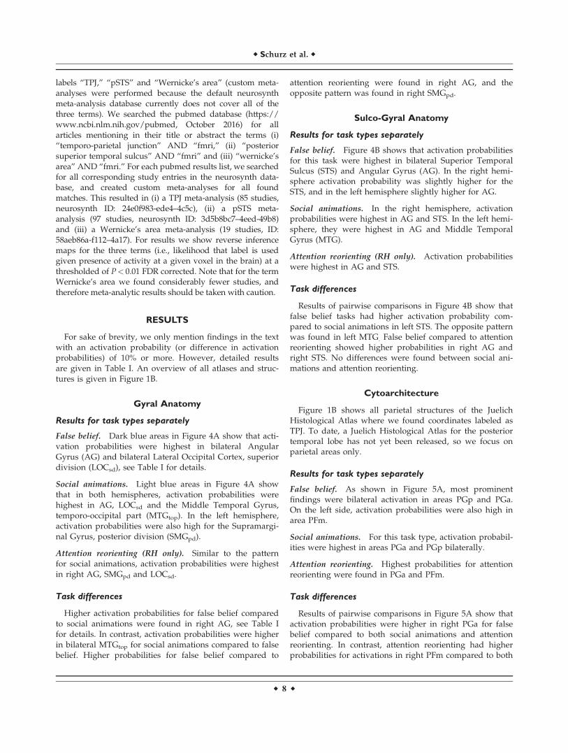
Figure 1B shows all parietal structures of the JuelichHistological Atlas where we found coordinates labeled asTPJ. To date, a Juelich Histological Atlas for the posteriortemporal lobe has not yet been released, so we focus onparietal areas only.
Results for task types separately
False belief. As shown in Figure 5A, most prominentfindings were bilateral activation in areas PGp and PGa.On the left side, activation probabilities were also high inarea PFm.
Social animations. For this task type, activation probabil-ities were highest in areas PGa and PGp bilaterally.
Attention reorienting. Highest probabilities for attentionreorienting were found in PGa and PFm.
Task differences
Results of pairwise comparisons in Figure 5A show thatactivation probabilities were higher in right PGa for falsebelief compared to both social animations and attentionreorienting. In contrast, attention reorienting had higherprobabilities for activations in right PFm compared to both
r Schurz et al. r
r 8 r
TA
BL
EI.
Deta
iled
pro
bab
ilis
tic
lab
elin
gre
sult
s
Fal
seB
elie
fS
oci
alA
nim
atio
ns
Att
enti
on
Reo
rien
tin
gF
Bv
s.S
AF
Bv
s.A
RS
Av
s.A
R
HL
abel
M(%
)95
%C
IM
(%)
95%
CI
M(%
)95
%C
IM
d(%
)95
%C
IM
d(%
)95
%C
IM
d(%
)95
%C
I
Harv
ard
-Oxfo
rdG
yra
lA
tlas
RA
G57.5
[45;
68]
17.6
[8;
26]
20.3
[14;
27]
39.9
[27;
54]
37.2
[23;
48]
22.
7[-
14;
9]S
MG
,p
d1.
5[1
;2]
8.4
[2;
17]
13.9
[9;
19]
26.
9[-
15;
21]
-12.4
[-18
;2
7]2
5.5
[-14
;4]
LO
C,
sd13.8
[5;
25]
22.3
[9;
38]
16.4
[9;
25]
28.
5[-
26;
9]2
2.5
[-14
;11
]6.
0[-
10;
23]
MT
G,
top
4.2
[1;
8]14.5
[5;
26]
8.7
[3;
15]
-10.2
[-23
;2
1]2
4.5
[-12
;2]
5.8
[-6;
18]
LO
C,
id1.
3[0
;4]
0.5
[0;
1]1.
6[0
.4;
4]0.
7[-
1;0]
20.
3[-
3;3]
21.
1[-
3;0]
ST
G,
pd
0.0
0.3
[0;
0.7]
0.3
[0.3
;1]
20.
3[-
1;0]
20.
3[-
1;0]
0.0
[-0.
9;1]
LA
G26.7
[17;
37]
15.1
[7;
25]
11.3
[-1;
25]
SM
G,
pd
4.6
[2;
9]13.3
[5;
21]
28.
7[-
18;
21]
LO
C,
sd36.1
[24;
49]
22.4
[8;
38]
13.6
[-6;
34]
MT
G,
top
0.3
[0;1
]12.1
[3;
22]
-11.7
[-22
;2
3]L
OC
,id
0.6
[1;
2]2.
6[0
;6]
22.
0[-
5;1]
ST
G,
pd
0.2
[0;
1]2.
0[0
;4]
21.
8[-
4;0]
Su
lco
-gy
ral
Atl
as
inT
AL
space
RA
G21.3
[14;
29]
12.8
[3;
26]
11.0
[7;
15]
8.4
[-7;
23]
10.2
[2;
19]
1.8
[-8;
15]
SM
G0.
3[0
;1]
0.9
[0;
3]4.
7[2
;8]
20.
6[-
2;1]
24.
4[-
8;2
2]2
3.8
[-8;
21]
SP
L0.
0[0
;0]
0.1
[0;
0]2.
5[1
;4]
20.
1[-
0.4;
0]2
2.5
[-5;
21]
22.
4[-
5;2
1]S
TG
,la
t0.
0[0
;0]
0.7
[0;
1]1.
4[0
;3]
20.
7[-
1;0]
21.
4[-
3;0]
20.
7[-
2;1]
MT
G0.
8[0
;2]
2.2
[0;
7]2.
6[1
;5]
21.
4[-
6;1]
21.
8[-
4;0]
20.
3[-
4;5]
Jen
s.S
ulc
.1.
4[1
;2]
0.1
[0;
0]2.
5[1
;4]
1.2
[1;
2]2
1.1
[-3;
21]
22.
4[-
4;2
1]IP
S0.
9[0
;1]
3.7
[0;
9]5.
3[2
;9]
22.
8[-
9;1]
24.
5[-
8;2
1]2
1.6
[-7;
6]S
TS
30.0
[23;
37]
27.2
[13;
40]
13.6
[9;
19]
2.8
[-12
;18
]16.3
[8;
25]
13.6
[-1;
28]
LA
G19.6
[7;
13]
11.5
[5;
21]
8.1
[4;
19]
SM
G2.
4[1
;4]
2.0
[1;
4]0.
3[-
2;3]
SP
L0.
0[0
;0]
1.8
[0;
6]2
1.8
[-7;
21]
ST
G,
lat
0.1
[0;
3]7.
3[3
;13
]2
7.2
[-13
;2
2]M
TG
1.0
[0;
2]15.4
[6;
26]
-14.4
[-25
;2
5]Je
ns.
Su
lc.
4.3
[2;
7]1.
0[0
;2]
3.3
[1;
6]IP
S0.
7[0
;1]
0.2
[0;
1]0.
5[0
;1]
ST
S18.1
[12;
24]
7.5
[4;
13]
10.6
[3;
18]
Jueli
chH
isto
log
ical
Atl
as
RP
Ga
53.8
[42;
65]
12.5
[5;
20]
13.5
[8;
20]
41.2
[26;
55]
40.2
[27;
54]
21.
0[-
10;
8]P
Gp
16.7
[6;
30]
16.3
[4;
32]
6.9
[1;
14]
0.3
[-18
;22
]9.
8[-
3;24
]9.
5[-
5;25
]P
Fm
1.1
[0;
2]3.
7[0
;11
]15.7
[8;
25]
22.
6[-
9;1]
-14.6
[-23
;2
6]-1
2.0
[-23
;2
1]h
IP1
3.7
[2;
6]1.
3[1
;4]
3.4
[1;
6]2.
5[-
1;5]
0.3
[-3;
3]2
2.1
[-5;
2]h
IP2
0.0
0.0
3.1
[1;
7]0.
02
3.1
[-6;
0]2
3.1
[-6;
21]
hIP
30.
02.
3[2
;7]
3.5
[1;
7]2
2.3
[-9;
22]
23.
5[-
7;2
1]2
1.2
[-6;
5]P
F0.
00.
9[1
;3]
3.1
[1;
7]2
0.9
[-3;
21]
23.
1[-
7;2
1]2
2.2
[-6;
1]
r Brain Anatomy Underlying Temporo-Parietal Junction r
r 9 r
TA
BL
EI.
(co
nti
nu
ed
).
Fal
seB
elie
fS
oci
alA
nim
atio
ns
Att
enti
on
Reo
rien
tin
gF
Bv
s.S
AF
Bv
s.A
RS
Av
s.A
R
HL
abel
M(%
)95
%C
IM
(%)
95%
CI
M(%
)95
%C
IM
d(%
)95
%C
IM
d(%
)95
%C
IM
d(%
)95
%C
I
LP
Ga
29.5
[21;
38]
10.5
[4;
19]
19.0
[6;
31]
PG
p22.2
[12;
34]
14.1
[4;
26]
8.2
[-8;
24]
PF
m11.9
[6;
18]
5.7
[2;
10]
6.2
[-1;
14]
hIP
11.
4[0
;3]
0.4
[0;
1]1.
0[0
;3]
hIP
20.
00.
00.
0h
IP3
0.0
0.0
0.0
PF
2.8
[0;
7]1.
8[0
;5]
1.0
[-4;
6]
Co
nn
ect
ivit
y-b
ase
dp
arc
ell
ati
on
Atl
as
RT
PJp
39.6
[19;
61]
19.1
[6;
40]
14.2
[5;
25]
20.4
[-8;
50]
25.3
[3;
48]
4.9
[-14
;29
]T
PJa
4.2
[1;
9]5.
8[4
;22
]11.0
[4;
20]
21.
6[-
15;
8]2
6.9
[-16
;2]
25.
2[-
18;
11]
IPL
B11.1
[5;
18]
0.0
5.7
[2;
10]
11.1
[5;
18]
5.4
[-3;
13]
25.
7[-
10;
22]
IPL
C8.
3[3
;14
]1.
7[1
;6]
16.4
[8;
25]
6.7
[0;
13]
28.
0[-
19;
2]-1
4.7
[-24
;2
6]IP
LD
6.2
[1;
12]
6.7
[3;
17]
8.9
[3;
16]
20.
4[-
12;
10]
22.
7[-
11;
6]2
2.2
[-12
;9]
IPL
E6.
2[1
;15
]8.
3[2
;19
]11.4
[5;
19]
22.
1[-
14;
10]
25.
2[-
15;
7]2
3.0
[-14
;10
]S
PL
A0.
02.
5[2
;9]
3.9
[1;
8]2
2.5
[-9;
22]
23.
9[-
8;2
1]2
1.4
[-7;
5]S
PL
C0.
03.
3[2
;13
]5.
7[1
;11
]2
3.3
[-13
;2
2]2
5.7
[-11
;2
1]2
2.4
[-10
;7]
SP
LD
0.0
2.5
[2;
9]9.
6[3
;18
]2
2.5
[-9;
22]
29.
6[-
17;
23]
27.
1[-
16;
2]S
PL
E0.
02.
5[2
;9]
5.0
[2;
10]
22.
5[-
9;2
2]2
5.0
[-9;
21]
22.
5[-
9;5]
M,
Mea
np
rob
abil
ity
;C
I,C
on
fid
ence
Inte
rval
;M
d,
Mea
nd
iffe
ren
ce.
Un
der
lin
edn
um
ber
sp
rob
abil
itie
sar
eab
ov
ech
ance
lev
el(i
.e.,
con
fid
ence
inte
rval
sd
on
ot
incl
ud
e0,
rou
nd
edto
wh
ole
nu
mb
ers)
.B
old
un
derl
ined
nu
mb
ers
pro
bab
ilit
ies
are
abo
ve
chan
cele
vel
and>
10%
,co
rres
po
nd
ing
tom
ain
fin
din
gs
rep
ort
edin
the
man
usc
rip
t.
r Schurz et al. r
r 10 r

false belief and social animations. In the left hemisphere,probabilities in PGa were higher for false belief comparedto social animations.
Connectivity-Based Parcellation (RH Only)
Probabilistic atlases of white matter connectivity-basedparcellation of the right TPJ [Mars et al., 2012], right IPLand SPL [Mars et al., 2011] were used, see Figure 1B. Marset al. [2012] defined the outlines of TPJ by the intra-parietal sulcus (IPS) dorsally, STS ventrally, and MNI
coordinates y 5 232 and y 5 264 on the anterior-posterioraxis. Similarly, the outline of lateral parietal cortex wasalso based on macroanatomical boundaries.
Results for task types separately
False belief. As shown in Figure 5B, activation probabili-ties for false belief tasks were highest for the posterior par-cellation of TPJ – TPJp, and a more anterior parcellation ofthe inferior parietal lobule – IPL B.
Figure 4.
Results of probabilistic labeling of TPJ activations based on (A)
gyral anatomy and (B) sulco-gyral anatomy. Polar plots show
mean activation probabilities (%), that is, the chance that
reported peaks are falling into a particular structure. Black
dashes indicate that activation probability is significantly above
chance for an area. Significant probability differences are only
shown if exceeding 10%, for a more detailed report, see Table I.
[Color figure can be viewed at wileyonlinelibrary.com]
r Brain Anatomy Underlying Temporo-Parietal Junction r
r 11 r
Social animations. Activation probabilities were againhighest for TPJp, see Figure 5B.
Attention reorienting. For this task type, probabilitieswere highest in TPJp, TPJa, IPL C and IPL E.
Task differences
For connectivity parcellations, we found higher activa-tion probabilities in area TPJp for false belief compared toattention reorienting. Moreover, activation probabilities inIPL B were higher for false belief compared to social ani-mations. Activation probabilities in IPL C were higher forattention reorienting compared to social animations.
Spatial Variability
The spatial variability within task type (mean of theEuclidian distances from each coordinate to all other coor-dinates in the same hemisphere) differed significantlybetween false belief, social animations and attention reor-ienting. The smallest variability was found bilaterally forfalse belief (RH: M 5 9.9, CI 5 [8.56; 11.76], LH: M 5 12.95,CI 5 [11.77; 14.12]). Social animations showed a signifi-cantly higher spatial variability compared to false belieffor both left and right TPJ (RH: M 5 23.45, CI 5 [20.68;26.40], LH: M 5 24.33, CI 5 [21.59; 27.47]). Largest variabil-ity was found for attention reorienting, differing signifi-cantly from both other task types (RH: M 5 28.59,
Figure 5.
Results of probabilistic labeling of TPJ activations based on (A) cytoarchitecture and (B) a
connectivity-based parcellation. Black dashes indicate that activation probability is significantly
above chance for an area. Significant probability differences are only shown if exceeding 10%, for
a more detailed report, see Table I. [Color figure can be viewed at wileyonlinelibrary.com]
r Schurz et al. r
r 12 r

CI 5 [26.95; 30.27]). Details on spatial variability are givenin Supporting Information Table I.
DISCUSSION
We used probabilistic brain atlases to characterize theanatomical basis of brain activation labeled as TPJ. Theseatlases [Caspers et al., 2006, 2008; Desikan et al., 2006; Des-trieux et al., 2010; Mars et al., 2012] are taking into accountinterindividual variability in anatomy when labeling braincoordinates, which addresses the issue of high interindi-vidual variability in macroanatomy of the TPJ.
Main Findings of Probabilistic Atlas Labeling
Figure 6A shows right hemispheric areas where wefound commonalities or differences in our review, andillustrates the overlap between them for the three MNI-space atlases we used (gyral-, cytoarchitecture-, connectiv-ity-based parcellation). Figure 6B additionally illustratesmain areas identified by the sulco-gyral atlas (shown sepa-rately as provided in TAL space).
Main commonalities
In the gyral atlas, common TPJ activations were mainlyassigned to bilateral Angular Gyrus (AG) and the LateralOccipital Cortex, superior division (LOCsd). In the sulco-gyral atlas, we found activation mainly in bilateral AG andSuperior Temporal Sulcus (STS). The only exception to thesefindings was little activation in left STS for social animations.In terms of cytoarchitecture, common activation was foundin bilateral areas PGp and PGa, in particular for ToM tasks(whereas relatively little activation was found in PGp forattention reorienting). For connectivity-based parcellations,common activation fell into right TPJp for all task types.
Main differences
Main differences between ToM tasks. In the left hemi-sphere, TPJ activation for false belief compared to socialanimations was more strongly linked to area STS in thesulco-gyral atlas and area PGa in the cytoarchitectonicatlas. In the gyral atlas, we only observed nonsignificanttrend in left AG and LOCsd. In the right hemisphere, acti-vation for false belief> social animations was linked to theAG in the gyral atlas, area PGa in the cytoarchitectonicatlas, and IPL B in the connectivity-based parcellationatlas. In the sulco-gyral atlas, AG was found for this com-parison only as a nonsignificant trend.
In contrast, activation for social animations compared to
false belief was linked to right MTG in the gyral atlas, and
left MTG in both the gyral and the sulco-gyral atlas. No
such corresponding differences were detected in the other
two atlases, which could be due to the facts that (i) both
atlases are not covering (all) posterior temporal structures
and (ii) the connectivity-parcellation atlas is only covering
the right hemisphere.
Main differences between ToM and attention. TPJ activa-tion for false belief compared to attention reorienting fellmore strongly into right AG (gyral and sulco-gyral atlas)and right STS (sulco-gyral atlas only). In terms of cytoarch-itecture, stronger activations for false belief> attentionreorienting fell into right area PGa, and in terms ofconnectivity-parcellations to right TPJp. On the otherhand, activation for attention reorienting was more closelylinked to right SMG in terms of gyral anatomy (comparedto false belief), in right area PFm in terms of cytoarchitec-ture (compared to false belief and social animations) andin right IPLC in terms of connectivity parcellations (com-pared to social animations).
Sulco-Gyral Atlas
Our sulco-gyral atlas findings are of interest, as in ToMresearch the two labels “TPJ” [e.g., Saxe and Kanwisher,2003; Schaafsma et al., 2015; Spunt and Adolphs, 2014; Spuntet al., 2016] and “pSTS” [Carringtonand Bailey, 2009; Frithand Frith, 2003, 2008; Lieberman, 2007; Singer, 2006] havebeen prominently linked to social cognition. In someaccounts, TPJ and pSTS were defined as two distinct areas ofthe ToM/social-cognition network [see e.g., Adolphs, 2009;Carrington and Bailey, 2009; Gobbini et al., 2007; Koster-Haleand Saxe, 2013; Saxe et al., 2004; Van Overwalle and Baetens,2009], while other accounts made a less strong distinction[e.g., Carter and Huettel, 2013; Corbetta et al., 2008; Decetyand Lamm, 2007; Heyes and Frith, 2014].
Since most ToM studies used gyral brain parcellationsfor labeling, they did not distinguish between the pSTSand surrounding gyri. One recent exception is a study byDeen et al. [2015], where a surface-based analysis ofsingle-subject fMRI data was performed. The authorscould show an anterior–posterior organization of the STSfor different social tasks. The most posterior part of STS,adjacent to the IPL, showed selective activation for a ToMtask (false belief) compared to other social but non-ToMtasks studied. Results from our review are consistent withthis aspect of Deen et al.’s [2015] findings, as we alsoshow that a considerable part of activation coordinates forfalse belief tasks fall into STS bilaterally.
With respect to the two ToM tasks we analyzed in ourreview, previous researchers linked activity for social ani-mations to “pSTS” and activity for false belief to “TPJ”[Bahnemann et al., 2010; Gobbini et al., 2007; Saxe, 2010].Interestingly, we found equally high probabilities of activa-tion in right STS for both tasks, and only a nonsignificanttrend was found in right Angular Gyrus (AG) for falsebelief> social animations (in the sulco-gyral atlas). How-ever, when considering a gyral atlas (i.e., leaving out sulcalinformation), this difference in right AG reached signifi-cance. In the left hemisphere, we found higher activationprobability for false belief compared to social animations in
r Brain Anatomy Underlying Temporo-Parietal Junction r
r 13 r

Figure 6.
(A) Illustration of the overlap of main findings in different atlases
(MNI space). Three columns show overlaps for three different
saggital sections. Within each column, areas in purple show the
overlap between gyral anatomy (blue) and cytoarchitecture (red),
areas in turquoise show the overlap between gyral anatomy
(blue) and connectivity-parcellations (green) and areas in yellow
show the overlap between cytoarchitecture (red) and
connectivity-parcellations (green). (B) Main findings in the sulco-
gyral atlas, illustrated in red (TAL space). (C) Overlap between
main findings from atlas review in gyral anatomy (black outlines)
and neurosynth meta-analyses for the labels “TPJ” (red), “pSTS”
(green) and “Wernicke’s Area” (blue). For display purposes, we
thresholded the probabilistic atlas maps of connectivity-
parcellations and cytoarchitectonics at 0.50, of gyral-parcellations
at 0.25 and of sulco-gyral parcellations at 0.20. [Color figure can
be viewed at wileyonlinelibrary.com]

the STS. However, the opposite task-difference was foundin left middle temporal gyrus (MTG), an area lying just lat-eral/ventral to the STS in the sulco-gyral atlas.
Cytoarchitecture and Connectivity-Based Parcel-
lation Atlases
Cytoarchitectonic and connectivity-based parcellationatlases provide additional information about the functionalorganization of the TPJ, as both modalities have been foundto closely reflect regional information processing. Withrespect to cytoarchitecture, combined electrophysiologicaland architectonic studies with experimental animals foundthat response properties of neurons change at the borderbetween cytoarchitectonic areas [e.g., Luppino et al., 1991].With respect to connectivity, studies showed that single-subject connectivity patterns from diffusion weighted imag-ing [Osher et al., 2016; Saygin et al., 2012, 2016] and resting-state fMRI [Tavor et al., 2016] predict if an area is activatedduring task-based fMRI on a voxel-by-voxel level; this find-ing that was replicated across several cognitive (task)domains. Connectivity-based parcellations have been foundrecently not only within the TPJ [Bzdok et al., 2013b; Marset al., 2012, 2013], but also within several other areas of thesocial brain, such as the medial prefrontal cortex [Bzdoket al., 2013a; Eickhoff et al., 2016; Neubert et al., 2015; Salletet al., 2013], posterior medial cortex/precuneus [Bzdoket al., 2015; Margulies et al., 2009], and the inferior parietallobule [Bzdok et al., 2016; Wang, et al., 2016, 2017].
In terms of cytoarchitectonics and connectivity-based par-cellations, we found that right PGa/TPJp was particularlystrongly linked to false belief tasks, whereas attention reor-ienting was more strongly linked to right PFm/IPLC. Marset al. [2012] and Bzdok et al. [2013b] found that TPJp is pri-marily connected to inferior parietal areas, precuneus,medial prefrontal cortex and middle temporal gyrus. Usingmeta-analytic decoding, Bzdok et al. [2013b] further showedthat TPJp’s connectivity network is mainly engaged in thecognitive domains ToM, memory encoding and episodicmemory retrieval. This supports the idea that part of TPJ’sfunctioning in ToM builds on a process shared with epi-sodic memory retrieval, as put forward in accounts of theareas function in terms of self-projection [Bucknerand Car-roll, 2007; Spreng et al., 2009] or processing of internallygenerated information [Bzdok et al., 2013b; see also Kanskeet al., 2015]. Results from the present review suggest thatthis common process hosted by TPJp is more stronglylinked to false belief tasks than the other tasks in our review(attention reorienting and social animations—although onlyas a nonsignificant trend for the latter).
Relation to Other Labels for Parietal and
Posterior Temporal Activations
In Figure 6C we illustrate the relation between key areasfound in our review (for the gyral atlas) and neurosynthmeta-analysis maps for the labels “TPJ,” “pSTS” and
“Wernicke’s area.” For the neurosynth map of TPJ, wefocus on results in the right hemisphere.2 Outlines of thefour atlas labels identified in our review (black lines in Fig-ure 6C) are in good correspondence to neuroynth maps forthe label TPJ (red) and the intersection of labels TPJ andpSTS (yellow). This shows how findings from our reviewgeneralize beyond the studied task types, as neurosynthmeta-analyses cover functional imaging studies of all topicsthat contain the terms “TPJ” or “pSTS” in their abstract/title. However, our labeling review found that only AG andLOCsd were characterized by common activation for alltask types, whereas SMGpd and MTGtop showed taskrelated activity differences (right SMGpd: attention> falsebelief, bilateral MTGtop: social animations> false belief).
Furthermore, we explored the neurosynth map for theterm “Wernicke’s area,” which is another important func-tional label for lateral posterior activations. For this term,the left hemisphere is of central interest.3 Figure 6C showsthat activation for Wernicke’s area overlapped with activa-tion for pSTS alone (shown in turquoise) as well as activa-tion for both TPJ and pSTS (which is shown in white).These findings are in line with results from a recent meta-analytic coactivation and connectivity-parcellation analysis[Bzdok et al., 2016], which found that two ventral subareasof the left inferior parietal lobule show convergent activa-tion for social cognition and language tasks. Furthermore,functional decoding of these findings suggested a potentialcommon role of complex semantic processing in the twocognitive domains.
CONCLUSION
Our analysis found that brain activity labeled by the term“TPJ” is linked to specific atlas areas in both hemispheres—and not randomly or unsystematically distributed acrossparietal and posterior temporal lobes. To illustrate with anexample, in a gyral atlas [Desikan et al., 2006] the majorityof reported “TPJ” activations fall either in Angular Gyrusor the superior division of the Lateral Occipital Cortex.Moreover, we found that “TPJ” activations systematicallyfall in distinct atlas areas for different task types. Takentogether, these findings suggest that adding neuroanatomi-cal labels to functional activations under the broad term“TPJ” (for both hemispheres) can reveal systematic andmeaningful differences, not only in terms of brain macroa-natomy, but also connectivity-based parcellations andcytoarchitecture. As these areal properties are important
2Note that the term TPJ commonly refers to activation in the righthemisphere, and thus contralateral (i.e., LH) activations found in theneurosynth map may reflect coactivations reported in studies thatfocused on right TPJ (and not “left TPJ”).3While neurosynth meta-analyses show bilateral activation for theterms TPJ, pSTS and Wernicke’s area, we focus on the RH for TPJand the LH for Wernicke’s area. pSTS can be linked to bothhemispheres.
r Brain Anatomy Underlying Temporo-Parietal Junction r
r 15 r

determinants of functional specialization [e.g., connectivity-patterns: Osher et al., 2016; Saygin et al., 2016, cytoarchitec-ture: Luppino et al., 1991], we conclude that using suchatlas information for labeling and discussing findingsaround the TPJ is a powerful tool for refining functionalaccounts of the area—both within and across differentstudy fields. The present atlas mappings of functional acti-vations found for ToM can serve as a reference for futureimaging studies, enabling a comparison of new imagingfindings to a neuroanatomical description of ToM. Theatlases used for the present review are freely available inFSL (http://fsl.fmrib.ox.ac.uk/fsl/fslview/) and AFNI(https://afni.nimh.nih.gov/afni/) software.
ACKNOWLEDGMENTS
We thank Rutvik Desai for supporting us in using the 3Dtransformed sulco-gyral atlas TT_desai_ddpmaps, andBenjamin Kubit and Anthony Jack for sharing the coordi-nate data underlying the meta-analysis of attention reor-ienting reported in Kubit and Jack [2013].
REFERENCES
Adolphs R (2009): The social brain: neural basis of social knowl-edge. Annu Rev Psychol 60:693–716.
Amunts K, Schleicher A, B€urgel U, Mohlberg H, Uylings HBM,Zilles K (1999): Broca’s region revisited: Cytoarchitecture andinter-subject variability. J Comp Neurol 412:319–341.
Amunts K, Schleicher A, Zilles K (2007): Cytoarchitecture of thecerebral cortex–more than localization. NeuroImage 37:1061–1065. discussion 1066–8.
Bahnemann M, Dziobek I, Prehn K, Wolf I, Heekeren HR (2010):Sociotopy in the temporoparietal cortex: Common versus dis-tinct processes. Soc Cogn Affect Neurosci 5:48–58.
Binder JR, Desai RH, Graves WW, Conant LL (2009): Where is thesemantic system? A critical review and meta-analysis of 120functional neuroimaging studies. Cereb Cortex 19:2767–2796.
Blanke O, Ortigue S, Landis T, Seeck M (2002): Stimulating illu-sory own-body perceptions. Nature 419:269–270.
Buckner RL, Carroll DC (2007): Self-projection and the brain.Trends Cogn Sci 11:49–57.
Bzdok D, Schilbach L, Vogeley K, Schneider K, Laird AR, LangnerR, Eickhoff SB (2012): Parsing the neural correlates of moralcognition: ALE meta-analysis on morality, theory of mind, andempathy. Brain Struct Funct 217:783–796.
Bzdok D, Langner R, Schilbach L, Engemann DA, Laird AR, FoxPT, Eickhoff SB (2013a): Segregation of the human medial pre-frontal cortex in social cognition. Front Hum Neurosci 7:232.
Bzdok D, Langner R, Schilbach L, Jakobs O, Roski C, Caspers S,Laird AR, Fox PT, Zilles K, Eickhoff SB (2013b): Characteriza-tion of the temporo-parietal junction by combining data-drivenparcellation, complementary connectivity analyses, and func-tional decoding. NeuroImage 81:381–392.
Bzdok D, Heeger A, Langner R, Laird AR, Fox PT, Palomero-Gallagher N, Vogt BA, Zilles K, Eickhoff SB (2015): Subspecializa-tion in the human posterior medial cortex. NeuroImage 106:55–71.
Bzdok D, Hartwigsen G, Reid A, Laird AR, Fox PT, Eickhoff SB(2016): Left inferior parietal lobe engagement in social cogni-tion and language. Neurosci Biobehav Rev 68:319–334.
Carrington SJ, Bailey AJ (2009): Are there theory of mind regionsin the brain? A review of the neuroimaging literature. HumBrain Mapp 30:2313–2335.
Carter RM, Huettel SA (2013): A nexus model of the temporal-parietal junction. Trends Cogn Sci 17:328–336.
Caspers S, Geyer S, Schleicher A, Mohlberg H, Amunts K, Zilles K(2006): The human inferior parietal cortex: Cytoarchitectonic par-cellation and interindividual variability. NeuroImage 33:430–448.
Caspers S, Eickhoff SB, Geyer S, Scheperjans F, Mohlberg H,Zilles K, Amunts K (2008): The human inferior parietal lobulein stereotaxic space. Brain Struct Funct 212:481–495.
Caspers S, Eickhoff SB, Rick T, von Kapri A, Kuhlen T, Huang R,Shah NJ, Zilles K (2011): Probabilistic fibre tract analysis ofcytoarchitectonically defined human inferior parietal lobuleareas reveals similarities to macaques. NeuroImage 58:362–380.
Caspers S, Schleicher A, Bacha-Trams M, Palomero-Gallagher N,Amunts K, Zilles K (2013): Organization of the human inferiorparietal lobule based on receptor architectonics. Cereb Cortex23:615–628.
Castelli F, Happ�e F, Frith U, Frith C (2000): Movement and mind: afunctional imaging study of perception and interpretation of com-plex intentional movement patterns. Neuroimage 12:314–325.
Chambers CD, Stokes MG, Mattingley JB (2004): Modality-specificcontrol of strategic spatial attention in parietal cortex. Neuron44:925–930.
Corbetta M, Shulman GL (2002): Control of goal-directed andstimulus-driven attention in the brain. Nat Rev Neurosci 3:201–215.
Corbetta M, Patel G, Shulman GL (2008): The reorienting systemof the human brain: From environment to theory of mind.Neuron 58:306–324.
Dale AM, Fischl B, Sereno MI (1999): Cortical surface-based analy-sis. I. Segmentation and surface reconstruction. NeuroImage 9:179–194.
Decety J, Lamm C (2007): The role of the right temporoparietaljunction in social interaction: How low-level computational pro-cesses contribute to meta-cognition. Neuroscientist 13:580–593.
Deen B, Koldewyn K, Kanwisher N, Saxe R (2015): FunctionalOrganization of Social Perception and Cognition in the Supe-rior Temporal Sulcus. Cereb Cortex 25:4596–4609.
Denny BT, Kober H, Wager TD, Ochsner KN (2012): A meta-analysis of functional neuroimaging studies of self- and otherjudgments reveals a spatial gradient for mentalizing in medialprefrontal cortex. J Cogn Neurosci 24:1742–1752.
Desikan RS, Segonne F, Fischl B, Quinn BT, Dickerson BC, BlackerD, Buckner RL, Dale AM, Maguire RP, Hyman BT, Albert MS,Killiany RJ (2006): An automated labeling system for subdivid-ing the human cerebral cortex on MRI scans into gyral basedregions of interest. NeuroImage 31:968–980.
Destrieux C, Fischl B, Dale A, Halgren E (2010): Automatic parcel-lation of human cortical gyri and sulci using standard anatom-ical nomenclature. NeuroImage 53:1–15.
Devlin JT, Poldrack RA (2007): In praise of tedious anatomy. Neu-roImage 37:1033–1041.
Efron, B., Tibshirani, R. (1993) An Introduction to the Bootstrap.London: Chapman and Hall.
Eickhoff SB, Stephan KE, Mohlberg H, Grefkes C, Fink GR,Amunts K, Zilles K (2005): A new SPM toolbox for combiningprobabilistic cytoarchitectonic maps and functional imagingdata. NeuroImage 25:1325–1335.
Eickhoff SB, Thirion B, Varoquaux G, Bzdok D (2015): Connectiv-ity-based parcellation: Critique and implications. Hum BrainMapp 36:4771–4792.
r Schurz et al. r
r 16 r

Eickhoff SB, Laird AR, Fox PT, Bzdok D, Hensel L (2016): Func-
tional segregation of the human dorsomedial prefrontal cortex.
Cereb Cortex 26:304–321.Fischl B, Sereno MI, Dale AM (1999): Cortical surface-based analy-
sis. II: Inflation, flattening, and a surface-based coordinate sys-
tem. NeuroImage 9:195–207.Frith CD, Frith U (2008): Implicit and explicit processes in social
cognition. Neuron 60:503–510.Frith U, Frith CD (2003): Development and neurophysiology of
mentalizing. Philos Trans R Soc London Ser B Biol Sci 358:
459–473.Geng JJ, Vossel S (2013): Re-evaluating the role of TPJ in atten-
tional control: Contextual updating? Neurosc Biobehav Rev 37:
2608–2620.Gobbini MI, Koralek AC, Bryan RE, Montgomery KJ, Haxby JV
(2007): Two takes on the social brain: A comparison of theory
of mind tasks. J Cogn Neurosci 19:1803–1814.Henssen A, Zilles K, Palomero-Gallagher N, Schleicher A,
Mohlberg H, Gerboga F, Eickhoff SB, Bludau S, Amunts K
(2016): Cytoarchitecture and probability maps of the human
medial orbitofrontal cortex. Cortex 75:87–112.Heyes CM, Frith CD (2014): The cultural evolution of mind read-
ing. Science 344:1243091.Igelstr€om, KM, Webb TW, Graziano MS (2015): Neural processes
in the human temporoparietal cortex separated by localized
independent component analysis. J Neurosci 35:9432–9445.Igelstr€om KM, Webb TW, Kelly YT, Graziano MS (2016): Topograph-
ical organization of attentional, social, and memory processes in
the human temporoparietal cortex. eNeuro 3:e0060–e16.2016.Johansen-Berg H, Behrens TE, Robson MD, Drobnjak I,
Rushworth MF, Brady JM, Smith SM, Higham DJ, Matthews
PM (2004): Changes in connectivity profiles define functionally
distinct regions in human medial frontal cortex. Proc Natl
Acad Sci U S A 101:13335–13340.Kanske P, B€ockler A, Trautwein FM, Singer T (2015): Dissecting
the social brain: Introducing the EmpaToM to reveal distinct
neural networks and brain-behavior relations for empathy and
theory of mind. NeuroImage 122:6–19.Koster-Hale J, Saxe R (2013): Theory of mind: A neural prediction
problem. Neuron 79:836–848.Krall SC, Rottschy C, Oberwelland E, Bzdok D, Fox PT, Eickhoff
SB, Fink GR, Konrad K (2015): The role of the right temporo-
parietal junction in attention and social interaction as revealed
by ALE meta-analysis. Brain Struct Funct 220:587–604.Krall SC, Volz LJ, Oberwelland E, Grefkes C, Fink GR, Konrad K
(2016): The right temporoparietal junction in attention and
social interaction: A transcranial magnetic stimulation study.
Hum Brain Mapp 37:796–807.Kr€onlein, R. U. (1886). €Uber die Trepanation bei Blutungen aus
der A. meningea media und geschlossener Sch€adelkapsel.
Deutsch Zeitschr f Chir 23(3–4):209–222.Kubit B, Jack AI (2013): Rethinking the role of the rTPJ in attention
and social cognition in light of the opposing domains hypothe-
sis: Findings from an ALE-based meta-analysis and resting-state
functional connectivity. Front Hum Neurosci 7:323.Lamm C, Decety J, Singer T (2011): Meta-analytic evidence for com-
mon and distinct neural networks associated with directly expe-
rienced pain and empathy for pain. NeuroImage 54:2492–2502.Lancaster JL, Tordesillas-Gutierrez D, Martinez M, Salinas F,
Evans A, Zilles K, Mazziotta JC, Fox PT (2007): Bias between
MNI and Talairach coordinates analyzed using the ICBM-152
brain template. Hum Brain Mapp 28:1194–1205.
Lee SM, McCarthy G (2016): Functional heterogeneity and conver-gence in the right temporoparietal junction. Cereb Cortex 26:1108–1116.
Liebenthal E, Desai RH, Humphries C, Sabri M, Desai A (2014):The functional organization of the left STS: A large scale meta-analysis of PET and fMRI studies of healthy adults. Front Neu-rosci 8:289.
Lieberman MD (2007): Social cognitive neuroscience: A review ofcore processes. Annu Rev Psychol 58:259–289.
Luppino G, Matelli M, Camarda RM, Gallese V, Rizzolatti G(1991): Multiple representations of body movements in mesialarea 6 and the adjacent cingulate cortex: An intracorticalmicrostimulation study in the macaque monkey. J Comp Neu-rol 311:463–482.
Mar RA (2011): The neural bases of social cognition and storycomprehension. Annu Rev Psychol 62:103–134.
Margulies DS, Vincent JL, Kelly C, Lohmann G, Uddin LQ, BiswalBB, Villringer A, Castellanos FX, Milham MP, Petrides M (2009):Precuneus shares intrinsic functional architecture in humans andmonkeys. Proc Natl Acad Sci USA 106:20069–20074.
Mars RB, Jbabdi S, Sallet J, O’Reilly JX, Croxson PL, Olivier E,Noonan MP, Bergmann C, Mitchell AS, Baxter MG, BehrensTE, Johansen-Berg H, Tomassini V, Miller KL, Rushworth MF(2011): Diffusion-weighted imaging tractography-based parcel-lation of the human parietal cortex and comparison withhuman and macaque resting-state functional connectivity.J Neurosci 31:4087–4100.
Mars RB, Sallet J, Schuffelgen U, Jbabdi S, Toni I, Rushworth MF(2012): Connectivity-based subdivisions of the human right“temporoparietal junction area”: Evidence for different areasparticipating in different cortical networks. Cereb Cortex 22:1894–1903.
Mars RB, Sallet J, Neubert FX, Rushworth MF (2013): Connectivityprofiles reveal the relationship between brain areas for socialcognition in human and monkey temporoparietal cortex. ProcNatl Acad Sci USA 110:10806–10811.
Mars RB, Verhagen L, Gladwin TE, Neubert FX, Sallet J,Rushworth MF (2016): Comparing brains by matching connec-tivity profiles. Neurosci Biobehav Rev 60:90–97.
Mitchell JP (2008): Activity in right temporo-parietal junction isnot selective for theory-of-mind. Cereb Cortex 18:262–271.
Molenberghs P, Cunnington R, Mattingley JB (2009): Is the mirrorneuron system involved in imitation? A short review andmeta-analysis. Neurosci Biobehav Rev 33:975–980.
Molenberghs P, Johnson H, Henry JD, Mattingley JB (2016):Understanding the minds of others: A neuroimaging meta-analysis. Neurosci Biobehav Rev 65:276–291.
Mort DJ, Malhotra P, Mannan SK, Rorden C, Pambakian A,Kennard C, Husain M (2003): The anatomy of visual neglect.Brain 126:1986–1997.
Murray RJ, Schaer M, Debban�e M (2012): Degrees of separation: Aquantitative neuroimaging meta-analysis investigating self-specificity and shared neural activation between self- andother-reflection. Neurosci Biobehav Rev 36:1043–1059.
Nee DE, Wager TD, Jonides J (2007): Interference resolution:Insights from a meta-analysis of neuroimaging tasks. CognAffect Behav Neurosci 7:1–17.
Neubert FX, Mars RB, Sallet J, Rushworth MF (2015): Connectivityreveals relationship of brain areas for reward-guided learningand decision making in human and monkey frontal cortex.Proc Natl Acad Sci USA 112:E2695–E2704.
Osher DE, Saxe RR, Koldewyn K, Gabrieli JD, Kanwisher N,Saygin ZM (2016): Structural connectivity fingerprints predict
r Brain Anatomy Underlying Temporo-Parietal Junction r
r 17 r

cortical selectivity for multiple visual categories across cortex.Cereb Cortex 26:1668–1683.
€Ozdem C, Brass M, Van der Cruyssen L, Van Overwalle F (2017):The overlap between false belief and spatial reorientation inthe temporo-parietal junction: The role of input modality andtask. Soc Neurosci 12:207–217.
Perner J, Leekam S (2008): The curious incident of the photo thatwas accused of being false: Issues of domain specificity in devel-opment, autism, and brain imaging. Q J Exp Psychol 61:76–89.
Sallet J, Mars RB, Noonan MP, Neubert FX, Jbabdi S, O’Reilly JX,Filippini N, Thomas AG, Rushworth MF (2013): The organiza-tion of dorsal frontal cortex in humans and macaques.J Neurosci 33:12255–12274.
Saxe R (2010): The right temporo-parietal junction: A specific brainregion for thinking about thoughts. In Leslie A, German T, edi-tors. Handbook of Theory of Mind. Hillsdale, NJ: Erlbaum.
Saxe R, Kanwisher N (2003): People thinking about thinking peo-ple. The role of the temporo-parietal junction in “theory ofmind.” NeuroImage 19:1835–1842.
Saxe R, Carey S, Kanwisher N (2004): Understanding other minds:Linking developmental psychology and functional neuroimag-ing. Annu Rev Psychol 55:87–124.
Saygin ZM, Osher DE, Koldewyn K, Reynolds G, Gabrieli JD,Saxe RR (2012): Anatomical connectivity patterns predict faceselectivity in the fusiform gyrus. Nat Neurosci 15:321–327.
Saygin ZM, Osher DE, Norton ES, Youssoufian DA, Beach SD,Feather J, Gaab N, Gabrieli JD, Kanwisher N (2016): Connectiv-ity precedes function in the development of the visual wordform area. Nat Neurosci 19:1205–1205.
Schaafsma SM, Pfaff DW, Spunt RP, Adolphs R (2015): Decon-structing and reconstructing theory of mind. Trends Cogn Sci19:65–72.
Schleicher A, Amunts K, Geyer S, Morosan P, Zilles K (1999):Observer-independent method for microstructural parcellationof cerebral cortex: A quantitative approach to cytoarchitecton-ics. NeuroImage 9:165–177.
Scholz J, Triantafyllou C, Whitfield-Gabrieli S, Brown EN, Saxe R(2009): Distinct regions of right temporo-parietal junction areselective for theory of mind and exogenous attention. PLoSOne 4:e4869.
Schurz M, Radua J, Aichhorn M, Richlan F, Perner J (2014): Frac-tionating theory of mind: A meta-analysis of functional brainimaging studies. Neurosci Biobehav Rev 42:9–34.
Segal E, Petrides M (2012): The morphology and variability of thecaudal rami of the superior temporal sulcus. Eur J Neurosci36:2035–2053.
Seghier ML (2013): The angular gyrus: Multiple functions andmultiple subdivisions. Neuroscientist 19:43–61.
Singer T (2006): The neuronal basis and ontogeny of empathy andmind reading: Review of literature and implications for futureresearch. Neurosci Biobehav Rev 30:855–863.
Singer T, Lamm C (2009): The social neuroscience of empathy.Ann N Y Acad Sci 1156:81–96.
Spreng RN, Mar RA, Kim AS (2009): The common neural basis ofautobiographical memory, prospection, navigation, theory ofmind, and the default mode: A quantitative meta-analysis.J Cogn Neurosci 21:489–510.
Sprong M, Schothorst P, Vos E, Hox J, van Engeland H (2007):Theory of mind in schizophrenia: Meta-analysis. Br J Psychia-try 191:5–13.
Spunt RP, Adolphs R (2014): Validating the Why/How contrast forfunctional MRI studies of Theory of Mind. NeuroImage 99:301–311.
Spunt RP, Kemmerer D, Adolphs R (2016): The neural basis ofconceptualizing the same action at different levels of abstrac-tion. Soc Cogn Affect Neurosci 11:1141–1151.
Stenger C (1881): Syphilom des linken Centrum ovale, der rechtenPonsh€alfte. Archiv f€ur Psychiatrie und Nervenkrankheiten 11:194–200.
Sugranyes G, Kyriakopoulos M, Corrigall R, Taylor E, Frangou S(2011): Autism spectrum disorders and schizophrenia: Meta-analysis of the neural correlates of social cognition. PLoS One 6:e25322.
Tavor I, P, Jones O, Mars RB, Smith SM, Behrens TE, Jbabdi S(2016): Task-free MRI predicts individual differences in brainactivity during task performance. Science 352:216–220.
Tryon WW (2001): Evaluating statistical difference, equivalence,and indeterminacy using inferential confidence intervals: Anintegrated alternative method of conducting null hypothesisstatistical tests. Psychol Methods 6:371–386.
Tzourio-Mazoyer N, Landeau B, Papathanassiou D, Crivello F,Etard O, Delcroix N, Mazoyer B, Joliot M (2002): Automatedanatomical labeling of activations in SPM using a macroscopicanatomical parcellation of the MNI MRI single-subject brain.NeuroImage 15:273–289.
Uddin LQ, Supekar K, Amin H, Rykhlevskaia E, Nguyen DA,Greicius MD, Menon V (2010): Dissociable connectivity withinhuman angular gyrus and intraparietal sulcus: Evidence fromfunctional and structural connectivity. Cereb Cortex 20:2636–2646.
Van Essen DC (2005): A population-average, landmark- andsurface-based (PALS) atlas of human cerebral cortex. Neuro-Image 28:635–662.
Van Overwalle F (2009): Social cognition and the brain: A meta-analysis. Hum Brain Mapp 30:829–858.
Van Overwalle F, Baetens K (2009): Understanding others’ actionsand goals by mirror and mentalizing systems: A meta-analysis.NeuroImage 48:564–584.
Wang J, Zhang J, Rong M, Wei X, Zheng D, Fox PT, Eickhoff SB,Jiang T (2016): Functional topography of the right inferior pari-etal lobule structured by anatomical connectivity profiles.Hum Brain Mapp 37:4316–4332.
Wang J, Xie S, Guo X, Becker B, Fox PT, Eickhoff SB, Jiang T(2017): Correspondent functional topography of the human leftinferior parietal lobule at rest and under task revealed usingresting-state fMRI and coactivation based parcellation. HumBrain Mapp 38:1659–1675.
Yeo BT, Krienen FM, Sepulcre J, Sabuncu MR, Lashkari D,Hollinshead M, Roffman JL, Smoller JW, Zollei L, Polimeni JR,Fischl B, Liu H, Buckner RL (2011): The organization of thehuman cerebral cortex estimated by intrinsic functional con-nectivity. J Neurophysiol 106:1125–1165.
Yirmiya N, Erel O, Shaked M, Solomonica-Levi D (1998): Meta-analyses comparing theory of mind abilities of individualswith autism, individuals with mental retardation, and nor-mally developing individuals. Psychol Bull 124:283–307.
Young L, Camprodon JA, Hauser M, Pascual-Leone A, Saxe R(2010): Disruption of the right temporoparietal junction withtranscranial magnetic stimulation reduces the role of beliefs inmoral judgments. Proc Natl Acad Sci USA 107:6753–6758.
Zilles K, Schleicher A, Langemann C, Amunts K, Morosan P,Palomero-Gallagher N, Schormann T, Mohlberg H, Burgel U,Steinmetz H, Schlaug G, Roland PE (1997): Quantitative analysisof sulci in the human cerebral cortex: Development, regionalheterogeneity, gender difference, asymmetry, intersubject vari-ability and cortical architecture. Hum Brain Mapp 5:218–221.
Zlatkina V, Petrides M (2014): Morphological patterns of the intra-parietal sulcus and the anterior intermediate parietal sulcus ofJensen in the human brain. Proc R Soc Lond Ser B 281:493.
r Schurz et al. r
r 18 r