Embed Size (px)
Citation preview

Specific brain disorders
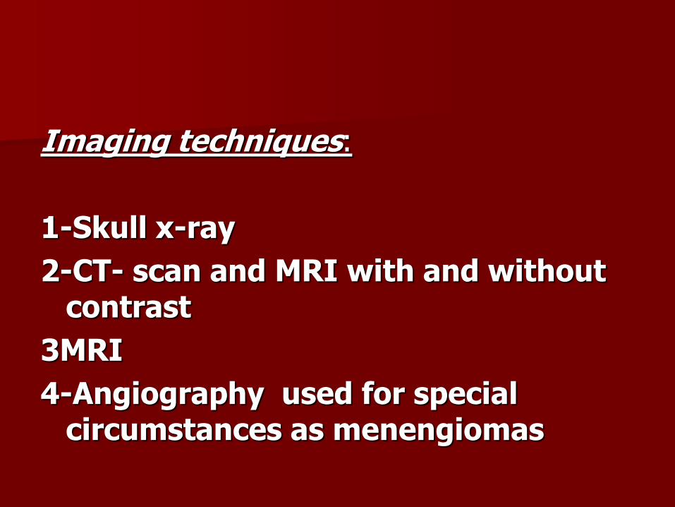
Imaging techniques:
1-Skull x-ray
2-CT- scan and MRI with and without contrast
3MRI
4-Angiography used for special circumstances as menengiomas
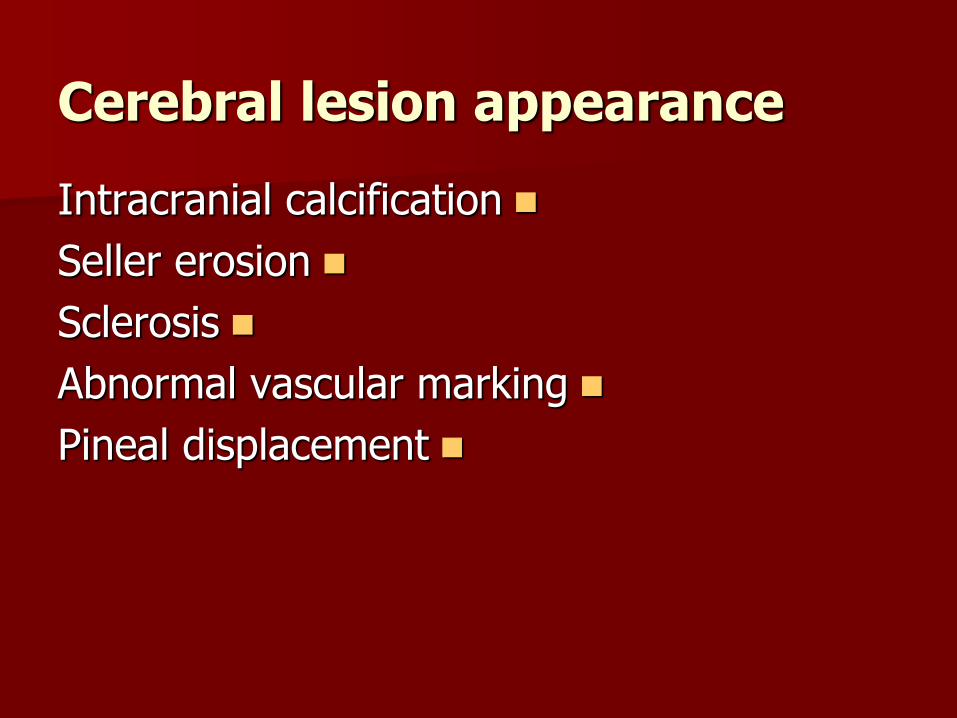
Cerebral lesion appearance
Intracranial calcification
Seller erosion
Sclerosis
Abnormal vascular marking
Pineal displacement
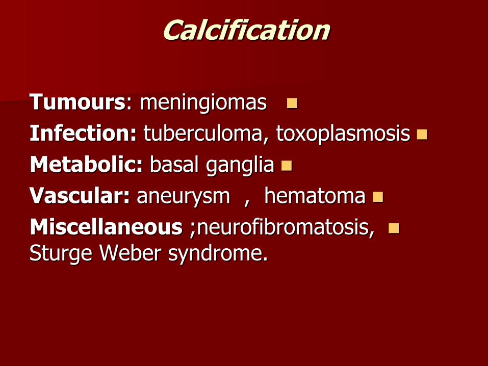
Calcification
Tumours: meningiomas
Infection: tuberculoma, toxoplasmosis
Metabolic: basal ganglia
Vascular: aneurysm , hematoma
Miscellaneous ;neurofibromatosis, Sturge Weber syndrome.


Sturge Weber

Erosion
. dermoid : Extracranial
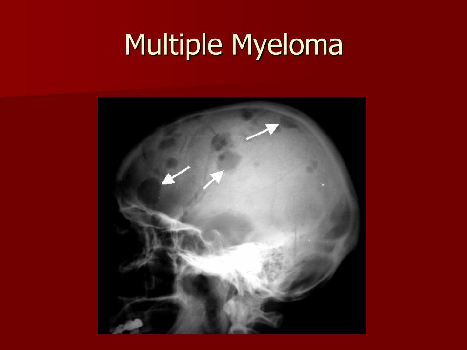
as multiple myeloma which appears as Bony lesionmultiple small rounded and clear cut defect.
Metastasis; extremely common and often multiple have characteristic mouth eaten appearance.
Intracranial with bony erosion
Pituitary adenoma ,expand and erode pituitary fosse, Acoustic neuroma lead to erosion of inner meat us
Non tumerous erosion Infection as osteomylitis .
Radiation .

Metastasis
Multiple Myeloma

Abnormal vascular marking

sclerosis
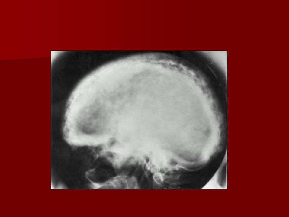
Generalize increase bone density Paget disease ; it may take form of multiple patchy
areas of increase density Localized
the commonest cause of bone sclerosis is hyperostosis frontalis intarna ,a condition of no clinical significance, in which there's irregular thickening of inner table of the skull in frontal region sparing midline
Meningioma Osteosarcoma


Cerebral neoplasm can be classified in several ways
According to the location :-
1-Intraxial (within the brain parenchyma)
2-Extra axial,out side the brain tissue (in the surrounding menenges,subarachnoid,subdural ,epidural or within the bony calvariam

Also the tumors can be localized as to compartment of brain within which it arise into supratentorial, sellar, suprasellar and infratentorial.
CT- scan
Observation are made of density of tumours prior to contrast and after it, most paranchymal tumours are hypodense indicating increase fluid with it ,hyper density in tumours raises one of three possibilities either

1-Bleeding(glioblastoma multiforma)
2-Calcification (craniopharangioma)
3-Hypercellular tumors (lymphoma)


Appearance by MRI
Usually sequences used in MRI are T1,T2 weighted images,most tumours apperars hypointense on T1 weighted images lower than the grey matter and hyperintense on T2 weighted images

Oligodendroglioma

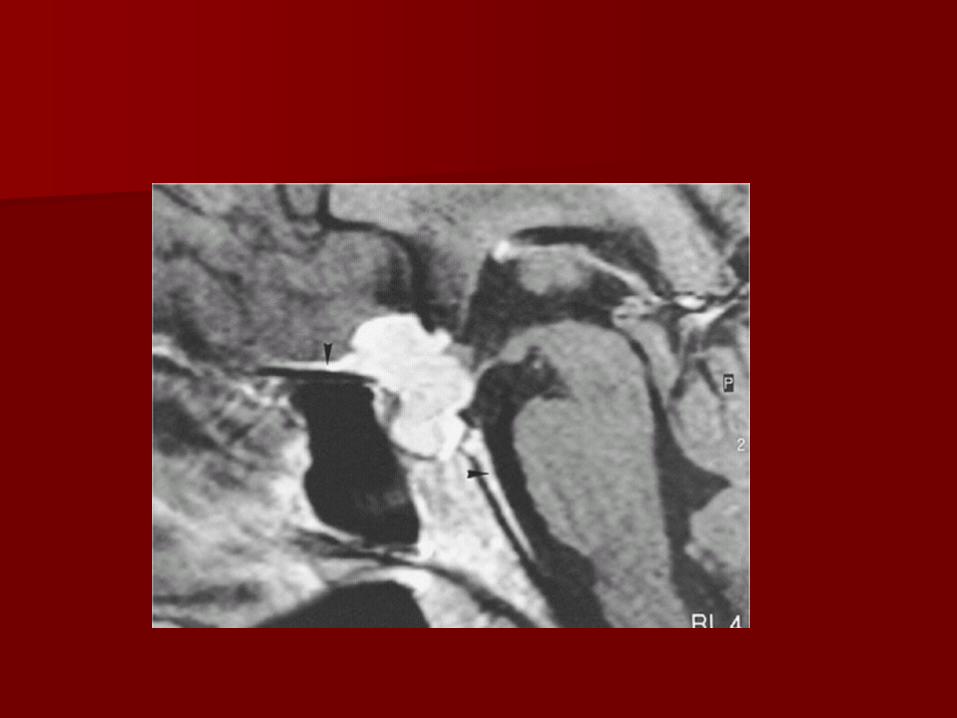
Metastasis

Metastasis to the brain, skull and menengies can occure in most form of systemic cancer .
CT-scan appearance
The CT- scan appearance of hematogenus metastasis in adult in grey-white matter junction ,seen most typically as zone of vasaogenic oedema within white matter surrounding much smaller areas that are denser,varying from hypodense to isodense to increase density relative to cortical grey matter.
Most of secondaries multiple.
Fallowing contrast ,most secondaries enhances ,
Pattern of enhancement quiet variable depending on whether secondaries is solid which is densly enhances or centerally necrotic which have ring like appearancec .

.Some secondaries solitary similar to primary tumour

MRI appearance
Appearance are variable but most are hypointense on T1 and hyper intense on T2 weighted images with similar CT –scan enhancement pattern.


Infection

In acute meningitis CT and MRI are usually normal.
CT scan -done prior to lumber puncture is only essential if theres evidence of raised intracranial pressure ,focal neurological signs or changes in conscious level .
The CT-scan may show hydrocephalus in such patients .

Encephalitis
Usually caused by virus,CT and MRI show unilateral or bilateral focal abnormal areas often in acharacteristic distribution appearing as low attenuation on CT and high signal on T2 weighted MRI images.

Abscess
An abscess can caused by pyogenic, tuberculous, fungal or parasitic organism.
Necrosis and pus formation occure in center of the abscess which appears as low density lesion in CT –scan .

The wall of abscess enhances with contrast and may be surrounded by oedema giving ring enhancement pattern ,similar changes seen in MRI


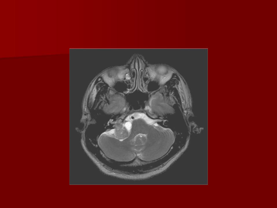
Multiple sclerosis
Multiple sclerosis
Disease of young adult characterise by disseminated plaques of demyelination and gliosis,their site usually in periventricular,optic pathway,brainstem,cerebbelar white matter and pudencle and spinal cord.
may show no abnormality but -: CTsometime low density areas may seen in acute stage
is far most sensitive than CT in -: MRIdemonstration of MS plaques,the charcterestic appearanceis that of preiventricular nodular hyperintense lesion on T2-weighted images.
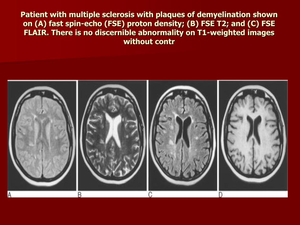
Patient with multiple sclerosis with plaques of demyelination shown on (A) fast spin-echo (FSE) proton density; (B) FSE T2; and (C) FSE FLAIR. There is no discernible abnormality on T1-weighted images
without contr

Ageing

Changes in CT and MRI are
1- Atrophy of brain occure resulting in dilitation of ventricle and cortical sulci .
2- Ischemia give rise low attenuation areas in deep white matter .

Dementia

In Alzheimer s disease ,the commonest form of dementia
CT and MRI shows dilated ventricles,widening cortical sulci and ill defind white matter abnormalitirs .
Atrophy of temporal lobe occure befor generalized atrophy .

Normal 65 year old