Embed Size (px)
Citation preview

DOI: 10.1093/brain/awh259 Brain (2004), 127, 2153–2172
REVIEW ARTICLE
Mitochondrial disorders
Massimo Zeviani1 and Stefano Di Donato2
Correspondence to: Massimo Zeviani, Stefano Di Donato,
Istituto Nazionale Neurologico ‘C. Besta’, Via Celoria
11 20133 Milan, Italy
E-mail: [email protected], [email protected]
1Division of Molecular Neurogenetics and 2Division
of Biochemistry and Genetics, Istituto Nazionale
Neurologico, Milan, Italy
SummaryInthemedical literaturetheterm‘mitochondrialdisorders’
is to a large extent applied to the clinical syndromes
associated with abnormalities of the common final pathway
of mitochondrial energy metabolism, i.e. oxidative
phosphorylation (OXPHOS). Faulty oxidative phos-
phorylation may be due to overall dysfunction of the
respiratory chain, a heteromultimeric structure embedded
in the inner mitochondrial membrane, or can be associatedwith single or multiple defects of the five complexes forming
therespiratorychainitself.Fromthegeneticstandpoint, the
respiratory chain is a unique structure of the inner
mitochondrial membrane formed by means of the
complementation of two separate genetic systems: the
nuclear genome and the mitochondrial genome. The nuclear
genome encodes the large majority of the protein subunits of
therespiratorycomplexesandmostofthemitochondrialDNA
(mtDNA) replication and expression systems, whereas the
mitochondrial genome encodes only 13 respiratory complex
subunits, and some RNA components of the mitochondrial
translational apparatus. Accordingly, mitochondrialdisorders due to defects in OXPHOS include both mendelian-
inherited and cytoplasmic-inherited diseases. This review
describes human genetic diseases associated with mtDNA
and nuclear DNA mutations leading to impaired OXPHOS.
Keywords: respiratory chain; oxidative phosphorylation; mitochondrial DNA mutations; nuclear DNA mutations
Abbreviations: adPEO = autosomal dominant progressive external ophthalmoplegia; CoQ10 = coenzyme Q10; COX =
cytochrome c oxidase; KSS = Kearns–Sayre syndrome; LHON = Leber’s hereditary optic neuropathy; MELAS = mitochondrial
encephalomyopathy with lactic acidosis and stroke-like episodes; MERRF = myoclonic epilepsy with ragged red fibres; MDS =
mitochondrial DNA depletion syndrome; mETC = mitochondrial electron transport chain; MNGIE = mitochondrial neuro-
gastro-intestinal encephalomyopathy; mtDNA = mitochondrial DNA; NARP = neuropathy ataxia and retinitis pigmentosa;
OXPHOS = oxidative phosphorylation; PEO = progressive external ophthalmoplegia; RRFs = ragged red fibres; SDH = succinate
dehydrogenase; SNHL = non-syndromic and aminoglycoside-induced sensorineural hearing loss; TK = thymidine kinase 2;
TP = thymidine phosphorylase.
Received March 23, 2004. Revised May 17, 2004. Accepted May 21, 2004. Advanced Access publication September 9, 2004
IntroductionNeurological syndromes are the most frequent clinical presen-
tations of mitochondrial disorders, a group of human diseases
characterized by defects of the mitochondrial energy output.
Mitochondria are cytoplasmic, double-membrane orga-
nelles, the main role of which is to synthesize ATP, the uni-
versal energy ‘currency’ of the cell. Because of the remarkable
expansion of knowledge on the molecular characterization of
human disorders associated with the energy pathways of mito-
chondria, the term ‘mitochondrial disorders’ is nowadays
restricted to indicate only the clinical syndromes associated
with abnormalities of oxidative phosphorylation (OXPHOS).
The respiratory chain is composed of five enzymatic multi-
heteromeric complexes (I, II, III, IV and V), embedded in the
inner membrane of mitochondria. The protein subunits of the
respiratory chain complexes are assembled together and with
prosthetic groups and metal-containing reactive centres by a set
of chaperones and assembly factors, some of which are specific
to each complex. Coenzyme Q (a lipoidal quinone) and cyto-
chromecarealsoinvolved inmitochondrial respiration,serving
as ‘electron shuttles’ between the complexes (Wallace, 1999).
The formation of the respiratory chain is under the control
of two separate genetic systems, the nuclear genome and the
Brain Vol. 127 No. 10 # Guarantors of Brain 2004; all rights reserved
Dow
nloaded from https://academ
ic.oup.com/brain/article-abstract/127/10/2153/404539 by guest on 05 April 2019

mitochondrial genome [mitochondrial DNA (mtDNA)]. In
particular, four of the five respiratory chain complexes (I, III,
IV and V) contain both nuclear-encoded and mtDNA-encoded
polypeptides (Fig. 1).
In terms of function, the first two linked events of respira-
tion, i.e. electron transfer and proton pumping, are carried
out by the mitochondrial electron transport chain (mETC),
a functional supramolecular structure located in the lipid
bilayer of the membrane, and composed of four complexes
(complex I–IV). In humans, complex I or NADH-ubiquinone
oxidoreductase, which accomplishes the oxidation of NADH
derived by the oxidation of fatty acids, pyruvate and amino
acids, contains seven subunits which are encoded by the
mtDNA (subunits ND1–ND6 and ND4L), plus at least 39
Fig. 1 Creative drawing of the respiratory chain and human mitochondrial DNA. Top: respiratory chain complexes. Mitochondriallyencoded subunits, embedded in the midst of nuclear-encoded subunits, are shown in different colours: complex I subunits = blue; complexIII subunit = green; complex IV subunits = red; complex V subunits = yellow. Pi = inorganic posphate; Cyt c = cytochrome c; CoQ =coenzyme Q. Bottom: mtDNA. myt genes: complex I genes = blue; complex III cytb gene = green; complex IV genes = red; complex Vgenes = yellow. syn genes: tRNA genes = grey; rRNA genes = purple. Cyt b = cytochrome b; COI = complex I; COII = complex II; COIII =complex III. (Courtesy of Dr Loredana Lamantea, Division of Molecular Neurogenetics).
2154 M. Zeviani and S. Di Donato
Dow
nloaded from https://academ
ic.oup.com/brain/article-abstract/127/10/2153/404539 by guest on 05 April 2019

nuclear-encoded subunits of complex I (Smeitink, 2001;
Carroll et al., 2003). Complex II or succinate-ubiquinone
oxidoreductase, which accomplishes the oxidation of
FADH2 derived from fatty acid and the Krebs’ cycle, is
composed of only four subunits, all encoded by the nuclear
genome. Complex III or ubiquinol-ferricytochrome c oxidor-
eductase holds one subunit, cytochrome b, encoded by the
mitochondrial genome and 10 subunits encoded by the
nuclear genome. Complex IV or cytochrome c oxidase
(COX) is composed of 13 subunits, three of which are
encoded by mtDNA (COX I–III) and the other 10 by nuclear
DNA. In addition, mETC contains two highly hydrophobic,
mobile, small electron carriers, coenzyme Q10 and cyto-
chrome c, both synthesized by nuclear genes (Fig. 1). In
substance the mETC is especially built to accept electrons
from NADH and FADH2, transfer them through a series of
oxidation–reduction reactions to molecular oxygen to produce
water and to simultaneously coupling this exergonic reaction
to the translocation of protons across the inner membrane
(Saraste, 1999; Di Donato, 2000).
Synthesis of ATP from ADP is the second fundamental
reaction of the mitochondrial respiratory chain, a process
performed by complex V or ATP synthase. ATP synthase
is also a genetic mosaic, since it is composed of two
mtDNA-encoded subunits (ATPase 6 and 8), and at least
13 nuclear DNA-encoded subunits (Fig. 1). As mentioned,
the proton electrochemical gradient generated at the mETC
level during electron transfer to oxygen creates a polarization
of the inner membrane which is changed back by the proton
flux through a proton channel which resides in the F0
component of ATP synthase. The proton flux drives the
condensation of ADP and inorganic phosphate into ATP
(Saraste, 1999; Wallace, 1999). Electron transfer across the
mETC and ATP synthesis are coupled, or linked. In fact, the
respiratory chain works as a proton pump which generates a
proton gradient and a membrane potential of about 180 mV
across the inner membrane with a negative polarity at the
matrix side of the inner membrane. The proton gradient is
utilized by the ATP synthase to phosphorylate matrix ADP.
During this process the proton gradient is decreased and this
activates respiration, i.e. electron transfer (Saraste, 1999).
Hence, the fundamental reaction of life, i.e. oxygen activation
and the conservation of energy in cell respiration, is essen-
tially a function of the integrity of the inner membrane
respiratory chain (Babcock and Wilkstrom, 1992).
Notably, energy production in mitochondria requires not
only a full assembly of functional protein at the level of the
inner mitochondrial membrane, but also a bidirectional flow
of information between the nuclear genome and the mitochon-
drial genome to adjust energy production in tissues to differ-
ent energetic demands (Poyton and McEwan, 1996).
Accordingly, many different mutations in mtDNA- and
nuclear DNA-encoding subunits, components or regulators
of the respiratory chain function can produce a wide range
of OXPHOS diseases (DiMauro and Schon, 2003; Zeviani
and Carelli, 2003).
Clinical aspectsGiven the complexity of mitochondrial genetics and biochem-
istry, the clinical manifestations of mtDNA disorders are
extremely heterogenous. They range from lesions of single
tissues or structures, such as the optic nerve in Leber’s here-
ditary optic neuropathy (LHON), or the cochlea in maternally
inherited non-syndromic deafness, to more widespread
lesions including myopathies, encephalomyopathies, cardio-
pathies, or complex multisystem syndromes with onset ran-
ging from neonatal to adult life (Table 1).
Adult patients usually show signs of myopathy associated
with variable involvement of the CNS (ataxia, hearing loss,
seizures, polyneuropathy, pigmentary retinopathy and, more
rarely, movement disorders). Some patients complain only of
muscle weakness and/or wasting with exercise intolerance
(Zeviani and Carelli, 2003). Several morphological and bio-
chemical hallmarks characterize many, albeit not all, of these
syndromes. The best known morphological finding is perhaps
the transformation of scattered muscle fibres into ‘ragged red
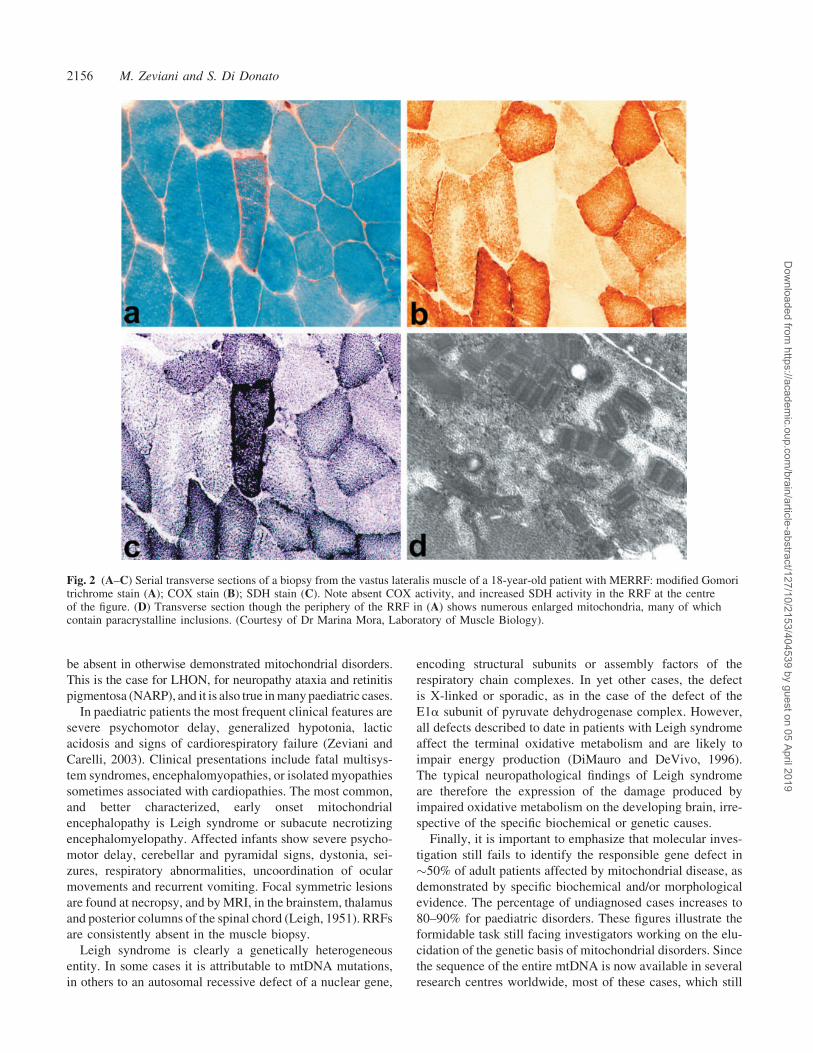
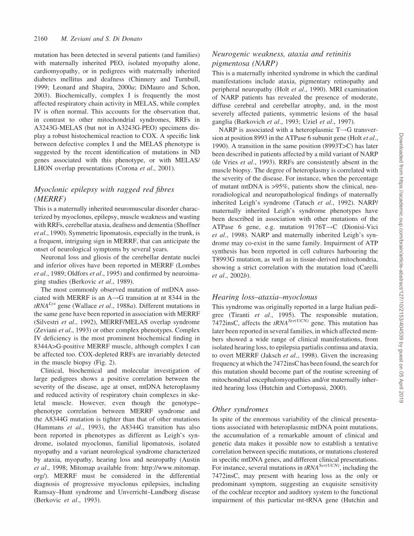
fibres’ (RRFs) (Fig. 2A). RRFs are characterized by the accu-
mulation of abnormal mitochondria under the sarcolemmal
membrane (Fig. 2D). The latter phenomenon is clearly
demonstrated by an intense subsarcolemmal reaction to a
respiratory chain-specific mitochondrial enzyme such as
succinate dehydrogenase (SDH) (Fig. 2C). Another common
finding is the presence of muscle fibres that stain negative
to the histochemical reaction to COX (respiratory complex
IV) (Fig. 2B). However, typical ‘mitochondrial’ clues may
Table 1 Phenotypic expression of mitochondrial diseases
Neurological manifestations Systemic manifestations
Neuromuscular HeartOphtalmoplegia CardiomyopathyMyopathy Cardiac conduction defectsExercise intolerancePeripheral sensory–motorneuropathy
Endocrine systemDiabetes
CNS Exocrine pancreas dysfunctionMyelopathy HypoparathyrodismHeadache Multiple endocrinopathyStroke Short statureSeizuresDementia Blood
PancytopeniaMovement disorders Sideroblastic anaemiaAtaxiaDystonia Mesenchymal organsParkinsonism HepatopathyMyoclonus Nephropathy
Intestinal pseudo-obstructionEyeBlindness MetabolismOptic neuropathy Metabolic acidosisPigmentary retinopathy Nausea and vomitingCataractEarSensorineural deafness
Mitochondrial disorders 2155
Dow
nloaded from https://academ
ic.oup.com/brain/article-abstract/127/10/2153/404539 by guest on 05 April 2019
be absent in otherwise demonstrated mitochondrial disorders.
This is the case for LHON, for neuropathy ataxia and retinitis
pigmentosa (NARP), and it is also true in many paediatric cases.
In paediatric patients the most frequent clinical features are
severe psychomotor delay, generalized hypotonia, lactic
acidosis and signs of cardiorespiratory failure (Zeviani and
Carelli, 2003). Clinical presentations include fatal multisys-
tem syndromes, encephalomyopathies, or isolated myopathies
sometimes associated with cardiopathies. The most common,
and better characterized, early onset mitochondrial
encephalopathy is Leigh syndrome or subacute necrotizing
encephalomyelopathy. Affected infants show severe psycho-
motor delay, cerebellar and pyramidal signs, dystonia, sei-
zures, respiratory abnormalities, uncoordination of ocular
movements and recurrent vomiting. Focal symmetric lesions
are found at necropsy, and by MRI, in the brainstem, thalamus
and posterior columns of the spinal chord (Leigh, 1951). RRFs
are consistently absent in the muscle biopsy.
Leigh syndrome is clearly a genetically heterogeneous
entity. In some cases it is attributable to mtDNA mutations,
in others to an autosomal recessive defect of a nuclear gene,
encoding structural subunits or assembly factors of the
respiratory chain complexes. In yet other cases, the defect
is X-linked or sporadic, as in the case of the defect of the
E1a subunit of pyruvate dehydrogenase complex. However,
all defects described to date in patients with Leigh syndrome
affect the terminal oxidative metabolism and are likely to
impair energy production (DiMauro and DeVivo, 1996).
The typical neuropathological findings of Leigh syndrome
are therefore the expression of the damage produced by
impaired oxidative metabolism on the developing brain, irre-
spective of the specific biochemical or genetic causes.
Finally, it is important to emphasize that molecular inves-
tigation still fails to identify the responsible gene defect in
�50% of adult patients affected by mitochondrial disease, as
demonstrated by specific biochemical and/or morphological
evidence. The percentage of undiagnosed cases increases to
80–90% for paediatric disorders. These figures illustrate the
formidable task still facing investigators working on the elu-
cidation of the genetic basis of mitochondrial disorders. Since
the sequence of the entire mtDNA is now available in several
research centres worldwide, most of these cases, which still
Fig. 2 (A–C) Serial transverse sections of a biopsy from the vastus lateralis muscle of a 18-year-old patient with MERRF: modified Gomoritrichrome stain (A); COX stain (B); SDH stain (C). Note absent COX activity, and increased SDH activity in the RRF at the centreof the figure. (D) Transverse section though the periphery of the RRF in (A) shows numerous enlarged mitochondria, many of whichcontain paracrystalline inclusions. (Courtesy of Dr Marina Mora, Laboratory of Muscle Biology).
2156 M. Zeviani and S. Di Donato
Dow
nloaded from https://academ
ic.oup.com/brain/article-abstract/127/10/2153/404539 by guest on 05 April 2019

await molecular characterization, are likely to be due to muta-
tions in (unknown) nuclear genes related to OXPHOS.
Defects of the mtDNA genesGene organization of the mitochondrial genomeHuman mtDNA is a 16.5 kb circular minichromosome, com-
posedof twocomplementarystrands, theheavyandlightstrands.
All of the coding sequences are contiguous with each other with
no introns (Anderson et al., 1981). The only non-coding stretch
of mtDNA is the displacement-loop (D-loop), a region of about
1 kb which contains the promoters for light and heavy strand
transcription (Fig. 1). Replication of mtDNA was believed to
proceed asynchronously andasymmetrically, starting from two
spatially separated replication origins, one for each strand. This
model, proposed by Clayton (1991), has recently been chal-
lenged by experimental evidence supporting the existence of
conventional, strand-coupled replication of mammalian
mtDNA (Holt et al., 2000; Yang et al., 2002).
Since the mtDNA genetic code differs from the universal
code, expression of mtDNA genes must rely upon
mitochondrion-specific protein synthesis, carried out through
the interplay of nuclear-encoded transcriptional and transla-
tional factors with tRNAs and rRNAs synthesized in situ from
the corresponding mitochondrial genes. Thus, human mtDNA
contains both protein-encoding genes (analogous to the yeast
mit genes), and protein synthesis genes (analogous to the
yeast syn genes). An important progress in the understanding
of the mitochondrial transcriptional machinery has been the
discovery that two novel transcriptional factors, TFB1M
and TFB2 M, cooperate with mitochondrial RNA polymerase
and mitochondrial transcription factor A to carry out basal
transcription of mammalian mtDNA (Falkenberg et al., 2002).
The 13 mit genes specify seven ND subunits of NADH-
ubiquinone reductase, three subunits of COX (complex IV),
subunits 6 and 8 of ATPsynthase (complex V), and apocyto-
chrome b, which is part of ubiquinol-cytochrome c reductase
(complex III). SDH-ubiquinone reductase (complex II) is com-
posed of four subunits, all encoded by nuclear genes. The syn
genes of mtDNA encode two rRNAs (12 and 16S rRNA)
and 22 tRNAs that are involved in protein translation of the
mit gene products. (see Fig. 1).
Clinical geneticsThe genetics of mtDNA differs from that of nuclear DNA in
the following unique properties (Zeviani et al., 2003).
The mitochondrial genome is maternally inherited. Paternal
mtDNA does not contribute to mitochondrial inheritance
despite a few sperm mitochondria entering the egg (Schwartz
and Vissing, 2002). Only the mother transmits her oocyte
mtDNA to all of her offspring, and her daughters transmit
their mtDNA to the next generation (Giles et al., 1980; Ankel-
Simons and Cummins, 1996). Mitochondria are polyploid.
Each human cell has hundreds of mitochondria, each
containing 2–10 mtDNA molecules. At cell division,
mitochondria and their genomes are randomly distributed
to daughter cells.
Normally, the mitochondrial genotype of an individual is
composed of a single mtDNA species, a condition known as
homoplasmy. However, the intrinsic propensity of mtDNA to
mutate randomly can occasionally determine a transitory con-
dition known as heteroplasmy, where the wild-type and the
mutant genomes co-exist intracellularly. Because of mito-
chondrial polyploidy, during mitosis the two mtDNA species
are stochastically distributed to daughter cells (Jenuth et al.,
1996). This phenomenon can account for the drastic change in
mutation loads observed in different generations of families
carrying heteroplasmic mtDNA, and increases the remarkable
variability in the phenotypic presentations of mitochondrial
disorders. Because of mitotic mtDNA segregation and poly-
ploidy, a threshold effect dictates the phenotypic expression
of a mtDNA-associated character (Jenuth et al., 1997). For a
given heteroplasmic mutation, only when mutated gene
copies accumulate over a certain threshold, the deleterious
effects of the mutation will no longer be complemented by
the co-existing wild-type mtDNA, and will be expressed
phenotypically as a cellular dysfunction leading to disease
(Thorbum and Dahl, 2001). A major breakthrough in the
understanding of mitochondrial disorders has been the
discovery of an impressive number of mutations of
mtDNA (available from: http://www.mitomap.org/).
The variability in clinical manifestations of mtDNA stems
from a number of factors, including the nature of the mutation,
i.e. its intrinsic pathogenicity, and the gene specifically
affected, the mutation load and its tissue distribution, and
the relative reliance of each organ system on the mitochon-
drial energy supply. In general, the visual and auditory sys-
tems, the CNS and PNS, the heart, muscle, endocrine
pancreas, kidney and liver are, in that order, the organs
most sensitive to OXPHOS failure (Table 1). However,
almost 15 years after the first reports on human mtDNA muta-
tions, and the many more that have been discovered after-
wards, the intimate molecular and cellular mechanisms which
link a given mtDNA change to a specific clinical presentation
are still largely unknown (Zeviani and Carelli, 2003).
Mutations of mtDNA are divided into large-scale rearrange-
ments (i.e. partial deletions or duplications) and inherited point
mutations. Both groups have been associated with well-defined
clinicalsyndromes.Whilelarge-scalerearrangementsareusually
sporadic, point mutations are usually maternally inherited. Simi-
lartorho0-petitephenotypeinyeast, large-scalerearrangements
include several genes and are invariably heteroplasmic. In
contrast, point mutations may be heteroplasmic or homoplas-
mic, and affect individual mit or syn genes (Table 2).
Large-scale rearrangements of mtDNASingle, large-scale rearrangements of mtDNA can be single par-
tial deletions, or partial duplications. Rearranged molecules,
lacking a portion of the mitochondrial genome, can be detected
as an independent mtDNA species (single mtDNA deletion) or
Mitochondrial disorders 2157
Dow
nloaded from https://academ
ic.oup.com/brain/article-abstract/127/10/2153/404539 by guest on 05 April 2019

joined to a wild-type molecule in a 1 : 1 ratio, as partially
duplicated mtDNA. Frequently, a mixture of the two rearran-
gements co-exists in the same cell or tissue (Zeviani et al.,
1988; Poulton et al., 1989).
Three main clinical phenotypes are associated with these muta-
tions:Kearns–Sayresyndrome(KSS),sporadicprogressiveexter-
nal ophthalmoplegia (PEO) and Pearson’s syndrome (Table 2).
KSS is a (usually) sporadic disorder characterized by the
triad of: (i) chronic progressive external ophthalmoplegia; (ii)
onset before age of 20 years; and (iii) pigmentary retinopathy.
Cerebellar syndrome, heart block, increased CSF protein con-
tent, diabetes and short stature are also part of the syndrome.
Patients with this disease invariably show RRFs in muscle
biopsy (Mita et al., 1989). KSS is characterized by neuro-
radiological abnormalities affecting the deep structures of the
brain and the subcortical white matter (Barkovich et al., 1993).
Single deletions/duplications can also result in milder phe-
notypes as PEO, characterized by late-onset progressive
external ophtalmoplegia, proximal myopathy and exercise
intolerance. In both KSS and PEO, diabetes mellitus and
hearing loss are frequent additional features, that may occa-
sionally precede, by years, the onset of neuromuscular symp-
toms (Shoffner et al., 1989).
Finally, large-scale single deletions/duplications of mtDNA
may cause Pearson’s bone-marrow–pancreas syndrome, a rare
disorder of early infancy characterized by connatal sideroblastic
pancytopenia and, less frequently, severe exocrine pancreatic
insufficiency with malabsorption (Rotig et al., 1990). Interest-
ingly, infants surviving into childhood or adolescence may
develop the clinical features of KSS (Shanske et al., 2002).
The majority of single large-scale rearrangements of
mtDNA are sporadic and are therefore believed to be the
result of the clonal amplification of a single mutational
event, occurring in the maternal oocyte or early during the
development of the embryo (Schon et al., 1989; Chen et al.,
1995). It is not yet understood why in multisystem disorders
such as KSS, in which D-mtDNAs are virtually ubiquitous,
mutations are not transmitted through female gametes to the
progeny. One possibility is that the germinal cells containing
deleted genomes are not viable for gametogenesis and/or fer-
tilization. However, mother-to-offspring transmission has
occasionally been documented in KSS/PEO. Hence, the recur-
rency risk for these mtDNA abnormalities can no longer be
considered absent. Until a reliable epidemiological survey of
PEO or KSS due to single rearrangements of mtDNA is avail-
able, we suggest a prudential figure of 5% recurrency risk in the
genetic counselling of affected women (Chinnery et al., 2000).
Molecular pathogenesisThe relative amount and tissue distribution of the
molecular lesion dictate the onset and severity of the disease.
Table 2 Mitochondrial OXPHOS diseases due to mtDNA mutations
Large-scale rearrangementsof mtDNA
Phenotype mtDNA mutation
KSS Ataxia, neuropathy, PEO,pigmentary retinal degeneration,cardiomyopathy and conduction block,short stature, high CSF protein
Single deletions or duplications(mostly sporadic)
Pearson’s syndrome Frequent death in infancy.Refractory sideroblastic anaemia withvacuolization of marrow precursors.
PEO Late-onset bilateral ptosis and ophtalmoplegia,proximal muscle weakness andwasting, and exercise intolerance
Point mutations of mtDNAMELAS Stroke-like episodes due to focal brain lesions
in the parieto-occipital lobes, lacticacidosis and/or RRFs
Heteroplasmic point mutations(maternally inherited)
MERRF Myoclonus, epilepsy, muscle weakness andwasting with RRFs, cerebellar ataxia,deafness and dementia
NARP Ataxia, pigmentary retinopathy, peripheralneuropathy and distal neurogenic weakness
Hearing loss–ataxia–myoclonus Syndromic hearing loss, myoclonus epilepsy,ataxia, myopathy
LHON Loss of central vision, large centro-caecal absolutescotoma, circumpapillary telangiectaticmicroangiopathy
Homoplasmic point mutations(maternally inherited)
SNHL Non-syndromic and aminoglycoside-inducedhearing loss
2158 M. Zeviani and S. Di Donato
Dow
nloaded from https://academ
ic.oup.com/brain/article-abstract/127/10/2153/404539 by guest on 05 April 2019

Transmitochondrial cybrids, obtained by introducing deleted
mtDNAs into mtDNA-less rho0 cells, showed impaired
respiration (Hayashi et al., 1991). A threshold of >60% rear-
ranged mtDNA molecules is enough for OXPHOS failure to
occur. The more widespread is the tissue distribution of the
lesion, the more severe is the clinical syndrome, from PEO, to
KSS, to Pearson’s syndrome. This notion is also relevant for
the diagnosis: for instance, deletions are confined to the
muscle biopsy in PEO, but in KSS they can also be found in
blood, albeit in lesser amounts, while in Pearson’s syndrome
the amount is comparable in blood and muscle.
Most rearrangements occur across direct repeats of variable
length (Schon et al., 1989; Mita et al., 1989), suggesting a
mechanism based on illegitimate homologous recombination.
Defective OXPHOS of mitochondria containing D-mtDNA
is due to the loss of both mit and syn genes contained within
the deletion. In particular, because the lack of tRNA genes
results in incompetency for translation (Mariotti et al., 1994),
mitochondria containing only D-mtDNA are rho0-mutants,
which cannot synthesize functional OXPHOS enzymes.
However, partial correction of the rho0-phenotype can be
accomplished through complementation by mRNAs and
tRNAs synthesized from wild-type mtDNA, provided that
D-mtDNA and wild-type mtDNA co-segregate in the same
organelles.
Point mutations of mtDNAIn contrast to large-scale rearrangements, mtDNA point
mutations are usually maternally inherited. Given the very
high mutational rate of mtDNA and the presence of numerous
‘private’ or population-specific polymorphisms, the distinc-
tion between non-deleterious and pathogenic mutations may
not be easy. The following features are frequently present in
pathogenic mutations: (i) high conservation of the affected
nucleotide/amino acid or loss of function of the gene product
(e.g. a stop mutation in a mit gene); (ii) segregation with
phenotype; (iii) quantitative correlation between phenotype
and heteroplasmy, if present; and (iv) identification of the
mutation in affected families from ethnically distinct
human populations (Zeviani and Carelli, 2003).
Point mutations involving tRNA syn genes cause a reduced
availability of functional tRNAs that may impair the overall
mitochondrial protein synthesis. Marked reduction of both
mitochondrial protein synthesis and respiration has been
documented for some mutations, when a threshold of 80–
90% of mutant mtDNA is reached. Mutations involving
protein-encoding mit genes affect specifically the function
of the respiratory chain complexes to which the corresponding
protein belongs (Mariotti et al., 1994).
It is worth mentioning that the clinical and biochemical
variability of many mtDNA mutations may be due to dif-
ferent mitochondrial and/or nuclear ‘gene backgrounds’. For
instance, the fate and expression of mutations in cultures
appears to be strongly influenced by the different nuclear
backgrounds of the cell types (Dunbar et al., 1995). It has
also been proposed that nucleotide changes in mtDNA that are
not intrinsically pathogenic may predispose to, modulate the
effects of, or reflect a propensity for the occurrence of
deleterious mutations. In turn, deleterious mutations may
promote the accumulation of somatic changes, through the
generation of OXPHOS-related mutagens. This phenomenon
could trigger a positive feed-back loop contributing to the
progression of the mitochondrial dysfunction (Luft, 1994).
Given their different pathophysiology and genetic features,
the most frequent heteroplasmic and homoplasmic mtDNA
point mutations will be discussed separately (Table 2).
Heteroplasmic point mutations
Mitochondrial encephalomyopathy with lacticacidosis and stroke-like episodes (MELAS)This is defined by the presence of (i) stroke-like episodes
due to focal brain lesions, often localized in the parieto-
occipital lobes; and (ii) lactic acidosis and/or RRFs. Other
signs of CNS involvement include dementia, recurrent
headache and vomiting, focal or generalized seizures, pig-
mentary retinopathy and deafness. Ataxia can be observed in
some patients. Diabetes, intestinal pseudo-obstructions and
cardiomyopathy may complicate single cases (Hirano et al.,
1992).
Infarct-like lesions widespread in the cerebral cortex are
associated with diffuse fibrillary gliosis in the cerebral and
cerebellar white matter. Multiple focal lesions with demye-
lination and numerous spheroids have been reported in the
pontocerebellar fibres, together with marked degeneration of
the posterior columns and spinocerebellar tracts (Mizukami
et al., 1992). Electron microscopic examination shows accu-
mulations of abnormal mitochondria in smooth muscle cells
and endothelium of the cerebral and cerebellar blood vessels,
suggesting a ‘mitochondrial angiopathy’. However, the pre-
sence of diffuse, prominent white matter gliosis of the CNS
and cerebellar cortical degeneration of granular cell type
may indicate morphologically widespread cellular dysfunc-
tion, not restricted to either neuronal or vascular derangement
(Mizukami et al., 1992; Tsuchiya et al., 1999). MRI exam-
ination typically shows that the signal abnormalities in the
brain do not correspond to well-defined vascular territories
(Barkovich et al., 1993). The stroke-like lesions may be
transient and resolve after a few months. The recurrent
occurrence of stroke-like episodes eventually leads to
permanent lesions.
MELAS was first associated with a heteroplasmic point
mutation in the tRNALeu(UUR), an A!G transition at position
3243 (Goto et al., 1990). Many other MELAS-associated
point mutations were later reported, although the 3243A>G
remains by far the most frequent one (Mitomap available
from: http://www.mitomap.org/). The genotype–phenotype
correlation of the A3243G mutation is rather loose, since
the observed clinical manifestations are not limited to the
full-blown MELAS syndrome. For instance, the 3243A>G
Mitochondrial disorders 2159
Dow
nloaded from https://academ
ic.oup.com/brain/article-abstract/127/10/2153/404539 by guest on 05 April 2019
mutation has been detected in several patients (and families)
with maternally inherited PEO, isolated myopathy alone,
cardiomyopathy, or in pedigrees with maternally inherited
diabetes mellitus and deafness (Chinnery and Turnbull,
1999; Leonard and Shapira, 2000a; DiMauro and Schon,
2003). Biochemically, complex I is frequently the most
affected respiratory chain activity in MELAS, while complex
IV is often normal. This accounts for the observation that,
in contrast to other mitochondrial syndromes, RRFs in
A3243G-MELAS (but not in A3243G-PEO) specimens dis-
play a robust histochemical reaction to COX. A specific link
between defective complex I and the MELAS phenotype is
suggested by the recent identification of mutations in ND
genes associated with this phenotype, or with MELAS/
LHON overlap presentations (Corona et al., 2001).
Myoclonic epilepsy with ragged red fibres(MERRF)This is a maternally inherited neuromuscular disorder charac-
terized by myoclonus, epilepsy, muscle weakness and wasting
with RRFs, cerebellar ataxia, deafness and dementia (Shoffner
et al., 1990). Symmetric lipomatosis, especially in the trunk, is
a frequent, intriguing sign in MERRF, that can anticipate the
onset of neurological symptoms by several years.
Neuronal loss and gliosis of the cerebellar dentate nuclei
and inferior olives have been reported in MERRF (Lombes
et al., 1989; Oldfors et al., 1995) and confirmed by neuroima-
ging studies (Berkovic et al., 1989).
The most commonly observed mutation of mtDNA asso-
ciated with MERRF is an A!G transition at nt 8344 in the
tRNALys gene (Wallace et al., 1988a). Different mutations in
the same gene have been reported in association with MERRF
(Silvestri et al., 1992), MERRF/MELAS overlap syndrome
(Zeviani et al., 1993) or other complex phenotypes. Complex
IV deficiency is the most prominent biochemical finding in
8344A>G-positive MERRF muscle, although complex I can
be affected too. COX-depleted RRFs are invariably detected
in the muscle biopsy (Fig. 2).
Clinical, biochemical and molecular investigation of
large pedigrees shows a positive correlation between the
severity of the disease, age at onset, mtDNA heteroplasmy
and reduced activity of respiratory chain complexes in ske-
letal muscle. However, even though the genotype–
phenotype correlation between MERRF syndrome and
the A8344G mutation is tighter than that of other mutations
(Hammans et al., 1993), the A8344G transition has also
been reported in phenotypes as different as Leigh’s syn-
drome, isolated myoclonus, familial lipomatosis, isolated
myopathy and a variant neurological syndrome characterized
by ataxia, myopathy, hearing loss and neuropathy (Austin
et al., 1998; Mitomap available from: http://www.mitomap.
org/). MERRF must be considered in the differential
diagnosis of progressive myoclonus epilepsies, including
Ramsay–Hunt syndrome and Unverricht–Lundborg disease
(Berkovic et al., 1993).
Neurogenic weakness, ataxia and retinitispigmentosa (NARP)This is a maternally inherited syndrome in which the cardinal
manifestations include ataxia, pigmentary retinopathy and
peripheral neuropathy (Holt et al., 1990). MRI examination
of NARP patients has revealed the presence of moderate,
diffuse cerebral and cerebellar atrophy, and, in the most
severely affected patients, symmetric lesions of the basal
ganglia (Barkovich et al., 1993; Uziel et al., 1997).
NARP is associated with a heteroplasmic T!G transver-
sion at position 8993 in the ATPase 6 subunit gene (Holt et al.,
1990). A transition in the same position (8993T>C) has later
been described in patients affected by a mild variant of NARP
(de Vries et al., 1993). RRFs are consistently absent in the
muscle biopsy. The degree of heteroplasmy is correlated with
the severity of the disease. For instance, when the percentage
of mutant mtDNA is >95%, patients show the clinical, neu-
roradiological and neuropathological findings of maternally
inherited Leigh’s syndrome (Tatuch et al., 1992). NARP/
maternally inherited Leigh’s syndrome phenotypes have
been described in association with other mutations of the
ATPase 6 gene, e.g. mutation 9176T!C (Dionisi-Vici
et al., 1998). NARP and maternally inherited Leigh’s syn-
drome may co-exist in the same family. Impairment of ATP
synthesis has been reported in cell cultures harbouring the
T8993G mutation, as well as in tissue-derived mitochondria,
showing a strict correlation with the mutation load (Carelli
et al., 2002b).
Hearing loss–ataxia–myoclonusThis syndrome was originally reported in a large Italian pedi-
gree (Tiranti et al., 1995). The responsible mutation,
7472insC, affects the tRNASer(UCN) gene. This mutation has
later been reported in several families, in which affected mem-
bers showed a wide range of clinical manifestations, from
isolated hearing loss, to epilepsia partialis continua and ataxia,
to overt MERRF (Jaksch et al., 1998). Given the increasing
frequency at which the 7472insC has been found, the search for
this mutation should become part of the routine screening of
mitochondrial encephalomyopathies and/or maternally inher-
ited hearing loss (Hutchin and Cortopassi, 2000).
Other syndromesIn spite of the enormous variability of the clinical presenta-
tions associated with heteroplasmic mtDNA point mutations,
the accumulation of a remarkable amount of clinical and
genetic data makes it possible now to establish a tentative
correlation between specific mutations, or mutations clustered
in specific mtDNA genes, and different clinical presentations.
For instance, several mutations in tRNASer(UCN), including the
7472insC, may present with hearing loss as the only or
predominant symptom, suggesting an exquisite sensitivity
of the cochlear receptor and auditory system to the functional
impairment of this particular mt-tRNA gene (Hutchin and
2160 M. Zeviani and S. Di Donato
Dow
nloaded from https://academ
ic.oup.com/brain/article-abstract/127/10/2153/404539 by guest on 05 April 2019
Cortopassi, 2000). The pathogenetic mechanisms underlying
this well-established observation are presently unknown. Like-
wise, mutations in tRNAIle are mainly associated with cardio-
myopathy (Santorelli et al., 2001) or PEO, while mutations in
cytochrome b are mainly associated with isolated myopathy
with high serum creatine kinase or myoglobinuria (Andreu
et al., 1999). Cytochrome b mutations are usually restricted
to skeletal muscle and, in contrast to most of the other point
mutations, are not transmitted maternally. A number of differ-
ent clinical presentations, ranging from infantile Leigh syn-
drome to adult-onset motor neuron disease, to complex
multisystem disorders, have been reported with different
point mutations of the three genes encoding complex IV,
while point mutations of the genes encoding complex I subunits
are usually associated with MELAS, LHON or overlap syn-
dromes (Mitomap available from: http://www.mitomap.org/).
However, an increasing number of Leigh-like, infantile ence-
phalopathic syndromes have been reported recently in associa-
tion with the 13513G >A mutation in ND5 (Chol et al., 2003).
Homoplasmic mtDNA mutationsGeneral featuresIn contrast to many heteroplasmic mutations, the clinical
expression of disorders associated with homoplasmic muta-
tions is often stereotypical and mainly restricted to a single
tissue. In this group of disorders, the presence of a pathogenic
mtDNA mutation is necessary but not sufficient to induce
disease (Table 2). As a consequence, penetrance is incomplete
and possibly controlled by environmental factors, additional
mitochondrial polymorphisms, or the effect of nuclear gene(s)
(Howell and Mackey, 1998). However, the specific molecular
mechanisms underlying these contributions are still largely
unkown.
LHONThis was the first maternally inherited disease to be associated
with a mtDNA point mutation (Wallace et al., 1988b). LHON
typically affects young adults, more often males. Visual
acuity deteriorates over a period of days/weeks as a conse-
quence of rapid, painless loss of central vision in one eye,
usually followed by the other eye. Stable residual values at or
below 20/200 are reached in a few months, associated with a
large centro-caecal absolute scotoma. Characteristic fundus
changes include circumpapillary telangiectatic microangiopa-
thy with tortuosity of peripapillary arterioles, swelling of the
nerve fibre layer and hyperaemic optic disc, and absence of
leakage on fluorescein angiography (Smith et al., 1973;
Nikoskelainen et al., 1983). Axonal loss in the papillomacular
bundle, leading to an early and prevalent temporal atrophy of
the optic disc, is a pathognomonic feature of LHON (Kwittken
and Barest, 1958; Smith et al., 1973).
Histopathological investigations show loss of retinal gang-
lion cell and nerve fibre layers, while the remaining layers
appear virtually normal. Ultrastructural investigations in
genetically proven LHON optic nerves showed degenerative
features in both axoplasm and myelin sheaths. Patchy accu-
mulations of mitochondria suggested an impairment of
axoplasmic transport. Variability in myelin thickness was
also evident, some axons being almost denuded of myelin
sheath. Morphometric investigation showed a preferential
loss of the smallest axons, corresponding to the P-cell
population which provides central vision (Sadun et al.,
2000; Carelli et al., 2002a).
Approximately 90% of the worldwide LHON patients carry
one of the three most frequent mtDNA mutations associated
with LHON, namely the 11778G>A, 3460A>G and
14484T>C mutations (Wallace et al., 1988b; Howell et al.,
1991; Chinnery et al., 2001). A further group of rare, but well-
established pathogenic mutations have been found only in a
few families; also, prognosis depends on the type of mutation
(Mackey and Howell, 1992; Kim et al., 2002). Other muta-
tions, found only in single cases or families, still await con-
firmatory identification from multiple independent cases.
All the LHON mutations which have been proved to be
pathogenic affect different mtDNA-encoded subunits of com-
plex I. Mutations are usually homoplasmic, although hetero-
plasmy can occasionally be found in some families or
singleton cases.
Variable expression of LHON may be due to the association
of pathogenic mutations with specific mtDNA haplogroups.
For instance, the European-specific haplogroup J is found
more frequently in 11778- or 14484-positive LHON patients
than in ethnically matched control populations, suggesting that
this haplogroup may increase the penetrance of the disease
(Brown et al., 2002; Hofman et al., 1997; Torroni et al.,
1997). Environmental factors seem also to play a role as
risk factors, in particular tobacco smoke (Tsao et al., 1999).
Finally, a nuclear modifier is thought to be a major determinant
for both disease expression and male prevalence. However,
search for an X-linked nuclear modifier has been unsuccessful
to date (Chalmers et al., 1996).
Additional puzzling features of LHON are the exquisite
tissue specificity and the subtle and ill-defined biochemical
abnormalities found in this condition. The unique anatomical
and physiological features of the optic nerve may explain its
vulnerability to the decreased bioenergetic efficiency and
increased oxidative stress associated with LHON mutations
(Bristow et al., 2002; Wong et al., 2002).
LHON-like optic atrophy may be part of more complex
syndromes including dystonia, Leigh syndrome and MELAS
(Shoffner et al., 1995). Private or infrequent mutations, again
affecting complex I subunit genes, have been reported in these
cases (Carelli et al., 2002a).
Non-syndromic and aminoglycoside-inducedsensorineural hearing loss (SNHL)This has been both associated with a unique, maternally inher-
ited point mutation at position 1555 (A!G) of the 12S rRNA
Mitochondrial disorders 2161
Dow
nloaded from https://academ
ic.oup.com/brain/article-abstract/127/10/2153/404539 by guest on 05 April 2019

gene (Prezant et al., 1993). Similar to LHON, this mutation is
almost invariably homoplasmic, and variable penetrance and
clinical severity have been documented (Jaber et al., 1992;
Estivill et al., 1998) A two-locus model, including a primary
mitochondrial mutation associated with a nuclear modifier
gene, was suggested to explain incomplete penetrance.
Bykhovskaya and colleagues reported the identification of
a locus on chromosome 8 for a putative nuclear modifier
gene, but this finding has not been confirmed by other studies
(Bykhovskaya et al., 2000; Finnila and Majamaa, 2003). In
addition, a paraomomycin resistance mutation in yeast,
homologous to the human 1555 mutation, expresses a respira-
tory-deficient phenotype only in the presence of a nuclear
mutation in one of two genes, Mss1 and Mto1 (Hu et al.,
1991). The human analogues of Mss1 and Mto1 are obvious
candidates as nuclear modifier genes in the 1555-related
SNHL. The 1555 mutation affects a highly conserved region
of the 12S rRNA gene, homologous to the bacterial domain
that binds aminoglycosides, and increases the similarity of the
human 12S rRNA to its bacterial counterpart. The growth rate
of mutant cells is markedly reduced when they are exposed to
aminoglycosides, confirming their sensitivity to this drug
(Inoue et al., 1996). However, 1555-positive subjects
who were never exposed to aminoglycosides can also
become deaf. Therefore, the 1555 mutation is now considered
as a frequent genetic cause of both non-syndromic and
aminoglycoside-induced post-lingual SNHL. The hair cells
of the cochlea are very energy dependent and local gene
expression may also play a relevant role in the strict
tissue specificity observed with the 1555 mutation and with
other mutations of mtDNA which are predominantly
characterized by hearing loss. A second homoplasmic
mutation (1494C>T) in the mtDNA 12S rRNA gene
has recently been associated with maternally inherited,
aminoglycoside-induced, non-syndromic deafness in a large
Chinese family (Zhao et al., 2004).
Other homoplasmic mutationsHomoplasmic mutations are frequently found during systema-
tic screening of mtDNA in mitochondrial patients, but their
pathogenicsignificanceremainsuncertain.Awell-documented
case is a mutation at position 1624 in the tRNAVal gene
(McFarland et al., 2002). This homoplasmic mutation was
found in a clinically normal woman, who had six stillbirths
and one surviving child with Leigh syndrome, from different
partners. Biochemical investigations demonstrated a pro-
found respiratory chain deficiency in both the apparently
healthy woman and her child. A second, homoplasmic muta-
tion (in tRNAIle) has later been identified in a family composed
of a healthy mother and three affected daughters. Both pri-
mary fibroblast cell cultures and transmitochondrial cybrid
derivatives from several members of this family showed a
profound defect in complexes I and IV (Limongelli et al.,
2004). A third mutation, again in the tRNAIle gene
(4300G>A), has been found in a few families with maternally
inherited congestive cardiomyopathy of variable penetrance
(Casali et al., 1995). Interestingly, this mutation was shown to
be associated with very low steady-state levels of the tRNAIle
transcript in heart, but not in skeletal muscle. Accordingly, a
severe, combined defect in complexes I and IV was detected
in the heart muscle, but not in the skeletal muscle of an index
case (Taylor et al., 2003).
These results strongly support the novel concept that
homoplasmic mutations in tRNA genes can be responsible
for mitochondrial disorders characterized by extremely vari-
able penetrance. Homoplasmic pathogenic mutations in the
mitochondrial genome represent a potentially vast, still lar-
gely overlooked and poorly understood area of mitochondrial
medicine, and will stand as a new challenge in the
nosological and physiopathological definition of these disor-
ders in the future.
Nuclear gene mutationsA clinical–genetic classification can now be proposed for
these defects, as follows (Leonard and Schapira, 2000b;
DiMauro and Schon, 2003; Zeviani et al., 2003): (i) disorders
due to gene defects altering the stability of mtDNA (Table 3);
(ii) disorders due to nuclear gene defects encoding structural
components or assembly factors of the OXPHOS complexes
(Table 3); (iii) disorders due to defects in non-protein com-
ponents of the respiratory chain (Table 3) and (iv) disorders
due to gene defects encoding proteins indirectly related to
OXPHOS (Table 4).
Disorders due to gene defects altering thestability of mtDNAAutosomal dominant progressive external ophthalmoplegia
(adPEO) is a mendelian disorder characterized by the accu-
mulation of multiple deletions of mtDNA in patient’s tissues
(Zeviani et al., 1989). The typical clinical feature of adPEO is
progressive muscle weakness, most severely affecting the
external eye muscles. Skeletal muscle shows RRFs and a
mild reduction in the activities of respiratory chain enzymes.
Ataxia, depression, hypogonadism, hearing loss, peripheral
neuropathy and cataract are present in some families (Servidei
et al., 1991; Hirano et al., 2001).
Most of the adPEO families carry heterozygous muta-
tions in one of three genes: ANT1, encoding the muscle-
heart-specific mitochondrial adenine nucleotide translocator
(Kaukonen et al., 2000), Twinkle, encoding a putative mtDNA
helicase (Spellbrink et al., 2001), and POLG1, encoding the
catalytic subunit of the mtDNA-specific polymerase gamma
(Van Goethem et al., 2001). Mutations in both POLG1 alleles
were also found in autosomal recessive PEO sibships with
multiple affected members and in apparently sporadic cases
(Lamantea et al., 2002). A prevalent mutation, the Y955C,
dramatically reduces the apparent binding affinity for
nucleoside triphosphates in vitro and also the accuracy for
base pair substitutions (Ponomarev et al., 2002).
2162 M. Zeviani and S. Di Donato
Dow
nloaded from https://academ
ic.oup.com/brain/article-abstract/127/10/2153/404539 by guest on 05 April 2019

Table 3 Mitochondrial OXPHOS diseases due to nuclear mutations
Phenotype Disease
Genes controlling the stability of mtDNAANT1 Multiple deletions mtDNA, PEO,
muscle weakness, ataxia, depression,hypogonadism, hearing loss, peripheralneuropathy
adPEO
TwinklePOLG1TP (autosomal dominant
or recessive)Multiple deletion/depletion mtDNA,ophthalmoparesis, peripheral neuropathy,leucoencephalopathy, and gastrointestinalsymptoms with intestinal dismotility
MNGIE
TK2 Fatal infantile congenital myopathy with orwithout a DeToni–Fanconi renal syndrome
MDS
DGUOK Fatal infantile hepatopathy leading to rapidlyprogressive liver failure
Deoxynucleotide carrier Congenital microcephaly of Amish
Genes encoding protein respiratory chain componentsComplex I NDUFS1 Leigh syndrome, complex I deficiency Autosomal recessive mutationsComplex I NDUFS2 Cardiomyopathy–encephalomyopathy Autosomal recessive mutationsComplex I NDUSFS4 Leigh-like syndrome Autosomal recessive mutationsComplex I NDUFS7 Leigh syndrome Autosomal recessive mutationsComplex I NDUFS8 Leigh syndrome Autosomal recessive mutationsComplex I NDUFV1 Leigh syndrome, leucodystrophy, myoclonus Autosomal recessive mutationsComplex II SDHA Leigh syndrome Autosomal recessive mutationsComplex II SDHB Phaeochromocytoma, cervical paraganglioma Autosomal dominant or sporadicComplex II SDHC and SDHD Hereditary paraganglioma Autosomal recessive mutationsComplex III UQCRB genesubunit VII
Hypokalaemia and lactic acidosis Autosomal recessive homozygousdeletion
Defects of non-protein respiratorychain constituentsCoenzyme Q deficiency Ataxia, seizures, myopathy (?)Tafazzin (cardiolipin acyltransferase?) Barth syndrome X-linked recessive
Genes encoding respiratory chain assembly componentsSURF1 COX� Leigh syndrome Autosomal recessive mutationsSCO1 COX� hepathopathy and ketoacidotic coma Autosomal recessive mutationsSCO2 COX� infantile cardiomyopathy Autosomal recessive mutationsCOX10 COX� leucodystrophy and renal tubulopathy Autosomal recessive mutationsCOX15 COX� hypertrophic cardiomyopathy Autosomal recessive mutationsBCS1L Complex III-deficient encephalopathy,
liver failure, renal tubulopathyAutosomal recessive mutations
LRPPRC (mRNA-binding protein) COX� Leigh syndrome Autosomal recessive mutationsATP12 Complex V deficiency–encephalopathy Autosomal recessive mutations
Table 4 Mitochondrial diseases due to nuclear mutations of genes indirectly involved in OXPHOS
Disease Phenotype Nuclear DNA mutation
Freidreich’s ataxia (FRDA1 gene) Ataxia, loss of DTR, sensoryneuropathy, Babinski sign,cardiomyopathy, diabetes
Autosomal recessive mutation in thefrataxin gene (iron handler iron–sulfurcluster assembly)
X-linked ataxia and sideroblasticanaemia
Ataxia, sideroblastic anaemia Autosomal recessive mutation in theABC7 iron exporter
Hereditary spastic paraplegia Spastic paraplegia Autosomal recessive mutation in the SPG7gene encoding a metalloprotease
X-linked deafness–dystonia syndrome Deafness and dystonia X-linked recessive mutation in the DDP1 geneencoding protein mitochondrial transporter
Autosomal dominant optic atrophy(OPA1 gene)
Optic atrophy and visual failure Autosomal dominant mutations in the OPA1gene encoding a dynamin-related protein
Mitochondrial disorders 2163
Dow
nloaded from https://academ
ic.oup.com/brain/article-abstract/127/10/2153/404539 by guest on 05 April 2019

Another disease in this series, mitochondrial neuro-
gastro-intestinal encephalomyopathy (MNGIE), is a
devastating disorder of juvenile onset, characterized by
ophthalmoparesis, peripheral neuropathy, leucoencephalopa-
thy and gastrointestinal symptoms with intestinal dismotility,
and histologically abnormal mitochondria in muscle
(Hirano et al., 1994). Mutations in the gene encoding
thymidine phosphorylase (TP), leading to loss of activity of
the enzyme, are associated with MNGIE (Nishino et al.,
1999). TP is an important factor involved in the control
and maintenance of the pyrimidine nucleoside pool of the
cell. Defects of TP are thought to produce an excess of
dTTP, resulting in the imbalance of dNTP pools that
can ultimately affect both the rate and fidelity of mtDNA
replication. This is reflected by the molecular phenotype
of MNGIE, which is characterized by both multiple
deletions and partial depletion of muscle mtDNA (Nishino
et al., 2000).
mtDNA depletion syndrome (MDS) is a heterogeneous
group of disorders characterized by a reduction in mtDNA
copy number (Moraes et al., 1991). Clinically, they include a
fatal infantile congenital myopathy with or without DeToni–
Fanconi renal syndrome, fatal infantile hepatopathy leading to
rapidly progressive liver failure, and late infantile or child-
hood myopathy, with onset after 1 year of age, characterized
by a progressive myopathy causing respiratory failure and
death by 3 years of age.
The presence of affected siblings born from healthy parents
suggested an autosomal recessive mode of inheritance, pos-
sibly affecting a nuclear gene involved in the control of the
mtDNA copy number. An important contribution to the
elucidation of the genetic bases of mtDNA depletion has
recently come from studies on selected families. MDS has
been linked to mutations in two genes involved in dNTP
metabolism: thymidine kinase 2 (TK2) and deoxy-guanosine
kinase, which are responsible for the myopathic form and the
hepatoencephalopathic form of MDS, respectively (Mandel
et al., 2001; Saada et al., 2001). The first reports on these
genes have later been confirmed by studies on larger cohorts
of MDS patients (Mancuso et al., 2002; Salviati et al., 2002a).
Correction of the original TK2 gene sequence and biochem-
ical investigations in vitro on the kinetic properties of mutant
TK2 proteins have also been reported (Spinazzola et al.,
2002). However, defects in TK2 or guanosine kinase are
responsible for only a minor fraction of MDS cases, indicating
that the condition is genetically heterogeneous. Both guano-
sine kinase and TK2 genes are involved in the formation of the
mitochondrial nucleotide pool, as is TP, responsible for
MNGIE. Biochemical investigations in patients’ cells do sug-
gest that derangement of balanced availability of dNTPs can
affect mtDNA integrity and maintenance (Spinazzola et al.,
2002). However, the pathogenetic relationship between
reduction of TP activity, increased levels of thymidine in
blood and accumulation of mtDNA lesions remains unclear.
This issue is further complicated by the absence of mtDNA
abnormality recently reported in knockout mice deficient in
either the TP gene or in both TP and uridine phosphorylase
genes (Haraguchi et al., 2002).
Adding interest to the role of nucleotide supply in mito-
chondrial biogenesis and disease is the discovery that a
recently identified mitochondrial deoxynucleotide carrier is
responsible for a rare form of congenital microcephaly, found
in interrelated Old Order Amish. These data indicate that
mitochondrial deoxynucleotide transport may be essential
for fetal brain development (Rosenberg et al., 2002).
Genes encoding protein subunits of therespiratory complexesIsolated deficiency of complex I is relatively frequent among
mitochondrial disorders. The primary genetic defect may be
either at the mtDNA or at the nuclear DNA level. There are
seven mtDNA-encoded and at least 39 nuclear-encoded sub-
units of complex I (Carroll et al., 2003), for a total of 46 genes,
which represents a truly formidable challenge for a systematic
genetic screening even in highly selected patients. Neverthe-
less, several disease-associated complex I mutations have
been discovered recently (Triepels et al., 2001) (Table 3).
In most of these cases, the clinical presentation is that of
an early onset progressive neurological disorder with lactic
acidosis, most often Leigh syndrome, occasionally compli-
cated by cardiomyopathy, or multisystem involvement
(Morris et al., 1996). However, no mutation in structural
genes has been found in many cases of complex I deficiency,
suggesting that still unknown assembly factors for complex I,
or other gene products involved in its formation and activity
may be responsible for these forms (Smeitink et al., 2001).
Complex II is an FAD-dependent enzyme at a cross-point
between OXPHOS and Krebs cycle pathways. It is composed
of four protein subunits, all encoded by nuclear genes (SDH-
A, -B, -C, -D). Mutations in SDHA, the largest subunit of com-
plex II, are a rare cause of Leigh syndrome or late-onset neuro-
degenerative disease (Bougeron et al., 1995). However, the
most interesting discovery concerning defects of complex II is
their association with inherited paragangliomas (Baysal,
2002). In 10–15% of the cases, these usually benign neuroec-
todermal tumours are inherited in an autosomal dominant fash-
ion with incomplete penetrance. It now appears that mutations
in SDHB, SDHC and SDHD are responsible for the majority of
familial paragangliomas and also for a significant fraction
of non-familial tumours, including phaeochromocytomas
(tumours of the adrenal medulla) (Baysal et al., 2002). The
inactivation of the SDHD gene is associated with stimulation
of the angiogenic pathway, a mechanism that could be
involved in the pathogenesis of neoplasm (Gimenez-Roqueplo
et al., 2001). Finally, the first mutation in a nuclear gene
encoding a subunit of complex III has recently been identified
in an infant with hypoglycaemic episodes and lactic acidosis.
A homozygous 4-bp deletion in the UQCRB gene, encoding
subunit QP-C (or subunit VII), was associated with an isolated
defect of complex III and reduced amount of cytochrome b
content in isolated mitochondria (Haut et al., 2003).
2164 M. Zeviani and S. Di Donato
Dow
nloaded from https://academ
ic.oup.com/brain/article-abstract/127/10/2153/404539 by guest on 05 April 2019

Genes involved in the assembly of respiratorycomplexesThis group comprises, so far, defects of genes encoding assem-
bly factors of COX (complex IV), ubiquinol-cytochrome
c reductase (complex III) and ATP synthase (complex V).
Human COX is composed of 13 subunits: the three largest
ones are encoded by mtDNA genes, while the remaining
subunits are encoded by nuclear genes. In infancy, the
most frequent manifestation of isolated, profound COX defi-
ciency is Leigh syndrome, although other phenotypes, includ-
ing leucoencephalopathy, severe cardiomyopathy or complex
encephalocardiomyopathies have also been reported
(Shoubridge, 2001). COX defects have been associated
with mutations of mtDNA tRNA genes, and also with a
few mutations in mtDNA genes encoding COX subunits.
No mutation in any of the nucleus-encoded subunits of
COX has been reported, while all of the nuclear gene defects
of COX so far identified are due to mutations in assembly
factors of the enzyme, including SURF1, SCO1, SCO2,
COX10 and COX15. SURF1 is a 30 kDa hydrophobic protein
located in the inner membrane of mitochondria. Mutations in
SURF1 are relatively frequent, accounting for the majority of
the Leigh syndrome cases due to COX deficiency (Tiranti
et al., 1998). Absence of SURF1 causes the accumulation
of early assembly intermediates and the drastic reduction of
fully assembled COX (Tiranti et al., 1999). This phenomenon
has been observed in different organisms carrying null muta-
tions of SURF1, including yeast strains (Nijtmans et al., 2001;
Barientosetal., 2002),humanpatients (Tirantietal., 1999)and,
more recently, SURF1 knockout mice (Agostino et al., 2003).
Mutations in other COX assembly genes are much rarer
and, in some cases, they have been reported in only a few
families or singleton cases.
Human SCO1 and SCO2 are nuclear-encoded copper-
binding proteins, presumed to be responsible for the
insertion of Cu into the COX holoenzyme. While mutations
in SCO1 were found in only one family (Valnot et al., 2000a),
mutations in SCO2 are more frequent (Papadopoulou et al.,
1999). The usual clinical presentation is that of an early-onset,
fatal cardio-encephalo-myopathy with COX deficiency, but
clinical variants have been reported resembling early-onset
(type 1) spinal muscular atrophy (Salviati et al., 2002b). Stu-
dies in yeast, bacteria and, more recently, humans, have
shown that Cu supplementation can restore COX activity
in cells harbouring mutations in genes involving Cu transport,
including SCO2 (Jaksch et al., 2001; Salviati et al., 2002c).
Also the product of the COX10 gene, mapping like the
SCO1 gene on chromosome 17p13, is involved in a crucial
step of COX maturation. COX10 encodes haem A: farnesyl-
transferase, which catalyses the first step in the conversion of
protohaem to the haem A prosthetic groups of the enzyme. A
homozygous missense mutation in the COX10 gene was found
in the affected members of a consanguineous family with an
isolated COX defect leading to an early-onset leucoencepha-
lopathy (Valnot et al., 2000b).
Similar to COX10, COX15 is involved in the synthesis of
haem A, the prosthetic group for COX. Antonicka and col-
leagues recently identified the first deleterious mutations in
COX15, in a patient with fatal, infantile hypertrophic
cardiomyopathy (Antonicka et al., 2002). This study
establishes COX15 as an additional cause, along with
SCO2, of fatal infantile, hypertrophic cardiomyopathy
associated with isolated COX deficiency. However, mutations
in COX15 may also cause Leigh syndrome.
Finally, mutations in LRPPRC (leucine-rich motif-PPR
containing) have been found in infants with a COX-deficiency
syndrome. Sequence analysis identified two mutations on two
independent haplotypes, providing definitive genetic proof
that genetic mutation in LRPPRC is a cause of Leigh syn-
drome (Mootha et al., 2003). LRPPRC encodes an mRNA-
binding protein which is likely to be involved with mtDNA
transcript processing, suggesting an additional mechanism of
mitochondrial pathophysiology.
Complex III catalyses electron transfer from succinate and
nicotinamide adenine dinucleotide-linked dehydrogenases to
cytochrome c. Complex III is made up of 11 subunits, of
which all but one (cytochrome b) are encoded by nuclear
DNA. Although several pathogenic mutations in the gene
encoding mitochondrial cytochrome b have been described
(Andreu et al., 1999), mutations in only one nuclear DNA-
encoded subunit of complex III (subunit VII or QP-C) has
been reported in a single infant patient affected by hypogly-
caemia and lactic acidosis. BCS1L, a mitochondrial inner-
membrane protein, is a chaperone necessary for the assembly
of mitochondrial respiratory chain complex III. Mutations in
BCS1L have been shown in infantile cases of complex III
deficiency associated with neonatal proximal tubulopathy,
hepatic involvement and encephalopathy (de Lonlay et al.,
2001), also called GRACILE (growth retardation, aminoaci-
duria, cholestasis, iron overload, lactacidosis and early death)
syndrome (Visapaa et al., 2002).
Finally, the first mutation in ATP12, an assembler of mito-
chondrial ATP synthase, has been identified in a single infant
patient with lactic acidosis, dysmorphic features and rapidly
progressive encephalopathy. Deficiency of complex V activ-
ity was associated with marked reduction of immunodetect-
able complex V subunits in both muscle and liver
mitochondria (De Meirleir et al., 2004).
Defects of non-protein constituents ofmitochondriaCoenzyme Q10 (CoQ10) deficiencyCoQ10, or ubiquinone, is a lipophilic component of the
electron transport chain, which transfers to complex III
(ubiquinone-cytochrome c reductase) electrons derived
from complex I, complex II, and from the oxidation of
fatty acids and branched-chain amino acids via flavin-linked
dehydrogenases. CoQ10 also plays a role as an antioxidant
and as a membrane stabilizer.
Mitochondrial disorders 2165
Dow
nloaded from https://academ
ic.oup.com/brain/article-abstract/127/10/2153/404539 by guest on 05 April 2019

Primary CoQ10 deficiency was first described (Ogasahara
et al., 1989) in two sisters aged 14 and 12 years with abnormal
fatiguability and slowly progressive weakness of proximal
limb and trunk muscles, seizures and myoglobinuria. In mus-
cle specimens from both patients, all type-1 RRFs also
showed marked lipid excess. Biochemical analysis of the
respiratory chain in muscle mitochondria revealed normal
activities of complexes I, II, III and IV, while the combined
activities of complexes I–III and II–III were reduced. These
results pointed to a defect of CoQ10 which was confirmed in
both sisters by direct assay of CoQ10. Treatment with oral
CoQ10 improved the muscle weakness, ataxia, learning dis-
ability and lactic acidosis in both sisters. A similar syndrome
characterized by the triad of recurrent myoglobinuria, brain
involvement (seizures, ataxia, mental retardation) and RRFs/
lipid storage in muscle has been also reported (Sobreira et al.,
1997).
A more widespread genetic defect of the respiratory chain
associated with severe coenzyme Q deficiency was described
in two siblings in whom coenzyme Q deficiency was general-
ized and present in muscle, blood cells and skin fibroblasts
resulting in severe encephalomyopathy and renal failure. Both
children had substantial improvement under oral ubidecare-
none supplementation (Rotig et al., 2000). A variant pheno-
type with clinical and MRI features of an adult-onset Leigh
syndrome has also recently been described in two sisters (Van
Maldergem et al., 2002).
Finally, a syndrome characterized by low coenzyme Q in
muscle, unexplained cerebellar ataxia, pyramidal signs, and
seizures, unspecific myopathic change and no myoglobinuria
has been reported (Musumeci et al., 2001). Irrespective of
the genetic causes of this defect, which are presently
unknown, early recognition of coenzyme Q deficiency is
important, because supplementation of CoQ10 can lead to
substantial clinical improvement.
Barth’s syndromeAn abnormality of cardiolipin metabolism has been found in
Barth syndrome (X-linked mitochondrial myopathy, cardio-
pathy, neutropenia, short stature and 3-methyl glutaconic
aciduria). The product of the mutated gene in Barth syndrome,
called tafazzin (Bione et al., 1996), is homologous to phos-
pholipid acyltransferases. Cardiolipin is a major component
of the phospholipid milieu of the mitochondrial inner mem-
brane (Valianpour et al., 2002) where it plays a modulatory
role on the activities of several respiratory chain complexes,
including complexes I and IV.
Genes encoding mitochondrial factorsindirectly related to OXPHOSOther neurodegenerative disorders have been attributed to
mutations in several mitochondrial proteins, which are not
obviously linked to overt OXPHOS defects, yet indirectly
related to respiration and energy production (Di Donato,
2000) (Table 4). This observation further broadens the con-
cept of mitochondrial disease and extends the possible invol-
vement of mitochondrial energy metabolism in a previously
unsuspected large number of important clinical phenotypes.
This group includes paraplegin, a mitochondrial metallopro-
tease associated with autosomal recessive spastic paraplegia
(Casari et al., 1998); ABC7, an iron mitochondrial exporter,
which controls the generation of cytosolic iron–sulfur proteins
and is responsible of X-linked sideroblastic anaemia and
ataxia (Allikmets et al., 1999); frataxin, a mitochondrial pro-
tein which is responsible for Friedreich’s ataxia, also puta-
tively involved in iron handling and iron–sulfur protein
manteinance (Campuzano et al., 1996; Puccio et al. 2001);
and DDP1, a component of the import machinery for mito-
chondrial carrier proteins, which is responsible of X-linked
deafness–dystonia syndrome, the Mohr–Tranebjaerg syn-
drome (Koehler et al., 1999; Roesch et al., 2002).
Mutations in OPA1, a gene encoding a dynamin-related
protein embedded in the mitochondrial inner membrane
(Olichon et al., 2003), have been found in autosomal domi-
nant optic neuropathy of the Kjehr type (Delettre et al., 2002).
Haplo-insufficiency of the gene seems to be a common patho-
genetic mechanism in OPA1 mutations. In addition,
polymorphisms in the OPA1 gene have been associated
with another ocular condition, normal tension glaucoma
(Aung et al., 2002; Buono et al., 2002). Down-regulation
of OPA1 gene expression in HeLa cells by RNA interference
experiments induced fragmentation of the mitochondrial net-
work, concomitant dissipation of membrane potential, disor-
ganization of the cristae, release of cytochrome c and
activation of caspase-dependent apoptosis (Olichon et al.,
2003). These findings on OPA1 are similar to those showing
that cells carrying LHON-associated mtDNA mutations are
more prone to apoptosis (Ghelli et al., 2003), and suggest the
existence of a common pathogenetic mechanism for these
hereditary optic neuropathies.
Finally, to further expand the spectrum of neurodegenera-
tive disorders associated with impairment of mitochondrial
biogenesis and OXPHOS stands the recent observation that
missense mutations in MFN2, a gene encoding mitofusin 2,
lead to Charcot–Marie–Tooth neuropathy type 2A (Zuchner
et al., 2004). Mitofusins are GTPase proteins regulating the
fission–fusion dynamics of the mitochondrial network. This is
a fundamental process in mitochondrial biogenesis, required
for establishing a uniform membrane potential of the orga-
nelles, for even energy supply throughout the cell.
New strategies for the discovery of diseaseloci and genesIdentification of nuclear OXPHOS disease genes is compli-
cated by the scarcity of large-size families and consangui-
neous families, and by the great heterogeneity of the
disorders, that may prevent the possibility of carrying out a
genome-wide search for disease loci based on traditional
strategies, including linkage analysis and homozygosity
2166 M. Zeviani and S. Di Donato
Dow
nloaded from https://academ
ic.oup.com/brain/article-abstract/127/10/2153/404539 by guest on 05 April 2019
mapping. Therefore, new strategies based, for instance, on
functional complementation of OXPHOS phenotype
expressed in cell culture have been applied in several
cases to elucidate the genetic aetiology of these disorders.
An interesting, successful development of these strategies has
recently been reported (de Lonley et al., 2002). A functional
complementation approach was developed by: (i) growing the
patient’s fibroblasts in a highly selective medium; and (ii)
transferring human chromosome fragments into respiratory
chain-deficient fibroblasts by microcell-mediated transfer.
In the absence of carbohydrates in the culture medium,
OXPHOS-deficient cells rapidly disappeared unless they
were rescued by a chromosome fragment carrying the disease
gene. This method, applied on two cell lines with complex II
or complex I + IV defects, allowed the mapping of the disease-
causing genes to small intervals (4 and 12 Mb) on chromo-
somes 12p13 and 7p21, respectively. This approach makes the
physical mapping of the disease genes feasible in sporadic
cases of OXPHOS deficiency.
The availability of the entire genome of the yeast
Saccharomyces cerevisiae, and the near completion of the
human genome project, including the establishment of expres-
sion profiles of human gene clusters in different tissues, will
make it possible to use high-throughput strategies, which
combine in vitro and in silico investigations, to assess the
human mitochondrial proteome and identify new disease
genes. Validation of these new strategies has actually been
provided by two recently published papers. A first strategy
(Steinmetz et al., 2002) exploited the high similarity between
yeast and human mitochondria to perform a systematic func-
tional screen based on both database interrogation and micro-
array expression analysis on the whole-genome pool of yeast
deletion mutants. Human orthologues were then identified,
many of which encode novel proteins, and some of them
were linked to heritable diseases using genomic map posi-
tions. A second strategy exploited the availability of whole-
genome data sets of RNA and protein expression to identify
the gene causing Leigh syndrome, French-Canadian type, a
human COX deficiency that maps to chromosome 2p16–21.
By intersecting information derived from RNA expression
data sets and a large survey of organellar proteomics with
the relevant genomic region, a single clear candidate gene was
identified and then proven to be mutated in affected indi-
viduals (Mootha et al., 2003). A similar strategy has been
adopted more recently to identify the gene responsible for
ethylmalonic encephalopathy, a complex mitochondrial dis-
order of infancy characterized by persistent lactic acidosis,
abnormal excretion of ethylmalonic acid, progressive neuro-
degenerative lesions in the brainstem, persistent diarrhoea,
peripheral vasculopathy and cytochrome c oxidase deficiency
in skeletal muscle (Tiranti et al., 2004).
Animal modelsMice carrying mtDNA with pathogenic mutations would pro-
vide a system in which to study how mutant mtDNAs are
transmitted and distributed in tissues, resulting in expression
of mitochondrial diseases. The first mouse carrying a hetero-
plasmic mtDNA deletion has been obtained by isolating
respiration-deficient cybrids with mtDNA carrying a deletion
and introducing this mtDNA into fertilized eggs (Inoue et al.,
2000). The mutant mtDNA was transmitted maternally, and
its accumulation induced mitochondrial dysfunction in vari-
ous tissues. Moreover, most of these mice died because of
renal failure, suggesting the involvement of mtDNA muta-
tions in the pathogeneses of new diseases.
Two interesting mouse models lacking mtDNA in selected
tissues have been developed by knocking out mtTFA, the
main transcription/replication activator of mtDNA. Condi-
tional mice were developed lacking mtTFA and mtDNA in
frontal cortex neurons during embryonic development (Wang
et al., 1999). Knockout mice survived for several months
before dying from massive apoptosis of cortical neurons. A
similar strategy was also adopted to create conditional
mtDNA-less mice in skeletal muscle, which developed a typi-
cal mitochondrial myopathy with RRFs and COX depletion
(Li et al., 2000).
The importance of mitochondrial defects in degenerative
diseases and ageing has been demonstrated using different
mouse models of mitochondrial disease (Melov et al.,
1999), including knockouts for Ant1 the adenine nucleotide
translocator, Surf1 and the mitochondrial manganese
superoxide dismutase. More recently, a mtDNA mutation
imparting chloramphenicol resistance to mitochondrial pro-
tein synthesis has been transferred into mice and resulted in
growth retardation and cardiomyopathy (Levy et al., 1999).
TreatmentNo effective therapy is available for mitochondrial disorders.
This is due to the lack, until recently, of suitable animal
models, as well as to the rarity and heterogeneity of the dis-
orders. However, several supportive measures, such as
improvement of nutrition, surgical correction of ptosis, treat-
ment of seizures and other complications, correction of lactic
acidosis, etc., can ameliorate specific problems and improve
the quality of life in several cases.
Gene therapyA number of experimental strategies are currently being pur-
sued. These include the introduction of modified genes or
gene products into mitochondria via the protein import
machinery (Manfredi et al., 2002) and inhibition of replica-
tion of mutant mtDNA by sequence-specific antigenomic
peptide-nucleic acids (Taylor et al., 1997). These approaches
are not yet clinically relevant. In selected, isolated myopathy
cases, reduction of heteroplasmic mutant load was obtained
by controlled muscle fibre damage and regeneration by muta-
tion-free satellite cells, using myotoxic drugs (Irwin et al.,
2002). However, this treatment was not effective in improving
ptosis in five patients with PEO.
Mitochondrial disorders 2167
Dow
nloaded from https://academ
ic.oup.com/brain/article-abstract/127/10/2153/404539 by guest on 05 April 2019

Metabolic therapyCreatine, the latest compound proposed for treatment of mito-
chondrial disorders, is the substrate for the synthesis of phos-
phocreatine, the most abundant energy storage compound in
muscle, heart and brain. An open trial of 81 patients with
various neuromuscular disorders (including 17 with mito-
chondrial diseases) showed significant improvement of
ischaemic isometric handgrip strength and non-ischaemic iso-
metric dorsiflexion torque. Another placebo-controlled,
double-blind, randomized crossover trial in 16 patients with
chronic progressive external ophthalmoplegia or mitochon-
drial myopathy, however, did not find significant effects on
exercise performance, eye movements, or activities of daily
life (Chinnery and Turnbull, 2001). Taken together, these data
suggest that creatine may be effective in some, but not all
mitochondrial diseases. As creatine is virtually free of adverse
effects, its administration may be warranted in patients with
muscle weakness even before a large controlled trial resolves
the issue of its efficacy. While CoQ10 is not effective in
mtDNA-associated mitochondrial disease (Bresolin et al.,
1990) it leads to marked improvement in ‘primary’ CoQ10
deficiency (see above). Idebenone, a shorter chain analogue of
CoQ10, appears to be effective in halting or even improving
the hypertrophic cardiomyopathy in Friedreich’s ataxia
(Rustin et al., 1999; Mariotti et al., 2003). Further trials
are currently underway to confirm this exciting preliminary
observation.
AcknowledgementsWe are gratefully indebted to Dr Loredana Lamantea, Divi-
sion of Molecular Neurogenetics, for the artwork of Fig. 1,
and to Dr Marina Mora, Laboratory of Muscle Biology,
Istituto Nazionale Neurologico Besta, for the serial transverse
sections of a muscle biopsy in Fig. 2. This work was supported
by Fondazione Telethon-Italy (grant no. GGP030039), Fon-
dazione Pierfranco e Luisa Mariani (Ricerca 2000), Ricerca
Finalizzata Ministero della Salute RF-2002/158, Ricerca
Finalizzata Ministero della Salute RF 2002-03/and MitEuro
network grant from the European UnionFramework Program
5 (M.Z.), and Ricerca Finalizzata Ministero della Salute RF
2002/157 (S.D.D.).
References
Agostino A, Invernizzi F, Tiveron C, Fagiolari G, Prelle A, Lamantea E, et al.
Constitutive knockout of Surf1 is associated with high embryonic lethality,
mitochondrial disease and cytochrome c oxidase deficiency in mice. Hum
Mol Genet 2003; 12: 399–413.
Allikmets R, Raskind WH, Hutchinson A, Schueck ND, Dean M, Koeller DM.
Mutation of a putative mitochondrial iron transporter gene (ABC7) in X-
linked sideroblastic anemia and ataxia (XLSA/A). Hum Mol Genet 1999; 8:
743–9.
Anderson S, Bankier AT, Barrell BG, de Bruijn MH, Coulson AR, Drouin J,
et al. Sequence and organization of the human mitochondrial genome.
Nature 1981; 290: 457–65.
Andreu AL, Hanna MG, Reichmann H, Bruno C, Penn AS, Tanji K, et al.
Exercise intolerance due to mutations in the cytochrome b gene of
mitochondrial DNA. N Engl J Med 1999; 341: 1037–44.
Ankel-Simons F, Cummins JM. Misconceptions about mitochondria and
mammalian fertilization: implications for theories on human evolution.
Proc Natl Acad Sci USA 1996; 93: 13859–63.
Antonicka H, Mattman A, Carlson CG, Glerum DM, Hoffbuhr KC, Leary SC,
et al. Mutations in COX15 produce a defect in the mitochondrial heme
biosynthetic pathway, causing early-onset fatal hypertrophic cardiomyo-
pathy. Am J Hum Genet 2003; 72: 101–14.
Aung T, Ocaka L, Ebenezer ND, Morris AG, Krawczak M, Thiselton DL, et al.
A major marker for normal tension glaucoma: association with polymorph-
isms in the OPA1 gene. Hum Genet 2002; 110: 52–6.
Austin SA, Vriesendorp FJ, Thandroyen FT, Hecht JT, Jones OT, Johns DR.
Expanding the phenotype of the 8344 transfer RNAlysine mitochondrial
DNA mutation. Neurology 1998; 51: 1447–50.
Babcock GT, Wikstrom M. Oxygen activation and the conservation of energy
in cell respiration. Nature 1992; 356: 301–9.
Barkovich A, Good W, Koch T, Berg B. Mitochondrial disorders: analysis of
their clinical and imaging characteristics. AJNR Am J Neuroradiol 1993;
14: 1119–37.
Baysal BE. Hereditary paraganglioma targets diverse paraganglia. J Med
Genet 2002; 39: 617–22.
Baysal BE, Willett-Brozick JE, Lawrence EC, Drovdlic CM, Savul SA,
McLeod DR, et al. Prevalence of SDHB, SDHC, and SDHD germline
mutations in clinic patients with head and neck paragangliomas. J Med
Genet 2002; 39: 178–83.
Berkovic SF, Carpenter S, Evans A, Karpati G, Shoubridge EA, Andermann F,
et al. Myoclonus epilepsy and ragged-red fibres (MERRF). A clinical,
pathological, biochemical, magnetic resonance spectrographic and positron
emission tomographic study. Brain 1989; 112: 1231–60.
Berkovic SF, Cochius J, Andermann E, Andermann F. Progressive
myoclonus epilepsies: clinical and genetic aspects. Epilepsia 1993; 34
Suppl 3: S19–30.
Bione S, D’Adams P, Maestrini E, Gedeon AK, Bolhuis PA, Toniolo D. A
novel X-linked gene, G4.5 is responsible for Barth syndrome. Nat Genet
1996; 12: 385–9.
Bourgeron T, Rustin P, Chretien D, Birch-Machin M, Bourgeois M,
Viegas-Pequignot E, et al. Mutation of a nuclear succinate dehydrogenase
gene results in mitochondrial respiratory chain deficiency. Nat Genet 1995;
11: 144–9.
Bresolin N, Doriguzzi C, Ponzetto C, Angelini C, Moroni I, Castelli E, et al.
Ubidecarenone in the treatment of mitochondrial myopathies: a multi-
center double-blind trial. J Neurol Sci 1990; 100: 70–8.
Bristow EA, Griffiths PG, Andrews RM, Johnson MA, Turnbull DM. The
distribution of mitochondrial activity in relation to optic nerve stricture.
Arch Ophthalmol 2002; 120: 791–6.
Brown MD, Starikovskaya E, Derbeneva O, Hosseini S, Allen JC,
Mikhailovskaya IE, et al. The role of mtDNA background in disease
expression: a new primary LHON mutation associated with Western
Eurasian haplogroup J. Hum Genet 2002; 110: 130–8.
Buono LM, Foroozan R, Sergott RC, Savino PJ. Is normal tension glaucoma
actually an unrecognized hereditary optic neuropathy? New evidence from
genetic analysis. Curr Opin Ophthalmol 2002; 13: 362–70.
Bykhovskaya Y, Estivill X, Taylor K, Hang T, Hamon M, Casano RA, et al.
Candidate locus for a nuclear modifier gene for maternally inherited deaf-
ness. Am J Hum Genet 2000; 66: 1905–10.
Campuzano V, Montermini L, Molto MD, Pianese L, Cossee M, Cavalcanti F,
et al. Friedreich’s ataxia: autosomal recessive disease caused by an intronic
GAA triplet repeat expansion. Science 1996; 271: 1423–7.
Carelli V, Ross-Cisneros FN, Sadun AA. Optic nerve degeneration and mito-
chondrial dysfunction: genetic and acquired optic neuropathies. Neuro-
chem Int 2002a; 40: 573–84.
Carelli V, Baracca A, Barogi S, Pallotti F, Valentino ML, Montagna P,
et al. Biochemical–clinical correlation in patients with different loads of
the mitochondrial DNA T8993G mutation. Arch Neurol 2002b; 59:
264–70.
Carroll J, Fearnley IM, Shannon RJ, Hirst J, Walker JE. Analysis of the
subunit composition of complex I from bovine heart mitochondria. Mol
Cell Proteomics 2003; 2: 117–26.
2168 M. Zeviani and S. Di Donato
Dow
nloaded from https://academ
ic.oup.com/brain/article-abstract/127/10/2153/404539 by guest on 05 April 2019

Casali C, Santorelli FM, D’Amati G, Bernucci P, DeBiase L, DiMauro S. A
novel mtDNA point mutation in maternally inherited cardiomyopathy.
Biochem Biophys Res Commun 1995; 213: 588–93.
Casari G, De Fusco M, Ciarmatori S, Zeviani M, Mora M, Fernandez P, et al.
Spastic paraplegia and OXPHOS impairment caused by mutations in para-
plegin, a nuclear-encoded mitochondrial metalloprotease. Cell 1998; 93:
973–83.
Chalmers RM, Davis MB, Sweeney MG, Wood NW, Harding AE. Evidence
against an X-linked visual loss susceptibility locus in Leber hereditary optic
neuropathy. Am J Hum Genet 1996; 59: 103–8.
Chen X, Prosser R, Simonetti S, Sadlock J, Jagiello G, Schon EA. Rearranged
mitochondrial genomes are present in human oocytes. Am J Hum Genet
1995; 57: 239–47.
Chinnery PF, Turnbull DM. Mitochondrial DNA and disease. Lancet 1999;
354 Suppl I: SI17–21.
Chinnery PF, Turnbull DM. Epidemiology and treatment of mitochondrial
disorders. Am J Med Genet 2001; 106: 94–101.
Chinnery PF, Johnson MA, Wardell TM, Singh-Kler R, Hayes C, Brown DT,
et al. The epidemiology of pathogenic mitochondrial DNA mutations. Ann
Neurol 2000; 48: 188–93.
Chinnery PF, Brown DT, Andrews RM, Singh-Kler R, Riordan-Eva P,
Lindley J, et al. The mitochondrial ND6 gene is a hot spot for mutations
that cause Leber’s hereditary optic neuropathy. Brain 2001; 124: 209–18.
Chol M, Lebon S, Benit P, Chretien D, de Lonlay P, Goldenberg A, et al. The
mitochondrial DNA G13513A MELAS mutation in the NADH dehydro-
genase 5 gene is a frequent cause of Leigh-like syndrome with isolated
complex 1 deficiency. J Med Genet 2003; 40: 188–91.
Clayton DA. Replication and transcription of vertebrate mitochondrial DNA.
Annu Rev Cell Biol 1991; 7: 453–78.
Corona P, Antozzi C, Carrara F, D’Incerti L, Lamantea E, Tiranti V, et al.
A novel mtDNA mutation in the ND5 subunit of complex I in two MELAS
patients. Ann Neurol 2001; 49: 106–10.
de Lonlay P, Valnot I, Barrientos A, Gorbatyuk M, Tzagoloff A,
Taanman JW, et al. A mutant mitochondrial respiratory chain assembly
protein causes complex III deficiency in patients with tubulopathy, ence-
phalopathy and liver failure. Nat Genet 2001; 29: 57–60.
de Lonlay P, Mugnier C, Sanlaville D, Chantrel-Groussard K, Benit P,
Lebon S, et al. Cell complementation using Genebridge 4 human:rodent
hybrids for physical mapping of novel mitochondrial respiratory chain
deficiency genes. Hum Mol Genet 2002; 11: 3273–81.
De Meirleir L, Seneca S, Lissens W, De Clercq I, Eyskens F, Gerlo E, et al.
Respiratory chain complex V deficiency due to a mutation in the assembly
gene ATP12. J Med Genet 2004; 41: 120–4.
de Vries DD, van Engelen BG, Gabreels FJ, Ruitenbeek W, van Oost BA. A
second missense mutation in the mitochondrial ATPase 6 gene in Leigh’s
syndrome. Ann Neurol 1993; 34: 410–12.
Delettre C, Lenaers G, Pelloquin L, Belenguer P, Hamel CP. OPA1 (Kjer
type) dominant optic atrophy: a novel mitochondrial disease. Mol Genet
Metab 2002; 75: 97–107.
Di Donato S. Disorders related to mitochondrial membranes: pathology of the
respiratory chain and neurodegeneration. J Inherit Metab Dis 2000; 23:
247–63.
DiMauro S, De Vivo DC. Genetic heterogeneity in Leigh syndrome. Ann
Neurol 1996; 40: 5–7.
DiMauro S, Schon E Mitochondrial respiratory-chain diseases. N Engl J Med
2003; 348: 2656–68.
Dionisi-Vici C, Seneca S, Zeviani M, Fariello G, Rimoldi M, Bertini E, et al.
Fulminant Leigh syndrome and sudden unexpected death in a family with
the T9176C mutation of the mitochondrial ATPase 6 gene. J Inherit Metab
Dis 1998; 21: 2–8.
Dunbar DR, Moonie PA, Jacobs HT, Holt IJ. Different cellular backgrounds
confer a marked advantage to either mutant or wild-type mitochondrial
genomes. Proc Natl Acad Sci USA 1995; 92: 6562–6.
Estivill X, Govea N, Barcelo E, Badenas C, Romero E, Moral L, et al. Familial
progressive sensorineural deafness is mainly due to the mtDNA A1555G
mutation and is enhanced by treatment with aminoglycosides. Am J Hum
Genet 1998; 62: 27–35.
Falkenberg M, Gaspari M, Rantanen A, Trifunovic A, Larsson NG,
Gustafsson CM. Mitochondrial transcription factors B1 and B2 activate
transcription of human mtDNA. Nat Genet 2002; 31: 289–94.
Finnila S, Majamaa K. Lack of a modulative factor in locus 8p23 in a
Finnish family with nonsyndromic sensorineural hearing loss associated
with the 1555A>G mitochondrial DNA mutation. Eur J Hum Genet 2003;
11: 652–8.
Ghelli A, Zanna C, Porcelli AM, Schapira AH, Martinuzzi A, Carelli V, et al.
Leber’s hereditary optic neuropathy (LHON) pathogenic mutations induce
mitochondrial-dependent apoptotic death in transmitochondrial cells incu-
bated with galactose medium. J Biol Chem 2003; 278: 4145–50.
Giles RE, Blanc H, Cann HM, Wallace DC. Maternal inheritance of
human mitochondrial DNA. Proc Natl Acad Sci USA 1980; 77:
6715–19.
Gimenez-Roqueplo AP, Favier J, Rustin P, Mourad JJ, Plouin PF, Corvol P,
et al. The R22X mutation of the SDHD gene in hereditary paraganglioma
abolishes the enzymatic activity of complex II in the mitochondrial respira-
tory chain and activates the hypoxia pathway. Am J Hum Genet 2001; 69:
1186–97.
Goto Y, Nonaka I, Horai S. A mutation in tRNALeu(UUR) gene associated with
the MELAS subgroup of mitochondrial encephalomyopathies. Nature
1990; 348: 651–3.
Hammans SR, Sweeney MG, Brockington M, Lennox GG, Lawton NF,
Kennedy CR, et al. The mitochondrial DNA transfer RNA(Lys)A!G(8344) mutation and the syndrome of myoclonic epilepsy with ragged
red fibres (MERRF). Relationship of clinical phenotype to proportion of
mutant mitochondrial DNA. Brain 1993; 116: 617–32.
Haraguchi M, Tsujimoto H, Fukushima M, Higuchi I, Kuribayashi H,
Utsumi H, et al. Targeted deletion of both thymidine phosphorylase and
uridine phosphorylase and consequent disorders in mice. Mol Cell Biol
2002; 22: 5212–21.
Haut S, Brivet M, Touati G, Rustin P, Lebon S, Garcia-Cazorla A, et al.
A deletion in the human QP-C gene causes a complex III deficiency
resulting in hypoglycaemia and lactic acidosis. Hum Genet 2003; 113:
118–22.
Hayashi J, Ohta S, Kikuchi A, Takemitsu M, Goto Y, Nonaka I. Introduction
of disease-related mitochondrial DNA deletions into HeLa cells lacking
mitochondrial DNA results in mitochondrial dysfunction. Proc Natl Acad
Sci USA 1991; 88: 10614–18.
Hirano M, Ricci E, Koenigsberger MR, Defendini R, Pavlakis SG, DeVivo DC,
et al. MELAS: an original case and clinical criteria for diagnosis. Neuro-
muscul Disord 1992; 2: 125–35.
Hirano M, Silvestri G, Blake DM, Lombes A, Minetti C, Bonilla E, et al.
Mitochondrial neurogastrointestinal encephalomyopathy (MNGIE): clini-
cal, biochemical, and genetic features of an autosomal recessive mitochon-
drial disorder. Neurology 1994; 44: 721–7.
Hirano M, Marti R, Ferreiro-Barros C, Vila MR, Tadesse S, Nishigaki Y, et al.
Defects of intergenomic communication: autosomal disorders that cause
multiple deletions and depletion of mitochondrial DNA. Semin Cell Dev
Biol 2001; 12: 417–27.
Hofmann S, Jaksch M, Bezold R, Mertens S, Aholt S, Paprotta A, et al.
Population genetics and disease susceptibility: characterization of central
European haplogroups by mtDNA gene mutations, correlation with D
loop variants and association with disease. Hum Mol Genet 1997; 6:
1835–46.
Holt IJ, Harding AE, Petty RHK, Morgan-Hughes JA. A new mitochondrial
disease associated with mitochondrial DNA heteroplasmy. Am J Hum
Genet 1990; 46: 428–33.
Holt IJ, Lorimer HE, Jacobs HT. Coupled leading- and lagging-strand synth-
esis of mammalian mitochondrial DNA. Cell 2000; 100: 515–24.
Howell N, Mackey DA. Low-penetrance branches in matrilineal pedigrees
with Leber hereditary optic neuropathy. Am J Hum Genet 1998; 63:
1220–4.
Howell N, Bindoff LA, McCullough DA, Kubacka I, Poulton J, Mackey D, et al.
Leber hereditary optic neuropathy: identification of the same
mitochondrial ND1 mutation in six pedigrees. Am J Hum Genet 1991; 49:
939–50.
Mitochondrial disorders 2169
Dow
nloaded from https://academ
ic.oup.com/brain/article-abstract/127/10/2153/404539 by guest on 05 April 2019

Hu DN, Qui WQ, Wu BT, Fang LZ, Zhou F, Gu YP, et al. Genetic aspects of
antibiotic induced deafness: mitochondrial inheritance. J Med Genet 1991;
28: 79–83.
Hutchin TP, Cortopassi GA. Mitochondrial defects and hearing loss. Cell Mol
Life Sci 2000; 57: 1927–37.
Inoue K, Takai D, Soejima A, Isobe K, Yamasoba T, Oka Y, et al. Mutant
mtDNA at 1555 A to G in 12S rRNA gene and hypersusceptibility of
mitochondrial translation to streptomycin can be co-transferred to r
HeLa cells. Biochem Biophys Res Commun 1996; 223: 496–501.
Inoue K, Nakada K, Ogura A, Isobe K, Goto Y, Nonaka I, et al. Generation of
mice with mitochondrial dysfunction by introducing mouse mtDNA carry-
ing a deletion into zygotes. Nat Genet 2000; 26: 176–81.
Irwin W, Fontaine E, Agnolucci L, Penzo D, Betto R, Bortolotto S, et al.
Bupivacaine myotoxicity is mediated by mitochondria. J Biol Chem. 2002;
277: 12221–7.
Jaber L, Shohat M, Bu X, Fischel-Ghodsian N, Yang HY, Wang SJ, et al.
Sensorineural deafness inherited as a tissue specific mitochondrial disorder.
J Med Genet 1992; 29: 86–90.
Jaksch M, Klopstock T, Kurlemann G, Dorner M, Hofmann S, Kleinle S, et al.
Progressive myoclonus epilepsy and mitochondrial myopathy associated
with mutations in the tRNA(Ser(UCN)) gene. Ann Neurol 1998; 44:
635–40.
Jaksch M, Paret C, Stucka R, Horn N, Muller-Hocker J, Horvath R, et al.
Cytochrome c oxidase deficiency due to mutations in SCO2, encoding a
mitochondrial copper-binding protein, is rescued by copper in human myo-
blasts. Hum Mol Genet 2001; 10: 3025–35.
Jenuth JP, Peterson AC, Fu K, Shoubridge EA. Random genetic drift in the
female germline explains the rapid segregation of mammalian mitochon-
drial DNA. Nat Genet 1996; 14: 146–51.
Jenuth JP, Peterson AC, Shoubridge EA. Tissue-specific selection for
different mtDNA genotypes in heteroplasmic mice. Nat Genet 1997; 16:
93–5.
Kaukonen J, Juselius JK, Tiranti V, Kyttala A, Zeviani M, Comi GP, et al.
Role of adenine nucleotide translocator 1 in mtDNA maintenance. Science
2000; 289: 782–5.
Kim JY, Hwang J-M, Park SS. Mitochondrial DNA C4171A/ND1 is a novel
primary causitive mutation of Leber’s hereditary optic neuropathy with a
good prognosis. Ann Neurol 2002; 51: 630–4.
Koehler CM; Leuenberge D, Merchant S, Renold A, Junne T, Schatz, G.
Human deafness dystonia syndrome is a mitochondrial disease. Proc Natl
Acad Sci USA 1999; 96: 2141–6.
Kwittken J, Barest HD. The neuropathology of hereditary optic atrophy
(Leber’s disease); the first complete anatomic study. Am J Pathol 1958;
34: 185–207.
Lamantea E, Tiranti V, Bordoni A, Toscano A, Bono F, Servidei S, et al.
Mutations of mitochondrial DNA polymerase gammaA are a frequent
cause of autosomal dominant or recessive progressive external ophthalmo-
plegia. Ann Neurol 2002; 52: 211–19.
Leigh D Subacute necrotizing encephalomyelopathy in an infant. J Neuro-
chem 1951; 14: 216–21.
Leonard JV, Schapira AVH. Mitochondrial respiratory chain disorders I:
mitochondrial DNA defects. Lancet 2000a; 355: 299–304.
Leonard JV, Schapira AVH. Mitochondrial respiratory chain disorders II:
neurodegenerative disorders and nuclear gene defects. Lancet 2000b;
355: 389–94.
Levy SE, Waymire KG, Kim YL, MacGregor GR, Wallace DC. Transfer of
chloramphenicol-resistant mitochondrial DNA into the chimeric mouse.
Transgenic Res 1999; 8: 137–45.
Li H, Wang J, Wilhelmsson H, Hansson A, Thoren P, Duffy J, et al. Genetic
modification of survival in tissue-specific knockout mice with mitochon-
drial cardiomyopathy. Proc Natl Acad Sci USA 2000; 97: 3467–72.
Limongelli A, Schaefer J, Jackson S, Invernizzi F, Kirino Y, Suzuki T, et al.
Variable penetrance of a familial progressive necrotising encephalopathy
due to a novel tRNA(Ile) homoplasmic mutation in the mitochondrial
genome. J Med Genet 2004; 41: 342–349.
Lombes A, Mendell JR, Nakase H, Barohn RJ, Bonilla E, Zeviani M, et al.
Myoclonic epilepsy and ragged-red fibers with cytochrome oxidase
deficiency: neuropathology, biochemistry, and molecular genetics. Ann
Neurol 1989; 26: 20–33.
Luft R. The development of mitochondrial medicine. Proc Natl Acad Sci USA
1994; 91: 8731–8.
Mackey D, Howell N. A variant of Leber hereditary optic neuropathy char-
acterized by recovery of vision and by an unusual mitochondrial genetic
etiology. Am J Hum Genet 1992; 51: 1218–28.
Mancuso M, Salviati L, Sacconi S, Otaegui D, Camano P, Marina A, et al.
Mitochondrial DNA depletion: mutations in thymidine kinase gene with
myopathy and SMA. Neurology 2002; 59: 1197–202.
Mandel H, Szargel R, Labay V, Elpeleg O, Saada A, Shalata A, et al. The
deoxyguanosine kinase gene is mutated in individuals with depleted hepa-
tocerebral mitochondrial DNA. Nat Genet 2001; 29: 337–41.
Manfredi G, Fu J, Ojaimi J, Sadlock JE, Kwong JQ, Guy J, et al. Rescue of a
deficiency in ATP synthesis by transfer of MTATP6, a mitochondrial
DNA-encoded gene, to the nucleus. Nat Genet 2002; 30: 394–9.
Mariotti C, Tiranti V, Carrara F, Dallapiccola B, DiDonato S, Zeviani M.
Defective respiratory capacity and mitochondrial protein synthesis in trans-
formants cybrids harboring the tRNALeu(UUR) mutation associated with
maternally inherited myopathy and cardiomyopathy. J Clin Invest 1994;
93: 1102–107.
Mariotti C, Solari A, Torta D, Marano L, Fiorentini C, Di Donato S. Idebe-
none treatment in Friedreich patients: one-year-long randomized placebo-
controlled trial. Neurology 2003; 60: 1676–98.
McFarland R, Clark KM, Morris AA, Taylor RW, Macphail S,
Lightowlers RN, et al. Multiple neonatal deaths due to a homoplasmic
mitochondrial DNA mutation. Nat Genet 2002; 30: 145–6.
Melov S, Coskun PE, Wallace DC. Mouse models of mitochondrial disease,
oxidative stress, and senescence. Mutat Res 1999; 434: 233–42.
Mita S, Schmidt B, Schon EA, DiMauro S, Bonilla E. Detection of ‘deleted’
mitochondrial genomes in cytochrome-c oxidase-deficient muscle fibers of
a patient with Kearns–Sayre syndrome. Proc Natl Acad Sci USA 1989; 86:
9509–13.
Mizukami K, Sasaki M, Suzuki T, Shiraishi H, Koizumi J, Ohkoshi N, et al.
Central nervous system changes in mitochondrial encephalomyopathy: a
light and electron microscopic study. Acta Neuropathol (Berl) 1992; 83:
449–52.
Mootha VK, Lepage P, Miller K, Bunkenborg J, Reich M, Hjerrild M, et al.
Identification of a gene causing human cytochrome c oxidase deficiency by
integrative genomics. Proc Natl Acad Sci USA 2003; 100: 605–10.
Moraes CT, Shanske S, Tritschler HJ, Aprille JR, Andreetta F, Bonilla E, et al.
mtDNA depletion with variable tissue expression: a novel genetic
abnormality in mitochondrial diseases. Am J Hum Genet 1991; 48:
492–501.
Morris AA, Leonard JV, Brown GK, Bidouki SK, Bindoff LA,
Woodward CE, et al. Deficiency of respiratory chain complex I is a com-
mon cause of Leigh disease. Ann Neurol 1996; 40: 25–30.
Musumeci O, Naini A, Slonim AE, Skavin N, Hadjigeorgiou GL, Krawiecki
N, et al. Familial cerebellar ataxia with muscle coenzyme Q10 deficiency.
Neurology 2001; 56: 849–55.
Nijtmans LG, Artal Sanz M, Bucko M, Farhoud MH, Feenstra M,
Hakkaart GA, et al. Shy1p occurs in a high molecular weight complex
and is required for efficient assembly of cytochrome c oxidase in yeast.
FEBS Lett 2001; 498: 46–51.
Nikoskelainen E, Hoyt WF, Nummelin K. Ophthalmoscopic findings in
Leber’s hereditary optic neuropathy. II. The fundus findings in the affected
family members. Arch Ophthalmol 1983; 101: 1059–68.
Nishino I, Spinazzola A, Hirano M. Thymidine phosphorylase gene
mutations in MNGIE, a human mitochondrial disorder. Science 1999;
283: 689–92.
Nishino I, Spinazzola A, Papadimitriou A, Hammans S, Steiner I, Hahn CD,
et al. Mitochondrial neurogastrointestinal encephalomyopathy: an
autosomal recessive disorder due to thymidine phosphorylase mutations.
Ann Neurol 2000; 47: 792–800.
Ogasahara S, Engel AG, Frens D, Mack D. Muscle coenzyme Q deficiency in
familial mitochondrial encephalomyopathy. Proc Natl Acad Sci USA 1989;
86: 2379–82.
2170 M. Zeviani and S. Di Donato
Dow
nloaded from https://academ
ic.oup.com/brain/article-abstract/127/10/2153/404539 by guest on 05 April 2019

Oldfors A, Holme E, Tulinius M, Larsson NG. Tissue distribution and disease
manifestations of the tRNA(Lys) A!G(8344) mitochondrial DNA muta-
tion in a case of myoclonus epilepsy and ragged red fibres. Acta Neuro-
pathol (Berl) 1995; 90: 328–33.
Olichon A, Baricault L, Gas N, Guillou E, Valette A, Belenguer P, et al. Loss
of OPA1 perturbates the mitochondrial inner membrane structure and
integrity, leading to cytochrome c release and apoptosis. J Biol Chem
2003; 278: 7743–6.
Papadopoulou LC, Sue CM, Davidson MM, Tanji K, Nishino I, Sadlock JE,
et al. Fatal infantile cardioencephalomyopathy with COX deficiency
and mutations in SCO2, a COX assembly gene. Nat Genet 1999; 23:
333–7.
Ponamarev MV, Longley MJ, Nguyen D, Kunkel TA, Copeland WC. Active
site mutation in DNA polymerase gamma associated with progressive
external ophthalmoplegia causes error-prone DNA synthesis. J Biol
Chem 2002; 277: 15225–8.
Poulton J, Deadman ME, Gardiner RM. Duplications of mitochondrial DNA
in mitochondrial myopathy. Lancet 1989; 1: 236–40.
Poyton RO, McEwen JE. Crosstalk between nuclear and mitochondrial gen-
omes. Annu Rev Biochem 1996; 65: 563–607.
Prezant TR, Agapian JV, Bohlman MC, Bu X, Oztas S, Qiu WQ, et al.
Mitochondrial ribosomal RNA mutation associated with both antibiotic-
induced and non-syndromic deafness. Nat Genet 1993; 4: 289–94.
Puccio H, Simon D, Cossee M, Criqui-Filipe P, Tiziano F, Melki J, et al.
Mouse models for Friedreich ataxia exhibit cardiomyopathy, sensory nerve
defect and Fe–S enzyme deficiency followed by intramitochondrial iron
deposits. Nat Genet 2001; 27: 181–6.
Roesch K, Curran SP, Tranebjaerg L, Koehler CM. Human deafness dystonia
syndrome is caused by a defect in assembly of the DDP1/TIMM8a–
TIMM13 complex. Hum Mol Genet 2002; 11: 477–86.
Rosenberg MJ, Agarwala R, Bouffard G, Davis J, Fiermonte G, Hilliard MS,
et al. Mutant deoxynucleotide carrier is associated with congenital micro-
cephaly. Nat Genet 2002; 32: 175–9.
Rotig A, Cormier V, Blanche S, Bonnefont JP, Ledeist F, Romero N, et al.
Pearson’s marrow–pancreas syndrome. A multisystem mitochondrial
disorder in infancy. J Clin Invest 1990; 86: 1601–8.
Rotig A, Appelkvist EL, Geromel V, Chretien D, Kadhom N, Edery P, et al.
Quinone-responsive multiple respiratory chain dysfunction due to wide-
spread coenzyme Q10 deficiency. Lancet 2000; 356: 391–5.
Rustin P, von Kleist-Retzow JC, Chantrel-Groussard K, Sidi D, Munnich A,
et al. Effect of idebenone on cardiomyopathy in Friedreich’s ataxia: a
preliminary study. Lancet 1999; 354: 477–9.
Saada A, Shaag A, Mandel H, Nevo Y, Eriksson S, Elpeleg O. Mutant
mitochondrial thymidine kinase in mitochondrial DNA depletion myopa-
thy. Nat Genet 2001; 29: 342–4.
Sadun AA, Win PH, Ross-Cisneros FN, Walker S, Carelli V. Leber’s her-
editary optic neuropathy differentially affects smaller axons in the optic
nerve. Trans Am Ophthalmol Soc 2000; 98: 223–32.
Salviati L, Sacconi S, Mancuso M, Otaegui D, Camano P, Marina A, et al.
Mitochondrial DNA depletion and dGK gene mutations. Ann Neurol
2002a; 52: 311–17.
Salviati L, Hernandez-Rosa E, Walker WF, Sacconi S, DiMauro S, Schon EA,
et al. Copper supplementation restores cytochrome c oxidase activity in
cultured cells from patients with SCO2 mutations. Biochem J 2002b; 363:
321–7.
Salviati L, Sacconi S, Rasalan MM, Kronn DF, Braun A, Canoll P, et al.
Cytochrome c oxidase deficiency due to a novel SCO2 mutation mimics
Werdnig–Hoffmann disease. Arch Neurol 2002c; 59: 862–5.
Santorelli FM, Tessa A, D’amati G, Casali C. The emerging concept of
mitochondrial cardiomyopathies. Am Heart J 2001; 141: E1.
Saraste M. Oxidative phosphorylation at the fin de siecle. Science 1999; 283:
1488–93.
Schon EA, Rizzuto R, Moraes CT, Nakase H, Zeviani M, DiMauro S. A direct
repeat is a hotspot for large-scale deletion of human mitochondrial DNA.
Science 1989; 244: 346–9.
Schwartz M, Vissing J. Paternal inheritance of mitochondrial DNA. N Engl
J Med 2002; 347: 576–80.
Servidei S, Zeviani M, Manfredi G, Ricci E, Silvestri G, Bertini E, et al.
Dominantly inherited mitochondrial myopathy with multiple deletions of
mitochondrial DNA: clinical, morphologic, and biochemical studies. Neu-
rology 1991; 41: 1053–9.
Shanske S, Tang Y, Hirano M, Nishigaki Y, Tanji K, Bonilla E, et al.
Identical mitochondrial DNA deletion in a woman with ocular myopathy
and in her son with Pearson syndrome. Am J Hum Genet 2002; 71:
679–683.
Shoffner JM, Lott MT, Voljavec AS, Soueidan SA, Costigan DA, Wallace DC.
Spontaneous Kearns–Sayre/chronic external ophthalmoplegia plus
syndrome associated with a mitochondrial DNA deletion: a slip-
replication model and metabolic therapy. Proc Natl Acad Sci USA
1989; 86: 7952–56.
Shoffner JM, Lott MT, Lezza AM, Seibel P, Ballinger SW, Wallace DC.
Myoclonic epilepsy and ragged-red fiber disease (MERRF) is associated
with a mitochondrial DNA tRNALys mutation. Cell 1990; 61: 931–7.
Shoffner JM, Brown MD, Stugard C, Jun AS, Pollock S, Haas RH, et al.
Leber’s hereditary optic neuropathy plus dystonia is caused by a mitochon-
drial DNA point mutation. Ann Neurol 1995; 38: 163–9.
Shoubridge EA. Cytochrome c oxidase deficiency. Am J Med Genet 2001;
106: 46–52.
Silvestri G, Moraes CT, Shanske S, Oh SJ, DiMauro S. A new mtDNA
mutation the tRNALys gene associated with myoclonic epilepsy and
ragged-red fibers (MERRF). Am J Hum Genet 1992; 51: 1213–17.
Smeitink J, van den Heuvel L, DiMauro S. The genetics and pathology of
oxidative phosphorylation. Nat Rev Genet 2001; 2: 342–52.
Smith JL, Hoyt WF, Susac JO. Ocular fundus in acute Leber optic neuropathy.
Arch Ophthalmol 1973; 90: 349–54.
Sobreira C, Hirano M, Shanske S, Keller RK, Haller RG, Davidson E, et al.
Mitochondrial encephalomyopathy with coenzyme Q10 deficiency.
Neurology 1997; 48: 1238–43.
Spelbrink JN, Li FY, Tiranti V, Nikali K, Yuan QP, Tariq M, et al. Human
mitochondrial DNA deletions associated with mutations in the gene encod-
ing Twinkle, a phage T7 gene 4-like protein localized in mitochondria. Nat
Genet 2001; 28: 223–31.
Spinazzola A, Marti R, Nishino I, Andreu AL, Naini A, Tadesse S, et al.
Altered thymidine metabolism due to defects of thymidine phosphorylase.
J Biol Chem. 2002; 277: 4128–33.
Steinmetz LM, Scharfe C, Deutschbauer AM, Mokranjac D, Herman ZS,
Jones T, et al. Systematic screen for human disease genes in yeast. Nat
Genet 2002; 31: 400–4.
Tatuch Y, Christodoulou J, Feigenbaum A, Clarke JT, Wherret J, Smith C,
et al. Heteroplasmic mtDNA mutation (T!G) at 8993 can cause Leigh
disease when the percentage of abnormal mtDNA is high. Am J Hum Genet
1992; 50: 852–8.
Taylor RW, Chinnery PF, Turnbull DM, Lightowlers RN. Selective inhibition
of mutant human mitochondrial DNA replication in vitro by peptide nucleic
acids. Nat Genet 1997; 15: 212–15.
Taylor RW, Giordano C, Davidson MM, d’Amati G, Bain H, Hayes CM, et al.
A homoplasmic mitochondrial transfer ribonucleic acid mutation as a cause
of maternally inherited hypertrophic cardiomyopathy. J Am Coll Cardiol
2003; 41: 1786–96.
Thorburn DR, Dahl HH. Mitochondrial disorders: genetics, counseling,
prenatal diagnosis and reproductive options. Am J Med Genet 2001;
106: 102–14.
Tiranti V, Chariot P, Carella F, Toscano A, Soliveri P, Girlanda P, et al.
Maternally inherited hearing loss, ataxia and myoclonus associated with a
novel point mutation in mitochondrial tRNASer(UCN) gene. Hum Mol Genet
1995; 4: 1421–7.
Tiranti V, Hoertnagel K, Carrozzo R, Galimberti C, Munaro M, Granatiero M,
et al. Mutations of SURF-1 in Leigh disease associated with cytochrome c
oxidase deficiency. Am J Hum Genet 1998; 63: 1609–21.
Tiranti V, Galimberti C, Nijtmans L, Bovolenta S, Perini MP, Zeviani M.
Characterization of SURF-1 expression and Surf-1p function in normal and
disease conditions. Hum Mol Genet. 1999; 8: 2533–40.
Tiranti V, D’Adamo P, Briem E, Ferrari G, Mineri R, Lamantea E, et al.
Ethylmalonic encephalopathy is caused by mutations in ETHE1, a gene
Mitochondrial disorders 2171
Dow
nloaded from https://academ
ic.oup.com/brain/article-abstract/127/10/2153/404539 by guest on 05 April 2019

encoding a mitochondrial matrix protein. Am J Hum Genet 2004; 74:
239–52.
Torroni A, Petrozzi M, D’Urbano L, Sellitto D, Zeviani M, Carrara F, et al.
Haplotype and phylogenetic analyses suggest that one European-specific
mtDNA background plays a role in the expression of Leber hereditary optic
neuropathy by increasing the penetrance of the primary mutations 11778
and 14484. Am J Hum Genet 1997; 60: 1107–21.
Triepels RH, van Den Heuvel LP, Trijbels JM, Smeitink JA. Respiratory chain
complex I deficiency. Am J Med Genet 2001; 106: 37–45.
Tsao K, Aitken PA, Johns DR. Smoking as an aetiological factor in a pedigree
with Leber’s hereditary optic neuropathy. Br J Ophthalmol 1999; 83:
577–81.
Tsuchiya K, Miyazaki H, Akabane H, Yamamoto M, Kondo H, Mizusawa H,
et al. MELAS with prominent white matter gliosis and atrophy of the
cerebellar granular layer: a clinical, genetic, and pathological study.
Acta Neuropathol (Berl) 1999; 97: 520–4.
Uziel G, Moroni I, Lamantea E, Fratta GM, Ciceri E, Carrara F, et al. Mito-
chondrial disease associated with the T8993G mutation of the mitochon-
drial ATPase 6 gene: a clinical, biochemical, and molecular study in six
families. J Neurol Neurosurg Psychiatry 1997; 63: 16–22.
Valianpour F, Wanders RJ, Overmars H, Vreken P, Van Gennip AH, Baas F,
et al. Cardiolipin deficiency in X-linked cardioskeletal myopathy and neu-
tropenia (Barth syndrome, MIM 302060): a study in cultured skin fibro-
blasts. J Pediatr 2002; 141: 729–33.
Valnot I, Osmond S, Gigarel N, Mehaye B, Amiel J, Cormier-Daire V, et al.
Mutations of the SCO1 gene in mitochondrial cytochrome c oxidase defi-
ciency with neonatal-onset hepatic failure and encephalopathy. Am J Hum
Genet 2000a; 67: 1104–9.
Valnot I, von Kleist-Retzow JC, Barrientos A, Gorbatyuk M, Taanman JW,
Mehaye B, et al. A mutation in the human heme A:farnesyltransferase gene
(COX10) causes cytochrome c oxidase deficiency. Hum Mol Genet 2000b;
9: 1245–9.
Van Goethem G, Dermaut B, Lofgren A, Martin JJ, Van Broeckhoven C.
Mutation of POLG is associated with progressive external ophthalmoplegia
characterized by mtDNA deletions. Nat Genet 2001; 28: 211–12.
Van Maldergem L,TrijbelsF,DiMauroS, SindelarPJ, Musumeci O, JanssenA,
et al. Coenzyme Q-responsive Leigh’s encephalopathy in two sisters. Ann
Neurol 2002; 52: 750–4.
Visapaa I, Fellman V, Vesa J, Dasvarma A, Hutton JL, Kumar V, et al.
GRACILE syndrome, a lethal metabolic disorder with iron overload,
is caused by a point mutation in BCS1L. Am J Hum Genet 2002; 71:
863–76.
Wallace DC. Mitochondrial diseases in man and mouse. Science 1999; 283:
1482–7.
Wallace DC, Zheng XX, Lott MT, Shoffner JM, Hodge JA, Kelley RI, et al.
Familial mitochondrial encephalomyopathy (MERRF): genetic, pathophy-
siological, and biochemical characterization of a mitochondrial DNA dis-
ease. Cell 1988a; 55: 601–10.
Wallace DC, Singh G, Lott MT, Hodge JA, Schurr TG, Lezza AM, et al.
Mitochondrial DNA mutation associated with Leber’s hereditary optic
neuropathy. Science 1988b; 242: 1427–30.
Wang J, Wilhelmsson H, Graff C, Li H, Oldfors A, Rustin P, et al. Dilated
cardiomyopathy and atrioventricular conduction blocks induced by heart-
specific inactivation of mitochondrial DNA gene expression. Nat Genet
1999; 21: 133–7.
Wong A, Cavelier L, Collins-Schramm HE, Seldin MF, McGrogan M,
Savontaus ML, et al. Differentiation-specific effects of LHON
mutations introduced into neuronal NT2 cells. Hum Mol Genet 2002;
11: 431–8.
Yang MY, Bowmaker M, Reyes A, Vergani L, Angeli P, Gringeri E, et al.
Biased incorporation of ribonucleotides on the mitochondrial L-strand
accounts for apparent strand-asymmetric DNA replication. Cell 2002;
111: 495–505.
Yao J, Shoubridge EA. Expression and functional analysis of SURF1 in Leigh
syndrome patients with cytochrome c oxidase deficiency. Hum Mol Genet
1999; 8: 2541–9.
Zeviani M, Carelli V. Mitochondrial disorders. Curr Opin Neurol 2003; 16:
585–94.
Zeviani M, Moraes CT, DiMauro S, Nakase H, Bonilla E, Schon EA, et al.
Deletions of mitochondrial DNA in Kearns–Sayre syndrome. Neurology
1988; 38: 1339–46.
Zeviani M, Servidei S, Gellera C, Bertini E, DiMauro S, Di Donato S. An
autosomal dominant disorder with multiple deletions of mitochondrial
DNA starting at the D-loop region. Nature 1989; 339: 309–11.
Zeviani M, Muntoni F, Savarese N, Serra G, Tiranti V, Carrara F, et al. A
MERRF/MELAS overlap syndrome associated with a new point
mutation in the mitochondrial DNA tRNALys gene. Eur J Hum Genet
1993; 1: 80–7.
Zeviani M, Spinazzola A, Carelli V. Nuclear genes in mitochondrial dis-
orders. Curr Opin Genet Dev 2003; 13: 262–70.
Zhao H, Li R, Wang Q, Yan Q, Deng JH, Han D, et al. Maternally inherited
aminoglycoside-induced and nonsyndromic deafness is associated with the
novel C1494T mutation in the mitochondrial 12S rRNA gene in a large
Chinese family. Am J Hum Genet 2004; 74: 139–52.
Zuchner S, Mersiyanova IV, Muglia M, Bissar-Tadmouri N, Rochelle J,
Dadali EL, et al. Mutations in the mitochondrial GTPase mitofusin 2
cause Charcot–Marie–Tooth neuropathy type 2A. Nat Genet 2004; 36:
449–51.
2172 M. Zeviani and S. Di Donato
Dow
nloaded from https://academ
ic.oup.com/brain/article-abstract/127/10/2153/404539 by guest on 05 April 2019