Embed Size (px)
Citation preview

Introduction
Radiotherapy has long been in clinical use for cancer treat-ment. The DNA of tumors and healthy cells is injured by ion-izing radiation, resulting in complex biochemical reactions, prolonged abnormal cell function, and, eventually, cellular death. Beams of ionizing photons such as X-rays or gamma-rays have been used for treating various types of cancer. Cur-rently, the widely available X-ray beam therapy (XRT) is con-sidered as the “conventional” radiation treatment method in clinical practice.
Charged particle beam therapy (i.e., particle therapy) has been emerging clinically as a branch of radiotherapy from the late twentieth century [1]. The initial clinical implementations were conducted at the University of Tsukuba and Loma Lin-da University, which started clinical centers for proton thera-py in 1983 and 1990, respectively [2,3] and the National Insti-tute of Radiological Sciences in Japan, which treated patients with the carbon ion therapy in 1994 [4]. Particle therapy with protons and heavier charged particles has significant physical and biological advantages over conventional therapy [5], thus allowing them to potentially achieve more effective tumor control while sparing the surrounding normal tissues.
Proton therapy is being used worldwide, including in two centers in Korea, but carbon ion therapy is available only in a few countries, namely, Germany, Italy, Austria, Japan, and
China [6]. Carbon ion therapy facilities in Korea are also under construction in two centers [7], and we expect that particle therapy will be of more use to many cancer patients in the near future. However, non-radiological oncologists or even trainees in radiation oncology are unfamiliar with par-ticle therapy, compared with conventional radiotherapy. In this paper, we introduce the basics of physical and biological characteristics of particle therapy for oncologists and focus on some recent issues, especially proton and carbon ion thera-pies.
Definition and Clinical Aspects of Particle Therapy 1. Definition of particle therapy
Particle therapy for cancer treatment is a form of external beam radiotherapy using protons, neutrons, or other heavier ions (e.g., helium or carbon ions). The type of a specific par-ticle therapy is generally based on the particles to generate beams for therapy.
2. Particle therapy in clinicsPhysically, particle beams yield the benefit of precise dose
localization, compared with X-rays. Particle beams deposit sharply increased energy at the last part and a very small
611│ https://www.e-crt.org │ Copyright ⓒ 2021 by the Korean Cancer Association This is an Open-Access article distributed under the terms of the Creative Commons Attribution Non-Commercial License (http://creativecommons.org/licenses/by-nc/4.0/)
which permits unrestricted non-commercial use, distribution, and reproduction in any medium, provided the original work is properly cited.
Special Article
Cancer Res Treat. 2021;53(3):611-620https://doi.org/10.4143/crt.2021.066
pISSN 1598-2998, eISSN 2005-9256
Particle therapy is a promising and evolving modality of radiotherapy that can be used to treat tumors that are radioresistant to conventional photon beam radiotherapy. It has unique biological and physical advantages compared with conventional radiotherapy. The characteristic feature of particle therapy is the “Bragg peak,” a steep and localized peak of dose, that enables precise delivery of the radiation dose to the tumor while effectively sparing normal organs. Especially, the charged particles (e.g., proton, helium, car-bon) cause a high rate of energy loss along the track, thereby leading to high biological effectiveness, which makes particle therapy attractive. Using this property, the particle beam induces more severe DNA double-strand breaks than the photon beam, which is less influenced by the oxygen level. This review describes the general biological and physical aspects of particle therapy for oncologists, including non-radiation oncologists and beginners in the field.Key words Particle therapy, Radiotherapy, Neoplasms, Radiation injuries
Hwa Kyung Byun1, Min Cheol Han1, Kyungmi Yang2, Jin Sung Kim1, Gyu Sang Yoo2, Woong Sub Koom1, Yong Bae Kim1
1Department of Radiation Oncology, Yonsei Cancer Center, Yonsei University College of Medicine, Seoul, 2Department of Radiation Oncology, Samsung Medical Center, Sungkyunkwan University School of Medicine, Seoul, Korea
Physical and Biological Characteristics of Particle Therapy for Oncologists
Correspondence: Yong Bae Kim Department of Radiation Oncology, Yonsei Cancer Center, Yonsei University College of Medicine, 50-1 Yonsei-ro, Seodaemun-gu, Seoul 03722, KoreaTel: 82-2-2228-8095 Fax: 82-2-2227-7823 E-mail: [email protected]
Received January 15, 2021 Accepted June 14, 2021Published Online June 16, 2021
Co-correspondence: Woong Sub KoomDepartment of Radiation Oncology, Yonsei Cancer Center, Yonsei University College of Medicine, 50-1 Yonsei-ro, Seodaemun-gu, Seoul 03722, KoreaTel: 82-2-2228-8095 Fax: 82-2-2227-7823 E-mail: [email protected]
*Hwa Kyung Byun, Min Cheol Han, and Kyungmi Yang contributed equally to this work.

dose in the tissue over the beam. This results in a decreased radiation dose delivered to normal tissues, compared to that in XRT, at the entry site of the radiation field and beyond the target area. For these reasons, radiation oncologists would expect radiation-induced morbidity from normal tissue damage to be smaller. It might be possible to deliver higher ablative doses of charged particles to the tumor area while reducing damage to the normal tissue. This property is par-ticularly attractive for inoperable case or tumors adjacent to critical structures. Recently, some clinical studies reported that proton therapy might be beneficial not only for tumors in adjacent organs but also to counteract the systemic com-plications from radiotherapy. Less low-dose exposure dur-ing proton therapy might affect the level of lymphocytes, which are regarded as a marker of immunity and therapeutic responses [8]. In a phase II randomized study, proton therapy reduced the rate of severe radiation-induced lymphopenia in glioblastoma patients from 39% to 14%, compared with XRT [9]. Additionally, retrospective studies have supported this result [10]. Particle therapy has the potential to reduce another complication, secondary cancer. A recent study of 450,000 patients conducted using the Chinese national data-base reported that the risk of secondary cancer during pro-ton therapy had an odds ratio of 0.31 compared with that in intensity-modulated radiotherapy [11]. In a Japanese study, the risk of secondary malignancy after carbon ion therapy for prostate cancer was compared to that after photon radiother-apy or surgery alone [12]. The results revealed that carbon ion therapy conferred significantly lower risk of developing secondary malignancy, with a hazard ratio of 0.80, than pho-ton radiotherapy. The risk of secondary malignancy in car-bon ion therapy was also not increased compared to that of surgery alone. Clinical studies for particle therapy have been gradually increasing, mostly based on proton therapy, or car-bon ion therapy. Although there were fewer studies on other
heavy particle therapies, centers using helium ion therapies have been showing good results [13,14].
Physics in Particle Therapy
1. Physical characteristics of particle therapyPhysically, charged particle beams deposit energy along
their paths when traveling in the body and exhibit a unique depth-dose distribution, termed the “Bragg peak” (Fig. 1). This special physical characteristic distinguishes particle therapy from X-ray. The particles deposit most of their energy in the final millimeters of their trajectory as they slow down. This results in a steep and localized peak of dose. Photon beams or X-rays do not have a Bragg peak and, thus, deliver the maximum dose to the tissues upon entry, follow-ing which, they are gradually attenuated as they pass through the body. Nevertheless, a substantial dose is still delivered deep inside the body. This is because X-rays are a form of electromagnetic radiation that has no mass or charge; there-fore, they easily pass through the body and deposit energy along the whole length of their path. Due to the nature of X-rays, multi-focus beams such as intensity-modulated radio-therapy are usually needed to irradiate deep-seated tumors with external beam X-rays with useful conformity. Because of the multi-focus beams, normal tissues around the target receive low doses of radiation. Consequently, compared with intensity-modulated radiotherapy, particle therapy can pro-duce steeper dose gradients and a more conformal dose dis-tribution without increasing the dose delivered to the normal tissue, using a smaller number of beams, as shown in Fig. 2.
As shown in Fig. 1, the Bragg peak used in particle therapy is too sharper and thinner than that used in conventional therapy. To utilize the particle beams in radiation therapy, it is necessary to achieve broadening of the beam, termed the
Cancer Res Treat. 2021;53(3):611-620
Fig. 1. (A) Depth-dose distributions for photons, protons, and carbon ions. (B) A spread-out Bragg peak of a carbon ion beam (bold line) for a single-entry port.
Rela
tive
dose
Depth in water (mm)
1
0
4
2
3
5
0 250 30020015050 100
Photons (6 MV)Protons (170 MeV)Carbon ions (322 MeV/u)
A
Rela
tive
dose
Depth in water (mm)
0.2
0
0.8
0.4
0.6
1.0
0 70 8060504010 20 30
B
612 CANCER RESEARCH AND TREATMENT

“spread-out Bragg Peak (SOBP),” to extend the uniform dose region to treat tumors of different depths. SOBP is the sum of several individual Bragg peaks at staggered depths.
The physical characteristics vary according to the type of particle. From the point of view of dose distribution, the beam quality depends on its energy spread, range strag-gling, and lateral sharpness, all being smaller in magnitude with increasing particle mass. For example, carbon ion beam shows a higher physical dose concentration with a narrow penumbra compared to proton beam, as shown in Fig. 3. Another difference between heavier ions and protons is the fragmentation mechanism. Compared with that of protons, fragmentation of carbon ions (e.g., boron, beryllium, lithium, and helium) occurs because of nuclear interactions between the atoms of the irradiated tissue. The energy of the fragmen-tation is deposited beyond the range of the carbon ions in the so-called tail region (Fig. 1). The biological effect of this frag-mentation is small because the tail contains only fragments with a low atomic number; nevertheless, this tail region of carbon ion beams should be checked through the radiation planning system if organs at risk surround the target.
2. Beam delivery systems for particle therapy Accelerators are one of the major devices used in radio-
therapy that produce and shape an electric field to accelerate charged particles. In conventional XRT, a single-pass-type accelerator (i.e., linear accelerator) is generally used to accelerate electron beams through the linear path. Unfortu-nately, due to the heavier mass of charged particles compared to that of electrons, the small size of the linear accelerator cannot produce a sufficient electric field for particle therapy. At present, the available option for heavier particle therapy involves efficient reuse of the electric field using circular (or multi-pass-type) accelerators (e.g., cyclotrons, synchrotrons, synchrocyclotrons) instead of a linear accelerator to reach the required energy for clinical use of particle beams.
The first particle accelerator was built in the early 1950s as
a synchrocyclotron [15,16]. The first patient was treated in 1957 using proton beams at the Lawrence Berkeley National Laboratory [17-19]. As of December 2020, 57 cyclotrons, 41 synchrotrons, and 13 synchrocyclotrons have been installed in various particle therapy centers for clinical use (e.g., 250 MeV for protons and 440 MeV/u for carbon ions) [20]. In general, these circular accelerators require large space for installation compared to conventional linear accelerators. Nevertheless, during the treatment process, the time the accelerator operates is much shorter than the time for patient setup or beam alignment in the treatment room. Consequent-ly, most institutes operate several treatment rooms per accel-erator to save time and optimize treatment schedules.
Particle therapy can be delivered using two delivery sys-tems: (1) a beam scattering method using a passive system and (2) a beam scanning method using an active system. In the passive system, the narrow peaks are swept over a wide area by a peak filter to create an SOBP, corresponding to the target volume size. This method simultaneously uses a band modulator, a collimator, and a compensator. In the active sys-tem, the peak position is moved within the target by varying
Hwa Kyung Byun, Physical and Biological Aspects of Particle Therapy
Fig. 2. Screenshot of plan comparison between carbon ion therapy (A) and conventional X-ray intensity-modulated radiotherapy (B). Note that carbon ion beams can produce steeper dose gradients and a more conformal dose distribution without increasing the dose delivered to the normal tissue, with a smaller number of beams.
A B
Fig. 3. Comparison of the lateral penumbra between protons and carbon ions. The penumbra of a carbon beam is much sharp-er than that of a proton beam of comparable range.
Rela
tive
dose
Lateral distance (mm)
0.50
0
1.00
0.75
0.25
1.25
–150 100 150500–100 –50
Protons (95 MeV)Carbon ions (200 MeV/u)
VOLUME 53 NUMBER 3 JULY 2021 613

the beam energy in the accelerator or by changing the beam’s penetration using absorbers at a dose sufficient to conform precisely to the target volume [21]. Fig. 4 schematically dem-onstrates the concepts of passive scattering beam and active scanning beam delivery systems used in particle therapy.
3. Issues in beam delivery for particle therapy In particle therapy, an important issue, based on the
respective physical properties, is the range uncertainty in the beam path length. Characterized by steep dose gradients, anatomical changes (including organ movements) might cause an important issue for the robustness of the clinical tar-get coverage. To avoid these uncertainties, charged particle therapy should also include the verification of plan robust-ness with respect to anatomical changes. Some of the robust optimization methods are distributional robustness, proba-bilistic robustness, worst case robustness, and voxel-wise worst-case robustness [22].
Another issue for active beam delivery systems is the interplay effect. The interplay effect is a dynamic character-istic of the particle beam that combines body motions (such as breathing) and spatiotemporal difference during beam delivery, resulting in a disagreement between the planned dose and the delivered dose [23]. During the early days of proton therapy for passive beam delivery or fixed target organs, this was not much of an issue. However, in active beam delivery or the scanning-type of beam in advanced sys-tems, this should be considered for moving the target organs such as the lung and liver. As the worst-case scenario, missing
of the target organs due to the interplay effect is a concerning issue for radiation oncologists, despite setting up everything perfectly. Nevertheless, researchers reported that the effect could be reduced through multiple fractionation schedules [24]. A study on hepatocellular carcinoma confirmed that proton therapy using pencil-beam scanning with approxi-mately 10 fractions showed no significant effect in terms of local control compared with proton therapy using passive scattering [25]. We can make more robust plans to consider uncertainties such as organ motions by increasing the target margin, or by using planned algorithms to optimize robust-ness. Also, motion control via abdominal compression or breath-holding might be effective in some patients. Another strategy is real-time tracking of beam delivery. Some tracking methods to tackle the effects of such interplay have been sug-gested. The most advanced method is “beam gating,” where the irradiation is gated by a pertinent signal generated from the patient. Another system under investigation is the “fast rescanning delivery,” where beam delivery to a homogene-ous target irradiation is restored by multiple scans, lead-ing to averaging of the dose inhomogeneity introduced by interplay effects. The last technique is “target tracking,” which aims at real-time tracking of the target motion with the scanning beam. Tracking is the most demanding option because it requires real-time adjustment of not only the lat-eral beam position but also the beam energy.
Cancer Res Treat. 2021;53(3):611-620
614 CANCER RESEARCH AND TREATMENT
Fig. 4. Schematic designs of passive scattering beam (A) and active scanning beam (B) delivery systems used in particle therapy.
TargetMonitoring systemScanning magnets
Passive scattering beam
Active scanning beam
Rangemodulator
RangecompensatorScattering system
Collimator
Target
A
B
Biology in Particle Therapy
1. Linear energy transfer and relative biological effective-ness of particle beams
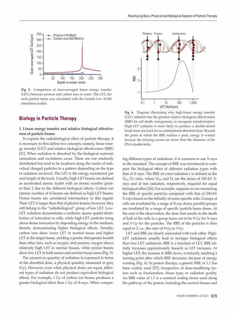
To explain the radiobiological effect of particle therapy, it is necessary to first define two concepts, namely, linear ener-gy transfer (LET) and relative biological effectiveness (RBE) [21]. When radiation is absorbed by the biological material, ionizations and excitations occur. These are not randomly distributed but tend to be localized along the tracks of indi-vidual charged particles in a pattern depending on the type of radiation involved. The LET is the energy transferred per unit length of the track. Usually, high-LET beams are defined as accelerated atomic nuclei with an atomic number great-er than 2 due to the different biological effects. Carbon ion (atomic number of 6) beams are defined as high-LET beams. Proton beams are considered intermediary in this regard. Their LET is larger than that of photon beams; however, they still belong to the “radiobiological” group of low LET. Low-LET radiation demonstrates a uniform, sparse spatial distri-bution of ionization in cells, while high-LET particles bring about dense ionization by depositing energy in the medium, thereby demonstrating higher biological effects. Notably, carbon ions show lower LET in normal tissue and higher LET in the target tissue, yielding a greater therapeutic benefit than other ions, such as oxygen, and protons; oxygen shows relatively high LET in normal tissues, while proton beams show low LET in both tumor and normal tissue areas (Fig. 5).
The amount or quantity of radiation is expressed in terms of the absorbed dose, a physical quantity measured in gray (Gy). However, even when physical doses are equal, differ-ent types of radiation do not produce equivalent biological effects. For example, 1 Gy of carbon ion beams produces a greater biological effect than 1 Gy of X-rays. When compar-
ing different types of radiations, it is common to use X-rays as the standard. The concept of RBE was introduced to com-pare the biological effect of different radiation types with that of X-rays. The RBE of a test radiation r is defined as the D250/Dr ratio, where D250 and Dr are the doses of 250-kV X-rays and of test radiation, respectively, required for equal biological effect [26]. For example, suppose we are measuring the RBE of specific particles compared with that of 250-kV X-rays based on the lethality of some specific cells. Groups of cells are irradiated by a range of X-ray doses; parallel groups are irradiated by a range of specific particle beam doses. At the end of the observation, the dose that results in the death of half of the cells in a group turns out to be 9 Gy for X-rays and 3 Gy for the particles. The RBE of the particles is then equal to 3, i.e., the ratio of 9 Gy to 3 Gy.
LET and RBE are closely associated with each other. High-LET radiations usually lead to stronger biological effects than low-LET radiations. RBE is a function of LET. RBE ini-tially increases approximately linearly as LET increases. At higher LET, the increase in RBE slows, eventually reaching a turning point after which RBE decreases, because of energy wasting (Fig. 6). In proton therapy, a generic RBE of 1.1 has been widely used [27], irrespective of dose-modifying fac-tors such as fractionation, tissue type, or radiation quality. An RBE value of 1.1 is a constant scaling factor used along the pathway of the proton, including the normal tissues and
Hwa Kyung Byun, Physical and Biological Aspects of Particle Therapy
Fig. 5. Comparison of dose-averaged linear energy transfer (LETd) between protons and carbon ions in water. The LETd for each particle beam was calculated with the Geant4 (ver. 10.06) simulation toolkit.
Dose
ave
rage
d LE
T (k
eV/µ
m)
Depth in water (mm)
100
0
250
150
200
300
50
350
0 250 30020015050 100
Protons (170 MeV)Carbon ions (322 MeV/u)
Fig. 6. Diagram illustrating why high-linear energy transfer (LET) radiation has the greatest relative biological effectiveness (RBE) for cell death, mutagenesis, or oncogenic transformation. High-LET radiation is most likely to produce a double-strand break from one track for an administered absorbed dose. Beyond the point at which the RBE reaches a peak, energy is wasted because the ionizing events are closer than the diameter of the DNA double helix.
RBE
LET (keV/µm)
2
1
3
0.1 100 1,000101
Photons Protons Carbon ions
VOLUME 53 NUMBER 3 JULY 2021 615

tumors; therefore, it does not provide benefits in terms of an increased therapeutic window. However, the actual RBE of the proton beam is known to vary with LET, particularly at the distal part of the range of the monoenergetic proton beam penetration, where LET increases. Thus, it should be avoided to locate critical normal structures at that distal end of the proton beams. In addition, experimental RBE of proton beams is calculated differently depending on the type of cell lines [28]. It was thought that the variance of RBE occurred according to the characteristics of the radiation repair pro-cess, and DNA damage response signaling showed different activation durations related to RBE. Thus, the invariant RBE value used currently has been criticized, and optimal ways to replace the standard RBE value are being investigated [29,30].
In contrast, the RBE of carbon ions is not a constant value but a function of position within the treatment beam. RBE tends to increase as the particle penetrates deeper into the target lesion. The depth-dose profile for each SOBP field was designed to yield a constant biological effect within the SOBP area by compensating for the increase in LET along its path. Deeper regions in the SOBP, where the LET is high, receive a lower physical dose; in contrast, shallower regions, where the LET is low, receive a higher physical dose (Fig. 7). Fur-thermore, at the entry site (normal tissue), the RBE value is lower than that in the SOBP, which eventually widens the therapeutic window of carbon ion therapy. In addition, sev-eral other radiation dose-modifying factors such as fractiona-tion, tissue type, and radiation quality are taken into account to determine the RBE.
The biological RBE was determined by a specific biologi-cal system and a biological endpoint and then scaled to the specific irradiation conditions of patients to obtain a “clini-cal RBE” (Fig. 7). The clinical RBE describes the ratio of pre-scribed absorbed doses of a photon to a high-LET irradiation, which are believed to result in clinically equivalent results. The RBE thus obtained was validated using the tumor con-trol probability from clinical data. Because of the complex-ity of RBE of carbon ion therapy, a biomathematical model is needed to consider the inconstant biological effect appro-priate to calculate the RBE in treatment planning. Radiation oncologists have tried to improve the models to reach a “uni-versal” definition of RBE-weighted dose, although it is not yet feasible to fully simulate the underlying biological pro-cesses. The use of in vitro data for RBE models is also a major weakness because the biological effectiveness is affected by general patient condition and the tumor microenvironment.
2. Biological characteristics of high-LET radiationsSolid tumors are often characterized by hypoxia. Acute
(perfusion-limited) hypoxia is caused by temporary distur-bance in perfusion, resulting in fluctuating microvascular oxygen supply. Chronic hypoxia arises due to the over-pro-liferation of cancer cells with poor vasculature. The increased distance between cells and the nearest blood vessel limits oxygen diffusion from tumor microvessels into the sur-rounding tissue. Tumor hypoxia is known to correlate with poor prognosis in cancer patients [31]. Low-LET radiation mostly causes DNA damage due to the presence of free radi-cals, which is enhanced by oxygen. Hence, tumor hypoxia has been considered to be one of the major mechanisms of radioresistance in cancer cells. The ratio of doses to hypoxic and normoxic tissues (oxygen enhancement ratio [OER]) can be close to 3 with low-LET radiation such as gamma-rays and X-rays, making tumor control by radiation difficult in the presence of hypoxia [32]. One method to overcome this obstacle in radiotherapy with low-LET radiation is multiple fractionations. Multiple fractionations allow for the supply of oxygen to the surviving, previously hypoxic volumes, between fractions, through a process termed reoxygenation.
In contrast, high-LET radiation strikes the DNA molecule directly and disrupts the molecular structure. This extensive damage is less influenced by oxygen levels. The OER is lower in high-LET beams. When high-LET radiation is delivered, the hypoxic tumor sites tend to not be affected by oxygen levels, demonstrating similar radiosensitivity. Therefore, heavy ion beams can have a better effect on hypoxic tumors such that the need for fractionation for reoxygenation is diminished. Fractionated irradiation is a basic concept of radiotherapy leading to improved therapeutic ratio. Various biological effects account for the benefits of fractionated irra-
Cancer Res Treat. 2021;53(3):611-620
Fig. 7. Physical, biological, and clinical depth-dose distributions for carbon beam spread-out Bragg peak (SOBP). The biological model was obtained using an in vitro model of a human sali-vary gland tumor cell line. The physical dose×relative biological effectiveness (RBE) is supposed to be constant within the SOBP. Note that the physical dose line is curved within the SOBP area. Accordingly, the RBE within the SOBP area is not a constant val-ue but rather a function of depth.
Dose
per
frac
tion
(Gy
or G
y (R
BE))
Depth in water (mm)
6
2
0
10
8
4
12
0 60 804020
Clinical dose Biological dosePhysical dose
3.834 Gy
RBE2.613
RBE1.781
616 CANCER RESEARCH AND TREATMENT

diation: (1) repair, (2) repopulation, (3) redistribution, and (4) reoxygenation, known as the “4Rs.” Repair and repopulation promote the recovery of damaged normal tissue, and redis-tribution and reoxygenation promote tumor control. The 4Rs are an important issue in conventional XRT, although they are of little importance for high-LET radiations. For example, sublethal damage repair, which promotes cell survival, is not as obvious in high-LET beams as in low-LET ones. The effect of high-LET radiation is uniform irrespective of the cell cycle, while the effect of low-LET radiation is affected by the cell cycle. The 4Rs’ effects in particle beams are small enough to neglect these factors. For these reasons, carbon ion therapy requires a lower number of fractions, shorter than XRT.
3. Special topics according to biology in particle therapyRadiogenomics is the study of the link between germline
or somatic genetic variations and the clinical variability observed in response to radiotherapy. The Radiogenomics Consortium, which included 133 institutions from 33 coun-tries as of April 2019 [33], has worked on patient samples by using single-nucleotide polymorphisms (SNPs) to perform genome-wide association studies (GWAS). SNPs are DNA sequence variations that occur when a single nucleotide within a gene locus is altered. The principle of a GWAS is to genotype between 300,000 and 1,000,000 tag SNPs, which represent most of the known common variations within the genome. The associations between SNPs and radiation-relat-ed toxicity have been identified and validated by the con-sortium, although the effects are not yet clinically actionable. For example, in prostate cancer, the TANC1 locus at 2q24.1 was found to be associated with overall urinary and bowel toxicity after radiation [34]; the KDM3B locus at 5q31.2 was associated with increased urinary frequency after radiation [35]; and the DNAH5 locus at 5p15.2 was associated with uri-nary retention after radiation [35].
The radiogenomic data of the response of mammalian cells to charged particles, predominantly protons and carbon ions, is less mature compared with X-ray exposures. Whether patients who receive particle therapy have different biomark-ers of normal tissue toxicity than those who receive conven-tional radiotherapy is an open question. Although radiation generally activates the genes associated with inflammatory pathways, DNA repair, and cell cycle progression, the spe-cific genes activated by X-rays, proton, and carbon ions can be different [36]. Efforts are underway to establish cohorts of patients with prostate cancer treated at the National Institute of Radiological Sciences with carbon ion therapy or proton therapy for radiogenomic studies [37]. Among the results of experimental studies of gene expression after exposure to carbon ions, the downregulation of genes involved in motil-ity due to carbon ion therapy is of particular interest [38,39],
which are, in contrast, generally upregulated by X-ray expo-sure [40-42]. Carbon ion therapy appears to suppress migra-tion, invasion, and metastasis of cancer cells [43,44] and does not lead to the induction of hypoxia-inducible factor-1 [45] and stem cell factor expression [39], both of which are asso- ciated with angiogenesis. Reduced tumor cell migration and invasion and reduced angiogenesis may be some of the major benefits of carbon ion therapy. However, the impact of these molecular signatures requires validation in animal models and in human studies.
In addition to dosimetric analyses, tests for germline and tumor genetic variants can be incorporated into clinical decision-making regarding particle therapy. For example, patients who carry the NF1 gene mutation are radiosensitive and predisposed to a number of cancers. NF1 mutations can be used as indicators in patients receiving proton therapy to reduce the risk of secondary radiation-related malignan-cies [46]. Furthermore, the identification of radioresistance to low-LET radiation using the GWAS approach can improve the selection of patients eligible for carbon ion therapy; how-ever, this approach needs to be validated in prospective clini-cal studies.
Another special issue with particle therapy is immuno-therapy. Immunotherapy has recently emerged as a prom-ising set of new cancer treatments. Immune checkpoint blockade, which increases antitumor immunity by block-ing inhibitory checkpoints, has gained an important place in the treatment of various types of cancer. However, the overall response rates with immune checkpoint blockade monotherapy are modest. For example, the response rates of melanoma after ipilimumab and pembrolizumab or nivolumab therapy range from 11% to 19% [47,48] and 33% to 44% [48-50], respectively. Thus, novel strategies that aug-ment systemic immune responses will be potentially critical in the curative management of the disease. The addition of radiotherapy to immunotherapy for patients with predomi-nantly widespread metastases has gained substantial inter-est. Radiation-induced cancer cell death results in the release of pro-inflammatory signals such as damage-associated molecular patterns danger signals and inflammatory cyto-kines, thereby triggering the innate immune system to acti-vate tumor-specific T cells. Radiation also has an effect on the tumor microenvironment, by promoting the infiltration of activated T cells, and can overcome some of the barriers to tumor rejection [51]. The advantages and outcomes of com-bined particle therapy with immunotherapy are still open for investigation. Preclinical studies support the immunogenic potential of proton therapy and suggest that proton therapy may actually have a wider range of immunogenic applica-tions than photon therapy. For example, in vitro studies have reported that protons mediated a greater increase in the
Hwa Kyung Byun, Physical and Biological Aspects of Particle Therapy
VOLUME 53 NUMBER 3 JULY 2021 617

expression of calreticulin on the cell surface than photons, increasing cross-priming to cytotoxic T lymphocyte killing [52].
Photon and carbon ion therapies induce different DNA damage and repair pathways, and this difference is based on the differential biological response to low- and high-LET beams. Depending on LET and dose, ionizing radiation causes a variety of different DNA lesions, including single- and double-strand breaks, DNA-protein cross-links, and DNA base damages [53]. This variation can be important because programmed death-ligand 1 expression in can-cer cells is upregulated in response to DNA double-strand breaks [54]. Because carbon ion therapy is performed in few sites worldwide, only limited experimental information is currently available. The typical endpoints of these experi-ments include the abscopal effect, defined as a reaction of the organism’s cells that had not been directly exposed to irradiation, which causes regression of the non-irradiated tumors and the growth of distal metastases [55]. Matsunaga et al. [56] reported that carbon ion therapy administered to a poorly immunogenic squamous cell carcinoma cell line induces reduction of tumor formation after secondary tumor challenge at the contralateral site in mouse models. It has been shown that carbon ion therapy induces systemic antitu-mor immunity. Combined carbon ion therapy and dendritic cell injection generated more prominent cytolytic activity than carbon ion therapy alone. Ando et al. [57] reported that a combination treatment of carbon ion therapy and intrave-nous dendritic cell administration enhances the suppression of lung metastases.
Conclusion
Particle therapy involves better dose distribution based on the Bragg peak, physically. Biologically, it shows higher LET and RBE, which is expected to be powerful from a clini-cal point of view. These properties enable the administration of higher radiation doses to the tumors while subjecting the normal organs to a reasonably low dose, thus widening the
therapeutic ratio. Because of the lack of clinical trials, it is not clearly demonstrated whether carbon ion therapy has superior clinical benefits over other radiotherapy modalities. Although further comparison studies are needed, conduct-ing randomized controlled trials comparing carbon ion ther-apy, proton therapy, and XRT seems to be difficult, mostly because of the differences in treatment costs and patients’ preferences. Nevertheless, we expect that the superior bio-logical and physical aspects of carbon ion therapy will lead to meaningful clinical benefit in cancer patients. Another uncertainty remains concerning the RBE. Currently, the con-stant RBE value of 1.1 for proton therapy is being criticized; thus, further investigation is required to determine the opti-mal RBE and its association with LET and dose. The RBE of carbon ion therapy has to be calculated by biomathematical models in treatment planning, which—in spite of all valida-tion efforts—still involve significant sources of uncertainty. Molecular biological research using highly advanced tech-nologies such as multi-omics or the effects of the combination of immunotherapy has to be incorporated into the particle therapy research field. By resolving unsolved issues regard-ing the physical and biological properties of particle therapy, we believe that the future of particle therapy is promising.
Author ContributionsConceived and designed the analysis: Kim JS, Koom WS, Kim YB.Collected the data: Byun HK, Han MC, Yang K, Koom WS.Contributed data or analysis tools: Kim JS, Yoo GS, Koom WS, Kim YB.Performed the analysis: Byun HK, Han MC, Yang K.Wrote the paper: Byun HK, Han MC, Yang K, Kim JS, Yoo GS, Koom WS, Kim YB.
Conflicts of InterestConflict of interest relevant to this article was not reported.
AcknowledgmentsThe authors thank Medical Illustration & Design, part of the Medi-cal Research Support Services of Yonsei University College of Medi-cine, for all artistic support related to this work.
Cancer Res Treat. 2021;53(3):611-620
618 CANCER RESEARCH AND TREATMENT
1. Halperin EC. Particle therapy and treatment of cancer. Lancet Oncol. 2006;7:676-85.
2. Slater JM, Archambeau JO, Miller DW, Notarus MI, Preston W, Slater JD. The proton treatment center at Loma Linda Uni-versity Medical Center: rationale for and description of its development. Int J Radiat Oncol Biol Phys. 1992;22:383-9.
3. Tsuji H, Akine Y, Okumura T. Proton radiotherapy: clinical
experience and outcome at Tsukuba. Radiol Nuclear Med. 1997;43:604-9.
4. Schaub L, Harrabi SB, Debus J. Particle therapy in the future of precision therapy. Br J Radiol. 2020;93:20200183.
5. Patel SH, Wang Z, Wong WW, Murad MH, Buckey CR, Mohammed K, et al. Charged particle therapy versus photon therapy for paranasal sinus and nasal cavity malignant dis-
References

Hwa Kyung Byun, Physical and Biological Aspects of Particle Therapy
eases: a systematic review and meta-analysis. Lancet Oncol. 2014;15:1027-38.
6. Particle therapy facilities in clinical operation (last update Feb 2021) [Internet]. Villigen: Particle Therapy Co-Operative Group; 2021 [cited 2021 Jun 14]. Available from: https://ptcog.ch/index.php/facilities-in-operation.
7. Particle therapy facilities under construction (last update Feb 2021) [Internet]. Villigen: Particle Therapy Co-Operative Group; 2021 [cited 2021 Jun 14]. Available from: https://ptcog.ch/index.php/facilities-under-construction.
8. Campian JL, Sarai G, Ye X, Marur S, Grossman SA. Associa-tion between severe treatment-related lymphopenia and pro-gression-free survival in patients with newly diagnosed squa-mous cell head and neck cancer. Head Neck. 2014;36:1747-53.
9. Mohan R, Liu AY, Brown PD, Mahajan A, Dinh J, Chung C, et al. Proton therapy reduces the likelihood of high-grade radiation-induced lymphopenia in glioblastoma patients: phase II randomized study of protons vs photons. Neuro Oncol. 2021;23:284-94.
10. Routman DM, Garant A, Lester SC, Day CN, Harmsen WS, Sanheuza CT, et al. A comparison of grade 4 lymphopenia with proton versus photon radiation therapy for esophageal cancer. Adv Radiat Oncol. 2019;4:63-9.
11. Xiang M, Chang DT, Pollom EL. Second cancer risk after pri-mary cancer treatment with three-dimensional conformal, intensity-modulated, or proton beam radiation therapy. Can-cer. 2020;126:3560-8.
12. Mohamad O, Tabuchi T, Nitta Y, Nomoto A, Sato A, Kasuya G, et al. Risk of subsequent primary cancers after carbon ion radiotherapy, photon radiotherapy, or surgery for localised prostate cancer: a propensity score-weighted, retrospective, cohort study. Lancet Oncol. 2019;20:674-85.
13. Saunders W, Castro JR, Chen GT, Collier JM, Zink SR, Pitluck S, et al. Helium-ion radiation therapy at the Lawrence Berke-ley Laboratory: recent results of a Northern California Oncol-ogy Group Clinical Trial. Radiat Res Suppl. 1985;8:S227-34.
14. Tessonnier T, Mairani A, Brons S, Sala P, Cerutti F, Ferrari A, et al. Helium ions at the heidelberg ion beam therapy center: comparisons between FLUKA Monte Carlo code predictions and dosimetric measurements. Phys Med Biol. 2017;62:6784-803.
15. Tobias CA, Anger HO, Lawrence JH. Radiological use of high energy deuterons and alpha particles. Am J Roentgenol Radium Ther Nucl Med. 1952;67:1-27.
16. Lawrence JH, Tobias CA, Born JL, Mc CR, Roberts JE, Anger HO, et al. Pituitary irradiation with high-energy pro-ton beams: a preliminary report. Cancer Res. 1958;18:121-34.
17. Castro JR, Quivey JM, Lyman JT, Chen GT, Phillips TL, Tobias CA. Radiotherapy with heavy charged particles at Lawrence Berkeley Laboratory. J Can Assoc Radiol. 1980;31:30-4.
18. Chen GT, Castro JR, Quivey JM. Heavy charged particle radiotherapy. Annu Rev Biophys Bioeng. 1981;10:499-529.
19. Chatterjee A, Alpen EL, Tobias CA, Llacer J, Alonso J. High energy beams of radioactive nuclei and their biomedical applications. Int J Radiat Oncol Biol Phys. 1981;7:503-7.
20. Particle Therapy Co-Operative Group [Internet]. Villigen: Par-
ticle Therapy Co-Operative Group; 2021 [cited 2021 Jun 14]. Available from: https://www.ptcog.ch/.
21. Tsujii H, Kamada T, Shirai T, Noda K, Tsuji H, Karasawa K. Carbon-ion radiotherapy: principles, practices, and treatment planning. Tokyo: Springer; 2014.
22. Fredriksson A. Robust optimization in radiation therapy. In: Terlaky T, Anjos MF, Ahmed S, editors. Advances and trends in optimization with engineering applications. Philadelphia, PA: SIAM; 2017. p. 1-12.
23. Han Y. Current status of proton therapy techniques for lung cancer. Radiat Oncol J. 2019;37:232-48.
24. Seco J, Robertson D, Trofimov A, Paganetti H. Breathing interplay effects during proton beam scanning: simulation and statistical analysis. Phys Med Biol. 2009;54:N283-94.
25. Yoo GS, Yu JI, Cho S, Jung SH, Han Y, Park S, et al. Compari-son of clinical outcomes between passive scattering versus pencil-beam scanning proton beam therapy for hepatocellu-lar carcinoma. Radiother Oncol. 2020;146:187-93.
26. Hall EJ, Giaccia AJ. Radiobiology for the radiologist. 7th ed. Philadelphia, PA: Lippincott Williams & Wolters Kluwer; 2010. p. 394-6.
27. Paganetti H, Niemierko A, Ancukiewicz M, Gerweck LE, Goitein M, Loeffler JS, et al. Relative biological effectiveness (RBE) values for proton beam therapy. Int J Radiat Oncol Biol Phys. 2002;53:407-21.
28. Choi C, Son A, Lee GH, Shin SW, Park S, Ahn SH, et al. Target-ing DNA-dependent protein kinase sensitizes hepatocellular carcinoma cells to proton beam irradiation through apoptosis induction. PLoS One. 2019;14:e0218049.
29. Jones B. Towards achieving the full clinical potential of pro-ton therapy by inclusion of LET and RBE models. Cancers (Ba-sel). 2015;7:460-80.
30. Paganetti H, Blakely E, Carabe-Fernandez A, Carlson DJ, Das IJ, Dong L, et al. Report of the AAPM TG-256 on the relative biological effectiveness of proton beams in radiation therapy. Med Phys. 2019;46:e53-78.
31. Bristow RG, Hill RP. Hypoxia and metabolism: hypoxia, DNA repair and genetic instability. Nat Rev Cancer. 2008;8:180-92.
32. Brown JM, Wilson WR. Exploiting tumour hypoxia in cancer treatment. Nat Rev Cancer. 2004;4:437-47.
33. West C, Rosenstein BS, Alsner J, Azria D, Barnett G, Begg A, et al. Establishment of a radiogenomics consortium. Int J Radiat Oncol Biol Phys. 2010;76:1295-6.
34. Fachal L, Gomez-Caamano A, Barnett GC, Peleteiro P, Car-ballo AM, Calvo-Crespo P, et al. A three-stage genome-wide association study identifies a susceptibility locus for late radiotherapy toxicity at 2q24.1. Nat Genet. 2014;46:891-4.
35. Kerns SL, Dorling L, Fachal L, Bentzen S, Pharoah PD, Barnes DR, et al. Meta-analysis of genome wide association studies identifies genetic markers of late toxicity following radiother-apy for prostate cancer. EBioMedicine. 2016;10:150-63.
36. Tinganelli W, Durante M. Carbon ion radiobiology. Cancers (Basel). 2020;12:3022.
37. West C. Translational radiobiology: radiogenomics. In: Pro-ceedings of the PTCOG-58; 2019 Jun 10-15; Manchester, UK.
38. Akino Y, Teshima T, Kihara A, Kodera-Suzumoto Y, Inaoka M,
VOLUME 53 NUMBER 3 JULY 2021 619
Cancer Res Treat. 2021;53(3):611-620
Higashiyama S, et al. Carbon-ion beam irradiation effectively suppresses migration and invasion of human non-small-cell lung cancer cells. Int J Radiat Oncol Biol Phys. 2009;75:475-81.
39. Kamlah F, Hanze J, Arenz A, Seay U, Hasan D, Juricko J, et al. Comparison of the effects of carbon ion and photon irradia-tion on the angiogenic response in human lung adenocarci-noma cells. Int J Radiat Oncol Biol Phys. 2011;80:1541-9.
40. Moncharmont C, Levy A, Guy JB, Falk AT, Guilbert M, Trone JC, et al. Radiation-enhanced cell migration/invasion process: a review. Crit Rev Oncol Hematol. 2014;92:133-42.
41. Sonveaux P, Brouet A, Havaux X, Gregoire V, Dessy C, Bal-ligand JL, et al. Irradiation-induced angiogenesis through the up-regulation of the nitric oxide pathway: implications for tumor radiotherapy. Cancer Res. 2003;63:1012-9.
42. Wild-Bode C, Weller M, Rimner A, Dichgans J, Wick W. Sub-lethal irradiation promotes migration and invasiveness of glioma cells: implications for radiotherapy of human glioblas-toma. Cancer Res. 2001;61:2744-50.
43. Ogata T, Teshima T, Kagawa K, Hishikawa Y, Takahashi Y, Kawaguchi A, et al. Particle irradiation suppresses metastatic potential of cancer cells. Cancer Res. 2005;65:113-20.
44. Takahashi Y, Teshima T, Kawaguchi N, Hamada Y, Mori S, Madachi A, et al. Heavy ion irradiation inhibits in vitro angiogenesis even at sublethal dose. Cancer Res. 2003;63:4253-7.
45. Subtil FS, Wilhelm J, Bill V, Westholt N, Rudolph S, Fischer J, et al. Carbon ion radiotherapy of human lung cancer attenuates HIF-1 signaling and acts with considerably enhanced thera-peutic efficiency. FASEB J. 2014;28:1412-21.
46. Sharif S, Ferner R, Birch JM, Gillespie JE, Gattamaneni HR, Baser ME, et al. Second primary tumors in neurofibromato-sis 1 patients treated for optic glioma: substantial risks after radiotherapy. J Clin Oncol. 2006;24:2570-5.
47. Hodi FS, Chesney J, Pavlick AC, Robert C, Grossmann KF, McDermott DF, et al. Combined nivolumab and ipilimumab versus ipilimumab alone in patients with advanced mela-noma: 2-year overall survival outcomes in a multicentre, ran-domised, controlled, phase 2 trial. Lancet Oncol. 2016;17:1558-
68.48. Wolchok JD, Chiarion-Sileni V, Gonzalez R, Rutkowski P,
Grob JJ, Cowey CL, et al. Overall survival with combined nivolumab and ipilimumab in advanced melanoma. N Engl J Med. 2017;377:1345-56.
49. Robert C, Long GV, Brady B, Dutriaux C, Maio M, Mortier L, et al. Nivolumab in previously untreated melanoma without BRAF mutation. N Engl J Med. 2015;372:320-30.
50. Robert C, Schachter J, Long GV, Arance A, Grob JJ, Mortier L, et al. Pembrolizumab versus ipilimumab in advanced mela-noma. N Engl J Med. 2015;372:2521-32.
51. Demaria S, Golden EB, Formenti SC. Role of local radiation therapy in cancer immunotherapy. JAMA Oncol. 2015;1:1325-32.
52. Gameiro SR, Malamas AS, Bernstein MB, Tsang KY, Vassan-tachart A, Sahoo N, et al. Tumor cells surviving exposure to proton or photon radiation share a common immunogenic modulation signature, rendering them more sensitive to T cell-mediated killing. Int J Radiat Oncol Biol Phys. 2016;95:120-30.
53. Bouquet F, Muller C, Salles B. The loss of gammaH2AX signal is a marker of DNA double strand breaks repair only at low levels of DNA damage. Cell Cycle. 2006;5:1116-22.
54. Sato H, Niimi A, Yasuhara T, Permata TB, Hagiwara Y, Isono M, et al. DNA double-strand break repair pathway regulates PD-L1 expression in cancer cells. Nat Commun. 2017;8:1751.
55. Schmid TE, Multhoff G. Non-targeted effects of photon and particle irradiation and the interaction with the immune sys-tem. Front Oncol. 2012;2:80.
56. Matsunaga A, Ueda Y, Yamada S, Harada Y, Shimada H, Hasegawa M, et al. Carbon-ion beam treatment induces sys-temic antitumor immunity against murine squamous cell carcinoma. Cancer. 2010;116:3740-8.
57. Ando K, Fujita H, Hosoi A, Ma L, Wakatsuki M, Seino KI, et al. Intravenous dendritic cell administration enhances suppres-sion of lung metastasis induced by carbon-ion irradiation. J Radiat Res. 2017;58:446-55.
620 CANCER RESEARCH AND TREATMENT