Embed Size (px)
Citation preview

Site-specific integrase-mediated transgenesis in micevia pronuclear injectionBosiljka Tasica, Simon Hippenmeyera, Charlene Wangb, Matthew Gamboab, Hui Zongc, Yanru Chen-Tsaib,1,and Liqun Luoa,1
aThe Howard Hughes Medical Institute and Department of Biology, Stanford University, Stanford, CA 94305; bTransgenic Facility, Stanford Cancer Center,Stanford University School of Medicine, Stanford, CA 94305; and cInstitute of Molecular Biology, University of Oregon, Eugene, OR 97403
Edited* by Matthew P. Scott, Stanford University/The Howard Hughes Medical Institute, Stanford, CA, and approved March 16, 2011 (received for reviewDecember 29, 2010)
Microinjection of recombinant DNA into zygotic pronuclei hasbeen widely used for producing transgenic mice. However, withthis method, the insertion site, integrity, and copy number of thetransgene cannot be controlled. Here, we present an integrase-based approach to produce transgenic mice via pronuclear in-jection, whereby an intact single-copy transgene can be insertedinto predetermined chromosomal loci with high efficiency (up to40%), and faithfully transmitted through generations. We showthat neighboring transgenic elements and bacterial DNAwithin thetransgene cause profound silencing and expression variability ofthe transgenic marker. Removal of these undesirable elementsleads to global high-level marker expression from transgenesdriven by a ubiquitous promoter. We also obtained faithful markerexpression from a tissue-specific promoter. The technique pre-sented here will greatly facilitate murine transgenesis and precisestructure/function dissection of mammalian gene function andregulation in vivo.
Production of transgenic mice via microinjection of DNA intozygotic pronuclei (1–3) has served mammalian genetics for
30 years. Although still the predominant method used to producetransgenic mice, it has several limitations: the insertion site, in-tegrity, and copy number of the transgene cannot be controlled.Insertion ofDNA into different chromosomal loci at random coulddisrupt the function of endogenous genes. Transgenes generated inthis manner can be influenced by the local chromatin environment(i.e., position effect) that can lead to transgene silencing or ectopicexpression (4–7). Moreover, transgenic DNA concatemerized intoa large array is subject to repeat-induced gene silencing (8).Single-copy transgenesis in mice can be achieved with retro-
viruses (9) and transposons (10, 11), but these approaches in-tegrate transgenes throughout the genome. As a result, thetransgenes are subjected to the local chromatin environment andcan cause endogenous gene disruption, although the mutagenicproperties of transposons can be desirable for particular appli-cations (10). These problems can be overcome by targeting thetransgene to a specific chromosomal locus via homologous re-combination in embryonic stem (ES) cells (12, 13). However, thismethod is significantly more laborious and time-consuming, as itinvolves creation of modified ES cells and mouse chimeras, aswell as eventual germline transmission of the transgene.Integrase enzymes from a variety of sources have been used
to catalyze integration of transgenes in heterologous systems(14, 15). Integrases catalyze irreversible recombination betweenappropriate attB and attP sites (14, 16). ϕC31 integrase froma Streptomyces phage has previously been used for transgene in-tegration in flies (17–19). In mice, ϕC31 integrase has been usedto catalyze integration of circular DNA into pseudo-attP sites inthe genome for gene therapy (20) or low-efficiency transgenesis(21), for transgenesis in mouse ES cells (22), and for removal ofundesirable transgene portions or reporter activation (23, 24).Here, we describe an integrase-mediated method for site-
specific transgenesis in mice via pronuclear microinjection, withintegration efficiencies as high as 40%. We use ϕC31 integraseto catalyze recombination between one or two attB sites in arecombinant DNA with one or more tandem attP sites that wepreviously inserted into specific loci in the mouse genome (Fig. 1).
We show that the plasmid bacterial backbone within the transgeneand nearby transgenic elements dramatically decrease expressionof our transgenes, and that the absence of these elements results inglobal, high-level transgene expression from a ubiquitous pro-moter. Finally, we show that a promoter for the murine tran-scription factor Hb9 integrated into one of the predetermined locidrives proper tissue-specific marker expression.
ResultsStrategy and Proof of Principle for Integrase-Mediated Site-SpecificTransgenesis. To generate embryos containing attP sites for ϕC31integrase-mediated transgenesis, we used standard homologousrecombination-based methods in mouse ES cells (12, 25). Weinserted three shortened tandem ϕC31 integrase attP sites(attPx3) or a single “full-length” attP site (14) into two loci: theRosa26 locus on mouse chromosome 6 (26) and an intergenicHipp11 (H11) locus on mouse chromosome 11 (27) (Fig. 1, Left,and SI Appendix, Fig. S1). The Rosa26 locus supports globalexpression of a single copy knock-in transgene driven by a com-bination of the CMV enhancer and the chicken β-actin promoter(pCA) (28, 29). Knock-in experiments confirmed that H11 sup-ports high-level global gene expression from the pCA promoterand a higher rate of mitotic (interchromosomal) recombinationcompared with Rosa26 (27). The latter property suggested thatH11 might allow better access to ϕC31 integrase than Rosa26.The modified ES cells were used to produce chimeric mice, andmice with germline-transmitted alleles were used to establishmouse colonies homozygous for the knock-in cassettes.For most experiments, we integrated transgenes into the H11
locus because homozygous insertions into this locus are not pre-dicted to disrupt any endogenous genes, and the resulting miceare completely healthy and fertile (27). We injected embryoshomozygous for attP or attPx3 at the H11 locus (H11P or H11P3,respectively) with ϕC31 mRNA together with attB-pCA-GFP,a minicircle DNA that was generated by removal of the plasmidbackbone to enable proper transgene expression (as detailedlater). The attB-pCA-GFP minicircle contains a full-length attBsite (14) and the sequence for a thermotolerant GFP (30, 31)driven by the ubiquitous pCA promoter (SI Appendix, SI Materialsand Methods). We obtained integration of the transgene (Fig. 1,Top Right, and Table 1, rows 1–3; SI Appendix, Table S1, providesmore details).As an alternative to insertion of DNA at a single attP site, we
also injected a plasmid in which pCA-GFP is flanked by two attB
Author contributions: B.T., H.Z., Y.C.-T., and L.L. designed research; B.T., S.H., C.W., andM.G. performed research; B.T. and L.L. analyzed data; S.H. contributed new reagents/analytic tools; and B.T. and L.L. wrote the paper.
Conflict of interest statement: Y.C.-T. is a consultant and co-founder of AppliedStemCell, Inc.
*This Direct Submission article had a prearranged editor.
Freely available online through the PNAS Open Access Option.1To whom correspondence may be addressed. E-mail: [email protected] or [email protected].
This article contains supporting information online at www.pnas.org/lookup/suppl/doi:10.1073/pnas.1019507108/-/DCSupplemental.
www.pnas.org/cgi/doi/10.1073/pnas.1019507108 PNAS Early Edition | 1 of 6
GEN
ETICS

sites (pattB-pCA-GFP-attB) into H11P3 homozygous embryos. Inprinciple, this approach should allow ϕC31 integrase to catalyzea recombinase-mediated cassette exchange reaction (Fig. 1,Bottom Right). We tested the integration of the circular pattB-pCA-GFP-attB plasmid into H11P3 and obtained successfulcassette exchange as confirmed by PCR (Fig. 1, Bottom Right,and Table 1, row 4). In these experiments, we never detectedinsertion of a full plasmid.The integrated transgenes were properly transmitted from
founders to progeny (SI Appendix, Table S2). Both strategiesresulted in broad and high-level GFP expression in pCA-GFPtransgenic mice (Fig. 1, Right, Fig. 2D, and Fig. S1C, Bottom).These data provide the proof of principle for our site-specificintegrase-mediated transgenesis in mice. In the subsequentsections, we describe in more detail the optimization processthat led to these results.
Transgenic Integrase Did Not Enable Site-Specific Integration. Ouroriginal knock-in cassettes contained a mammalian codon-optimizedϕC31 integrase (ϕC31o) (23) driven by a fragment of themouseVASA promoter sufficient for germline expression (32), and
a neomycin resistance gene, flanked by FRT5 sites (33) (SI Ap-pendix, Fig. S1). This “NVϕ cassette” (i.e., Neo-VASA-ϕC31o) wasdesigned to provide the integrase in embryos in situ. We injectedmouse embryos homozygous for the H11P3NVϕ knock-in witha plasmid containing the attB-pCA-GFP transgene (pattB-pCA-GFP) but did not obtain any site-specific integration (0/32 F0s;Table 1, row 5) despite occasional random integrations. However,coinjecting pattB-pCA-GFP with ϕC31o mRNA into homozygousH11P3NVϕ embryos produced site-specific integrations (Table 1,row 6). Thus, the VASA promoter does not promote sufficientϕC31o expression in situ to enable site-specific insertions.Because the NVϕ cassette did not perform as expected we re-
moved it from the H11P3NVϕ allele by FLP-mediated recom-bination to generate the H11P3 allele (SI Appendix, Fig. S1, andSI Appendix, SI Materials and Methods). Similarly, the H11P al-lele was derived from H11PNVϕ. Coinjection of pattB-pCA-GFPand ϕC31o mRNA into homozygous H11P or H11P3 embryosproduced site-specific integrants (Table 1, rows 8–10). Thus,we coinjected integrase mRNA with DNA for all subsequentexperiments.
Fig. 1. Schematic summary for site-specific ϕC31 integrase-mediatedtransgenesis via pronuclear injec-tion in mice. A single or threetandem attP sites were knockedinto the Hipp11 (H11) or Rosa26loci via homologous recombina-tion in ES cells (Left; for details, seeFig. S1). Mice homozygous for oneof the modified loci (H11 is shownas an example) served as embryodonors. A mix of DNA and in vitrotranscribed ϕC31 mRNA was injec-ted into a single pronucleus ofeach zygote. The integration ofplasmid bacterial backbone (BB)that decreases the transgene ex-pression was avoided either byinjecting a minicircle DNA with a single attB site (top branch; in this case, ϕC31 catalyzes a typical integration reaction), or by injecting plasmid DNA where thegene of interest (e.g., GFP) was flanked by two attB sites (bottom branch; in this case, ϕC31 catalyzes a recombinase-mediated cassette exchange reaction).Right, Middle: Green fluorescence of a representative transgenic F0 embryo and the corresponding PCR results that indicate site-specific insertion. The rednumbers for the PCR results correspond to the numbers on the H11P3-pCA-GFP transgene scheme below; the same PCR tests can be used for the transgeneabove. The particular embryo shown was obtained by cassette exchange. Inset: Bright-field image of the same embryo.
Table 1. Efficiency of site-specific integration
Row DNA* DNA typeDNA
size, kb Strain Background F0 (n) SS F0 (n) Significance¶SS, %(of F0)
R F0(n)
R, %(of F0)
1 attB-pCA-GFP Minicircle ~3 H11P Mix 21 1 NS vs. row 2 4.8 1 4.82 attB-pCA-GFP Minicircle ~3 H11P3 Mix 39 4 10.3 1 2.63 attB-pCA-GFP Minicircle ~3 H11P3 FVB N4 15 6 P < 0.05 vs. row 2 40.0 3 20.04 attB-pCA-GFP-attB Plasmid ~6 H11P3 FVB N4 38‡ 6k — 15.8 1 2.65 attB-pCA-GFP, no RNA Plasmid ~6 H11P3NVϕ Mix 32‡ 0 — 0.0 5 15.66 attB-pCA-GFP Plasmid ~6 H11P3NVϕ Mix 64§ 10 NS vs. row 7 15.6 4 6.37 attB-pCA-GFP Plasmid ~6 H11PNVϕ Mix 30‡ 2 6.7 0 0.08 attB-pCA-GFP-FRT5 Plasmid ~6 H11P Mix 51 5 NS vs. row 9 9.8 3 5.99 attB-pCA-GFP-(FRT5)† Plasmid ~6 H11P3 Mix 61 4 6.6 9 14.810 attB-pCA-GFP-FRT5 Plasmid ~6 H11P3 FVB N4 8 3 P < 0.05 vs. row 9 37.5 1 10.311 attB-pHB9-GFP-FRT5 Plasmid ~14 H11P3 FVB N4 66 2 — 3.0 2 3.012 attB-pCA-GFP Plasmid ~6 R26P3NVϕ Mix 22‡ 2 — 9.1 2 9.1
Abbreviations: F0, embryos or animals obtained from injections; SS, site-specific integration; R, random integration; mix, mixed background of 129, C57BL/6and DBA2; FVB N4, mice of the mixed background were outcrossed for 4 generations to the FVB strain and then intercrossed.*All DNA was coinjected with ϕC31o mRNA, except for row 5.†Both FRT and non-FRT versions of attB-pCA-GFP were used.‡F0s were analyzed only as E10 or E11 embryos.§F0s were analyzed either as E10 or E11 embryos or as live pups.kThe six founders listed contained pCA-GFP without the bacterial backbone; five more founders with cassette exchange contained only the bacterialbackbone. Therefore, the total number of founders with cassette exchange is 11 (29%).¶Fisher’s exact test. NS, not significant.
2 of 6 | www.pnas.org/cgi/doi/10.1073/pnas.1019507108 Tasic et al.

Bacterial Backbone and the NVϕ Cassette Affect Proper TransgeneExpression. Despite proper transmission of the site-specificallyintegrated transgenes produced from the pattB-pCA-GFP plas-mid, the progeny of these transgenic founders exhibited a widerange of GFP expression levels as seen in whole-mount embryos(SI Appendix, Fig. S1C). Moreover, the GFP expression in theprogeny was mosaic in several internal tissues, including theheart, brain, and particularly the liver (Fig. 2B and SI Appendix,Fig. S2). We first observed this variable and mosaic expressionwith H11P3NVϕ as the host. We suspected that the nearbygermline-specific VASA promoter and/or other elements withinthe NVϕ cassette (e.g., the neomycin resistance gene) could af-fect the expression of pCA-GFP inserted at H11. Indeed, trans-genic embryos containing pCA-GFP inserted at H11P3, whichlack the NVϕ cassette, produced more uniform GFP expression(Fig. 2C and SI Appendix, Fig. S2). However, considerable vari-ability of pCA-GFP expression still persisted especially in thelivers of N1 or N2 animals derived from a number of transgenicfounders (Fig. 2C and SI Appendix, Fig. S2).It has been reported that plasmid bacterial backbone can de-
crease the expression of integrated and episomal transgenes (34–37). To test if this is the case in our system, we developed an invitro method to produce minicircle DNA: circular DNA con-taining desired transgene elements, but devoid of the bacterialbackbone (SI Appendix, Fig. S3 and SI Appendix, SI Materials andMethods). We injected the minicircle DNA into H11P3 embryosto produce transgenic animals (SI Appendix, Fig. S1, and Table1). For simplicity, we designate hereafter the transgenic allelesderived from integration of the entire pCA-GFP plasmid (whichcontains the bacterial backbone) as pCA-GFP-BB (Fig. 2C), andthe mice derived from integration of the pCA-GFP minicircle aspCA-GFP (Fig. 2D). Transgenic animals derived from the pCA-GFP minicircle exhibited higher and more uniform expression inembryos and all adult tissues examined (Fig. 2D and SI Appendix,Fig. S2). The removal of bacterial backbone from the pCA-GFP-BB transgene by crossing to our newly generated GFP-FLPotransgenic mice (SI Appendix, SI Materials and Methods) alsoresulted in elevated transgene expression (SI Appendix, Fig. S4).These data demonstrate that pCA-GFP at the H11 locus canexpress GFP ubiquitously in the absence of the NVϕ cassette andthe bacterial backbone.Because the greatest variability was observed in the liver (Fig.
2 and SI Appendix, Fig. S2), we used it as a model to determinethe relative contributions of the NVϕ cassette and the bacterialbackbone to GFP expression variability in these transgenic mice.In addition, to test for the possible differences between a singleattP site and attPx3, we analyzed transgenic mice obtained byinsertion into the H11P allele (Table 1, rows 1 and 8). Finally, toprobe the effect of genetic background, we analyzed transgenesinserted into the H11P3 allele in mice that had been outcrossedto the FVB inbred strain for four generations (Table 1, rows 3and 10). We compared the total GFP fluorescence of liver sec-tions from different transgenic animals under identical con-ditions. We observed no statistically significant differences inGFP fluorescence between transgenes that differed only in thenumber of attP copies or in the genetic background of the mousestrain used for transgenesis (SI Appendix, Fig. S5). Therefore, wegrouped all data according to the presence of the NVϕ cassetteand/or the bacterial backbone (Fig. 2E). We found that, in the
Fig. 2. GFP expression in animals carrying site-specific pCA-GFP transgenesintroduced by ϕC31 integrase-mediated transgenesis. (A–D) Representativefluorescence images from embryonic day 11 embryos and adult livers, hearts,and cerebella of N1 or N2 transgenic animals corresponding to the geno-types and schematics of transgenes shown (Upper). Embryos or same tissueswere imaged under identical conditions, except that “5×-exp” designatesfivefold longer exposure time than for the rest of the images in the samecolumn. Whole-mount embryos were imaged for GFP fluorescence; corre-sponding bright-field images of each embryo are also shown (Insets). Thelivers and hearts are represented by epifluorescence images of 10-μm sec-tions stained only by DAPI (blue). The green signal is GFP fluorescence. Thecerebella are represented by confocal images of sections stained by anti-GFPantibody (green), anti-calbindin (red) for Purkinje cells, and DAPI (blue). TwoPurkinje cells labeled by asterisks appear negative for GFP. ϕC31 attL andattR are the product of recombination of an attP (black arrows) and attB site(purple) and are therefore half black and half purple. Half circles representFRT5 sites. Half white/half black triangle represent λ-integrase attB sitecreated during minicircle production. pSV40, SV40 promoter. pVASA, VASApromoter. U, unique sequence. pCA, CMV enhancer and β-actin promoter. G,GFP. pA, polyA signal. BB, plasmid bacterial backbone. (Scale bars: 1 mm forembryos, 100 μm for tissue sections.) (E) Average fluorescence in arbitrary
units (AU) in the GFP channel for liver sections from animals of genotypesshown below (no int., no integration; represents wt, H11P3NVϕ/wt, H11P3/wt, and H11P/wt genotypes). Each dataset is represented by mean ± SD. Thenumbers of individual animals and founders analyzed for each genotype arelisted below the genotypes. When samples from multiple founders werecombined to obtain an average, each founder was represented by the samenumber of animals except in the case labeled by a spade. The fluorescenceintensities differ significantly among the groups by one-way ANOVA [F(3,50) = 84.09, P < 0.0001]. Tukey’s post-hoc test was used for pair-wise com-parisons (ns, not significant; *P < 0.05 and ***P < 0.001).
Tasic et al. PNAS Early Edition | 3 of 6
GEN
ETICS

presence of both the NVϕ cassette and the bacterial backbone,GFP expression was detectable in the liver in a small number ofcells and at a very low level (Fig. 2B), but total fluorescence wasstatistically indistinguishable from negative controls (Fig. 2E,second column vs. first column). In other organs analyzed (heartand brain), GFP expression was apparent but mosaic (Fig. 2Band SI Appendix, Fig. S2). When the NVϕ cassette was removedbut the bacterial backbone was still present, average GFP fluo-rescence intensity became significantly higher (Fig. 2E, thirdcolumn vs. second column). Finally, when the bacterial backbonewas removed, average GFP fluorescence intensity became evenhigher (Fig. 2E, fourth column vs. third column). Thus, both theNVϕ cassette and the bacterial backbone significantly reducedtransgene expression. The reduction of total fluorescence in-tensity could be caused by low-level of expression in every cell,absence of expression in a subset of cells, or a combination of thetwo. As is evident from Fig. 2 A–D and SI Appendix, Fig. S2, bothmechanisms contributed to the reduced level of transgene ex-pression in the presence of the NVϕ cassette and/or the bacte-rial backbone.
H11 Can Support Tissue-Specific Expression. To test if a tissue-spe-cific promoter can provide appropriate expression using ourtransgenesis method, we integrated pattB-pHb9-GFP-FRT5 intoH11P3 (Table 1, row 11). This plasmid contains an approxi-mately 9-kb promoter fragment from the murine transcriptionfactor Hb9 gene that has been shown to be sufficient to directappropriate tissue- and cell-specific expression in transgenicanimals (38). We examined tissue-specific marker expressionbefore and after removal of the bacterial backbone by using theGFP-FLPo transgene (SI Appendix, SI Materials and Methods andFig. 3A). In agreement with the reported expression pattern (38),we observed GFP expression in motor neurons in the ventralspinal cord and the tail tip (Fig. 3B). In this case, the removal ofbacterial backbone did not appear to affect the expression levelof the transgene (Fig. 3C). Double-labeling with endogenousHb9 protein confirmed the motor neuron-specific expression ofthe transgene (Fig. 3C). These experiments indicate that ourintegrase-based strategy can be used for faithful tissue-specificexpression of transgenes.
Integration Efficiency. We compared the integration efficiency forattP-modified loci, expressed as the percentage of F0 animals withsite-specific integrations obtained from the total number of F0s(Table 1; SI Appendix, Table S1, provides more details). Althoughin pooled data (SI Appendix, Table S3), H11P3 (three copies ofshortened attP) appeared somewhat more efficient than H11P(one copy of the full-length attP), the efficiencies of site-specificinsertions into these two loci were statistically indistinguish-able (SI Appendix, Table S3, compare rows 1 vs. 2; and Table 1,
compare rows 1 vs. 2, 6 vs. 7, and 8 vs. 9). In contrast, outcrossingtheH11P3mice to the FVB strain for four generations (FVB N4)significantly increased the integration efficiency to approximately40% (Table 1, compare rows 2 vs. 3 and 9 vs. 10; and SI Appendix,Table S3, compare rows 2 vs. 3). This efficiency is comparable toor better than the efficiency of traditional transgenesis withrandom integration. Circular DNAs with sizes from 3 to 6 kbappeared to have similar efficiencies of integration (Table 1), butlarger DNA (14 kb) showed decreased integration efficiency(∼3%; Table 1, row 11). The efficiency of cassette exchange byusing H11P3 is approximately 30%, but because identical attBsites in the plasmid and identical attP sites in the genome wereused, cassette exchange could result in either integration of thetransgene of interest or the bacterial backbone. Therefore, onlyhalf of the cassette-exchange insertions (∼16%) contained GFPand the other half contained the plasmid backbone (Table 1,row 4).Although we used circular DNA for injections, we also ob-
served insertions at locations other than our intended attP sites(Table 1). In 20 of 23 founders that transmitted their site-specifictransgenes to the progeny, the site-specific integrants containeda single-copy transgene and did not contain a second randominsertion as judged by PCR and quantitative PCR (SI Appendix,SI Materials and Methods). In rare cases, when site-specific andrandom integration occurred in the same transgenic founder, thetwo distinct transgene integrations could be readily segregated inthe N1 progeny.
Site-Specific Integration into Rosa26. To test whether ϕC31-medi-ated integration is applicable to other genomic loci, we injectedpattB-pCA-GFP into embryos homozygous for R26P3NVϕ(attPx3+NVϕ integrated into the Rosa26 locus). We obtainedsite-specific integrants (Table 1, row 12). We have removed theNVϕ cassette from R26P3NVϕ by using Flpo, and have recentlycreated homozygous R26P3 mice to provide a second locus forintegrase-mediated transgenesis.
DiscussionHere we describe an efficient method for producing transgenicmice containing an intact, single-copy transgene integrated intoa predetermined locus via pronuclear injection. Our method isconsiderably simpler than transgenesis using homologous recom-bination in ES cells and offers many technical advantages com-pared with the current method of random integration of trans-genes via pronuclear injection (1–3). Transgenes produced fromour site-specific integrationmethod are intact, have a defined copynumber and chromosomal environment, and do not disrupt en-dogenous genes (at least at the H11 locus). These properties willfacilitate many transgenesis-based experiments and will increasetheir reliability and efficiency. For example, the relationships be-
Fig. 3. GFP expression in embryoscarrying a single copy of pHb9-GFPtransgene site-specifically integrated inH11. (A) Schematic representation ofthe generation of H11P3-pHb9-GFP al-lele. After site-specific integration ofthe plasmid pBT366 (SI Appendix, SIMaterials and Methods), the bacterialbackbone (BB) was removed by cross-ing to the GFP-FLPo transgenic line (SIAppendix, SI Materials and Methods).The embryos that inherited only theHb9 allele but not the Flpo transgenewere tested for GFP expression. (B) GFPexpression in a whole-mount repre-sentative embryonic day 11 embryocontaining the H11P3-pHb9-GFP allele.A WT littermate is also shown (Right).(Scale bar, 1mm.) (C) Immunofluorescenceof sections fromembryonic day 11 spinal cords at limb levelwith anti-GFP signal in green, anti-Hb9 signal in red, andDAPIin blue. Insets: Magnified bottom left portions of each image containing Hb9-positive nuclei. (Scale bar, 100 μm.)
4 of 6 | www.pnas.org/cgi/doi/10.1073/pnas.1019507108 Tasic et al.

tween amino acid sequences or domain structures of a protein andits in vivo biological functions can be more reliably compared ifa series of transgenes encoding different variants of a protein areexpressed at the same level. The regulatory elements that controlgene expression can also be systematically dissected when reportertransgenes from the same integration site are compared. Subtledifferences in levels or patterns of transgene expression that wouldbe overwhelmed by positional effects and differences in copynumbers in randomly integrated transgenes are more appropri-ately compared by using site-specific integration of transgenes.Recently, two other approaches for site-specific transgenesis
in mice using pronuclear injection were reported (39–41). Oneapproach relied on zinc-finger nucleases to create site-specificdouble stranded breaks that were repaired by injected recombi-nant DNA via homologous recombination (39, 41). The tworeports using this approach achieved transgene integration witha frequency of approximately 2.5% (two events among 80 em-bryos) into the Rosa26 locus (39), and approximately 5% (two of40 for GFP insertion) into theMdr1a locus (41). Both reports haveyet to demonstrate germline transmission and proper adult ex-pression of the transgenes, although proper germline transmissionfor the same technique in rats was reported (41). The other ap-proach used the Cre recombinase to catalyze cassette exchange inthe Rosa26 locus and a tissue-specific locus, H2-Tw3, at an aver-age frequency of approximately 4.3%, with proper expressionand germline transmission of the transgenes (40). One drawbackof the Cre-based method is that it cannot be used to generateCre-activated transgenes (containing loxP-STOP-loxP), which arefrequently used for reporting Cre activity and perturbing genefunction in cells in which Cre is expressed. The integration fre-quencies of these studies and the present study cannot be easilycompared, as the studies used different strains, loci, and con-structs. However, our approach with the FVB strain and the H11locus consistently produces higher integration efficiencies with3- to 6-kb plasmids than either of the other two approaches.The present study also revealed that plasmid bacterial backbone
and a nearby transgenic cassette (NVϕ) have profound effects onthe expression reliability of the GFP transgenes driven by a ubiq-uitous promoter. We did not observe any obvious change in HB9-GFP transgene expression upon removal of the bacterial back-bone; this observation could be the consequence of small numbersof animals compared, or a result of the possibility that the bacterialbackbone could have different effects on different promoters. Theeffect of bacterial backbone has been reported for randomly in-tegrated and episomal transgenes (34–37), and other native bac-terial sequences like the lacZ gene or the neomycin resistance genehave been linked to variegation in transgenic animals (40, 42–44).Our experiments based on site-specific integration enabled us tosystematically and quantitatively characterize these effects forsingle-copy chromosomally integrated transgenes.We have generated mice that allow integration at two defined
loci, the widely used Rosa26 locus (26) and the new Hipp11 locus(27), which support high-level ubiquitous expression of integratedtransgenes. We describe three different approaches to createtransgenes devoid of the bacterial backbone: (i) use of minicircleDNA for transgenesis, (ii) flanking the gene of interest with twoattB sites in a plasmid to enable cassette exchange, (iii) removingthe bacterial backbone from a transgene generated from a plas-mid by crossing the transgenic mice toGFP-Flpo transgenic mice.Although currently only half of the cassette exchange events aredesirable, the cassette exchange strategy removes the bacterialbackbone without the need to produce minicircle DNA or toremove the plasmid backbone by subsequent crossing to GFP-Flpomice. Therefore, the cassette exchange may be the approachof choice because of its combination of convenience and goodintegration efficiency. A future improvement could use two mu-tually noncompatible pairs of attB and attP sites to control for thedirection of insertion. In addition, to expand the application ofour method for producing transgenic mouse models, we are inthe process of introducing the attP sites into the frequently usedC57BL/6 genetic background. In summary, the present study fa-cilitates murine transgenesis, highlights the requirements for
gene expression reliability in mammals, and provides an efficientsystem for studies of gene expression and function in vivo.
Materials and MethodsRecombinant DNA. We used standard methods of recombinant DNA toconstruct all plasmids used in this study. Construction details are described inSI Appendix, SI Materials and Methods.
Gene Targeting in Mouse ES Cells. We used standard techniques to modifymouse ES cells (45). SI Appendix, SI Materials and Methods, providesmore details.
Mouse Breeding and Maintenance. All experimental procedures were carriedout in accordance with the Administrative Panel on Laboratory Animal Careprotocol and the institutional guidelines by the Veterinary Service Center atStanford University.
The F1 attP knock-in animals obtained from the cross of chimeras toB6D2F1/J females (stock no. 100006; Jackson Laboratories) were crossed to each otherto establish homozygous knock-inmouse lines. These linesweremaintainedbyintercrosses between homozygous animals. To outcross the mice to FVB(Charles River), we started from a homozygous transgenic male and bred himand his transgenicmale progeny to FVB females for a total of four generations.During the outcrossing, we preferentially selected transgenic mice of whitecoat color. The fourth generation outcrossed mice (FVB N4) were crossed toeach other to make homozygous males and females that were subsequentlyused to produce zygotes for microinjection. The FVB N4 homozygous line wassubsequently maintained by homozygous crosses. For testing transgenicfounders we crossed the founder (F0) animals to WT CD1 mice (Charles River)to generate the N1 generation. For the N2 and N3 generations, we continuedcrossing to CD1. We have generated homozygous mice from the founder E1(SI Appendix, Table S2), and they were healthy and fertile.
Preparation of mRNA and DNA for Microinjection. Capped mRNAs for ϕC31oand Flpo were generated by using a mMESSAGEmMACHINE in vitro tran-scription kit (Ambion) according to the manufacturer’s instructions fromBamHI-digested pBT317 (SI Appendix, SI Materials and Methods) and BssHII-digested pFlpo (23), respectively. The integrity of the RNA was assessed byelectrophoresis on a 1% agarose gel. Before loading on the gel, the RNA wasdenatured by using the loading buffer provided in the Ambion kit accordingto the manufacturer’s instructions.
Plasmid DNA was prepared using a modified Qiagen miniprep procedureand was subsequently extracted with phenol/chloroform (SI Appendix, SIMaterials and Methods). The DNA was diluted to 6 ng/μL by sterile micro-injection TE buffer (0.1 mM EDTA, 10 mM Tris, pH 7.5) and was kept at −80 °Cuntil the injection. The DNA was tested to be RNase-free by incubation withan in vitro transcribed RNA at 37 °C for 1 h and then by analyzing the mixon a 1% agarose gel. Before loading on the gel, the RNA was denatured asdescribed earlier. SI Appendix, SI Materials and Methods provides details onpreparation of minicircle DNA.
Microinjection for Generation of Site-Specific Integrants. Microinjection wasperformed with an established setup at the Stanford Transgenic Facility.Superovulated homozygous attP-containing females were bred to corre-sponding males to generate homozygous attP-containing zygotes. A DNA/mRNA mix of interest was microinjected into a single pronucleus and cyto-plasm of each zygote by using a continuous flow injection mode. The sur-viving zygotes were implanted into oviducts of pseudopregnant CD1(Charles River) recipient mothers. All injection mixes contained 3 ng/μL DNAand 48 ng/μL of in vitro transcribed ϕC31o mRNA in microinjection TE buffer(0.1 mM EDTA, 10 mM Tris, pH 7.5). The injection mixes were prepared freshbefore each injection by mixing equal volumes of 6 ng/μL DNA solution and96 ng/μL mRNA solution.
PCR. To test F0 animals for site-specific and random insertions, we performedthree PCRs: one for the 5′-end junction, one for the 3′-end junction, and oneinternal to the transgene. These PCRs cannot detect random insertions thatoccurred in mice with site-specific insertions. For that purpose, see PCRanalysis of N1 generation below. For testing integration into H11P or H11P3alleles, we used PCR1, PCR2, and PCR6 (SI Appendix, SI Materials andMethods). To test if a particular site-specific insertion into H11P or H11P3originated from appropriate cassette exchange or minicircle insertion, weused PCR3 and PCR4 or PCR4′ (SI Appendix, SI Materials and Methods). ThesePCRs demonstrated that all injections of minicircle DNA produced only
Tasic et al. PNAS Early Edition | 5 of 6
GEN
ETICS
minicircle insertions, suggesting that contamination of minicircle preps withfull-length plasmid was negligible.
To test germline transmission of both site-specific and random insertions toN1 animals, we performed three PCRs on the N1 progeny: one for the 5′-endjunction, one for the 3′-end junction, and onewith internal primers. Correlationof 100% between the GFP-specific and site-specific integration PCRs on DNAfrom N1 animals suggested that the corresponding F0 founder most likelycontained only a single site-specific insertion. This conclusion was reinforced byquantitative PCR (SI Appendix, SI Materials and Methods) for GFP to show thata selected number of N1 animals indeed had a single-copy transgene.
SI Appendix, SI Materials and Methods (46, 47), provides additional in-formation on PCR procedures used in this study, and SI Appendix, Table S4provides primer sequences.
Tissue Preparation and Immunohistochemistry. The procedures were per-formed essentially as described (29). SI Appendix, SI Materials and Methods,includes further details.
Quantification of GFP Fluorescence in Liver Sections. At least three individualimages were taken from randomly chosen 10-μm sections for each liver bya camera connected to a fluorescence microscope (Nikon) with a 20× ob-jective. The regions of interest were consistently chosen to contain minimalnumber of large blood vessels, so that the majority of every image would becovered by hepatocytes. All images were taken with the same exposure time(5 ms), same gain, and during two consecutive days of imaging. At this
condition, even the samples with brightest fluorescence had no saturatedpixels. Total fluorescence for each image was calculated by using ImageJ.Averaged total fluorescence from all images of the same liver was plottedon a graph (SI Appendix, Fig. S2). The fluorescence images shown in figuresrepresent the same fields that were used for the measurements, but exposedfour times longer for easier visualization.
Animal and Reagent Availability. Plasmids (containing attB sites or integrasecDNA) and H11P3 and R26P3 homozygous frozen embryos and mice will bedistributed through Applied StemCell, Inc. (www.appliedstemcell.com), forprices comparable to those of other distributors (e.g., Addgene for plasmids,Jackson Labs for mice). Applied StemCell will also provide services formaking customized, integrase-mediated site-specific transgenic mice.
ACKNOWLEDGMENTS. We thank Yanfeng Li, Jennifer Lin, Hong Zeng, YingJiang, and Carlota Manalac for technical support; Michele Calos for plasmids;Russell Fernald for real-time PCR machine; Silvia Arber for anti-Hb9 anti-body; Dritan Agalliu for help with embryo dissections; and KazunariMiyamichi and Tim Mosca for comments on the manuscript. This work issupported by National Institutes of Health Grant R01-NS050835. B.T. wasa Damon Runyon Fellow and was supported by Damon Runyon CancerResearch Foundation Grant DRG-1819-04. S.H. was supported by postdoc-toral fellowships from the European Molecular Biology Organization (ALTF851-2005), Human Frontier Science Program Organization (LT00805/2006-L),and Swiss National Science Foundation (PA00P3_124160). L.L. is an in-vestigator of The Howard Hughes Medical Institute.
1. Gordon JW, Scangos GA, Plotkin DJ, Barbosa JA, Ruddle FH (1980) Genetictransformation of mouse embryos by microinjection of purified DNA. Proc Natl AcadSci USA 77:7380–7384.
2. Gordon JW, Ruddle FH (1981) Integration and stable germ line transmission of genesinjected into mouse pronuclei. Science 214:1244–1246.
3. Brinster RL, et al. (1981) Somatic expression of herpes thymidine kinase in micefollowing injection of a fusion gene into eggs. Cell 27:223–231.
4. Milot E, et al. (1996) Heterochromatin effects on the frequency and duration of LCR-mediated gene transcription. Cell 87:105–114.
5. Pedram M, et al. (2006) Telomere position effect and silencing of transgenes neartelomeres in the mouse. Mol Cell Biol 26:1865–1878.
6. Gao Q, et al. (2007) Telomeric transgenes are silenced in adult mouse tissues and embryofibroblasts but are expressed in embryonic stem cells. Stem Cells 25:3085–3092.
7. Williams A, et al. (2008) Position effect variegation and imprinting of transgenes inlymphocytes. Nucleic Acids Res 36:2320–2329.
8. Garrick D, Fiering S, Martin DI, Whitelaw E (1998) Repeat-induced gene silencing inmammals. Nat Genet 18:56–59.
9. Lois C, Hong EJ, Pease S, Brown EJ, Baltimore D (2002) Germline transmission and tissue-specific expression of transgenes delivered by lentiviral vectors. Science 295:868–872.
10. Ding S, et al. (2005) Efficient transposition of the piggyBac (PB) transposon inmammalian cells and mice. Cell 122:473–483.
11. Mátés L, et al. (2009) Molecular evolution of a novel hyperactive Sleeping Beautytransposase enables robust stable gene transfer in vertebrates. Nat Genet 41:753–761.
12. Doetschman T, et al. (1987) Targetted correction of a mutant HPRT gene in mouseembryonic stem cells. Nature 330:576–578.
13. Thomas KR, Capecchi MR (1987) Site-directed mutagenesis by gene targeting inmouse embryo-derived stem cells. Cell 51:503–512.
14. Groth AC, Olivares EC, Thyagarajan B, Calos MP (2000) A phage integrase directsefficient site-specific integration in human cells. Proc Natl Acad Sci USA 97:5995–6000.
15. Keravala A, et al. (2006) A diversity of serine phage integrases mediate site-specificrecombination in mammalian cells. Mol Genet Genomics 276:135–146.
16. Thorpe HM, Smith MC (1998) In vitro site-specific integration of bacteriophage DNAcatalyzed by a recombinase of the resolvase/invertase family. Proc Natl Acad Sci USA95:5505–5510.
17. Groth AC, Fish M, Nusse R, Calos MP (2004) Construction of transgenic Drosophila byusing the site-specific integrase from phage phiC31. Genetics 166:1775–1782.
18. Venken KJ, He Y, Hoskins RA, Bellen HJ (2006) P[acman]: A BAC transgenic platform fortargeted insertion of large DNA fragments in D. melanogaster. Science 314:1747–1751.
19. Bischof J, Maeda RK, Hediger M, Karch F, Basler K (2007) An optimized transgenesissystem for Drosophila using germ-line-specific phiC31 integrases. Proc Natl Acad SciUSA 104:3312–3317.
20. Olivares EC, et al. (2002) Site-specific genomic integration produces therapeuticFactor IX levels in mice. Nat Biotechnol 20:1124–1128.
21. Hollis RP, et al. (2003) Phage integrases for the construction and manipulation oftransgenic mammals. Reprod Biol Endocrinol 1:79.
22. Belteki G, Gertsenstein M, Ow DW, Nagy A (2003) Site-specific cassette exchange andgermline transmission with mouse ES cells expressing phiC31 integrase. NatBiotechnol 21:321–324.
23. Raymond CS, Soriano P (2007) High-efficiency FLP and PhiC31 site-specific recombinationin mammalian cells. PLoS ONE 2:e162.
24. Sangiorgi E, Shuhua Z, Capecchi MR (2008) In vivo evaluation of PhiC31 recombinaseactivity using a self-excision cassette. Nucleic Acids Res 36:e134.
25. Capecchi MR (1989) Altering the genome by homologous recombination. Science 244:1288–1292.
26. Soriano P (1999) Generalized lacZ expression with the ROSA26 Cre reporter strain. NatGenet 21:70–71.
27. Hippenmeyer S, et al. (2010) Genetic mosaic dissection of Lis1 and Ndel1 in neuronalmigration. Neuron 68:695–709.
28. Zong H, Espinosa JS, Su HH, Muzumdar MD, Luo L (2005) Mosaic analysis with doublemarkers in mice. Cell 121:479–492.
29. Muzumdar MD, Tasic B, Miyamichi K, Li L, Luo L (2007) A global double-fluorescentCre reporter mouse. Genesis 45:593–605.
30. Siemering KR, Golbik R, Sever R, Haseloff J (1996) Mutations that suppress thethermosensitivity of green fluorescent protein. Curr Biol 6:1653–1663.
31. Okada A, Lansford R, Weimann JM, Fraser SE, McConnell SK (1999) Imaging cells inthe developing nervous system with retrovirus expressing modified green fluorescentprotein. Exp Neurol 156:394–406.
32. Gallardo T, Shirley L, John GB, Castrillon DH (2007) Generation of a germ cell-specificmouse transgenic Cre line, Vasa-Cre. Genesis 45:413–417.
33. Seibler J, Bode J (1997) Double-reciprocal crossover mediated by FLP-recombinase:A concept and an assay. Biochemistry 36:1740–1747.
34. Townes TM, Lingrel JB, Chen HY, Brinster RL, Palmiter RD (1985) Erythroid-specificexpression of human beta-globin genes in transgenic mice. EMBO J 4:1715–1723.
35. Chen ZY, He CY, Ehrhardt A, Kay MA (2003) Minicircle DNA vectors devoid of bacterialDNA result in persistent and high-level transgene expression in vivo. Mol Ther 8:495–500.
36. Chen ZY, He CY, Meuse L, Kay MA (2004) Silencing of episomal transgene expressionby plasmid bacterial DNA elements in vivo. Gene Ther 11:856–864.
37. Suzuki M, Kasai K, Saeki Y (2006) Plasmid DNA sequences present in conventionalherpes simplex virus amplicon vectors cause rapid transgene silencing by forminginactive chromatin. J Virol 80:3293–3300.
38. Arber S, et al. (1999) Requirement for the homeobox gene Hb9 in the consolidation ofmotor neuron identity. Neuron 23:659–674.
39. Meyer M, de Angelis MH, Wurst W, Kühn R (2010) Gene targeting by homologousrecombination in mouse zygotes mediated by zinc-finger nucleases. Proc Natl AcadSci USA 107:15022–15026.
40. Ohtsuka M, et al. (2010) Pronuclear injection-based mouse targeted transgenesis forreproducible and highly efficient transgene expression. Nucleic Acids Res 38:e198.
41. Cui X, et al. (2011) Targeted integration in rat and mouse embryos with zinc-fingernucleases. Nat Biotechnol 29:64–67.
42. Montoliu L, Chávez S, Vidal M (2000) Variegation associated with lacZ in transgenicanimals: a warning note. Transgenic Res 9:237–239.
43. Cohen-Tannoudji M, Babinet C, Morello D (2000) lacZ and ubiquitously expressedgenes: Should divorce be pronounced? Transgenic Res 9:233–235.
44. Fiering S, et al. (1995) Targeted deletion of 5’HS2 of the murine beta-globin LCRreveals that it is not essential for proper regulation of the beta-globin locus. GenesDev 9:2203–2213.
45. Nagy A, Rossant J, Nagy R, Abramow-Newerly W, Roder JC (1993) Derivation ofcompletely cell culture-derived mice from early-passage embryonic stem cells. ProcNatl Acad Sci USA 90:8424–8428.
46. Zhao S, Fernald RD (2005) Comprehensive algorithm for quantitative real-timepolymerase chain reaction. J Comput Biol 12:1047–1064.
47. Li L, et al. (2010) Visualizing the distribution of synapses from individual neurons inthe mouse brain. PLoS ONE 5:e11503.
6 of 6 | www.pnas.org/cgi/doi/10.1073/pnas.1019507108 Tasic et al.
Supporting Information SI Materials and Methods Recombinant DNA construction. All PCR for DNA construction was done with Phusion DNA polymerase (Finnzymes, Finland). All DNA fragments that were amplified by PCR were completely sequenced after cloning. For primer sequences used in construction see Table S4. pBT296 (pBS-U-attP-FRT5-pSV40-Neo-pA-FRT5): Into a modified pBluescript, we subcloned: 1) a unique sequence “U” from the promoter of yeast his3 gene: (GGTGATAGGTGGCAAGTGGTATTCCGTAAGGATATC). 2) the single “full-length” attP site from pTA-attP (gift of Michelle Calos) (Ref. 1). 3) FRT5 (GAAGTTCCTATTCCGAAGTTCCTATTCTTCAAAAGGTATAGGAACTTC) (Ref. 2, 3)-flanked neomycin resistance gene driven by an SV40 promoter. pBT298 (pBS-U-attPx3-FRT5-pSV40-Neo-pA-FRT5): Same as pBT296, but the “full-length” attP site was replaced by three sequential attP sites (70 bp each, sequence of a single site: CGGGAGTAGTGCCCCAACTGGGGTAACCTTTGAGTTCTCTCAGTTGGGGGCGTAGGGTCG), synthesized by Celtek Genes (Nashville, TN). pBT305 (pBS-U-attPx3-FRT5-pSV40-Neo-pA-PL-FRT5): PL represents a polylinker: SbfI-HpaI-AatII. The plasmid was generated by inserting annealed oligos PR402 and PR403 into the XbaI site of pBT298, thereby destroying the XbaI sites on both ends of the PL. pBT307 (pBS-U-attPx3-FRT5-pSV40-Neo-pA-!C31o-pA-FRT5): !C31o was amplified by PCR from pPGKPhiC31obpA (Addgene plasmid 13795) (Ref. 4) and subcloned into pBT305. pBT308b (pTOPO-pVasa): A previously described fragment of the murine VASA promoter (5) was amplified by PCR from genomic DNA of the FVB strain and TOPO-cloned into pTOPO (Invitrogen). pBT309a (pBS-U-attPx3-FRT5-pSV40-Neo-pA-pVasa-!C31o-pA-FRT5): pVasa was subcloned from pBT308b into pBT307. pBT310 (pBS-U-attP-FRT5-pSV40-Neo-pA-pVasa-!C31o-pA-FRT5): The NheI/AscI fragment from pBT309a was subcloned into the NheI/AscI-digested pBT296. pBT311 (pH11-U-attPx3-FRT5-pSV40-Neo-pA-pVasa-!C31o-pA-FRT5): The SwaI/AscI fragment from pBT309a was subcloned into the PmeI/AscI-digested pHipp11 (Ref. 6). pBT312 (pH11-U-attP-FRT5-pSV40-Neo-pA-pVasa-!C31o-pA-FRT5): The SwaI/AscI fragment from pBT310 was subcloned into the PmeI/AscI-digested pHipp11 (Ref. 6). pBT313 (pR26-U-attPx3-FRT5-pSV40-Neo-pA-pVasa-!C31o-pA-FRT5): The SwaI/AscI fragment from pBT309a was subcloned into the SwaI/AscI-digested pROSA26 (Ref. 7).
Tasic et al., Supporting Information 1

pBT314 (pR26-U-attP-FRT5-pSV40-Neo-pA-pVasa-!C31o-pA-FRT5): The SwaI/AscI fragment from pBT310 was subcloned into the SwaI/AscI-digested pROSA26 (Ref. 7). pBT316 (pattB-pCA-GFP-pA): The SalI fragment from pTA-attB (1) containing the “full-length” attB site was subcloned into the SalI site of pBT255 (pCA-GFP4m-pA). pBT317 (pET-!C31o-pA – for preparation of !C31o mRNA): !C31o gene was amplified by PCR from pPGK!C31obpA (Addgene plasmid 13795) (Ref. 4) using primers PR437 and PR438. The PCR was digested with BamHI and MseI and cloned into BamHI/NdeI-digested pET11!C31pA (8). pBT340 (pattB-pCA-GFP-pA-FRT5-pPGK-Flpo-pA). A fragment containing FRT5-pPGK-Flpo-pA was PCR amplified from pPGKFLPobpA (Addgene plasmid 13793) (Ref. 4) and cloned between NotI and AscI sites of pBT316. pBT344 (pattB-pCA-GFP-pA-FRT5 – for cloning any DNA fragment to be integrated as a full plasmid using !C31; the bacterial backbone can be subsequently removed by crossing to GFP-Flpo mice): The pPGK-Flpo-pA portion was removed from pBT340. pBT346 (p"-attB-pCA-GFP-pA – for producing attB-containing minicircle in vitro): I-SceI restriction site and the " attL1 were amplified from pENTR-TopoD using PR493 and PR494. The PCR product was digested with Acc65I and XhoI, and inserted into the Acc65I/XhoI-digested pBT316 to generate a construction intermediate. Subsequently, the " attR1 site was amplified from pENTR-TopoD using PR495 and PR496 and cloned into the SacI/NotI-digested construction intermediate to generate pBT346. pBT366 (pattB-Hb9-GFP-pA-FRT5). The filled-in Hb9-GFP XhoI fragment from pHB9-EGFP (Addgene plasmid 16275) (Ref. 9) was subcloned into the PacI/AscI digested and filled-in pBT344. pBT374 (pattB-pCA-GFP-pA-attBSwa): The filled-in SalI fragment containing the attB site from pBT316 was subcloned into the SwaI site of pBT316. This plasmid was used for initial cassette exchange tests, but for future use we recommend pBT378 below, as it contains more convenient restriction sites. pBT378 (pattB-pCA-GFP-pA-attB – for !C31-mediated cassette exchange): It contains more convenient restriction sites than pBT374 that enable replacement of the pCA-GFP-pA insert with an insert of choice (ClaI, HindIII, PacI, PmeI, PstI between the first attB and pCA, and SwaI, AscI, SpeI and NotI between the pA and the second attB). It was created by subcloning the filled-in SalI fragment containing the attB site from pBT316 into the BstXI-linearized and filled-in pBT316. Preparation of plasmid DNA by a modified Qiagen mini-prep procedure. We started from 4 ml of DH5" bacterial culture grown in LB broth for not more than 10 h at 37ºC. We collected the bacteria in 2 ml tubes by spinning 2 ml of culture twice in the same tube. We doubled the recommended volumes of P1, P2 and N3 (Qiagen). After loading the samples onto the Qiagen
Tasic et al., Supporting Information 2

mini-prep columns, we washed the columns twice with buffer PB and then twice with buffer PE (Qiagen). The PB washes diminish but do not abolish RNase contamination. We eluted the DNA in 3-fold diluted EB (Qiagen). The plasmid DNA yield from a single prep was usually in the range of 7.5 to 25 µg. To obtain more DNA, several preps can be performed at the same time and pooled. We determined the concentration using the Nanodrop spectrophotometer (Thermo Scientific) and used at least 5-20 µg in 200 µl of solution for subsequent extractions to remove residual RNase (see below). Phenol/chloroform extraction of plasmid DNA. At least 5 µg of DNA in 200 µl of solution were extracted twice with a phenol:chloroform (50:50) mix and twice with chloroform only. The DNA was mixed with 1/10 volume of 3M sodium-acetate pH 5.2, precipitated with 2.7 volumes of ethanol, and subsequently dissolved in sterile and RNase-free microinjection TE buffer (miTE; 0.1 mM EDTA, 10 mM Tris pH 7.5). The DNA was filtered through a sterile 0.2 µm filter (Millipore, Cat. No. SLGV004SL) and the concentration was determined using the Nanodrop spectrophotometer (Thermo Scientific). Preparation of minicircle DNA. We started from 4 µg of pBT346 plasmid DNA purified by the modified Qiagen mini-prep procedure above. The 200 µl-recombination reaction consisted of 40 µl of LR clonase II (Invitrogen, Cat. No. 11791-020) and 160 µl of the DNA diluted in miTE buffer. The reaction was incubated for 3 h at 25ºC in the PCR machine. The reaction was purified using the QIAquick PCR Purification kit (Qiagen) and the DNA was eluted in 35 µl of 3-fold diluted EB (Qiagen). The DNA was digested in a 50 µl reaction with 20U each of SacI and KpnI. The DNA was analyzed on 1% agarose gel (Figure S3) and the minicircle DNA was purified using the MinElute Gel Extraction kit (Qiagen). The DNA was eluted in 12 µl of 3-fold diluted EB (Qiagen), filtered through a sterile 0.2 µm filter (Millipore, Cat. No. SLGV004SL) and the concentration was determined using the Nanodrop spectrophotometer (Thermo Scientific). The overall yield of the DNA with this procedure is about 3% of the starting DNA. The DNA was diluted to 6 ng/µl in miTE buffer and stored at -80ºC before injection. We have noticed that this DNA is more difficult to microinject than plasmid DNA. Preparation of mouse genomic DNA No. 1 – for genotyping by long-range PCR. Tissue samples from mouse pups (~5 mm of each tail tip) were collected in 1.5-ml tubes. Each tail was digested in 0.5 ml of lysis buffer (TrisHCl pH 8-8.5, 100 mM; EDTA pH 8, 5 mM; SDS, 0.2%; NaCl, 200 mM; proteinase K, 0.2 mg/ml) at 55ºC overnight. The digestion was centrifuged on the next day for 5 min. at !10,000 g, and 450 µl of the supernatant were transferred to a new tube. After adding 450 µl of 5M NaCl, the tubes were rocked for 5 min. at room temperature. The samples were centrifuged at !10,000 g for 10 min. 750 µl of the supernatant were transferred to a new tube and precipitated with 750 µl of isopropanol. The samples were centrifuged for 15 min at !10,000 g at room temperature. The pellet was washed with 500 µl of 70% ethanol and the tubes were air dried for 5-10 min. The pellet was dissolved in 200 µl of TE (1 mM EDTA, 10 mM Tris pH 7.5), and extracted twice with a phenol:chloroform (50:50) mix and twice with chloroform only. The DNA was precipitated with 1/10 volume of 3M sodium-acetate pH 5.2 and 2.7 volumes of ethanol, and dissolved in 200 µl of TE. 1 µl of this solution was used as template in long-range PCR (see below).
Tasic et al., Supporting Information 3

Preparation of mouse genomic DNA No. 2 – for genotyping by short-range PCR. Tissue samples from embryos or pups (~2 mm of each tail tip) were collected in 96-well plates, so that many subsequent steps could be done with a multi-channel pipet. The plate was sealed with the plastic cover (ThermalSeal, E&K Scientific, Cat. No. 100-THER-PLT) and briefly centrifuged before the next step to make sure that the tissue samples were on the bottom of the wells. Each tissue sample was lysed with 120 µl of 50 mM NaOH. The plate was sealed with a new plastic cover, incubated in PCR machine at 95ºC for 38 min., briefly centrifuged to collect possible condensation, and the cover was peeled away. At this moment, some gas may be released from the samples and cause droplets of lysate to come close to the rim of the wells. We collected any solution that was close to the rim of the wells by blotting it away carefully with a kimwipe. The lysates were neutralized with 30 µl of 1 M Tris (pH 7.5), tightly sealed with a new plastic seal, vortexed (using a flat head vortex), and briefly centrifuged. 1 µl of this prep was used for PCR (Materials and Methods). Long-range genomic PCR. We used LA Taq (Takara Bio; Cat Nos. RR02AG and RR002M) and the following primers for H11 5’ arm: PR374 and PR432; H11 3’ arm: PR351 and PR422; Rosa26 5’ arm: Rosa3 and PR432, and Rosa26 3’ arm: PR351 and PR395. The complete PCR reactions had a volume of 20 µl, and contained 1 µl of genomic DNA that was prepared by the DNA preparation protocol No. 1 above. For H11 5’arm, we used the LA PCR buffer II and the following program: 94ºC, 3 min., 40 cycles of: [94ºC, 20 sec.; 60ºC, 30 sec.; 68ºC, 5 min. 30 sec.], 72ºC, 15 min. For H11 3’ arm, we used the GC buffer I and the following program: 94ºC, 3 min., 40 cycles of: [94ºC, 30 sec.; 56ºC, 30 sec.; 72ºC, 3 min. 30 sec.], 72ºC, 15 min. For Rosa26 3’ arm, we used the LA PCR buffer II and the following program: 94ºC, 3 min., 40 cycles of: [94ºC, 20 sec.; 58ºC, 30 sec.; 68ºC, 5 min.], 72ºC, 15 min. For Rosa26 5’ arm, we used the GC buffer I and the following program: 94ºC, 3 min., 40 cycles of: [94ºC, 30 sec.; 60ºC, 30 sec.; 72ºC, 2 min.], 72ºC, 5 min. Short-range genomic PCR. All short-range PCRs were performed in 20 µl reactions containing 1 µl of prepared DNA (see Preparation of mouse genomic DNA No. 2 above), using Taq polymerase (Qiagen), and the following program: 94ºC, 3 min.; 32 cycles of [94ºC, 20 sec., 60ºC, 25 sec., 72ºC, 45 sec.]; 72ºC, 5 min. Taq polymerase from Qiagen has proven more reliable than polymerases from other manufacturers with this particular DNA preparation. The products were analyzed on a 2% agarose gel.
Primer combinations and expected product sizes for the PCRs used in this study are: PCR1 in Fig. 1 and Fig. S1; 5’-junction: PR425 and PR436. Expected sizes are: 147 bp,
217 bp, 287 bp, and 244 bp, for insertion into the first attP, second attP, third attP, or full-length attP, respectively.
PCR2 in Fig. 1 and Fig. S1; 3’-junction: PR522 and PR387. Expected sizes are: 371 bp, 301 bp, 231 bp, and 313 bp, for insertion into the first attP, second attP, third attP, or full-length attP, respectively.
PCR3 in Fig. 1; 5’-junction: PR425 and PR551. Expected sizes are 395 bp, 465 bp, 535 bp, and 492 bp, for insertion into the first attP, second attP, third attP, or full-length attP, respectively.
PCR4 in Fig. 1; 3’-junction: PR488 and PR387. In the case of cassette exchange with pBT374, expected sizes are 502 bp, 432 bp, and 362 bp, for insertion into the first attP, second attP, and third attP, respectively. For insertions of the minicircle derived from pBT346, expected
Tasic et al., Supporting Information 4

sized are: 463 bp, 393 bp, 323 bp, and 405 bp for insertion into the first attP, second attP, third attP, and full-length attP, respectively.
PCR4’ in Fig. S1; 3’-junction: This PCR can be used instead of PCR4. It detects the same junction, but instead of PR488, it uses PR487. The expected sizes for minicircle insertions are: 544 bp, 474 bp, 404 bp, and 405 bp for insertion into the first attP, second attP, third attP, and full-length attP, respectively.
PCR5 in Fig. S1; 3’-junction: PR21 and PR387. Expected sizes are 498 bp, 428 bp, 358 bp, and 440 bp, for insertion into the first attP; second attP, third attP, or full-length attP, respectively. The products will be obtained only if the full plasmid is integrated.
PCR6 in Fig. S1; internal: FACS G5' and GFP2-Hermie. Expected size: 420 bp. This PCR amplifies a portion of the GFP cDNA.
PCR7+8 in Fig. S1: SH176, SH178 and PR432. The expected sizes are: 147 bp for any knockin or site-specifically integrated allele into H11 that has the unique sequence “U” at 5’ end (see plasmids above), 321 bp for wt, 726 bp for H11P, and 687 bp for H11P3.
PCR9 in Fig. S1; 3’ junction: PR522 and PR428. Expected sizes are: 178 bp, 248 bp, 318 bp, and 260 bp for insertion into the first attP; second attP, third attP, or full-length attP, respectively.
To confirm that a particular insertion into H11P(3) detected by PCR1 and PCR2 originated from the plasmid, we performed an additional PCR for the 3’ junction, PCR5 (Fig. S1). The products were indeed obtained only when the full plasmid was integrated. This PCR was also used to test the cassette exchange founders that were positive for PCR1 and PCR2 and negative for PCR3 and PCR4. Indeed, all those founders were positive for PCR5, thereby confirming that they contain only integration of the plasmid bacterial backbone. For detection of integration into the H11PNV! or H11P3NV! alleles we used PCR1 and PCR6, and instead of PCR2 we used PCR9. For detection of any knock-in or site-specifically integrated allele into H11 we used PCR7+8. For detection of the H11P or H11P3 Flp-out alleles we used PCR8.
For the majority of integrants that were analyzed in detail by sequencing of the recombinant junctions (22 out of 28 founders in Table S2), !C31 catalyzed precise recombination between attP and attB. In 6 cases, integration appeared to occur at two different attP sites, or it caused the deletion of one or more attP sites (for example, see Fig. S1B, top panel). These imprecise events occurred only when transgenesis was performed on embryos with three tandem attP sites.
For detection of site-specific integration into R26P3NV! we used PCR1. For detection of any knock-in or site-specifically integrated allele into Rosa26 we used primers: Rosa10, Rosa11, and PR432. Expected sizes are: 168 bp for any knockin or site-specifically integrated allele into Rosa26 that has the unique sequence “U” at 5’ end (see plasmids above), 330 bp for wt, and 696 bp for R26P3. For detection of the R26P or R26P3 Flp-out alleles we used primers Rosa10 and Rosa11. Quantitative PCR. Quantitative PCR was performed as previously described (46, 47). Each sample was tested in triplicate both for GFP and for the internal control. The primers used for GFP were: LL84 and LL85, and they generate a product of 187 bp. The internal control primers were: IMR0015 and IMR0016, and they generate a product of 200 bp. Gene targeting in mouse ES cells. After electroporation of targeting constructs pBT311, pBT312, pBT313, and pBT314 into mouse ES cells of 129 strain origin (10), individual G418-
Tasic et al., Supporting Information 5
resistant clones were evaluated for homologous recombination by long-range PCR (see above). The clones containing correctly recombined targeting vectors were used to generate mouse chimeras by injection into C57BL/6 blastocysts. The chimeras were crossed to B6D2 F1 females (F1 females from a cross between C57BL/6J and DBA2/J mice; Stock No. 100006, Jackson Lab). Agouti F1 progeny were genotyped for the presence of approproate knockin allele using the same long-range PCRs that were used for screening ES cells. Subsequent genotyping was performed with short-range PCR described above. Microinjection for generation of site-specific integrants, additional notes. We have tested Qiagen maxi-prep DNA that was filtered through the 0.2 µm filter for injections. We noticed that although this DNA was RNase-free, it was more difficult to inject. Phenol/chloroform extractions followed by filtration as described above greatly facilitated the injection of this DNA.
We would not recommend the use of homozygous attP-containing F0 animals that do not contain site-specific integrations and were obtained from injections in subsequent injections, as they may contain random insertions or conversions of attPx3 into attPx2 or a single attP, which, although infrequent, have been observed. The use of these animals for subsequent injections is recommended only after proper control PCRs exclude the animals with undesirable events mentioned above.
To test for integrity of RNA after each injection, we analyzed the remaining DNA/RNA injection mix on 1% agarose gel (after incubation with the Ambion loading buffer as described for the analysis of in vitro transcribed RNA). Microinjection of Flpo mRNA for generation of Flp-out alleles. To remove the NV! cassette, which is flanked by FRT5 sites (2, 3), we injected the capped in vitro transcribed Flpo mRNA obtained from pFlpo (Addgene plasmid 13792) (Ref. 4) at 48 ng/µl in miTE into the cytoplasm of attP-homozygous embryos. The average efficiency of Flp-out was 25.6% (32 out of 125) for H11P(3)NV! and 7.4% (6 out of 81) for R26P(3)NV!. None of the animals obtained were homozygous Flp-outs (n=206) based on the PCR that can detect both the Flp-out and non-Flp-out alleles. We mated the animals containing the same Flp-out allele to each other to create homozygous Flp-out mouse lines. Generation of GFP-Flpo transgenic mice. GFP-Flpo mice were generated as random integrants from an experiment in which pBT340 was co-injected with !C31o mRNA into H11P3NV! homozygous embryos of mixed background in an attempt to achieve site-specific integration of this plasmid and subsequent removal of the bacterial backbone by Flpo-mediated self-excision. The site-specific integration was not successful (0/106 F0 founders screened). However, several random insertions were retained for further characterization, in order to select an efficient Flpo line that can be detected by ubiquitous GFP expression (the progeny can be screened for GFP fluorescence with a UV lamp). The activity of one of the GFP-Flpo lines was initially evaluated by analyzing the Flp-out frequency for the H11P3NV! allele (removal of the NV! cassette) to create the H11P3 allele. As all F1 progeny from this founder, which was generated by a random insertion of pBT340 into H11P3NV! homozygous embryos, were heterozygous for the H11 knock-in, we were able to establish the efficiency of Flp-out after a single cross to wt mice. We analyzed all F1 progeny that was negative for GFP-Flpo, and by the nature of the cross, heterozygous for the H11 knock-in allele to detect H11P3 and H11P3NV!
Tasic et al., Supporting Information 6

alleles. Based on this experiment, the efficiency of Flp-out was 100%, as only H11P3 and no H11P3NV! alleles could be detected among the F1 progeny. Removal of bacterial backbone from site-specific transgenes by crossing to GFP-Flpo mice. The bacterial backbone can be removed from site-specific transgenes if the plasmid that was used to generate the transgene contains an FRT5 site between the 3’ end of the transgene and the plasmid bacterial backbone (e.g., pBT344 or pBT366). This procedure requires two crosses: first one to create double heterozygous animals containing a site-specifically integrated allele and GFP-Flpo, and the second one to remove the GFP-Flpo transgene. For both H11P3-pCA-GFP-BB and H11P3-pHb9-GFP-BB, all progeny from the second cross that did not contain GFP-Flpo, but contained the site-specific integration allele, had the bacterial backbone removed (i.e., detected by genotyping as H11P3-pCA-GFP/wt and H11P3-pHb9-GFP/wt, respectively). Therefore, with this crossing scheme and our GFP-Flpo mice, the corresponding alleles without the bacterial backbone were generated at 100% efficiency.
We also examined the F1 progeny from the cross of H11P3-pHb9-GFP-BB/wt (male) to GFP-Flpo (female) for possible bacterial backbone excision by maternal contribution of the Flp recombinase. We did not detect any Flp-out in F1 animals from this cross that were positive only for the site-specifically inserted Hb9 allele.
We also compared the efficiency of our GFP-Flpo line with Rosa-Flpe (Jackson Labs, Stock No. 003946) (Ref. 11). Rosa-Flpe generated Flp-out only in a small minority of F2 progeny following the two-cross scheme described above. Tissue preparation and immunohistochemistry. Tissues were obtained from postnatal day 21 (± 2 days) mice that were transcardially perfused with 4% paraformaldehyde (PFA) in phosphate buffered saline (PBS). The tissues were post-fixed overnight in 4% PFA, washed once with PBS and cryoprotected in 30% sucrose overnight. The tissues were embedded in OCT (Tissue-Tek) and stored at -80ºC before cryosectioning. Tissue sectioning was performed using a Leica cryostat. The livers and hearts were sectioned coronally to obtain 10 µm-thick sections, washed in PBS, stained with DAPI and mounted in Fluorogel (Electron Microscopy Sciences, Cat. No. 17985-11). The livers and hearts were imaged with a fluorescence microscope (Nikon). The brains were sectioned sagittally to obtain 30 µm-thick sections, the sections were washed 3 times in PBS and incubated overnight at 4ºC with chicken anti-GFP antibody (Aves Labs) at 1:500 dilution and monoclonal mouse anti-calbindin antibody (Sigma) at 1:3000 dilution. Following incubation with fluorophore-conjugated secondary antibodies (Jackson ImmunoResearch) and DAPI, the slides were washed 3 times in PBS, mounted in Fluorogel, and imaged with a Zeiss confocal microscope.
E11 embryos were dissected in ice-cold PBS, fixed for 2 h at 4ºC with shaking, washed 3 times in ice-cold PBS, and cryoprotected in 30% sucrose overnight. Embryos were embedded into OCT coronally and sectioned at 12 µm thickness with a Leica cryostat. The sections were washed 3 times in PBS and incubated overnight at 4ºC with chicken anti-GFP antibody (Aves Labs) at 1:500 dilution and polyclonal rabbit anti-N-terminal Hb9 antibody (generous gift of S. Arber) (Refs. 9, 12) at 1:1000 dilution. Following incubation with fluorophore-conjugated secondary antibodies (Jackson ImmunoResearch) and DAPI, the slides were washed 3 times in PBS, mounted in Fluorogel, and imaged with a Zeiss confocal microscope.
Tasic et al., Supporting Information 7

References: 1. Groth AC, Olivares EC, Thyagarajan B, & Calos MP (2000) A phage integrase directs
efficient site-specific integration in human cells. Proc Natl Acad Sci U S A 97(11):5995-6000.
2. Seibler J & Bode J (1997) Double-reciprocal crossover mediated by FLP-recombinase: a concept and an assay. Biochemistry 36(7):1740-1747.
3. Seibler J, Schubeler D, Fiering S, Groudine M, & Bode J (1998) DNA cassette exchange in ES cells mediated by Flp recombinase: an efficient strategy for repeated modification of tagged loci by marker-free constructs. Biochemistry 37(18):6229-6234.
4. Raymond CS & Soriano P (2007) High-efficiency FLP and PhiC31 site-specific recombination in mammalian cells. PLoS ONE 2(1):e162.
5. Gallardo T, Shirley L, John GB, & Castrillon DH (2007) Generation of a germ cell-specific mouse transgenic Cre line, Vasa-Cre. Genesis 45(6):413-417.
6. Hippenmeyer S, et al. (2010) Genetic mosaic dissection of Lis1 and Ndel1 in neuronal migration. Neuron 68(4):695-709.
7. Srinivas S, et al. (2001) Cre reporter strains produced by targeted insertion of EYFP and ECFP into the ROSA26 locus. BMC Dev Biol 1:4.
8. Hollis RP, et al. (2003) Phage integrases for the construction and manipulation of transgenic mammals. Reprod Biol Endocrinol 1:79.
9. Arber S, et al. (1999) Requirement for the homeobox gene Hb9 in the consolidation of motor neuron identity. Neuron 23(4):659-674.
10. Nagy A, Rossant J, Nagy R, Abramow-Newerly W, & Roder JC (1993) Derivation of completely cell culture-derived mice from early-passage embryonic stem cells. Proc Natl Acad Sci U S A 90(18):8424-8428.
11. Farley FW, Soriano P, Steffen LS, & Dymecki SM (2000) Widespread recombinase expression using FLPeR (flipper) mice. Genesis 28(3-4):106-110.
12. Thaler J, et al. (1999) Active suppression of interneuron programs within developing motor neurons revealed by analysis of homeodomain factor HB9. Neuron 23(4):675-687.
Tasic et al., Supporting Information 8
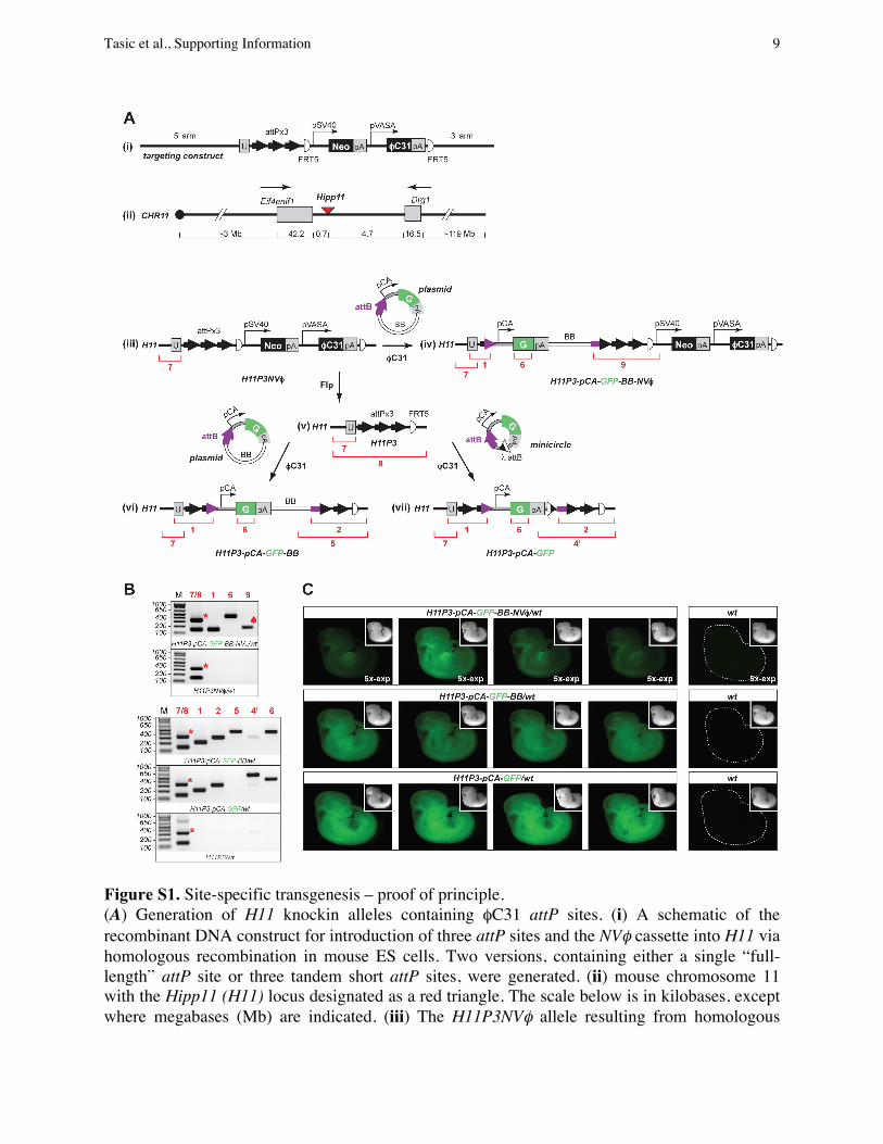
Figure S1. Site-specific transgenesis – proof of principle. (A) Generation of H11 knockin alleles containing !C31 attP sites. (i) A schematic of the recombinant DNA construct for introduction of three attP sites and the NV! cassette into H11 via homologous recombination in mouse ES cells. Two versions, containing either a single “full-length” attP site or three tandem short attP sites, were generated. (ii) mouse chromosome 11 with the Hipp11 (H11) locus designated as a red triangle. The scale below is in kilobases, except where megabases (Mb) are indicated. (iii) The H11P3NV! allele resulting from homologous
Tasic et al., Supporting Information 9

recombination between (i) and (ii). (iv) H11P3-pCA-GFP-BB-NV! allele obtained by !C31-catalyzed site-specific insertion of the pattB-pCA-GFP plasmid (pBT316) into the first attP site. All three attP sites are suitable recipients for the transgene and the site used in any particular case can be determined by PCR. (v) H11P3 locus that was generated from (iii) by Flpo mRNA injection into the cytoplasm of mouse embryos carrying (iii) (see SI Materials and Methods). The H11 locus with a single attP site and the Rosa26 locus with either a single or three attP sites were generated in the same manner. (vi) and (vii), two products obtained by !C31-catalyzed site-specific insertion of the pattB-pCA-GFP plasmid (pBT316, left) or the attB-pCA-GFP minicircle (generated from pBT346, right). The corresponding alleles are: H11P3-pCA-GFP-BB (left) and H11P3-pCA-GFP (right), respectively. (B) PCR results on N1 animals confirming site-specific integrations. The DNA template for each PCR panel was obtained from a mouse of the genotype designated below each gel. The red numbers correspond to the PCR products designated on the schemes by red brackets and numbers in (A). The primer set #9 amplified a band smaller than expected due to the deletion of two attP sites during integration (spade). The wt H11 locus is also amplified by primer set #8 to generate a 321 bp band (asterisk, see schematic (v)). (C) GFP expression in N2 mouse embryos at embryonic day 11. Each row shows representative embryos from a single pregnancy with genotypes designated above. Images were obtained under identical conditions, except that “5x-exp” designates five-fold longer exposure time than for the rest of the images. Insets represent the corresponding bright field images of each embryo. Abbreviations: pSV40, SV40 promoter; pVASA, VASA promoter; U, unique sequence; FRT5, a mutant version of FRT that is compatible with itself but not with wt FRT; pCA, "-actin promoter and CMV enhancer; G, GFP; pA, polyA signal; BB, plasmid bacterial backbone; attB and attP, !C31 attB and attP sites.
Tasic et al., Supporting Information 10

Figure S2. GFP expression in individual adult animals carrying pCA-GFP site-specific transgenes introduced by !C31 integrase-mediated transgenesis. Each column on the graph represents average fluorescence in arbitrary units (AU) in the GFP channel for liver sections from individual animals represented by numbers and genotypes below (“no integration” represents wt, H11P3NV!/wt,
Tasic et al., Supporting Information 11
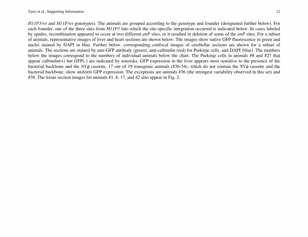
H11P3/wt and H11P/wt genotypes). The animals are grouped according to the genotype and founder (designated further below). For each founder, one of the three sites from H11P3 into which the site-specific integration occurred is indicated below. In cases labeled by spades, recombination appeared to occur at two different attP sites, or it resulted in deletion of some of the attP sites. For a subset of animals, representative images of liver and heart sections are shown below. The images show native GFP fluorescence in green and nuclei stained by DAPI in blue. Further below, corresponding confocal images of cerebellar sections are shown for a subset of animals. The sections are stained by anti-GFP antibody (green), anti-calbindin (red) for Purkinje cells, and DAPI (blue). The numbers below the images correspond to the numbers of individual animals below the chart. The Purkinje cells in animals #8 and #27 that appear calbindin(+) but GFP(-) are indicated by asterisks. GFP expression in the liver appears most sensitive to the presence of the bacterial backbone and the NV! cassette. 17 out of 19 transgenic animals (#36-54), which do not contain the NV! cassette and the bacterial backbone, show uniform GFP expression. The exceptions are animals #36 (the strongest variability observed in this set) and #38. The tissue section images for animals #1, 8, 17, and 42 also appear in Fig. 2.
Tasic et al., Supporting Information 12

Figure S3. Generation of minicircle DNA with ! integrase and excisionase (LR clonase, Invitrogen) in vitro. (A) From left to right: The starting plasmid (pBT346) contains ! attL and attR sites, which recombine in the LR clonase-catalyzed reaction to generate two minicircles: one (MC) contains the "C31 attB site and pCA-GFP, and the other contains the plasmid bacterial backbone (BB). After recombination, the DNA is treated with appropriate restriction endonucleases to selectively digest the BB minicircle and the starting plasmid. (B) The recombined and digested DNA is run on 1% agarose gel. MC DNA (orange arrow) migrates faster than the linear BB or plasmid DNA and is purified from the gel for microinjection.
Tasic et al., Supporting Information 13

Figure S4. GFP expression from a site-specifically integrated H11P3-pCA-GFP-BB transgene increases upon the removal of the bacterial backbone. Top, schematic of the generation of the H11P3-pCA-GFP-BB transgene (from pBT344) and subsequent derivation the H11P3-pCA-GFP transgene from it, by crossing to the GFP-Flpo transgenic mouse to remove the bacterial backbone (SI Materials and Methods). Below, four tail tips for each genotype designated above and a tail tip of a wt littermate were imaged for GFP fluorescence using identical imaging conditions. Bright field (BF) images of the same tails are shown further below. 10x-exp, the same tails above were imaged for GFP fluorescence with 10-times longer exposure. Scale bar, 1 mm.
Tasic et al., Supporting Information 14

Figure S5. Average GFP fluorescence in livers does not differ between mice containing an insertion of the same transgene into one of the three attP sites from H11P3 or insertion into a single attP site from H11P (compare 1st and 3rd columns in both graphs). The GFP fluorescence is also not affected by the genetic background of the injected embryos (compare 1st and 2nd columns in both graphs). Each dataset is represented by mean ± standard deviation. The numbers of individual animals and founders analyzed for each genotype are listed below the genotypes. When samples from multiple founders were combined to obtain an average, each founder was represented by the same number of animals except in the case labeled by a spade. Mouse designations are numbers used to represent each mouse in Fig. S2. Statistical comparisons were performed with one-way ANOVA.
Tasic et al., Supporting Information 15

Row DNAa DNA type
DNA size (kb) Strain
Back-ground
Embryos injected
(n)
Embryos implanted
(n)
Implanted/ Injected
(%) F0 (n)
F0/ Implan-ted (%)
SS (n)
SS% (of injected)
SS% (of implan-
ted)
SS% (of F0)
R (n)h
R% (of
implan-ted)
R% (of F0)
Experi-ments
(n) 1 attB-pCA-GFP minicircle ~3 H11P mix 141 115 82 21 18 1 0.7 0.9 4.8 1 0.9 4.8 4 2 attB-pCA-GFP minicircle ~3 H11P3 mix 168 136 81 39 29 4 2.4 2.9 10.3 1 0.7 2.6 4 3 attB-pCA-GFP minicircle ~3 H11P3 FVB N4 122 115 94 15 13 6 4.9 5.2 40.0 3 2.6 20.0 3 4 attB-pCA-GFP-attB plasmid ~6 H11P3 FVB N4 128 119 93 38c 32 6e 15.8 5.0 15.8 1 0.9 2.6 1 5 attB-pCA-GFP, no RNA plasmid ~6 H11P3NV! mix 160 78 49 32c 41 0 0.0 0.0 0.0 5 6.4 15.6 1 6 attB-pCA-GFP plasmid ~6 H11P3NV! mix 292 223 76 64d 29 10 3.4 4.5 15.6 4 1.8 6.3 3 7 attB-pCA-GFP plasmid ~6 H11PNV! mix 140 89 64 30c 34 2 1.4 2.2 6.7 0 0.0 0.0 2 8 attB-pCA-GFP-FRT5 plasmid ~6 H11P mix 264 232 88 51 22 5f 1.9 2.2 9.8 3g 0.9 5.9 5 9 attB-pCA-GFP-(FRT5)b plasmid ~6 H11P3 mix 142 129 91 61 47 4f 2.8 3.1 6.6 9g 9.9 14.8 4
10 attB-pCA-GFP-FRT5 plasmid ~6 H11P3 FVB N4 50 43 86 8 19 3f 6.0 7.0 37.5 1g 0.0 10.3 1 11 attB-pHB9-GFP-FRT5 plasmid ~14 H11P3 FVB N4 305 267 88 66d 25 2f 0.7 0.7 3.0 2g 0.7 3.0 2 12 attB-pCA-GFP plasmid ~6 R26P3NV! mix 83 63 76 22c 35 2 2.4 3.2 9.1 2 3.2 9.1 1
Abbreviations: F0, embryos or animals obtained from injections; SS, site-specific integration; R, random integration; mix, mixed background of 129, C57BL/6 and DBA2; FVB N4, mice of the mixed background were outcrossed for 4 generations to the FVB strain and then intercrossed. a All DNA was injected at the final concentration of 3 ng/!l. All DNA was coinjected with !C31o mRNA at the final concentration of 48 ng/!l, except for the experiment in row 5. b Both FRT and non-FRT versions of pattB-pCA-GFP (pBT316 or pBT344) were used. c F0s were analyzed only as E10 or E11 embryos. d F0s were analyzed either as E10 or E11 embryos or as live pups. e The 6 founders with cassette exchange listed here contain pCA-GFP without the bacterial backbone; 5 more founders with cassette exchange contained only the bacterial backbone. Therefore the total number of founders with cassette exchange is 11 (29%). f One F0 with site-specific insertion, also contains a random insertion. g One random insertion was found in a founder that also contains a site-specific insertion. h The number of random insertions may be somewhat underreported when F0 embryos were included in the analysis, as random insertions are not detectable by GFP-specific PCR if they occur in embryos that also contain site-specific insertions.
Table S1. Stepwise efficiency of site-specific integration
Tasic et al., Supporting Information 16

Transgene Founder Back-
ground Sex
Insertion into attP 1, 2 or 3
GT of SS
(Y/N)a N1 (n)
N1 SS (n)
N1 SS (%)c
N2/3 (n)
N2/3 SS (n)
N2/3d SS (%)
R (Y)
N1 R (n)
N1 R
(%) A1 mix M 1/3b Y 55 36 65.5 25 15 60.0 0 0 A2 mix M 3 Y 62 12 19.4*** 16 10 62.5 0 0 A3 mix M 1/3b Y 36 10 27.8* 0 0
H11P3-pCA-GFP-BB-NV!
A4 mix M 3 Y 63 29 46.0 14 4 28.6 0 0 B1 mix M 3 Y 143 19 13.3*** 48 20 41.7 Y 11 7.7 B2 mix M 2/3b Y 29 11 37.9 85 36 42.4 0 0 B3 mix Fem 2 Y 44 9 20.5*** 76 34 44.7 0 0 B4 mix Fem 2 Y 20 4 20.0* 0 0 B5 FVB N4 M 2 Y 24 9 37.5 0 0 B6 FVB N4 Fem 2 N
H11P3-pCA-GFP-
BB
B7 FVB N4 Fem 2/3b Y 8 3 37.5 Y 3 37.5 C1 mix Fem n/a Y 15 11 73.3 0 0 C2 mix M n/a Y 44 9 20.5*** 0 0 C3 mix Fem n/a Y 9 4 44.4 Y 2 22.2 C4 mix Fem n/a N 1 0 0 0 0
H11P-pCA-GFP-
BB
C5 mix M n/a Y 67 14 20.9*** 62 29 46.8 0 0 D1 mix Fem 1/2b Y 20 8 40.0 25 15 60.0 0 0 D2 mix M 3 Y 33 10 30.3 12 4 33.3 0 0 D3 mix Fem 3 Y 23 5 21.7* 0 0 D4 mix M 3 N 59 0 0*** 0 0 D5 FVB N4 Fem 3 Y 32 6 18.8** 0 0 D6 FVB N4 Fem 3 Y 41 10 24.4** 0 0 D7 FVB N4 M 3 N D8 FVB N4 Fem 3 Y 14 2 14.3* 0 0 D9 FVB N4 M 1/3b N
H11P3-pCA-GFP
D10 FVB N4 Fem 2 Y 27 8 29.6 20 10 50.0 0 0 H11P-
pCA-GFP E1 mix M n/a Y 39 11 28.2* 0 0 H11P-
pHB9-GFP F1 FVB N4 Fem 2 Y 45 9 20.0*** 0 0 Abbreviations: SS, site-specific integration; R, random integration; GT, germline transmission; mix, mixed background of 129, C57BL/6 and DBA2; FVB N4, the mice of the mixed background were outcrossed for 4 generations to the FVB strain and then intercrossed; N1, progeny from the first generation where a founder was crossed to CD1 wt animal; N2 or N3, progeny from the second or third generation where N1 or N2 animals were crossed to CD1 wt animals, respectively. a The founders that did not transmit were ether sterile (D7 and D9), died upon delivery (C4), or cannibalized the pups (B6). b Based on site-specific PCR, integration appeared to occur at two different attP sites, or it caused the deletion of one or more attP sites. c Frequency of germline transmission for site-specific integrations from some founders was sub-Mendelian, suggesting mosaicism in the founders (Fisher’s exact test, *, p<0.05; **, p<0.01; ***, p<0.001). d Frequency of transmission for site-specific integrations for subsequent generations (N2 and/or N3) is statistically indistinguishable from Mendelian transmission (Fisher’s exact test).
Table S2. Complete list of transgenic founders (n=28) and their germline transmission efficiency.
Tasic et al., Supporting Information 17
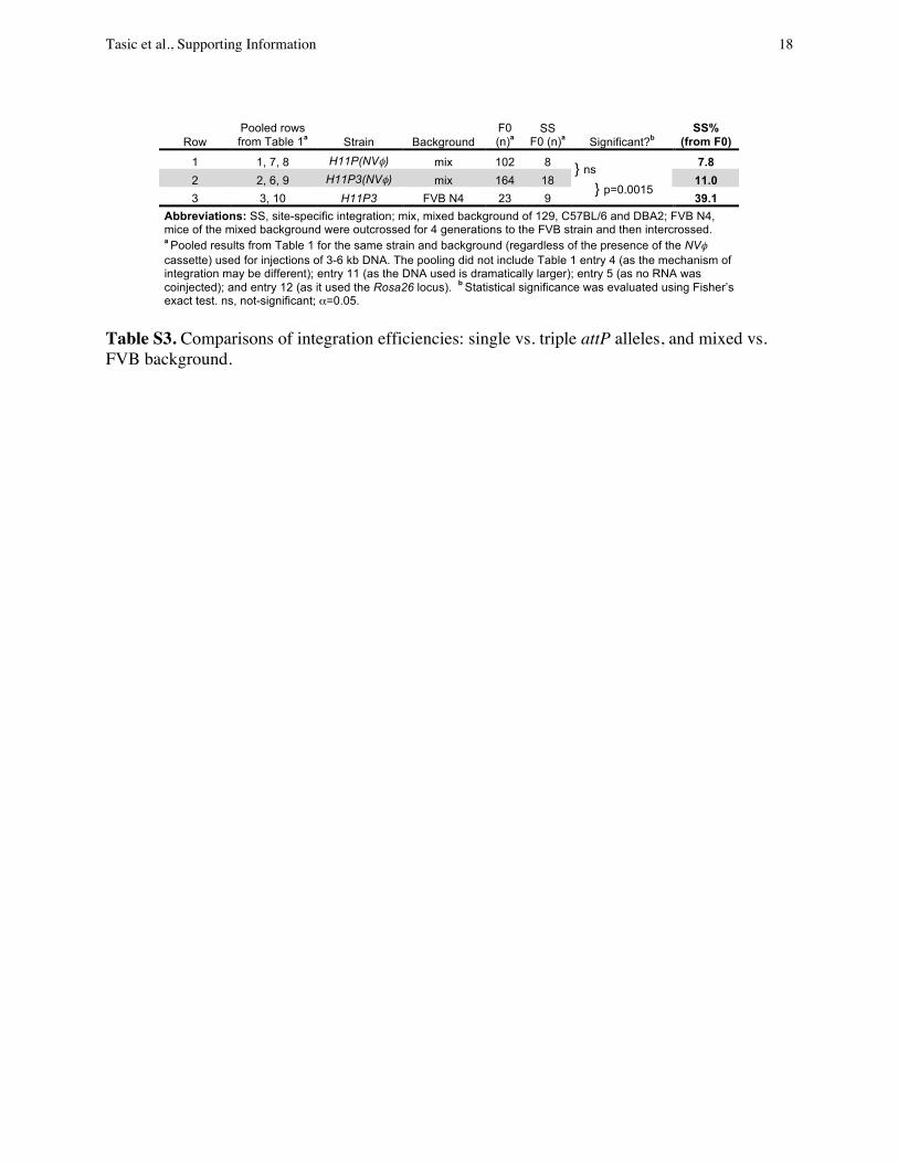
Row Pooled rows from Table 1a Strain Background
F0 (n)a
SS F0 (n)a Significant?b
SS% (from F0)
1 1, 7, 8 H11P(NV!) mix 102 8 7.8
2 2, 6, 9 H11P3(NV!) mix 164 18 11.0 3 3, 10 H11P3 FVB N4 23 9 39.1
Abbreviations: SS, site-specific integration; mix, mixed background of 129, C57BL/6 and DBA2; FVB N4, mice of the mixed background were outcrossed for 4 generations to the FVB strain and then intercrossed.
a Pooled results from Table 1 for the same strain and background (regardless of the presence of the NV! cassette) used for injections of 3-6 kb DNA. The pooling did not include Table 1 entry 4 (as the mechanism of integration may be different); entry 11 (as the DNA used is dramatically larger); entry 5 (as no RNA was coinjected); and entry 12 (as it used the Rosa26 locus). b Statistical significance was evaluated using Fisher’s exact test. ns, not-significant; !=0.05.
Table S3. Comparisons of integration efficiencies: single vs. triple attP alleles, and mixed vs. FVB background.
} ns } p=0.0015
Tasic et al., Supporting Information 18
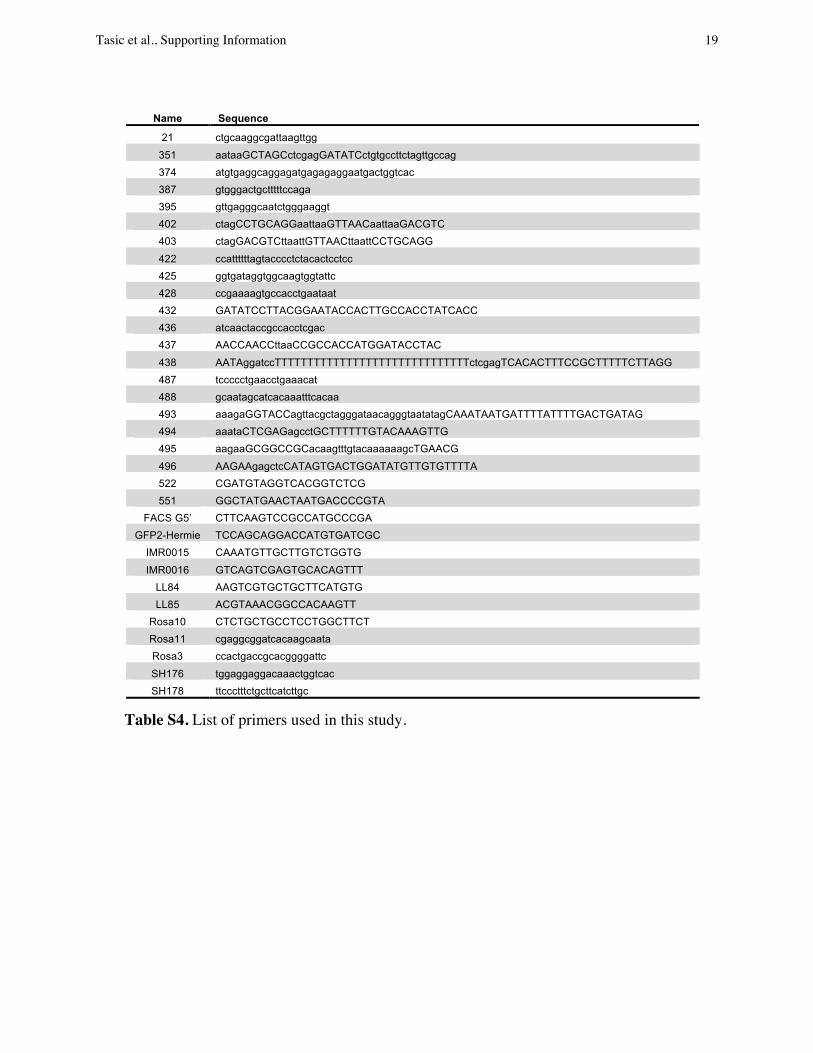
!Name Sequence
21 ctgcaaggcgattaagttgg
351 aataaGCTAGCctcgagGATATCctgtgccttctagttgccag 374 atgtgaggcaggagatgagagaggaatgactggtcac 387 gtgggactgctttttccaga 395 gttgagggcaatctgggaaggt 402 ctagCCTGCAGGaattaaGTTAACaattaaGACGTC 403 ctagGACGTCttaattGTTAACttaattCCTGCAGG
422 ccattttttagtacccctctacactcctcc 425 ggtgataggtggcaagtggtattc 428 ccgaaaagtgccacctgaataat 432 GATATCCTTACGGAATACCACTTGCCACCTATCACC 436 atcaactaccgccacctcgac 437 AACCAACCttaaCCGCCACCATGGATACCTAC
438 AATAggatccTTTTTTTTTTTTTTTTTTTTTTTTTTTTTTctcgagTCACACTTTCCGCTTTTTCTTAGG 487 tccccctgaacctgaaacat 488 gcaatagcatcacaaatttcacaa 493 aaagaGGTACCagttacgctagggataacagggtaatatagCAAATAATGATTTTATTTTGACTGATAG 494 aaataCTCGAGagcctGCTTTTTTGTACAAAGTTG 495 aagaaGCGGCCGCacaagtttgtacaaaaaagcTGAACG
496 AAGAAgagctcCATAGTGACTGGATATGTTGTGTTTTA 522 CGATGTAGGTCACGGTCTCG 551 GGCTATGAACTAATGACCCCGTA
FACS G5’ CTTCAAGTCCGCCATGCCCGA GFP2-Hermie TCCAGCAGGACCATGTGATCGC
IMR0015 CAAATGTTGCTTGTCTGGTG
IMR0016 GTCAGTCGAGTGCACAGTTT LL84 AAGTCGTGCTGCTTCATGTG LL85 ACGTAAACGGCCACAAGTT
Rosa10 CTCTGCTGCCTCCTGGCTTCT Rosa11 cgaggcggatcacaagcaata Rosa3 ccactgaccgcacggggattc
SH176 tggaggaggacaaactggtcac SH178 ttccctttctgcttcatcttgc
Table S4. List of primers used in this study.
Tasic et al., Supporting Information 19