Embed Size (px)
Citation preview

Simulated single molecule microscopy with SMeagol
Martin Linden, Vladimir Curic, Alexis Boucharin, David Fange, & Johan Elf†
Department of Cell and Molecular Biology, Uppsala University, Sweden. †[email protected]
Summary: SMeagol is a software tool to simulate highly realistic microscopy data based on spa-tial systems biology models, in order to facilitate development, validation, and optimization ofadvanced analysis methods for live cell single molecule microscopy data.Availability and Implementation: SMeagol runs on Matlab R2014 and later, and uses compiledbinaries in C for reaction-diffusion simulations. Documentation, source code, and binaries for re-cent versions of Mac OS, Windows, and Ubuntu Linux can be downloaded fromhttp://smeagol.sourceforge.net.Supplementary information: Supplementary data are available at Bioinformatics online.
Recent advances in single particle tracking (SPT) microscopy1 make it possible to obtain tensof thousands macromolecular trajectories from within a living cell in just a few minutes. Sincemolecules typically change their movement properties upon interactions, these trajectories containinformation about both locations and rates of intracellular reactions. This information is unfortu-nately obscured by physical limitations of the optical microscope and noise in detection systems,making statistical methods development for SPT analysis a very active research field. Unbiasedtesting and comparison of such methods are however difficult given the absence of in vivo data ofintracellular dynamics where the true states of interaction are known, a.k.a. the ground truth. Acommon resort is to instead use simulated, synthetic, data. However, tests using such data giveunrealistically optimistic results if the simplifying assumptions underlying the analysis method areexactly satisfied. The need for realistic simulations is long recognized in microscopy and systemsbiology2–12, but systematic combinations of the two are only currently emerging13,14.
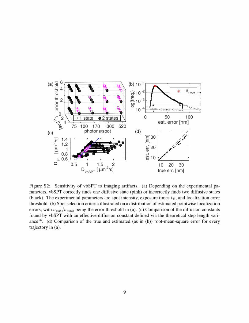
We present the SMeagol package, that has been developed to generate highly realistic singlemolecule microscopy time-lapse image series aimed primarily at single particle tracking applica-tions. The purpose of SMeagol is to enable realistic comparisons between the output of advancedanalysis methods and known ground truth. SMeagol includes an extended MesoRD11 version forsimulation of 3D diffusion in cellular compartments, diffusion limited reaction kinetics, surfaceadsorption, reactions in membranes, and other complex aspects of reaction diffusion kinetics thatdo occur in cells, but are not considered in SPT analysis algorithms. In addition to the molecules’trajectories, SMeagol integrates the 3D point spread function of the microscope, the kinetics ofphoto-activation, blinking and bleaching of the simulated fluorophores, background noise, andcamera specific parameters (Figure 1, movie S1,S2). Great flexibility is allowed by the possibil-ity to supply these characteristic parameters either as tabulated experimental data for a particularoptical setup, or as theoretical models. The combination of using reaction diffusion kinetics incellular geometries and physics-based simulations of the emission and detection processes makesthe images more realistic than the synthetic data used for example by5.
SMeagol can be used to optimize imaging conditions for specific systems in silico and to
1
arX
iv:1
509.
0403
1v2
[ph
ysic
s.bi
o-ph
] 5
May
201
7

benchmark methods for SPT analysis in analogy with the methods that has been developed tobenchmark localization methods for non-moving single particles7. In the supplementary material,we explore the robustness against localization errors and motional blur of the vbSPT software,which extracts multi-state diffusive models from SPT data15, and find that these effects can induceoverfitting under certain conditions. In addition, we provide a number of examples highlightingpossibilities, limitation and computational requirements of the SMeagol simulation engine.
When combined with increasingly refined simulations of intracellular processes, photo-physicsand optics; live-cell microscopy is moving closer to methods in fundamental physics, where com-bined simulation of physical processes and detection systems have guided experimental design anddata analysis for a long time.
time
brig
htn
ess
space-discretized particle trajectory
time
posi
tion
illumination illuminationdiffusive path
(a)
(b)
5 s
2.7 μm
Min oscillations
experimentalbackground
simulatedfluorophores+
single particle tracking4 ms exposure
4 m
s
2.7 μm
time-lapse1 s exposure
5 s
Simulated microscopy
- stochastic reaction/diffusion simulations- diffusion-limited mesoscopic kinetics - arbitrary geometry, multiple compartments
- stochastic fluorophore photophysics- background & camera noise models - point-spread function of the optical system
(c)
Browian bridgeinterpolation
PSF
zr
zr
zr
PSF PSF
Simulated reaction-diffusion kinetics
2.7 μm
illumination
time
posi
tion
Figure 1: Simulated microscopy with SMeagol. (a) Workflow from stochastic reaction-diffusionsimulations to images. (b) The microscopy simulation starts from trajectories generated by stochas-tic reaction-diffusion simulations, fills in stochastic motion and photon emission events betweenthe trajectory points, and finally combines PSF and camera noise models to simulate realistic im-ages. (c) Simulated microscopy of fluorescently labeled MinE proteins in the Min oscillatory sys-tem. Left: Stochastic reaction-diffusion simulation. Mid columns: Simulated SPT microscopy us-ing an actual experimental background noise movie with continuous illumination and 4 ms/frame.Right: A simulation of continuous illumination and 1 s/frame renders a conventional (non-singlemolecule) fluorescence microscopy time-lapse movie. See also Supplementary movies S1,S2, andthe Supplementary material for further details.
2

Acknowledgements
We thank Fredrik Persson and Elias Amselem for helpful discussions about microscopy.
Funding
This work was supported by the European Research Council, Vetenskapsrdet, the Knut and AliceWallenberg Foundation, the Foundation for Strategic Research, and the Swedish strategic researchprogramme eSSENCE.
References
1. Suliana Manley, Jennifer M. Gillette, George H. Patterson, Hari Shroff, Harald F. Hess, Eric Betzig,and Jennifer Lippincott-Schwartz. High-density mapping of single-molecule trajectories with photoac-tivated localization microscopy. Nat. Meth., 5(2):155–157, 2008. doi:10.1038/nmeth.1176.
2. Stephanie Fullerton, Keith Bennett, Eiji Toda, and Teruo Takahashi. Camera simulation engine enablesefficient system optimization for super-resolution imaging. volume 8228 of Proc. SPIE, pages 822811–7, 2012. doi:10.1117/12.906346.
3. Tristan S. Ursell, Eliane H. Trepagnier, Kerwyn Casey Huang, and Julie A. Theriot. Analysis of surfaceprotein expression reveals the growth pattern of the gram-negative outer membrane. PLoS Comput.Biol., 8(9):e1002680, 2012. doi:10.1371/journal.pcbi.1002680.
4. Cox, S. et. al. Bayesian localization microscopy reveals nanoscale podosome dynamics. Nat. Meth., 9(2):195–200, 2012. doi:10.1038/nmeth.1812.
5. Chenouard, N. et. al. Objective comparison of particle tracking methods. Nat. Meth., 11(3):281–289,2014. doi:10.1038/nmeth.2808.
6. Jzsef Sink, Rbert Kkonyi, Eric Rees, Daniel Metcalf, Alex E. Knight, Clemens F. Kaminski, GborSzab, and Mikls Erdlyi. TestSTORM: Simulator for optimizing sample labeling and image acquisi-tion in localization based super-resolution microscopy. Biomed. Opt. Express, 5(3):778–787, 2014.doi:10.1364/BOE.5.000778.
7. Daniel Sage, Hagai Kirshner, Thomas Pengo, Nico Stuurman, Junhong Min, Suliana Manley, andMichael Unser. Quantitative evaluation of software packages for single-molecule localization mi-croscopy. Nat. Meth., 12(8):717–724, 2015. doi:10.1038/nmeth.3442.
8. Boris M. Slepchenko, James C. Schaff, John H. Carson, and Leslie M. Loew. Computational cellbiology: spatiotemporal simulation of cellular events. Annu. Rev. Biophys. Biomol. Struct., 31:423–441, 2002. doi:10.1146/annurev.biophys.31.101101.140930.
9. Rex A. Kerr, Thomas M. Bartol, Boris Kaminsky, Markus Dittrich, Jen-Chien Jack Chang, Scott B.Baden, Terrence J. Sejnowski, and Joel R. Stiles. Fast Monte Carlo simulation methods for biologicalreaction-diffusion systems in solution and on surfaces. SIAM J. Sci. Comput., 30(6):3126–3149, 2008.doi:10.1137/070692017.
3

10. K. Takahashi, S. Tanase-Nicola, and P. R. ten Wolde. Spatio-temporal correlations can drasticallychange the response of a MAPK pathway. Proc. Natl. Acad. Sci. U.S.A., 107(6):2473–2478, 2010.doi:10.1073/pnas.0906885107.
11. D. Fange, A. Mahmutovic, and J. Elf. MesoRD 1.0: Stochastic reaction-diffusion simulations in themicroscopic limit. Bioinformatics, 28(23):3155–3157, 2012. doi:10.1093/bioinformatics/bts584.
12. Steven S. Andrews. Spatial and stochastic cellular modeling with the Smoldyn simulator. In Jacquesvan Helden, Ariane Toussaint, and Denis Thieffry, editors, Bacterial Molecular Networks, volume 804,pages 519–542. Springer, New York, NY, 2012.
13. Juan Angiolini, Nicolas Plachta, Esteban Mocskos, and Valeria Levi. Exploring the dynamics of cellprocesses through simulations of fluorescence microscopy experiments. Biophys. J., 108(11):2613–2618, 2015. doi:10.1016/j.bpj.2015.04.014.
14. Masaki Watabe, Satya N. V. Arjunan, Seiya Fukushima, Kazunari Iwamoto, Jun Kozuka, Satomi Mat-suoka, Yuki Shindo, Masahiro Ueda, and Koichi Takahashi. A computational framework for bioimagingsimulation. PLoS ONE, 10(7):e0130089, 2015. doi:10.1371/journal.pone.0130089.
15. Fredrik Persson, Martin Lindn, Cecilia Unoson, and Johan Elf. Extracting intracellular diffusivestates and transition rates from single-molecule tracking data. Nat. Meth., 10(3):265–269, 2013.doi:10.1038/nmeth.2367. Software: http://sourceforge.net/projects/vbspt/.
16. Winston C. Chow. Brownian bridge. WIREs Comp. Stat., 1(3):325–332, 2009. doi:10.1002/wics.38.
17. Maximilian H Ulbrich and Ehud Y Isacoff. Subunit counting in membrane-bound proteins. Nat. Meth.,4(4):319–321, 2007. doi:10.1038/NMETH1024.
18. A. Finney and M. Hucka. Systems biology markup language: Level 2 and beyond. Biochem. Soc. T.,31(6):1472–1473, 2003. doi:10.1042/bst0311472.
19. S F Gibson and F Lanni. Experimental test of an analytical model of aberration in an oil-immersionobjective lens used in three-dimensional light microscopy. J. Opt. Soc. Am. A, 9(1):154–166, 1992.doi:10.1364/JOSAA.9.000154.
20. H. Kirshner, F. Aguet, D. Sage, and M. Unser. 3-D PSF fitting for fluorescence microscopy: im-plementation and localization application. J. Microsc., 249(1):13–25, 2013. doi:10.1111/j.1365-2818.2012.03675.x.
21. William H. Press, Brian P. Flannery, Saul A. Teukolsky, and William T. Vetterling. Numerical Recipesin C: The Art of Scientific Computing. Cambridge University Press, Cambridge ; New York, 2 edition,1992.
22. K. C. Huang, Y. Meir, and N. S. Wingreen. Dynamic structures in Escherichia coli: Spontaneousformation of MinE rings and MinD polar zones. Proc. Natl. Acad. Sci. U.S.A., 100(22):12724–12728,2003. doi:10.1073/pnas.2135445100.
23. David Fange and Johan Elf. Noise-induced Min phenotypes in E. coli. PLoS Comput. Biol., 2(6):e80,2006. doi:10.1371/journal.pcbi.0020080.
4

24. David Fange, Otto G. Berg, Paul Sjberg, and Johan Elf. Stochastic reaction-diffusion ki-netics in the microscopic limit. Proc. Natl. Acad. Sci. U.S.A., 107(46):19820–19825, 2010.doi:10.1073/pnas.1006565107.
25. Sang-Hyuk Lee, Jae Yen Shin, Antony Lee, and Carlos Bustamante. Counting single photoactivat-able fluorescent molecules by photoactivated localization microscopy (PALM). Proc. Natl. Acad. Sci.U.S.A., 109(43):17436–17441, 2012. doi:10.1073/pnas.1215175109.
26. Andrew J. Berglund. Statistics of camera-based single-particle tracking. Phys. Rev. E, 82(1):011917,2010. doi:10.1103/PhysRevE.82.011917.
27. Hendrik Deschout, Kristiaan Neyts, and Kevin Braeckmans. The influence of movement on the local-ization precision of sub-resolution particles in fluorescence microscopy. J. Biophotonics, 5(1):97–109,2012. doi:10.1002/jbio.201100078.
28. Johan Elf, Gene-Wei Li, and X Sunney Xie. Probing transcription factor dynamics at the single-molecule level in a living cell. Science, 316(5828):1191–1194, 2007. doi:10.1126/science.1141967.
29. Kim I. Mortensen, L. Stirling Churchman, James A. Spudich, and Henrik Flyvbjerg. Optimized lo-calization analysis for single-molecule tracking and super-resolution microscopy. Nat. Meth., 7(5):377–381, 2010. doi:10.1038/nmeth.1447.
30. David MacKay. Information theory, inference, and learning algorithms. Cambridge University Press,2003.
31. Alexandre Lazarescu and Kirone Mallick. An exact formula for the statistics of the current in theTASEP with open boundaries. J. Phys. A: Math. Theor., 44(31):315001, 2011. doi:10.1088/1751-8113/44/31/315001.
5

Supplementary material
S1 Simulated microscopy with SMeagol
SMeagol is a Matlab software suite that simulates microscopy images of randomly moving par-ticles using two main ingredients: diffusive motion and stochastic photon emission events. Inaddition, noise from various sources (camera, background, optics, blinking and photobleaching,etc.) can be included in a modular and flexible way. This makes it possible to evaluate how dif-ferent aspects of the biological-, reporter- and detection- system influence the overall result of theexperiment.
Reactions and random motion in arbitrary geometries The microscopy simulation part ofSMeagol uses input trajectories in the form of a list of times, positions, particle id numbers, anddiffusive states, and can also include the time for creation and destruction of particles. The inputtrajectories are interpolated using Brownian bridges16 to generate individual emission positions ofevery simulated photon. Brownian bridges simulate free diffusion, but the input data need not.Thus, with a fine time step one can use SMeagol to simulate general types of motion. For anexample, see Sec. S5b and Fig. S4.
We have extended the reaction-diffusion simulation software MesoRD11 to keep track ofindividual molecules and write trajectories in the appropriate input format, and incorporated it inSMeagol, but it is also possible to use indata from other sources. SMeagol’s trajectory data formatis described in the software manual.
Tunable photophysics In parallel with the diffusion process, each particle in the simulation goesthrough a simulated stochastic photophysical process which includes activation, Markovian transi-tions between multiple photophysical states with different photon emission intensities, and eventu-ally irreversible photobleaching. Short exposures can be simulated by setting an exposure time tEshorter than the frame time ∆t, and photophysical effects of excitation, by specifying different pho-tophysical transition rates during the illuminated (0 ≤ t < tE) and non-illuminated (tE ≤ t < ∆t)phases. Separating photophysics and molecular diffusion makes it possible to simulate the samereaction-motion trajectory under a wide range of experimental conditions.
EMCCD noise and background The emitted photons are mapped to the camera chip using apoint-spread function (PSF) model, combined with simulated EMCCD17 and background noise.The microscopy image, which is written to tif-stacks for further analysis.
Flexible, modular and user-friendly SMeagol is designed to allow easy incorporation of exper-imental data and theoretical parameters at many levels. Thus, the user can specify arbitrary fluo-rophore activation and photophysical kinetics, and also incorporate custom-written Matlab routinesfor PSF and background models, by extending existing template files. It is also possible to use the
6

independently measured PSF for a specific optical set-up, or background movies from a specificsample. Stochastic reaction-diffusion models are described using the systems biology markup lan-guage (SBML)18 with extensions to spatial models11. The trajectories and the different buildingblocks of the microscopy simulation are then combined and parameterized using either a graphicaluser interface, parameter text files (runinput files), or Matlab structs.
S2 Point-spread function (PSF) model
For all microscopy simulations described here, we used a rotationally symmetric PSF model con-structed from the Gibson-Lanni model19, as implemented in PSFgenerator20.
We simulated the Gibson-Lanni PSF model with high resolution for 584 nm light, NA=1.4,and otherwise default settings PSFgenerator, computed the cumulative radial distribution function(CRDF) for different focal planes and radii up to 5 µm, and constructed a Matlab look-up table forthe inverse CRDF. An individual photon emitted at xem., yem., zem. were then simulated as detectedat position
xdet.=xem. + r cos ν, (S1)ydet.=yem. + r sin ν, (S2)
where the angle ν is uniformly distributed on [0, 2π], and r is sampled using the inverse transformmethod21, i.e.,
r = CRDF−1(u; zem.), (S3)
with u uniformly distributed on (0, 1). The total intensity of the spot did not vary significantly inthe region (|zem. − zfocus| < 400 nm) relevant for our simulations.
In focus, the above PSF model has a standard deviation of about 335 nm. This is largely dueto large shoulders of the PSF, and the width of the central peak is about 95 nm.
S3 Simulated experiments with Min oscillations
To generate Fig. 1c, we simulated a minimal stochastic model of the Min oscillation cycle22,23,implemented using scale-dependent mesoscopic reaction rate constants24.
Reaction-diffusion simulation To match experimental microscopy data, we choose an E. coli-like geometry consisting of a cylinder with spherical end caps, with outer diameter 1 µm and totallength 2.7 µm. The outer 35 nm of the cell was modeled as the membrane region. In this geometry,the oscillations have a period of 25-30 s.
We used a 10 nm spatial discretization, and initial conditions placing 1716 MinD and 483
7

MinE randomly in the membrane region at one half of the cell. After a 300 s simulation to reachsteady state, we ran a tracking production run, collecting snapshots (used for the left column ofFig 1c) every 0.25 s, and tracking positions of all species involving MinE every 5 ms.
MinE SPT simulation (Fig 1c, mid columns, and supplementary movie S1). We simulated a sin-gle particle tracking experiment with 4 ms frame rate and continuous illumination. The coordinatesystem of the input trajectories where rotated and translated to fit a brightfield image of an E colibacterium expressing no fluorophores (Fig. 1c), and a fluorescence time-lapse movie of the samecell was used as a background.
The Gibson-Lanni PSF model was used as described above, and an experimentally param-eterized blink/bleach model for mEos225, with a bright state intensity of 125000 photons/s thatyields on average 500 photons per spot and 4 ms frame if no blinking occurs. Photoactivationevents where simulated every 10 s, with an activation probability of 20% per unconverted molecule.
We simulated an EMCCD gain of 40 (SMeagol inverse gain camera.alpha=1/40), i.e.,every photon generates an exponentially distributed number of image counts with mean value 40.Offset and readout noise are in this case included in the experimental background.
MinE long-exposure simulation (Fig 1c, right column, and supplementary movie S2). For thelong-exposure simulations, we randomly activated 50% of the MinE-containing molecules with aconstant emission intensity of 60 photons/s and no blinking or bleaching. For background, we seta uniform intensity of 1 photon/pixel. We used continuous illumination and a frame rate of 1 Hz,the Gibson-Lanni PSF model described above, an EM gain of 40, offset 100, and readout noisewith std. 4.
The reaction-diffusion model and trajectory output files, and the SMeagol runinput files forthese simulations, are included in Supplementary dataset S1.
S4 Analysis of diffusion with blur and localization errors
To illustrate how SMeagol could be used to evaluate analysis methods for live cell single particletracking experiments, we explore the ability of the vbSPT software15 to correctly identify thenumber of diffusive states in SPT data. We simulated an SPT experiment with normal diffusionat a rate of D = 1 µm2 s−1 sampled every 3 ms in an E. coli geometry with varying fluorophorebrightness and exposure time (see Supplementary movie S3). vbSPT assumes that data comefrom a Markov model with state-dependent diffusion constants, i.e., a model that neglects, e.g.,z-dependent localization errors27, motional blur26,27, and confining effects of the cell boundaries.The simulation thus contains many real features not included in the analysis model, which couldlead vbSPT to overfit the data by incorrectly identifying more than one diffusive state. The question
8
true err. [nm]10 20 30
est.
err.
[nm
]
10
20
30(d)
520300170100754
2
64
2
0tE [m
s]
(a)
(c)
Def
f.[µ
m2 /s
]
DvbSPT [µm 2/s]0.5 1 1.5 2
0.60.8
11.21.4
photons/spot
erro
r thr
esho
ld (b)
1 state 2 states 0 50 100
log(
freq.
)
10 -4
10 -3
10 -2
10 -1
σmode
est. error [nm]
Figure S2: Sensitivity of vbSPT to imaging artifacts. (a) Depending on the experimental pa-rameters, vbSPT correctly finds one diffusive state (pink) or incorrectly finds two diffusive states(black). The experimental parameters are spot intensity, exposure times tE , and localization errorthreshold. (b) Spot selection criteria illustrated on a distribution of estimated pointwise localizationerrors, with σmax/σmode being the error threshold in (a). (c) Comparison of the diffusion constantsfound by vbSPT with an effective diffusion constant defined via the theoretical step length vari-ance26. (d) Comparison of the true and estimated (as in (b)) root-mean-square error for everytrajectory in (a).
9

Table S1: Settings for the microscopy simulations of simple diffusion (Fig. S2).
sample time : 3 ms per framephotophysics : constant emission intensity, no bleachingROI : 80 nm pixels, 55× 20 pixel ROI, focal plane in the mid-
: plane of the bacteriacamera : offset=100, readout noise std.=4,
: EM gain=20 counts/photonbackground : constant, on average 1 photon/pixel per frame
is under which experimental conditions this is likely to happen.
Reaction-diffusion simulation We simulated simple diffusion of a single fluorescent particle inan E coli-like geometry, built as a cylinder with length 3 µm and diameter of 0.8 µm, plus sphericalend caps. We used 10 nm voxels, and wrote particle positions every 7 ms.
SMeagol simulation We generated SPT movies with a frame duration of 3 ms in a range ofimaging conditions from the above diffusive trajectory, by varying the exposure time tE and theaverage number of photons per spot (Nphot. = tE × emission intensity). In particular, we used allcombinations of tE = 0.5 ms, 1 ms, 2 ms, 3 ms, and Nphot. = 75, 100, 300, 520. Other microscopyparameters used in all cases are summarized in table S1.
Estimated number of states Fig. S2a shows number of states learned by vbSPT as a function ofthree tuning parameters: the exposure time tE , the average number of photons per spot, and themaximally allowed pointwise localization error (Fig. S2b). The correct and overfitting conditionsare indicated in purple and black, respectively. In general, all three parameters influence the over-fitting tendency in a non-trivial way. Continuous illumination (exposure time=frame time) leadsto overfitting in almost all conditions, but a modest decrease in exposure time using, e.g., strobo-scopic illumination28, leads to significant improvement due to decreased motional blur. We alsonote that if the number of photons per localized molecule is limited, it is advantageous to includeonly positions with high localization accuracy.
Estimating the diffusion constant As the analysis model of vbSPT neglects both localizationerrors and motional blur, one should not take the numerical estimates of the diffusion constants atface value. However, the estimates can be interpreted using a theory of motional blur for diffusingparticles26. A closer inspection of the analysis algorithm15 shows that vbSPT effectively looks atthe step length variance, which in the absence of localization error and blur is simply⟨
∆x2⟩
= 2DvbSPT∆t. (S4)
10

A more detailed model that includes motional blur and localization errors26 instead predicts⟨∆x2
⟩= 2D∆t(1− 2R) + 2
⟨σ2x
⟩, (S5)
where R = tE3∆t
is the motional blur coefficient, and 〈σ2x〉 is the mean-square localization error.
Eliminating the step length variance from the above equations, we find that
DvbSPT = Deff. ≡ D(1− 2R) +〈σ2
x〉∆t
. (S6)
Fig. S2c plots the effective diffusion constantDeff. vs. the posterior mean ofDvbSPT for the differentdata sets (using our estimated average localization errors), and we see that the prediction of Eq. (S6)is reproduced well when a single diffusive state is correctly identified.
Point localization We localized the spots using a maximum-likelihood fit of a symmetric Gaus-sian plus constant background to a 7-by-7 fit region, using the EMCCD likelihood function of Ref.29, with the offset and gain settings of table S1. Each spot is thus described by 5 fit parameters:background b, spot amplitude N , spot standard deviation s, and spot position µx, µy. We usedMatlab’s built-in function fminunc for numerical optimization of the log likelihood, which wasparameterized to allow only positive values of b, N , and s2, and used the true spot positions, andthe average PSF width and amplitude to construct an initial guess for each fit. To minimize con-finement artifacts from the cell walls, we analyzed motion and uncertainties along the long cellaxis (x coordinate) only.
Estimating point-wise localization uncertainty Due to fluorophore motion during exposure, ran-dom photon emission, z-dependence of the PSF, etc., the quality of the fit varies from spot to spot.We used a Laplace approximation30 (also known as the saddle point approximation in statisticalphysics) of the likelihood function to estimate the localization uncertainty of individual spots, asfollows: Let IM denote the fit region of the image used for localization, and θ = (µx, µy, . . .)the fit parameters. We approximate the likelihood function p(IM |θ) by a Gaussian centered at themaximum likelihood estimate θ∗ using a Taylor expansion in (θ − θ∗),
p(IM |θ) ≈ exp(
ln p(IM |θ∗) +∇θ lnP (IM |θ)∣∣θ∗︸ ︷︷ ︸
=0
(θ − θ∗)
− 1
2(θ − θ∗)TΣ−1(θ − θ∗) + . . .
), (S7)
where the first order term disappears since θ∗ is a local maximum, and the second order term isgiven by the inverse covariance matrix,
Σ−1 = −∂2 ln p(IM |θ)
∂θ2
∣∣∣∣θ∗. (S8)
This can be interpreted as the Bayesian posterior distribution (with a flat prior). The uncertainty ofthe parameters are then characterized by their posterior covariances30. In particular, the posteriorvariance of µx is approximately given by Σµx,µx .
11

As a simple test of this estimator, we compare the true and estimated average root-mean-square (RMS) error for all points in every trajectory (Fig. S2d). We find it to be correct on average,i.e., ⟨
(µ∗x − µx,true)2⟩≈ 〈Σµx,µx〉 , (S9)
for true RMS errors . 20 nm, but biased downwards for larger errors, probably because the Gaus-sian approximation of the posterior density (Eq. (S7)) is inaccurate in those cases.
Selection criteria To build diffusion trajectories for vbSPT analysis, we first discarded spotswhere the numerical optimization failed. We then built a histogram of estimated standard errorsσx =
√Σµx,µx , and identified the most likely estimated error σmode. One such histogram is shown
in Fig. S2b. Finally, we discarded spots that had either σx < σmode/2 as being unrealistically pre-cise, or σx > σmode × (error threshold), using error thresholds in the range 1.3 − 6 as our thirdcontrol variable in Fig. S2a. The highest threshold of 6 included practically all spots.
The fraction of retained spots as well as average trajectory length vary with both simulationparameters and error threshold, but for this experiment we generated enough images to constructdata sets with 80 000 diffusive steps for all conditions.
vbSPT settings For the trajectory analysis, we used vbSPT 1.1.215. For each data set, we ran 20independent runs of the greedy model search algorithm with up to 15 hidden states. We used aninverse gamma prior with mean value 1 µm2 s−1 and strength 5 (std. ≈ 0.6 µm2 s−1) for the diffu-sion constants (with 80 000 diffusive steps in the trajectory, this prior is completely overwhelmedby the data), flat Dirichlet priors for the initial state and state-change probability distributions, anda Beta distribution with mean 0.02 s and std. 2 s for the mean dwell time of the hidden states. (Fordetailed definitions, we refer to the vbSPT manual).
S5 Misc. model examples
Here, we briefly describe some additional model examples, to illustrate the capability and limita-tions of SMeagol in various settings. Source files for these examples are included in Supplementarydata S5.
S5a. Affinity to cell poles To illustrate the geometric modeling capabilities of MesoRD andSMeagol, we construct a simplified model of a diffusing cytoplasmic protein P with specific affinityto binding partners localized at the cell poles.
We use an E coli-like rod shape, where cytoplasm is modeled by a union of a cylinder andtwo spheres with radii 465 nm. In the cytoplasm, the proteins diffuse with diffusion constant2.5 µm2 s−1. The membrane region is modeled as an additional 35 nm outer shell, from whichpolar regions in the form of small spherical caps are created, as shown in Fig. S3a. In these
12

a) b)
Figure S3: Simulating a diffusing protein with polar binding regions. a) Model geometry, with thecytosol (black) inside a thin membrane region (blue) that also contains polar caps (red,magenta)where binding receptors are localized. b) Snapshot from a simulated SPT experiment, with bothpolar bound (red arrows) and freely diffusing molecules visible.
a) b)
-400
-200
0
400
200
z
400
200
y
0
x
-200 15001000-400 500
focus belowfocus midcellfocus above
Figure S4: Simulating non-diffusive transport. (a) Snapshot from TASEP simulation (black, parti-cles) on a helical path (red). (b) Simulated images with focal plane placed near the upper, middle,and lower part of the helical curve emphasize different parts of the path.
caps, we assume a constant concentration [R] of receptors. Then, the binding rate can be writtenr = ka[R][P ], where ka is the association rate constant, and the unbinding rate constant is kd. Wechoose ka[R] = 20 s−1, kd = 0.01 s−1, and diffusion constant 0.01 µm2 s−1 for the bound complex(confined to the membrane caps).
We ran 6 s of stochastic reaction-diffusion simulations starting with 125 P molecules uni-formly distributed outside the polar caps, and after a burn-in of 2 s, wrote positions to file every1 ms. For the microscopy simulation, we randomly activated 5% of the molecules, and used thesame settings as in the simple diffusion experiment above (table S1), except slightly larger re-gion of interest, a fluorophore intensity corresponding to giving on average 270 photons/frame andfluorophore, and photobleaching with a mean lifetime of 1 s. A snapshot is shown in Fig. S3b.
13

a) c) 500 frame averageb) snapshot
15 30 60 120 240∆ x [nm]
10 -1
10 0
10 1
10 2
wal
l tim
e [m
in]
t ∝ ∆ x -2
RD allimage
d) e)
10 2 10 3
N
10 0
10 1
10 2
wal
l tim
e [m
in]
RD allRD noneRD 25image
quadr.linear
Figure S5: Simulating a diffusing transcription factor in a yeast cell. (a) The model geometryincludes a cytoplasm compartment with two buds (red) and a spherical nucleus (blue). Also shownis the bacterial model of Fig. S3 (black). (b,c) A snapshot and 500-frame average, respectively,from a 5 ms/frame SPT simulation. (d) Wall time versus voxel size for a 5 s stochastic reactiondiffusion simulation tracking all proteins (RD all), and microscopy image simulation. The formerclosely follows the expected 1/∆x2 scaling, while the latter is essentially constant. (e) Wall timesfor the ∆x = 30 nm case from (d), but with varying number of involved proteins. “RD none” and“RD 25” refers to stochastic reaction diffusion simulations with tracking deactivated and trackingonly 25 proteins irrespective of total copy number.
S5b. Non-diffusive transport along helical membrane filaments To illustrate the possibility ofsimulating more complex motion than diffusion using SMeagol, we constructed an active transportmodel of particles moving on a helical path (Fig. S4). More specifically, we wrote a Matlab scriptto simulate a totally asymmetric exclusion process (TASEP31) with open boundaries: Particles(black) are created at the left end of the helix in Fig. S4a, walk forwards in 36 nm steps along thehelical path (red) with a rate of 10 s−1 (under a site exclusion constraint), until they fall off at theright end. The insertion rate was 2 s−1, putting the TASEP in the low density phase31. We thenwrote particle coordinates to a trajectory text file at regular intervals (all in the same chemical state),and fall-off events as particle destructions in the reactions text file. For microscopy simulations,we set D = 0, which disables the Brownian bridges and leads to linear interpolation between thetrajectory coordinates. Simulation scripts and runinput files are included in data set S5.
14

S5c. A large cell To illustrate the computational requirements of SMeagol, we use a simplemodel of transcription factor (TF) motion in a budding yeast cell. The model has a cytoplasmiccompartment, made of two spheres joined to an ellipse with a spherical nucleus compartmentinside. As seen in Fig. S5a, it is several times larger than the bacterial-like examples described sofar.
The main computational load for fine-scale MesoRD simulations consists of diffusive motion(total diffusive hopping rate is 6D/∆x2), so we use a simple kinetic model where the TF has a’free’ state that diffuses with diffusion constant 2.5 µm2 s−1 in the entire cell, and can interconvertto a ’bound’ state (0.01 µm2 s−1) inside the nucleus. In addition, we simulate active transport intothe nucleus by setting the diffusion rate from the cytoplasm to the nucleus 20 times larger thanthat in the reverse direction. Protein accumulation in the nucleus is clearly visible in the simulatedimages (Fig. S5b,c).
Fig. S5d shows wall time1 for a 5 s stochastic reaction-diffusion simulation, starting withabout 375 uniformly distributed TFs and various voxel sizes, which fits very well with the expectedinverse quadratic scaling∝ ∆x−2. Second, we generated a 2.5 s simulated microscopy movie with5 ms frame time, 100 80 nm pixels, and 10% of the proteins activated and emitting on average270 photons/frame. This simulation is limited by evaluating Brownian bridges and parsing thetrajectory file, and thus independent of the discretization of the trajectories.
Secondly, we varied the number of proteins, while keeping the discretization constant at∆x = 30 nm. As seen in Fig. S5d, the image simulation part scales linearly as expected, while theRD simulation scales quadratically if all proteins are tracked, but linearly with tracking deactivatedor if tracking only a fixed number of proteins. Thus, we see that while fairly large cells can be sim-ulated, the RD simulations can be computationally demanding especially for models that includelarge cell size, tracking of many molecules, and small details that require fine discretizations.
Finally, we note that the total size in itself does not influence computing time significantly,as long as the number of subvolumes fit in the RAM memory of the computer. As an illustration,we rescaled all length in the ∆x = 30 nm case of Fig. S5d by a factor 0.3 (thus decreasing thevolume by 97%) but kept the discretization and number of molecules constant. However, the RDsimulation of the smaller model only took 25% less time.
S5d. Complex shapes To highlight SMeagol’s abilities to model geometries beyond the tractablepossibilities given by transformation and combinations of simple geometric primitives, we alsomodel particle diffusion in an erythrocyte shaped geometry, shown in Fig. S6a. The starting pointis here a freely available 3D model2, which we imported to and exported from Blender3 to make
1All computing times were measured on a 2.40GHz Intel Xeon CPU with 24 GB RAM.2 http://www.turbosquid.com/FullPreview/Index.cfm/ID/509576, accessed 2016-02-04)3www.blender.org
15

a) b) c)
x
z
y
x
150 x 150 px ROI
Figure S6: Modeling an erythrocyte. a) Rendering of the SBML model with 50 nm subvolumes.b) Alignment of the model (particle positions indicated by black dots) relative to the simulatedregion of interest (ROI) and focal plane of the simulation. c) Snapshot from a simulated moviewith 25 ms exposure time.
it a triangular mesh. The mesh was then converted to SBML compatible format using a customPython script, and incorporated into an SBML model, where about 70 particles diffuse freely (D=2µm2 s−1) within the erythrocyte.
For the microscopy simulation, we rotated the model to align it with the focal plane as shownin Fig. S6b and used similar settings as for the yeast example to produce the snapshot in Fig. S6c.
S6 Misc. supplementary material
• Supplementary movie S1 illustrates the simulated MinE single particle tracking experiment(Fig. 1c, mid columns), showing both true particle positions, the experimental background,and the simulated result in three separate panels.
• Supplementary movie S2 illustrates the simulated MinE fluorescence microscopy time-lapse movie (Fig. 1c, right column), as well as the true particle positions.
• Supplementary movie S3 shows simulated single particle tracking experiments that areanalyzed in Fig. S2.
• Supplementary dataset S4 contains the SBML files, reaction-diffusion trajectory output,and SMeagol runinput files for the simulated Min oscillation experiments (Fig. 1c).
• Supplementary dataset S5 contains model and runinput files, scripts, and source files forthe example models described in Sec. S5.
16