Embed Size (px)
Citation preview

Section 8 – SID 5
Scientific objectives
Objective 1: Delivery of an improved lateral flow device extraction method and Objective 2: In-house test and validation of LFD extraction method
In the first two phases of SE1796, we evaluated protein extraction methods with the aim of increasing the sensitivity of the Neogen lateral flow device (LFD) method. This LFD method uses antibodies raised against thermally-stable bovine Troponin I as a marker for the presence of ruminant processed animal proteins (PAPs). Two different methods of protein extraction were assessed to improve the sensitivity and specificity of detection of ruminant proteins in animal feed: ammonium sulphate salting-out (AAS) of proteins from solution and an acid-EDTA digestion of pre-concentrated bone fragments to release proteins bound to the bone hydroxyl-apatite.
1. Ammonium sulphate precipitation 1.1 Method :
40g 5g of spiked feed sample (0.5% spiked with bovine MBM 133oC) was mixed with 360ml of 0.1M, pH 7.2 phosphate buffer (PBS) and soaked for 15 minutes at room temperature. The sample was heated
at 95C 5C for 15 minute, filtered through 3 layers of muslin and the filtrate was collected. 120ml of filtrate was processed through ammonium sulphate precipitations for selective removal of plant and gelatine proteins (primary precipitation) and then the remaining proteins were concentrated for testing (secondary precipitation). Initially, a 45% saturation of ammonium sulphate was used for the primary precipitation (this has been shown to remove gelatine and a large proportion of the vegetable based proteins). The ammonium sulphate was mixed with the sample and shaken to dissolve. The sample was then incubated overnight
at 31C 5C. The unwanted, precipitated proteins were then centrifuged and discarded. The secondary level of ammonium sulphate was set at 75%. This was mixed with the sample and processed as for the primary phase. The resultant pellet, following centrifugation, was collected in PBS containing 0.5% soya flour and retained for testing. In order to remove residual ammonium sulphate from the pellets, dialysis was used. This was carried out
by taking lengths (6-8cm) of dialysis membrane (Cellu Sep H1, flat width 18mm, wall thickness 25m). The dialysis membrane was washed using deionised water and tied at one end. The ammonium sulphate extracted proteins were pipetted into the dialysis membrane, and the other end was securely tied. The samples were then suspended in a large beaker of de-ionized water. The samples were left dialysing for 4-6 hours, the water was changed 3 times and dialysed further for 1 hour. The samples were removed from the dialysis bag and the dialysed solutions were placed into sample tubes and tested according to
the manufacturers instructions. Dialysed extracts were tested at room temperature, 70C and 90C using Neogen LFDs; The following interpretation criteria were applied for reading the samples: if a line formed in the test zone and another line formed in the control zone within 10 minutes (2 lines in total), the sample was deemed positive. If there is no line in the control zone, the test was invalid and the sample was retested with another strip. If after the full 10 minutes there was no visible line in the test zone but a visible line in the control zone, the sample is negative.
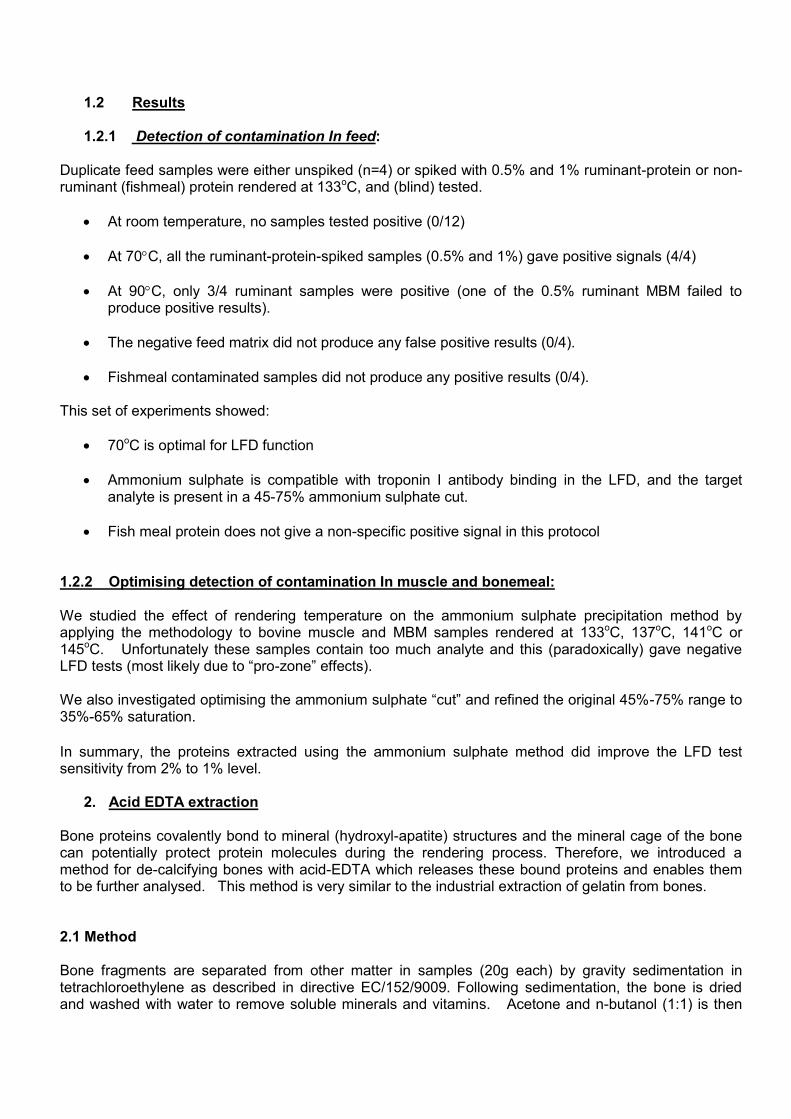
1.2 Results 1.2.1 Detection of contamination In feed:
Duplicate feed samples were either unspiked (n=4) or spiked with 0.5% and 1% ruminant-protein or non-ruminant (fishmeal) protein rendered at 133oC, and (blind) tested.
At room temperature, no samples tested positive (0/12)
At 70C, all the ruminant-protein-spiked samples (0.5% and 1%) gave positive signals (4/4)
At 90C, only 3/4 ruminant samples were positive (one of the 0.5% ruminant MBM failed to produce positive results).
The negative feed matrix did not produce any false positive results (0/4).
Fishmeal contaminated samples did not produce any positive results (0/4). This set of experiments showed:
70oC is optimal for LFD function
Ammonium sulphate is compatible with troponin I antibody binding in the LFD, and the target analyte is present in a 45-75% ammonium sulphate cut.
Fish meal protein does not give a non-specific positive signal in this protocol
1.2.2 Optimising detection of contamination In muscle and bonemeal:
We studied the effect of rendering temperature on the ammonium sulphate precipitation method by applying the methodology to bovine muscle and MBM samples rendered at 133oC, 137oC, 141oC or 145oC. Unfortunately these samples contain too much analyte and this (paradoxically) gave negative LFD tests (most likely due to “pro-zone” effects). We also investigated optimising the ammonium sulphate “cut” and refined the original 45%-75% range to 35%-65% saturation.
In summary, the proteins extracted using the ammonium sulphate method did improve the LFD test sensitivity from 2% to 1% level.
2. Acid EDTA extraction Bone proteins covalently bond to mineral (hydroxyl-apatite) structures and the mineral cage of the bone can potentially protect protein molecules during the rendering process. Therefore, we introduced a method for de-calcifying bones with acid-EDTA which releases these bound proteins and enables them to be further analysed. This method is very similar to the industrial extraction of gelatin from bones. 2.1 Method Bone fragments are separated from other matter in samples (20g each) by gravity sedimentation in tetrachloroethylene as described in directive EC/152/9009. Following sedimentation, the bone is dried and washed with water to remove soluble minerals and vitamins. Acetone and n-butanol (1:1) is then

added to the sediment and stirred for 1 minute and left for 5 minutes. The organic layer is discarded and the sediment is retained in a sedimentation glass. The sediment is then dried in a hot air oven at 70oC. Three times the weight of the sediment is added in EDTA free acid (to each 300 mg of sediment, add 900mg of EDTA free acid) along with 2ml of deionised water to every 100 mg of sediment. This is shaken for at least 16-18 hours on a shaker or rotor at 160rpm.
Phosphate-buffered saline (pH 7, 4 mL) is added to the bone sediment and mixed gently. The pH is adjusted to between 7.5 -8.0 using 20 N NaOH. The mixture is allowed to settle for 15 minutes and the supernatant is collected. The supernatants are diluted 1:4, 1:6, 1:8 with distilled water and incubated at 50oC for 10 minutes. The warm extracts are tested with RevealTM for ruminant protein in feed LFD dipsticks for 10 minutes. While testing, the sample is kept at 50°C.
2.2 Results 2.2.1 Evaluation of method: This extraction method was assessed in-house using a range of meat and bonemeal samples Table 1: Summary of results of MBM extraction using EDTA free acid
Sample extracts Bovine Ovine Porcine Avian Fish
133C + + - - -
137C + + - - -
141C + + - - -
145C + + - - -
145C+ minerals + + - - -
145C+ Vitamins + + - - -
The results presented above show that all the samples rendered at different temperatures were detected
correctly. While proteins extracted from feed samples rendered at 145C using ammonium sulphate
precipitation were not detected (data not shown), protein extracted from bone sediments of 145C rendered sample using the EDTA method were correctly identified, as were all the ruminant samples rendered at all temperatures. This shows that the proteins covalently bound to bone mineral structures appear to be stabilised from denaturation or fragmentation. These results were presented in a poster session of TSE Joint Funders meeting at the University of Warwick in September 2008.
8 feed samples spiked with 0.1% to 0.5% bovine MBM along with 1% porcine, 1% avian and 1% fish meal spiked samples were tested using this method. All of the ruminant spiked samples were detected positive and all the non-ruminant showed negative results. The thickness of band is gradually increased according to the level of contamination showing the sensitivity. The Neogen kit’s limit of detection is at the 1-2% level where as our improved method is able to detect at the 0.1% level. A further 18 negative feed matrix samples spiked with 0.1, 0.2, 0.3, 0.4, 0.5, 0.75 and 1% of bovine and ovine samples rendered at 133ºC and bovine sample rendered at 144ºC were tested along with 0.5% porcine, avian and fish meal samples. Again, all of the ruminant samples spiked above 0.2% levels were correctly identified. To test the ability of this method to identify blind samples, 21 blind samples spiked with various concentrations of ruminant MBM were tested using the EDTA extraction method and all the samples were identified correctly. Further, to test the ability of this method for detecting the presence of ruminant material in other pure processed animal protein (PAP) (i.e PAP in PAP eg. bovine MBM in pure porcine/avian/fish meal), 64 samples were tested. This method did not produce accurate results on samples containing PAP in PAP.

This could be due to steric hindrance (crowding of specific and nonspecific target protein) on proximity and orientation of antigen and antibody complex formation.
Table 2: In-house assessment of EDTA free acid extraction method
Sample number Composition Expected result Actual results
1 1% porcine - -
2 Negative - -
3 Negative - -
4 Negative - -
5 1% bovine in feed +5% porcine + +
6 8% bovine + +
7 5% porcine - -
8 1% bovine in feed +5% porcine + +
10 5% porcine - -
14 0.2% bovine + +
15 8% porcine - -
16 Negative - -
17 5% porcine - -
18 Negative - -
19 0.5% bovine + +
20 Negative - -
21 Negative - -
22 0.5% bovine + +
24 1% bovine + +
27 1% bovine in feed +5% porcine + +
29 1% bovine + +
Table 2 shows that all the blind samples were correctly identified. The negative feed matrix and non-ruminant samples gave negative results without producing any false positive results. All the ruminant samples were correctly identified in spite of non-ruminant sample co-existing in some of the feed samples. 2.2.2 Small inter-laboratory study: The bone extraction method with EDTA produced the desired results and increased the sensitivity of the dipstick testing from 2% to 0.2%. This study included VLA, CCL (Netherlands) and CRA-W (Belgium). Therefore, a small inter-laboratory study was carried out on 5 samples. Table 3 shows that 2 labs have identified all the positive and Negative samples correctly and one lab failed to detect a positive sample. Table 3: Small inter-laboratory study focussing on the transferability of the EDTA extraction method between laboratories
Sample number Composition
Netherland results
Belgium results
VLA results
1 0,5% bovine in NFM - + +
2 0,5% ovine in NFM + + +
3 0,5% porcine in NFM - - -
4 0,5% avian in NFM - - -
5 Negative Feed Matrix - - -
Following this study, the method has been passed to the CRL for animal protein detection for consideration for a larger scale study in 2010. 2.2.3 EDTA extraction application for other detection tests:

EDTA extracts were also tested for presence of proteins using an ELISA method. The commercially available ELISA kit produced by TaKaRa® detects carboxylated (Gla-OC) and uncarboxylated (Glu-OC) osteocalcin present in bone material. A panel of 69 bone extracts were tested on the kits and the data was scrutinised for any species related pattern. Using the osteocalcin data we were able to identify the ruminant samples from non ruminant samples based on the protein levels of carboxylated and uncarboxylated osteocalcin. Gla-OC levels and Glu-OC levels are high (>10ng/ml and >1.0ng/ml respectively) in both bovine and ovine bone extracts. Low levels of Gla-OC levels and Glu-OC are seen in avian MBM and fishmeal (<10ng/ml and <1.0ng/ml respectively). The porcine samples have shown isolated increased levels of Glu-OC (>10ng/ml) with low levels of Gla-OC (<1.0ng/ml). These analyses will help in identifying ruminant material in feed as well as presence of ruminant PAP in non-ruminant PAP. Table 4: Assessment of the EDTA extraction method using the TaKaRa Osteocalcin detection kit plus interpretation of results based on levels of carboxylated and uncarboxylated osteocalcin.
Sample type Gla-OC Glu-OC Interpretation
Bovine MBM 133oC 20.47 10.23 Ruminant
0.2% Bovine 133oC in Feed 15.17 2.49 Ruminant
Bovine MBM 141oC 20.47 6.61 Ruminant
0.1% Bovine141oC in Feed 12.21 1 Ruminant
0.5%Bovine141oC with mineral 18.05 4.14 Ruminant
Bovine MBM 144oC 16.19 1.3 Ruminant
0.5%Bovine133oC with 0.1%mineral 20.47 3.92 Ruminant
0.5%Bovine133oC in feed 19.08 3.32 Ruminant
0.5%Bovine133oC with 1%mineral 20.47 3.44 Ruminant
0.5%Bovine133oC with 0.5%mineral 16.69 3.09 Ruminant
0.5%Bovine133oC with 0.2%mineral 20.47 3.77 Ruminant
Bovine MBM137oC 20.47 7.76 Ruminant
Ovine MBM133oC 20.47 4.19 Ruminant
Avian MBM 133oC 6.33 0.21
Fishmeal 4.2 0.15
Porcine MBM 133oC 0.61 4.91
Coco Beans 0.255 0.18
10%Milk powder in confect. 0.38 0.18
1% vitamins in NFM 0.57 0.33
Cal. Phos 0.21 0.13
Bovine MBM 133oC 22.472 18.697 Ruminant
Bovine MBM 141oC 22.472 14.374 Ruminant
Bovine MBM 137oC 22.472 20.388 Ruminant
Bovine MBM 145oC 22.472 3.994 Ruminant
0.5% Bovine133oC MBM in feed 22.472 4.481 Ruminant
Ovine MBM 133oC 22.472 22.493 Ruminant
Ovine MBM 141oC 22.472 18.359 Ruminant
Ovine MBM 137oC 22.472 26.432 Ruminant
Ovine MBM 145oC 22.472 10.855 Ruminant
Avian MBM 133oC 1.546 0.44
Porcine MBM 133oC 0.375 26.432
Fishmeal 0.422 0.575
5% Vitamins in Negative feed 0.284 1.168
Negative feed 0.165 0.328
Sugar beet 0.169 0.331
1% Bovine MBM 133oC 30.893 14.674 Ruminant
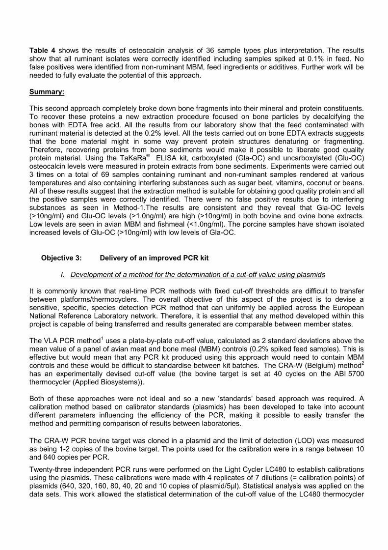
Table 4 shows the results of osteocalcin analysis of 36 sample types plus interpretation. The results show that all ruminant isolates were correctly identified including samples spiked at 0.1% in feed. No false positives were identified from non-ruminant MBM, feed ingredients or additives. Further work will be needed to fully evaluate the potential of this approach. Summary: This second approach completely broke down bone fragments into their mineral and protein constituents. To recover these proteins a new extraction procedure focused on bone particles by decalcifying the bones with EDTA free acid. All the results from our laboratory show that the feed contaminated with ruminant material is detected at the 0.2% level. All the tests carried out on bone EDTA extracts suggests that the bone material might in some way prevent protein structures denaturing or fragmenting. Therefore, recovering proteins from bone sediments would make it possible to liberate good quality protein material. Using the TaKaRa® ELISA kit, carboxylated (Gla-OC) and uncarboxylated (Glu-OC) osteocalcin levels were measured in protein extracts from bone sediments. Experiments were carried out 3 times on a total of 69 samples containing ruminant and non-ruminant samples rendered at various temperatures and also containing interfering substances such as sugar beet, vitamins, coconut or beans. All of these results suggest that the extraction method is suitable for obtaining good quality protein and all the positive samples were correctly identified. There were no false positive results due to interfering substances as seen in Method-1.The results are consistent and they reveal that Gla-OC levels (>10ng/ml) and Glu-OC levels (>1.0ng/ml) are high (>10ng/ml) in both bovine and ovine bone extracts. Low levels are seen in avian MBM and fishmeal (<1.0ng/ml). The porcine samples have shown isolated increased levels of Glu-OC (>10ng/ml) with low levels of Gla-OC.
Objective 3: Delivery of an improved PCR kit
I. Development of a method for the determination of a cut-off value using plasmids
It is commonly known that real-time PCR methods with fixed cut-off thresholds are difficult to transfer between platforms/thermocyclers. The overall objective of this aspect of the project is to devise a sensitive, specific, species detection PCR method that can uniformly be applied across the European National Reference Laboratory network. Therefore, it is essential that any method developed within this project is capable of being transferred and results generated are comparable between member states. The VLA PCR method1 uses a plate-by-plate cut-off value, calculated as 2 standard deviations above the mean value of a panel of avian meat and bone meal (MBM) controls (0.2% spiked feed samples). This is effective but would mean that any PCR kit produced using this approach would need to contain MBM controls and these would be difficult to standardise between kit batches. The CRA-W (Belgium) method2 has an experimentally devised cut-off value (the bovine target is set at 40 cycles on the ABI 5700 thermocycler (Applied Biosystems)). Both of these approaches were not ideal and so a new ‘standards’ based approach was required. A calibration method based on calibrator standards (plasmids) has been developed to take into account different parameters influencing the efficiency of the PCR, making it possible to easily transfer the method and permitting comparison of results between laboratories.
The CRA-W PCR bovine target was cloned in a plasmid and the limit of detection (LOD) was measured as being 1-2 copies of the bovine target. The points used for the calibration were in a range between 10 and 640 copies per PCR.
Twenty-three independent PCR runs were performed on the Light Cycler LC480 to establish calibrations using the plasmids. These calibrations were made with 4 replicates of 7 dilutions (= calibration points) of plasmids (640, 320, 160, 80, 40, 20 and 10 copies of plasmid/5µl). Statistical analysis was applied on the data sets. This work allowed the statistical determination of the cut-off value of the LC480 thermocycler

and the design of a protocol for use with a wider range of test platforms. The cut-off value is determined in such a way that there is no more than 5 % of chance that later signals are due to the presence of the target. Later signals are considered as insignificant but there is still up to 5% chance that a late amplification signal is a result of the DNA fragment that was targeted. Running such an extensive set of plasmids in order to establish cut-off values was impractical. It was found that acceptable results could be achieved with a limited number of calibrations performed using 3 to 5 independent runs and this was enough to fix the cut-off value. A protocol based on three sets of calibrators made up of 640 target copies per 5ul, 160 target copies per 5ul and 40 target copies per 5ul was developed and validated through inter-laboratory study (see objective 5).
II. Delivery of a SAFEED PCR test kit
The Safeed PCR kit was developed using the complementary aspects of both the VLA and CRA-W methods combined into an improved PCR kit. These included a sample extraction control and a multiplex inhibition control.
Extraction control: this control consists of a complete animal DNA virus detected with a specific PCR primers/probe set. This control is used to control DNA extraction and PCR procedures.
Inhibition control: the aim of this control is to avoid false negative PCR results due to an extraction procedure mistake and/or the presence of inhibitor factors in the PCR reaction. This control consists in a plasmid detected with a specific PCR primers/probe set.
A private molecular biology company (Diagenode) were charged with delivering the Safeed PCR method in kit form. The final kit contains :
Three calibrators used for standardizing cut-offs between platforms (640 target copies per 5ul, 160 target copies per 5ul and 40 target copies per 5ul).
Specific primers and probes for species detection and internal control (inhibition detection). Test controls (positive, negative and inhibition). Mastermix. Instruction covering suggested extraction approach and PCR method. Note: these will be finalized
based on inter-laboratory study performance.
Objective 4: Provision of a protocol for DNA extraction from large sample size A fundamental requirement of any feed screening method is to start with a large initial sample size. Official feed sampling methodologies set out in European regulation requires that 500g of feed sample is delivered to the laboratory for testing. This official sample is a composite of statistically sampled aliquots taken from throughout a feed consignment. Many PCR methods have been developed based on small sample portions (10-100mg). This significantly reduces the reproducibility of the assay due to the contaminant not being included in this small test portion. In this study, two methods, developed at VLA1 and CRA-W2 (Belgium), were assessed in a parallel study in order to determine the advantages and disadvantages of each method. The VLA method uses a 40g initial sample size from which DNA is extracted using a combination of a phosphate buffer, heating step and a Chelex resin sample clean up. With this method, the test portion is soaked in a phosphate buffer to release material from the sampled pellets. The soaked sample is pre-processed (heated at 95oC for 15 minutes) in order to release the DNA in to the buffer. A sub-aliquot of the buffer is treated with Chelex, vortexed for 20 seconds and then centrifuged for 10 minutes. The liquid is then removed and is ready for testing.

The CRA-W method involves grinding 100g of raw sample in a ZM200 mill (Retsch GmbH & Co., Haan, Germany) to obtain a powder of particles with a diameter < 500µm. This is then randomly sub-sampled to give two aliquots of 100mg which are used as test samples. Extraction and purification is achieved using with the commercial kit “Wizard® Magnetic DNA Purification System for Food” (Promega Corporation, Madison, WI, USA) according to supplier’s instructions and using the King Fisher Magnetic Particle Processor (Thermo Labsystems, Helsinki, Finland) as a semi-automatic device for performing these extractions. Final DNA extract was recovered in 300 µl (undiluted extract). DNA extraction was assessed using the VLA and CRA-W PCR method. The characteristics of both tests are given in the table 5.
Table 5 : Summary of the protocols used by CRA-W and VLA.
Step CRA-W VLA
DNA Extraction/purification protocol
Test portion size 100mg x 2 taken from 100g of ground feed (<1mm)
40g of un-ground feed
Method used Magnetic beads Heat treatment and Chelex resin
Amplification protocol
DNA target Mitochondrial DNA Mitochondrial DNA
Species detected/amplicon size
Bovine/68 bp Bovine/108 bp
Cut-off limit a
40 cycles About 35 cycles (calculated on a plate by plate basis)
a Limit to distinguish between positive and negative samples.
Samples were distributed between the CRA-W and the VLA laboratories and tested using both systems. The DNA obtained from the extractions was also exchanged between the two laboratories for analysis using each other’s testing platform. Table 6 presents the results obtained by the CRA-W with the sample set produced by the VLA. The two samples containing bovine material were detected whatever the extraction method. No false positive results, nor false negative ones were observed. The simultaneous presence of another MBM (a porcine MBM) does not seem to affect the detection of the bovine material (samples 2 and 7). The Ct values of the DNA extracted by the VLA method were very similar whereas the CRA-W extracts showed a gap of 2 cycles between the signals produced with only bovine MBM and that produced with a combination of bovine and porcine MBM. From the results on this samples set, it seems that the two methods of extraction produce DNA extracts free of PCR inhibitors. The Ct values obtained with the extraction method of the VLA are slightly earlier than the ones with the CRA-W extraction method but the differences observed are probably due to different amounts of DNA present in the reaction. Table 6: PCR results obtained with the CRA-W PCR target on DNAs produced with the VLA and the CRA-W extraction methods from the VLA samples set.
Sample Animal protein content Conclusion Conclusion
Undiluted 10 fold diluted
Replicates Mean Replicates Mean
1 Negative feed matrix 50.00 50.00 Negative 50.00 50.00 Negative
50.00 50.00 50.00 50.00
2 0.5 % bovine MBM 24.17 27.79 Positive 26.16 32.36 Positive
25.34 25.75 31.19 31.78
3 0.5 % ovine MBM 40.20 45.96 Negative 50.00 50.00 Negative
42.35 46.18 50.00 50.00
4 0.5 % porcine MBM 49.32 50.00 Negative 46.33 50.00 Negative
46.16 46.25 50.00 50.00
5 0.5 % avian MBM 50.00 50.00 Negative 50.00 50.00 Negative
50.00 50.00 50.00 50.00
6 0.5 % fishmeal 50.00 50.00 Negative 50.00 50.00 Negative
50.00 50.00 50.00 50.00
7 0.5 % bovine MBM + 0.5 % porcine MBM 24.08 27.08 Positive 26.63 32.14 Positive
27.68 27.16 34.17 33.16
8 0.5 % ovine MBM + 0.5 % avian MBM 50.00 44.65 Negative 38.85 50.00 Negative
45.02 41.94 50.00 50.00
9 0.5 % avian MBM + 0.5 % fishmeal 50.00 50.00 Negative 50.00 50.00 Negative
50.00 50.00 50.00 50.00
10 Negative feed matrix 50.00 50.00 Negative 50.00 50.00 Negative
50.00 50.00 50.00 50.00
* Ct values < 40 cycles are considered as significant
Ct values obtained with VLA extracts* Ct values obtained with CRA-W extracts*
Undiluted 10 fold diluted

A series of 500g samples were produced containing decreasing concentrations of bovine meat & bone meal. These samples were extracted using both the VLA and CRA-W extraction technique and were tested on the CRA-W platform (in order to standardise the PCR assay). Table 7 presents the results obtained with the DNAs extracted from this samples set. On this set of samples, DNA targets were detected at levels of bovine MBM contaminations lower than 0.1 % whatever the DNA extraction method. A level of 0.025 % is detected with the undiluted extract of both extraction methods. A level of 0.0125 % of bovine MBM is even detected with the DNA extracted according the CRA-W extraction protocol. This result must be considered cautiously as we are probably very close to the limit of detection, even if the two independent test portions were detected as positive.
Table 7: PCR results obtained with the CRA-W PCR target on DNAs produced with the VLA and the CRA-W extraction methods from the CRA-W samples set.
Sample Animal protein content Conclusion Conclusion
Undiluted 10 fold diluted
Replicates Mean Replicates Mean
1 Negative feed matrix 50.00 50.00 Negative 50.00 50.00 Negative
39.62 44.81 50.00 50.00
2 0.003125 % bovine MBM 42.26 50.00 Negative 49.57 50.00 Negative
50.00 49.79 50.00 50.00
3 0.0075 % bovine MBM 48.75 50.00 Negative 40.92 50.00 Negative
50.00 45.46 50.00 50.00
4 0.0125 % bovine MBM 50.00 40.56 Negative 39.33 50.00 Positive
38.56 38.95 50.00 50.00
5 0.025 % bovine MBM 38.61 50.00 Positive 38.16 50.00 Positive
36.70 37.43 45.63 47.82
6 0.050 % bovine MBM 40.28 50.00 Negative 36.62 41.84 Positive
36.37 36.50 50.00 45.92
7 0.075 % bovine MBM 37.47 39.55 Positive 36.57 40.74 Positive
36.49 36.53 40.91 40.83
8 0.100 % bovine MBM 38.43 50.00 Positive 34.97 39.76 Positive
35.76 35.37 41.58 40.67
9 0.100 % bovine MBM 39.00 42.21 Positive 37.84 44.46 Positive
36.37 37.11 38.91 41.69
10 0.200 % bovine MBM 37.87 50.00 Positive 35.16 38.73 Positive
35.62 35.39 38.89 38.81
* Ct values < 40 cycles are considered as significant
Ct values obtained with VLA extracts* Ct values obtained with CRA-W extracts*
Undiluted 10 fold diluted

Conclusion:
Based on the results obtained with these two sets of samples, the two extraction methods seem to have comparable efficiency. Both extraction approaches allow detection with the CRA-W PCR method at a level of contamination around 0.1 % or below. The grinding with the ZM200 mill (Retsch GmbH & Co., Haan, Germany) gives equally good homogenisation of the samples as the large sample VLA method. With such a treatment, duplicated limited test portions as 100 mg seem large enough to analyse the samples with a high sensitivity and a good repeatability. As both approaches were able to meet the required level of detection (<0.1% MBM contamination within a feedstuff) then it was decided that both approaches would be presented as acceptable and which one was used would depend on the situation at the testing laboratory.
Objective 5: In-house test and validation of improved PCR kit 1. Validation of the protocol for the determination of a cut-off value on PCR platforms: This was set up as an inter-laboratory study with the aim of validating the protocol developed within the project for the transfer of the PCR kit method to any platform (thermocycler, reagents and test environment), using a cut-off value derived using a plasmid. The cut-off value for a given platform is defined as the Ct value above which a signal has a high probability (95%) to be due to an aspecific amplification. This cut off is the value delimiting positive results (Ct <cut-off) from negative results (Ct >cut-off). This means that setting an accurate cut-off value is essential to have a low rate of false results. The Safeed PCR kit was used in this study but the approach would be equally valid for any PCR method. The PCR was run over 50 cycles and involved participants testing 3 sets of calibrators made up of 640 target copies per 5ul, 160 target copies per 5ul and 40 target copies per 5ul. In routine use it is anticipated that each calibrator will be used in triplicate on each test plate (9 wells in total). However, for the purpose of this study participants were asked to perform 6 sets of 3 for each of the calibrators (18 wells per calibrator). In addition, positive and negative test controls were also included. Twelve blind DNA extracts were also analysed in triplicate. The study took place between April and July 2009. Nineteen institutes (17 from the EU, 1 from Japan and 1 from Australia) took part in the study with a total of 23 thermocyclers from 4 major companies (Applied Biosystems, Bio-Rad, Roche Diagnostics and Stratagene). Based on the overall conclusions of the study, the protocol of transfer was considered fit for purpose. The cut-off values of the 23 platforms tested showed huge differences between raw data and justify the necessity of the approach to determine specifically the cut-off value of each platform. However, when using the respective cut-off values of each platform on the blind samples included in the trial, the 95% level of correct assignments corresponded to samples containing between 1 and 2 copies of targets which proves without any doubt that the transfer protocol is working correctly even though the obtained cut-off values are widely different. Conclusion of study: • The cut-off values of the 23 platforms determined using the proposed transfer protocol showed significant differences from one platform to another one (37.699 < Cut-off value< 43.636) and justify the usefulness of determining specifically the cut-off value of each platform. • The percentages of blocks detected as positive showed the accuracy of the calculated cut-off values and allowed discrimination of true positive results from unspecific amplifications. • Despite slight weaknesses of some labs based on different objective criteria (number of outliers, repeatability, reproducibility, z-score), the transfer protocol can be considered as robust as no laboratory was considered to be underperforming.

• The robustness of the protocol was successfully tested with thermocyclers from 4 companies. Nevertheless, the results obtained with Stratagene thermocyclers were more variable and can explain some weaker performances. • Based on these conclusions, the study can be considered as successful and the protocol of transfer as fit for purpose. The finalised inter-laboratory study report will be available early in 2010 and a lecture specially dedicated to the results of the inter-laboratory study will be presented to the NRL network in March 2010. 2. Protocol for the validation of a real time PCR method for the detection of cattle DNA in feed: The PCR kit protocol was evaluated through inter-laboratory study which aimed to validate the protocol for the detection of cattle DNA in feed. It focused on the following objectives: 1. Taking forward the transferability of the qualitative PCR method on any platform (thermocycler, master mix and test environment) through the determination of a cut-off value using plasmids as calibrators. 2. Based on the results provided by the participants, a limit of detection in copy number of the target will be determined. 3. The capacity of the method to detect the presence of 0.1 % (w/w) of cattle MBM in a feedingstuff will be also evaluated. Calibrators, samples, primers, probes and mastermix were supplied to each participant. Detailed instructions on handling test materials were also provided. As in the previous study (looking at the transferability of PCR methods) three plasmids were used to establish standard cut-offs and to mitigate problems between platforms. Three calibrants were supplied made up of 640 target copies per 5ul, 160 target copies per 5ul and 40 target copies per 5ul. In addition, two PCR test controls were provided with the kit and analysed four times on each test plate. In this particular study, 70 samples were analysed consisting of DNA extracted from feedingstuffs spiked (or not) with bovine MBM and containing low copy numbers of the target. Each sample was analysed in duplicate over 6 test plates (plate layouts supplied to participants along with the kits). The deadline for returning results of this study is January 2010 and the results will be officially reported early in 2010 by the Community Reference Laboratory for feedingstuffs (CRL_AP).
Objective 6: Selection of protein markers by bioinformatics/literature review/sample interrogation
A combination of approaches including database searches, literature review and actual test data were combined in order to evaluate possible protein targets. Note: a pure database driven approach did not generate data robust enough to formulate a target list due to the limited available information relating to processed protein structures. It is documented that Troponin I (TnI) is thermally stable at rendering temperatures 3&4 and it is used as a target in commercial test kits for meat and bone meal detection eg the Neogen LFD kit. For this reason, it made very good sense to include TnI as a target. The use of collagen as a protein marker in the archaeological field for speciating bone material5 also made this an attractive target for investigation.

Through the outputs of the bioinformatics and interrogation of databases (Swiss Prot and NCBI) the most abundant muscle proteins were checked for species-specific sequences. The most promising protein markers identified were Troponin, tropomyosin and myosin. BLAST searching of the theoretical peptide sequences showed that the initial single theoretical peptide for Osteocalcin, believed to be specific for the chicken protein and which was identified in three of the four samples of chicken MBM also matched the sequence for the Xenopus tropicalis osteocalcin. Osteopontin was also added to the list of potential target proteins recoverable from bone fragments. Even though osteopontin also occurs in milk products, its large degree of species variation (bovine and ovine sequences are almost identical but there is a lot of variation between ruminant, pig, chicken, mouse, rabbit) would merit further investigation and consideration as a target. However, no data was available as to the thermal stability of osteopontin and so it was necessary to established this prior to investing in its potential as a marker. The amino acid sequences of actin were found to be poor in terms of species specific character. This protein was therefore excluded from the target list. The product of these searches was combined with actual MS data using meat meals (MM) and meat and bone meals (MBM) in order to formulate the target list. The presence of species-specific haemoglobin sequences on MS analysis has lead to this protein being added to the list, although this was not progressed as a primary target. Table 8: Target protein list
Soft tissue Bone Blood
Myosin Collagen (Haemoglobin)
Troponin Osteocalcin
Tropomyosin Osteopontin
() will not be progressed directly within WP3
Objective 7: Protocol for extraction of species specific proteins
Based on the potential target protein list, formulated as a result of objective 6, methods have been developed to concentrate and extract targets. These extraction techniques fall into two categories: 1. extraction of targets from bone; and 2. extraction of soluble proteins from meat and bone meal. The former has significant advantages in that bone material can be easily sedimented from within a feed matrix using tetrachloroethylene (TCE) sedimentation6. This has the effect of physically concentrating the target material and reduces interfering effects of vegetable-based proteins. However, the current feed bans are not restricted to the detection of bone material and so it was appropriate to also consider soluble (non-bone derived) proteins. Extraction of bone proteins: The EU directive EC152/2009 (Microscopic analysis of feedstuffs) sets out a method for sedimenting bone fragments from within a feed matrix using TCE. The recovered bone fragments can then be digested using EDTA (see objective 1); heat plus ammonium bicarbonate (see section 9 &10); or by digesting with 0.6N Hydrochloric acid at 4oC overnight. Osteocalcin / Osteopontin: EDTA free-acid extraction were used for extraction of Osteocalcin and Osteopontin from sedimented bone fragments. Following digestion, targets were captured using Osteocalcin (3G8E1F11) - mouse monoclonal antibody raised against native osteocalcin of cow origin

and Osteopontin (FL-314) - rabbit polyclonal antibody raised against amino acids 1-314 representing full length Osteopontin. Both affinity capture methods were checked using protein standards and both were found to be effective in recovering targets. When applied to rendered bone material, proteins were recovered but it was not possible to detect species specific Osteocalcin or Osteopontin and it was concluded that both of these targets were not thermally stable enough to present species specific character post rendering. Both of these targets were subsequently deleted from the target list. Collagen: Both the heat plus ammonium bicarbonate method and the digestion with 0.6N hydrochloric acid at 4oC overnight approach were applied to the recovery of collagen targets from bone material. Both of these approaches proved successful in recovering collagen from bone material. Further information relating to this approach is given under objective 9 and 10. Extraction of soluble (non-bone) proteins: Troponin: Protein extraction methods for recovery of Troponin I were developed as a priority for the project, mainly driven by the knowledge that thermally stable Troponin I proteins were present in meat and bonemeal samples 3&4 and also by the availability of commercial immunoassay kits for detecting this target.
The following extractions were applied to raw, cooked (125oC) and rendered products: a) 50mM KCl then LiCl pH 4.5 extraction, further protein isolation by precipitation by raising, and later lowering, the pH prior to TCA precipitation b) NaCl extraction, TCA, de-fat with acetone, diethyl ether, butanol, followed by clean up by molecular weight cut off filtration c) NaCl extraction, followed by clean up by molecular weight cut off filtration d) KCl extraction followed by ammonium sulphate precipitation e) KCl extraction, EDTA dialysis, followed by ammonium sulphate precipitation f) KCl extraction, EDTA dialysis, sulpholane precipitation
Extracts were separated by SDS PAGE in duplicate. One batch of each sample was stained with coomassie blue and the duplicate batch was transferred to membrane and a Western blot was carried out using the antibody used in the production of the lateral flow device produced by Neogen corp.
The bands on a PAGE gel which corresponded to the bands detected by the antibody at the correct mass for troponin I were excised, digested with trypsin and analysed by MALDI-MS/MS for the bovine-specific sequence of the troponin I protein.
The following table summarises the outcome of this exercise:
Sample type Extraction methods generating Troponin stained bands
Raw beef KCl, EDTA dialysis, ammonium sulphate precipitation
Cooked muscle (125oC) KCl extraction followed by ammonium sulphate precipitation
Rendered Material (133oC) KCl extraction followed by ammonium sulphate precipitation
Through the use of this extraction technique, it has been possible to extract and identify bovine specific troponin. This extraction technique was taken forwards to the affinity purification phase of the project (see objective 8).
Myosin: Work carried out on recovery of Myosin targets was largely carried out at FERA and will be reported in full under a separate SID5 report. The following is a brief overview of what was achieved in this work.

Myosin is routinely extracted from fresh muscle tissue as the actomyosin complex in high ionic strength buffers. At the start of the project it was established that myosin or myosin fragments could be extracted, as determined by tryptic digestion followed by MS/MS, using buffers containing NaCl or the denaturant guanidine hydrochloride. Based on this, samples of MBM were extracted in four buffers with increasing chaotropic strength and the extracts immunoblotted with antimyosin antibody in order to determine the nature and size of myosin fragments in each of the extracts. The results showed that the least denaturing conditions, produced the clearest immunoblot banding for myosin, although these bands were very faint. The banding also appeared around 30kDa and between 65 and 90kDa, significantly lower than native myosin mass (220kDa) indicating that breakdown of the myosin had occurred. However the results did indicate that these myosin fragments behaved in a similar manner to native myosin in that they could be extracted in high ionic strength buffer from MBM.
The high ionic strength buffer (0.25M KH2PO4 0.05M Na pyrophosphate, 1.1M NaCl pH 7.5 with solid NaOH, containing 0.01% sodium azide) was then chosen to extract 1.5g of the four species of MBM. The results showed that myosin fragments were present between 95 and 90kDa in the extracts of all four species of MBM. In addition, higher mass fragments and possibly intact myosin were present in the ovine and avian sample extracts. In order to determine whether these fragments contained any species-specific sequence, the bands were cut from the gel and MALDI-TOF TOF analysis carried out. However, no myosin related peptides were detected in any of the samples. This suggests that although immunodetection methods could identify a number of myosin fragments in the MBM extracts there was generally insufficient protein in any one band detected on a gel to permit any identification of the myosin by MS. Affinity capture is needed to increase concentration of the myosin target (see objective 8).
Objective 8: Protocol for affinity purification of heat stable markers from animal feed
Troponin: Efforts to develop affinity purification of Troponin I targets were seriously hampered during the early phase of this project due to the limited availability of good quality antibodies to Troponin I. All efforts to develop a method based on commercial antibodies proved unsuccessful. Faced with this, two alternative routes were followed. Firstly, tests were carried out based on antibodies produced within the project (RIKILT, The Netherlands and CAHEC, China), and secondly by acquiring samples of antibodies used in the commercial Neogen kit. Antibodies raised within the project: Four monoclonal antibodies developed by the CAHEC laboratory in China and eight monoclonal antibodies raised at the RIKILT laboratory were assessed for their species specificity to troponin I. ELISA plates were prepared and antibodies differentiated into general capture and specific species capture. Two batches of general Troponin I capture antibodies (86-3A1 & 86-4H11) were coupled to CNBr activated Sepharose 4B overnight at 4oC and used for affinity capture from 1ml protein extracts (see objective 7). This approach generated the following protein concentrations: bovine 14.2mg/ml; ovine 10.3mg/ml; porcine 11.8mg/ml and avian 13.2mg/ml. Tryptic digestion of the bovine product followed by MALDI-MS/MS revealed bovine specific sequence of the troponin I protein. Antibodies obtained from Neogen corp.: following protracted discussions, VLA were able to obtain, under material transfer agreement, a small quantity of antibodies used in the production of Neogen’s lateral flow device for detection of Troponin I within feed. The release of these antibodies was for sole use within this project and for the characterisation of the LFD capture product. The release of this antibody was free of charge and thanks go to Frank Klien of Neogen corp. for his help and support in this work.
Using the Neogen antibodies, it has been possible to recover species specific troponin I from raw, low temperature and higher temperature bovine material. This was achieved using samples extracted by KCl, EDTA dialysis and ammonium sulphate precipitation followed by western blot with the Neogen

antibody to identify the location of target bands. Two troponin I (fast skeletal) bands were detected and the extracted proteins were digested with trypsin and analysed by MALDI-MS/MS. The two bands were identified as svmlqiaatelekeegr which is specific to bovine, rabbit and armadillo and idaaeeekydmeir which is bovine specific. This confirms that the Neogen antibody is, indeed detecting Troponin I (some questions were raised about whether the Neogen assay was in fact detecting Troponin I or some breakdown product of a larger protein) and that it is possible to identify the ruminant specific sequence using MALDI-MS/MS. With a supply of these antibodies it would be possible to use this approach to confirm the presence of species-specific troponin I in a test sample providing direct confirmation of results obtained the Neogen product. Myosin: As described in objective 7, it was necessary to affinity capture myosin from the protein extracts of meat and bone meal in order to recover MS detectable target.
An immunoaffinity matrix was prepared using CNBr activated Sepharose and the Sigma myosin polyclonal antibody. Four buffers were assessed based on their ability to retain myosin in solution while possibly being suitable for elution of myosin from the affinity matrix. The following buffer was found to be appropriate: 200mM phosphate buffer pH 6.8 containing 100mM sodium pyrophosphate, 1.5 M sodium chloride, 10% TMS and 15% glycerol. However, when this was applied to MBM extracts followed by SDS-PAGE of the eluted proteins and MALDI –TOF TOF analysis of the tryptic peptides the results showed that the product was heavily contaminated with rabbit serum albumin. These were derived from the polyclonal antibody used in the capture and bound sufficiently weakly that the elution buffer was able to wash this protein off the column. Methods of removing these proteins were attempted: progressive washing of the column to remove the unwanted protein; affinity purification of polyclonal antibody before immobilisation; chromatographic separation of serum protein from myosin fragments after enrichment; use of an immobilised monoclonal anti-myosin antibody. Of these approaches, purification of polyclonal antibody before immobilisation produced successful results. 150 µg of the myosin antibody was affinity purified using immobilised turkey myosin. 90 µg of affinity purified antibody protein was recovered and bound to 0.3g CNBr Sepharose. This was used to affinity purify myosin extracted from ovine MBM. The results showed that affinity purification of the polyclonal antibody significantly removed the albumin protein from the eluate and revealed a trace of a 30kDa protein, (a fragment of affinity purified ovine myosin, similar in mass to the fragments seen in the original western blots).
Affinity purification of the polyclonal antibody is a feasible approach. However, the affinity column prepared with the commercial antibody appeared to have a relatively short life, with only five elution cycles reducing the binding capacity of the column by 90%. This short lifespan of the columns would significantly limit the financial viability of the approach but for a confirmatory method it would be a suitable approach.
Objective 9: Protocol for suitable protein extraction from within an animal feed and Objective 10: Final procedure for the identification of species specific protein in animal feed
Introduction Prior to embarking on the Safeed project it was known that mass spectrometry approaches were being developed within the field of archaeology to speciate protein samples recovered from bone material5. Intact or partially degraded collagen molecules were being used as a target for this work. Collagen proteins are fibrous in nature and found throughout the body especially skin, bone, tendons, muscle and cartilage. Collagen is one of the most abundant proteins present in the body of mammals and makes up

about 25% of the total amount of protein in the body. The collagen molecule is approximately 300nm long and 1.5nm in diameter and is made up of three polypeptide strands each having a left handed helix confirmation. These three strands are twisted together forming a right handed triple helix stabilised by hydrogen bonds. This structure gives the molecule significant strength. The shear abundance of collagen within animal material, coupled with its species specific character and the ability to derive useful data from samples dating back many thousands of years made this target very attractive within the field of archaeology. The preservation of these molecules is thought to be largely down to the shielding nature of the bone material from within which these molecules are derived. The mineral cage surrounding the collagen helix, within a typical bone fragment, appears to physically confine the molecule preventing it from unwinding and subsequent denaturation. Within this project it has been possible to draw links between archaeologically recovered bone fragments and bone material processed through EU rendering plants. The degree of racemization of proteins within a rendered bone fragment is equivalent to archaeological samples dating back thousands of years. The similarity between archaeologically recovered collagen samples and those recovered post rendering suggested that the methodology used for speciating archaeological bone material could also be applied within the field of processed animal protein species identification. Rendering processes expose animal material to temperatures greater than 133oC at 3 bar pressure for a minimum of 20 minutes. These conditions are set as a minimum rendering standard across Europe and typical meat and bone meal samples are exposed to temperatures in excess of this (up to 145oC under temperature and pressure). Even under these extreme conditions, bone fragments remain intact and form between 25 and 40% of the typical meat and bone meal composition. Commonly used methods for recovering bone fragments from within meat and bone meal (and feed stuffs) exist within EU directive EC152/2009 (Microscopic analysis of feedstuffs)6. The ability to recover bone fragments across a wide range of temperatures, coupled with the shear abundance of collagen molecules within these samples it was hypothesised that a collagen detection approach should be able to offer a species identification of meat and bone meals heated to high temperatures under pressure. Basic Approach Initial work focused on taking the existing method developed by Dr Matthew Collins, BioArch, York University and applying it to recovered bone fragments. This initial method involved taking meat and bone meal samples, sedimenting with tetrachloroethylene to extract bone material from within the sample, drying and then using a cold acid digestion method to release the collagen from within the mineral matrix. The recovered collagen molecules were then digested with trypsin and identified using MALDI-MS in reflection mode. The direct application of this method to meat and bone meal samples was assessed in year one of this project using single species samples of bovine, ovine, porcine and avian meat and bone meal, followed by a series of mixed species samples. The method proved highly successful in identifying the origin of the single species samples confirming collagen as a good target which persists well relative to other proteins and maintains species character sufficient to discriminate bovine, ovine, porcine and avian species. However, this assessment did reveal that the method did not detect mixed species contamination and in all cases it was not possible to detect the lower concentration contaminant even when present at substantial levels (10%). This aspect of the work was particularly concerning as it would be possible to mask the presence of significant levels of risk material by incorporating as a mixed product. Based on this findings, efforts were focused on trying to refine the original method so that it could be applied to individual bone fragments (>250um). This approach is based on the premise that single bone fragments would only liberate collagen molecules of a single species and as such would not be affected by the issues identified above. Multiple fragments would need to be analysed in isolation to give a

statistical based assessment of the sample but this would still be appropriate for a confirmatory method and highly valuable when very few bone fragments were detected in a field sample. A method has been developed within this project to allow the recovery and characterisation, to a species level, of individual bone fragments recovered from within complex feed matrices. This method has been evaluated within the project and the results are presented below. Evaluation of MALDI MS method for detection and species level identification of meat and bone meal within feed matrices. Materials and Methods Samples: Animal feed matrices were formulated from vegetable material normally found in typically UK animal feeds. These included wheat, soya, rape, maize and sunflower. All individual feed ingredients were assessed to confirm the absence of any animal material using both microscopic analysis and PCR techniques. Samples of shell (oyster) and mineral (rock derived dicalcium phosphate) were also assessed to confirm the absence of animal material using microscopic analysis and PCR. Meat and bone meal samples rendered at 133oC and 141oC for 20 minutes at 3bar pressure were obtained from Prosper de Mulder (Doncaster, UK), a commercial rendering plant. As a result of the rendering process the product was crushed, ground and dried to a friable meat and bone meal powder. Meat and bone meal spiked samples were prepared under laboratory conditions by accurate weighing of meat and bone meal samples into pre-weighed negative feed matrices. This process took place on a sample by sample basis to ensure accuracy. Fish meal samples were purchased from United Fish Industries. Aquatic mammal samples (sea lion and dolphin) were supplied by VLA Truro, collected under veterinary supervision, and heated to 121oC under pressure before being crushed and ground. A panel of six field derived samples were also included in the study. Four of these were commercially available pet food samples containing animal materials, and two were historical positive samples collected from GB national feed audit surveillance. Bone Fragment Extraction: 50g of each sample was weighed into sterile sedimenting glasses. 75ml of tetrachloroethylene was carefully added to each sedimentation glass. The sample was stirred for 15 seconds and then left to stand for 15 minutes following which is was stirred again and left to stand for a further 15 minutes. During this time the dense bone fragments settle to the base of the glass while the plant component floated on the surface of the solvent. The liquid and plant components were then carefully decanted and discarded leaving only bone fragments at the base of the sedimenting glass. These bone fragments were left to dry. The dry bone fragments were sieved through a 250um sieve and the large fraction was transferred to a watch glass. The larger bone fragments were examined under a stereo microscope to confirm bone-like character and these were then collected in micro-titre tubes, coded and submitted for testing. Note: Samples of mineral and shell were included in the blinded samples even though it did not possess bone typical character when examined under stereo microscopy. In routine testing it is envisaged that these samples would be sifted out prior to the MS analysis phase and these are only included for robustness assessment. Collagen Recovery: De-mineralisation to break down the mineral cage was carried out on individual bone chips was achieved using 500ul 0.6M HCl for 4 hours. Following centrifugation and removal of the supernatant, the residue is gelatinised by incubation for four hours at 65oC in 50mM ammonium bicarbonate pH 8.0. The product was trypsinated with 1mg of trypsin overnight at 37oC. Following digestion, the samples were re-constituted in 0.1% trifluoroacetic acid and concentrated using equilabrated C18 zip-tips pipette tips (Millipore). Absorption and dilutions carried out according to
manufacturers instructions.

Each sample was spotted onto an Applied Bio Systems target template and cocrystalised with a cyano-hydroxy cinnamic acid matrix. Each spot was analysed by MALDI MS in reflection mode with m/z range 800-4000 using a calibrated Applied Bio Systems 4700 proteomic analyser. Note: as the majority of the extraction steps are carried out in a single tube the risk of protein loss or cross-contamination is limited. Analysis of Mass Spectromic Data: The resulting spectra were analysed using the BioArch database which hold collagen sequencing information collected from many species. This database has been made available to project SE1797 for analysis of collagen based data. Clearly visible peptide fragments have been identified capable of distinguishing between ruminant and non-ruminant gelatine and repeatedly observed in replicate analysis. The presence or absence of these peptides was used to differentiate between the target species. All interpretation was carried out by eye. Results Data was analysed by scoring the presence or absence of target peptides on the mass spectrometry data. No defined criteria were set prior to analysis as to when a peptide peak should be scored as being present. Initially the operator was asked to categorise the samples into bovine, ovine, porcine or avian target species based on the data present. Following this they were then asked to identify samples which generated poor quality spectra or spectra that were not suitable for analysis. The results of this analysis are presented in table 9.
Table 9: Species Identification of Blind Samples Following Sedimentation, De-mineralization, Tryptic Digestion and MS Analysis.
Sample Code
Nature of contaminant (in feed matrix) Reported results
Type % Temperature Species ID Comment
1 Shell 1% Poor spectra
2 Ovine 0.5% 133oC Ruminant
3 Bovine 0.5% 133oC Bovine
4 Avian 0.5% 141oC Avian
5 Common dolphin 1% 121oC Poor spectra
6 Ice fish 0.5% N/A Avian
7 Porcine 0.5% 133oC Porcine
8 Porcine 0.5% 141oC Porcine
9 Porcine 0.1% 137oC Porcine
10 Bovine 0.5% 141oC Bovine
11 Ovine 0.5% 133oC Ruminant
12 Ovine 0.5% 133oC Ovine
13 Ovine 0.5% 133oC Porcine
14 Ovine 0.1% 141oC Ovine
15 Bovine 0.1% 133oC Bovine
17 Ice fish 0.5% N/A Poor spectra
20 Ice fish 0.5% N/A Avian
25 Mineral 1% N/A Bovine
27 Bovine 0.1% 133oC Bovine
28 Bovine 0.1% 133oC Bovine
29 Sea lion 0.5% 121oC Poor spectra
32 Porcine 0.1% 141oC Porcine
34 Bovine 0.1% 133oC Bovine
36 Avian 0.1% 141oC Avian
39 Porcine 0.5% 133oC Porcine
40 Ovine 0.5% 133oC Porcine
44 Bovine 0.1% 141oC Bovine
47 Bovine 0.1% 133oC Bovine
48 Shell 1% N/A Poor spectra
49 Ice fish 0.5% N/A Poor spectra
50 Bovine 0.5% 133oC Bovine
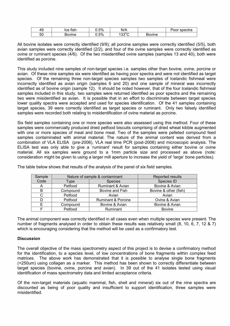
All bovine isolates were correctly identified (9/9); all porcine samples were correctly identified (5/5), both avian samples were correctly identified (2/2), and four of the ovine samples were correctly identified as ovine or ruminant species (4/6). Of the two misidentified ovine samples (samples 13 and 40), both were identified as porcine. This study included nine samples of non-target species i.e. samples other than bovine, ovine, porcine or avian. Of these nine samples six were identified as having poor spectra and were not identified as target species. Of the remaining three non-target species samples two samples of Icelandic fishmeal were incorrectly identified as avian origin (samples 6 and 20) and one sample of mineral was incorrectly identified as of bovine origin (sample 12). It should be noted however, that of the four Icelandic fishmeal samples included in this study, two samples were returned identified as poor spectra and the remaining two were misidentified as avian. It is possible that in an effort to discriminate between target species lower quality spectra were accepted and used for species identification. Of the 41 samples containing target species, 39 were correctly identified as target species or ruminant. Only two falsely identified samples were recorded both relating to misidentification of ovine material as porcine. Six field samples containing one or more species were also assessed using this method. Four of these samples were commercially produced dried petfood biscuits comprising of dried wheat kibble augmented with one or more species of meat and bone meal. Two of the samples were pelleted compound feed samples contaminated with animal material. The nature of the animal content was derived from a combination of VLA ELISA (pre-2006), VLA real time PCR (post-2006) and microscopic analysis. The ELISA test was only able to give a ‘ruminant’ result for samples containing either bovine or ovine material. All six samples were ground to a 1mm particle size and processed as above (note: consideration might be given to using a larger mill aperture to increase the yield of ‘large’ bone particles). The table below shows that results of the analysis of the panel of six field samples.
Sample Code
Nature of sample & contaminant Reported results
Type Species Species ID
A Petfood Ruminant & Avian Bovine & Avian
B Compound Bovine and Fish Bovine & other (fish)
C Petfood Avian Avian
D Petfood Ruminant & Porcine Ovine & Avian
E Compound Bovine & Avian Bovine & Avian
F Petfood Ruminant Bovine
The animal component was correctly identified in all cases even when multiple species were present. The number of fragments analysed in order to obtain these results was relatively small (8, 10, 6, 7, 12 & 7) which is encouraging considering that the method will be used as a confirmatory test. Discussion The overall objective of the mass spectrometry aspect of this project is to devise a confirmatory method for the identification, to a species level, of low concentrations of bone fragments within complex feed matrices. The above work has demonstrated that it is possible to analyse single bone fragments (>250um) using collagen as a marker. This method has been shown to correctly differentiate between target species (bovine, ovine, porcine and avian). In 39 out of the 41 isolates tested using visual identification of mass spectrometry data and limited acceptance criteria. Of the non-target materials (aquatic mammal, fish, shell and mineral) six out of the nine spectra are discounted as being of poor quality and insufficient to support identification, three samples were misidentified.

Based on this assessment it appears that the method has the potential to be used as a practical tool for supporting confirmation of contamination within complex feed matrices. It is however, evident that further work is required on data analysis and defining rigid criteria for accepting and rejecting spectra, on a statistical basis prior to this method being suitable for routine use. Analysis of the field samples is particularly encouraging and shows the correct species can be obtained even when faced with a small number of bone fragments. These samples were representative of typical feedstuffs in that they were processed, pellets/ biscuits and were available commercially within the UK. Analysis of single bone fragments ensures that the data relates to a single species, however, the downside to this approach is that many fragment may need to be identified, each in isolation, in order to get a full picture of the contaminating species within a sample. It should be remembered that this is intended as a confirmatory method rather than a routine screening tool and it would highly valuable in supporting microscopic analysis testing where single bone fragments can be visualised under the microscope but are commonly not present in high enough concentrations to liberate species level detection using PCR approaches. Based on this study and the detection of collagen molecules from within individual bone fragments would be suitable for confirmation of positive from routine screening tests applied for the detection of processed animal proteins in feeding stuffs. All work has been carried out on the private collagen database maintained by BioArch, York University. The routine application of this method would depend on access to the BioArch database or formulation of a similar (although potentially simpler) database for supporting interpretation of data.