Embed Size (px)
Citation preview
Multimedia Health Education
Disclaimer
Dr. Prem LoboG.P.O Box No. 635Sydney NSW-2001 Australia
Phone:+61-2-82057549Fax:+61-2-9475 1036 Email:[email protected]
USA
Holly Edmonds RN,Clnc1006 Triple Crown Drive IndianTrial,NC28079
Office:1.877.388.8569 (Toll Free)Fax:1.704.628.0233 E-mail:[email protected]
Greg EdenP O Box 17340 GreenlaneAuckland 1130
Phone:+64-9-636 3332Fax:+64-9-634 6282 E-mail:[email protected]
Australia New Zealand
This movie is an educational resource only and should not be used to make a decision on Shoulder Arthroscopy. All decisions about Arthroscopy must be made in conjunction with your surgeon or a licensed healthcare provider.
Shoulder Arthroscopy
Multimedia Health Education
MULTIMEDIA HEALTH EDUCATION MANUAL
TABLE OF CONTENTS
SEC TION C ONTENT PAG E
2 . Indications
1 . The Shoulder Joint
3 . Arthroscopy
Shoulder Arthroscopy
d. Points of Interest
a. Introduction
b. Procedure
c. Post -Op Recovery
INTRODUCTION
Multimedia Health Education
The information in this presentation has been intended to help consumers understand the structure and function of anatomical components and take charge of Orthopaedic health. The animated surgeries and procedures should help you understand Joint replacement procedures and help you to make a decision. Also, it explains the risks, complications and provides guidelines for living with surgeries, conditions and procedures.
Shoulder Arthroscopy

Multimedia Health EducationShoulder Arthroscopy
The Shoulder JointThe shoulder is the most �exible joint in the body making it the most susceptible to instability and injury.
Shoulder Joint Anatomy
Three bones, the collarbone (clavicle), the shoulder blade (scapula), and the upper arm bone (humerus) come together to form the shoulder joint.
Shoulder is a 'ball-and-socket' joint. A 'ball' at the top of the upper arm bone (the humerus) �ts neatly into a 'socket', called the glenoid, which is part of the shoulder blade (scapula).
Humerus
Scapula
Clavicle
Three bones, the collarbone (clavicle), the shoulder blade (scapula), and the upper arm bone (humerus) come together to form the shoulder joint.
Provides attachment to muscles of the upper arm. The humoral head forms the ball of the ball-and-socket shoulder joint.
Scapula (shoulder blade) is a �at, triangular bone providing attachment to the muscles of back and neck.
The clavicle is an S-shaped bone that connects the shoulder girdle to the trunk. It maintains the shoulder in a functional position with the axial skeleton and allows varied arm positions in sports.In addition to its structural function, the clavicle protects major underlying nerves and blood vessels as they pass from the neck to the axilla.
Shoulder is a 'ball-and-socket' joint. A 'ball' at the top of the upper arm bone (the humerus) �ts neatly into a 'socket', called the glenoid, which is part of the shoulder blade (scapula).
Multimedia Health Education
Indications
Shoulder Arthroscopy
Coracoid Process
Acromion
Glenoid
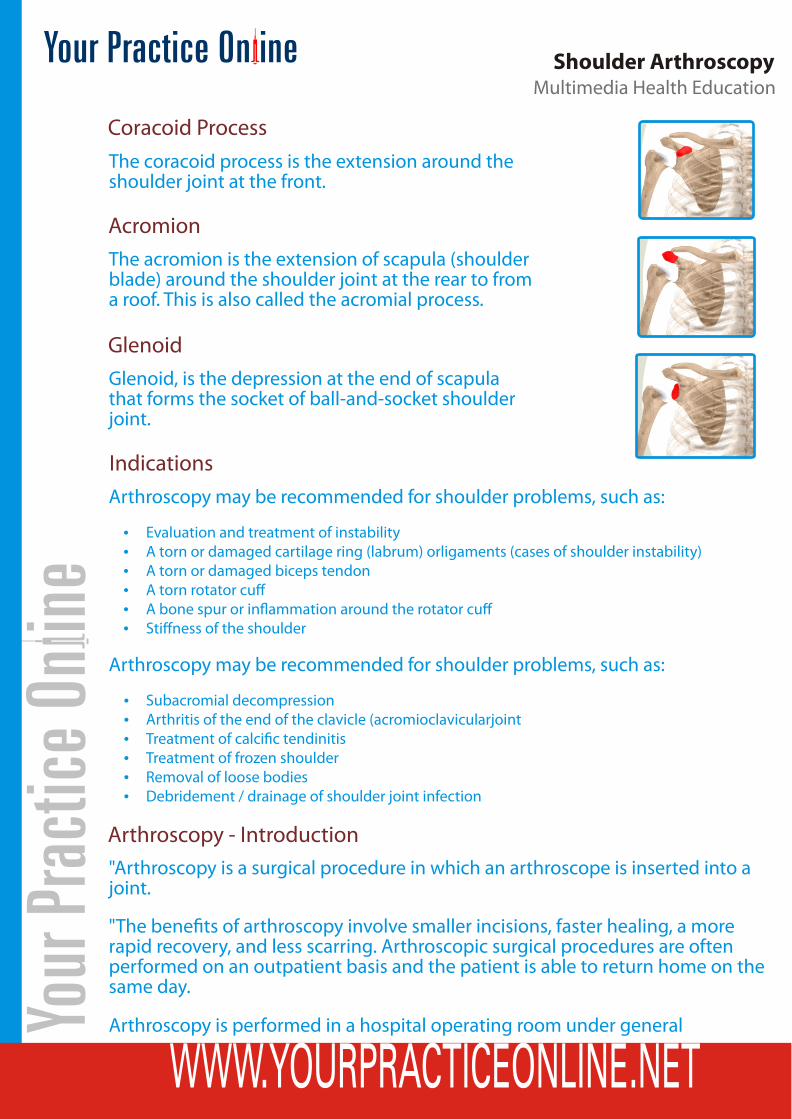
The coracoid process is the extension around the shoulder joint at the front.
The acromion is the extension of scapula (shoulder blade) around the shoulder joint at the rear to from a roof. This is also called the acromial process.
Glenoid, is the depression at the end of scapula that forms the socket of ball-and-socket shoulder joint.
Arthroscopy may be recommended for shoulder problems, such as:
Evaluation and treatment of instabilityA torn or damaged cartilage ring (labrum) orligaments (cases of shoulder instability)A torn or damaged biceps tendonA torn rotator cu�A bone spur or in�ammation around the rotator cu�Sti�ness of the shoulder
Arthroscopy may be recommended for shoulder problems, such as:
Subacromial decompressionArthritis of the end of the clavicle (acromioclavicularjointTreatment of calci�c tendinitisTreatment of frozen shoulderRemoval of loose bodiesDebridement / drainage of shoulder joint infection
"Arthroscopy is a surgical procedure in which an arthroscope is inserted into a joint.
"The bene�ts of arthroscopy involve smaller incisions, faster healing, a more rapid recovery, and less scarring. Arthroscopic surgical procedures are often performed on an outpatient basis and the patient is able to return home on the same day.
Arthroscopy is performed in a hospital operating room under general
Arthroscopy - Introduction
Multimedia Health EducationShoulder Arthroscopy
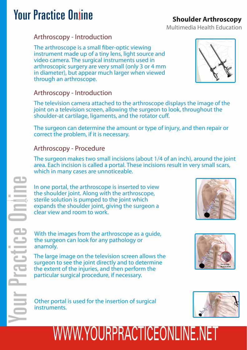
Arthroscopy - IntroductionThe arthroscope is a small �ber-optic viewing instrument made up of a tiny lens, light source and video camera. The surgical instruments used in arthroscopic surgery are very small (only 3 or 4 mm in diameter), but appear much larger when viewed through an arthroscope.
The television camera attached to the arthroscope displays the image of the joint on a television screen, allowing the surgeon to look, throughout the shoulder-at cartilage, ligaments, and the rotator cu�.
The surgeon can determine the amount or type of injury, and then repair or correct the problem, if it is necessary.
Arthroscopy - Introduction
The surgeon makes two small incisions (about 1/4 of an inch), around the joint area. Each incision is called a portal. These incisions result in very small scars, which in many cases are unnoticeable.
Arthroscopy - Procedure
In one portal, the arthroscope is inserted to view the shoulder joint. Along with the arthroscope, sterile solution is pumped to the joint which expands the shoulder joint, giving the surgeon a clear view and room to work.
With the images from the arthroscope as a guide, the surgeon can look for any pathology or anamoly.
The large image on the television screen allows the surgeon to see the joint directly and to determine the extent of the injuries, and then perform the particular surgical procedure, if necessary.
Other portal is used for the insertion of surgical instruments.

Multimedia Health EducationShoulder Arthroscopy
After treating the problem, the portals (incisions) are closed by suturing or by tape.
Arthroscopy is much less traumatic to the muscles, ligaments and tissues than the traditional method of surgically opening the shoulder with long incisions (open techniques).
Arthroscopy - Post -op RecoveryYou will wake up in the recovery room and then be transferred back to the ward.A bandage will be around the operated shoulder.Once you are recovered your drip will be removed and you will be shown a number of exercises to do.Your surgeon will see you prior to discharge and explain the �ndings of the operation and what was done during surgery.Pain medication will be provided and should be taken as directed.You can remove the bandage in 24 hours and place waterproof dressings (provided) over the wounds.It is NORMAL for the shoulder to swell after the surgery. Placing Ice-Packs on the shoulder will help reduce swelling (Ice-Packs on for 20 minutes 3-4 times a day until swelling has reduced).You are able to drive and return to work when comfortable unless otherwise instructed.Please make an appointment 7-10 days after surgery to monitor your progress.
Arthroscopy - Points of Interest What are the advantages ?
Diagnosing joint injuries and disease begins with a thorough medical history, physical examination, and usually X-rays. Additional tests such as an MRI, or CT also scan may be needed. Through the arthroscope, a �nal diagnosis is made which may be more accurate than through "open" surgery or from X-ray studies.
A surgical instrument is used to probe various parts within the joint to determine the extent of the problem.
If the surgeon sees an opportunity to treat a problem, a variety of surgical instruments can be inserted through this portal.
What are the joints that can be viewed with an Arthroscope ?
Although the inside of nearly all joints can be viewed with an arthroscope, six joints are most frequently examined with this instrument. These include the knee, shoulder, elbow, ankle, hip, and wrist.

Multimedia Health EducationShoulder Arthroscopy
As engineers make advances in electronic technology and orthopaedic surgeons develop new techniques, other joints may be treated more frequently in the future.
What are the conditions that can be treated by arthroscopy ?
Some problems associated with arthritis also can be treated. Several disorders are treated with a combination of arthroscopic and standard surgery. Disease and injuries can damage bones, cartilage, ligaments, muscles, and tendons. Some of the most frequent conditions found during arthroscopic examinations of joints are:
In�ammation - Synovitis - in�amed lining (synovium) in knee, shoulder, elbow, wrist, or ankle. Injury - acute and chronic - Shoulder - rotator cu� tendon tears, impingement syndrome, and recurrent dislocations Knee - meniscal (cartilage) tears, chondromalacia (wearing or injury of cartilage cushion), and anterior cruciate ligament tears with instability Wrist - carpal tunnel syndrome Loose bodies of bone and/or cartilage - knee, shoulder, elbow, ankle, or wrist Loose bodies of bone and/or cartilageknee, shoulder, elbow, ankle, or wrist
Although uncommon, complications do occur occasionally during or following arthroscopy. Anaesthetic complications are uncommon and may include Allergic reactions to medications and di�culty in breathing.
Local complications may include Infection, phlebitis (blood clots of a vein), excessive swelling or bleeding, damage to blood vessels or nerves, and instrument breakage are the most common complications, but occur in far less than 1 percent of all arthroscopic procedures.
What are the possible complications ?
Although arthroscopic surgery has received a lot of public attention because it is used to treat well-known athletes, it is an extremely valuable tool for all orthopaedic patients and is generally easier on the patient than "open" surgery.
Most patients have their arthroscopic surgery as outpatients and are home several hours after the surgery.
What are the advantages ?
The small puncture wounds take several days to heal. The operative dressing can usually be removed the morning after surgery and adhesive strips can be applied to cover the small healing incisions.
Recovery after arthroscopy
Multimedia Health EducationShoulder Arthroscopy
A good knowledge of this procedure will make the stress of undertaking the procedure easier for you to bear.The decision to proceed with the surgery is made because the advantages of surgery outweigh the potential disadvantages.It is important that you are informed of these risks before the surgery.
Summary
Although the puncture wounds are small and pain in the joint that underwent arthroscopy is minimal, it takes several weeks for the joint to maximally recover.
Multimedia Health EducationShoulder Arthroscopy
2009 YOUR SURGERY DATE
READ YOUR BOOK AND MATERIAL
VIEW YOUR VIDEO /CD / DVD / WEBSITE
PRE - HABILITATION
ARRANGE FOR BLOOD
MEDICAL CHECK UP
ADVANCE MEDICAL DIRECTIVE
PRE - ADMISSION TESTING
FAMILY SUPPORT REVIEW
Physician's Name :
Physician's Signature:
Date :
Patient’s Name :
Patient’s Signature:
Date :


![7-Practical Application of Shoulder Arthroscopy [唯讀] Application of Shoulder Arthroscopy ... neck & body are support in a neutral position ... Instrument for anchor fixation](https://img.dokumen.tips/doc/110x75/5aac4ef77f8b9aa9488cdea5/7-practical-application-of-shoulder-arthroscopy-application-of-shoulder.jpg)


![Shoulder Arthroscopy Surgery Ed...Shoulder Arthroscopy Surgery [page 2]What to Expect After Surgery You may be more comfortable resting or sleeping in a recliner type chair the first](https://img.dokumen.tips/doc/110x75/60b62a1953edc40b0032ab75/shoulder-arthroscopy-surgery-ed-shoulder-arthroscopy-surgery-page-2what-to.jpg)