Embed Size (px)
Citation preview
Plant Science Letters, 34 (1984) 219--223 219 Elsevier Scientific Publishers Ireland Ltd.
SHOOT REGENERATION FROM HORMONE-AUTONOMOUS CALLUS FROM SHOOT CULTURES OF SEVERAL SUGARBEET ( B E T A V U L G A R I S L.) GENOTYPES*
J.W. SAUNDERS a and M.E. DAUB b'** aResearch Geneticist, USDA-ARS and b Research Associate, Department of Crop and Soil Sciences, Michigan State University, East Lansing, M148824-1114 (U.S.A.)
(Received September 7th, 1983) (Revision received October 31st, 1983) (Accepted November 4th, 1983)
SUMMARY
Shoot cultures of six sugarbeet genotypes produced callus when grown in dim fluorescent light (10--15 ~Em -2 s- ' ) at 30°C on Mumshige-Skoog (MS) medium with 0.25 rag/1 6-benzyladenine (BA). Hormone au tonomy of the callus was indicated by sustained callus growth following multiple transfers onto MS medium lacking auxin and cytokinin. Shoots were regenerated from 1-month-old callus o f two genotypes after transfer to MS medium with 0.25--5.0 mg/1 BA at 30°C in dim fluorescent light (10 -15 #Em -2 s- ') .
K e y words: Hormone-autonomy -- Be ta vulgaris - - Callus -- Shoot regener- ation -- Shoot cultures
INTRODUCTION
The most noteworthy items about adventitious shoots on sugarbeet tissues to date have been the broad array of source tissues and the circum- stances surrounding their appearance. Adventitious shoots in sugarbeet in vitro have been observed at the base of flower buds [1 ], at the base of isolated stem axes [2], on leaf pieces from shoots grown in vitro [3], and on intact leaves of shoot cultures [4,5]. In addition, regeneration of shoots in vitro has been observed from callus derived from anthers [6], seedling
*Cooperative investigations of Agricultural Re,arch Service, USDA and Michigan Agri- cultural Experiment Station, EMt Lansing, MI 48824, U.S.A. Journal article 10724. **Present address: Department of Plant Pathology, North Carolina State University, Raleigh, NC 27650, U.S.A. Abbreviations: BA, 6-benzyladenine; IAA, indole-3-acetic acid; MS, Mursehige and Skoog; NAA, a-naphthaleneacetic acid.
0304-4211/84/$03.00 © 1984 Elsevier Scientific Publishers Ireland Ltd. Printed and Published in Ireland
220
explants [7,8], axillary buds in vitro [9], as well as in an habituated cell line [10]. Adventitious shoots in sugarbeet have also been reported to occur a t low frequency on leaf cuttings [11], and can also be induced on subsequent leaves of intact plants after benzyladenine treatment of seedlings [12]. We now report a reproducible method for induction of ho rmone autonomous callus in sugarbeet and regeneration of shoots from this callus.
MATERIALS AND METHODS
Shoot cultures of seven genotypes were established from seedlings or lateral buds of flower stalks [4,5]. Murashige and Skoog [13] medium (MS) with 0.25 rag/1 BA, 1.0 rag/1 thiamine--HC1, 0.5 mg/1 pyridoxine--HCl, 0.5 rag/1 nicotinic acid and 0.9% (w/w) agar was used for routine maintenance and experimental culture. The medium was contained in Falcon Optilux 100 × 20 mm petri dishes with 35 -40 ml per plate. Shoots were subcultured at monthly intervals, with three shoots transferred to each dish. Shoot cultures were maintained at 24 -+ 2°C in growth chambers with continuous fluorescent (General Electric 40D Malnlighter) light at 100--200 ~Em -2 s -1.
Callus was cultured in petri dishes or in 25 × 70 mm glass screw-top vials (seals removed) containing 10 ml of medium. All callus cultures were grown on MS medium with differing concentrations of growth regulators, in lighted growth chambers (20 ° or 30°C) or in dark incubators (24°C). Callus growth was measured by the following procedure: 8-ram s callus pieces were sul> cultured onto fresh medium. After I month, the length and width of the callus pieces were measured, and an approximate cross-sectional area was computed. Alternatively, callus fresh weight was recorded at subculture and again after 1 month. Shoots obtained in regeneration studies were subcultured on MS + 0.25 mg/l BA for 1 month, rooted on MS + 3.0 mgfl a-naphthalene acetic acid (NAA) for 5 weeks, then potted in soil in the greenhouse.
RESULTS
Callus formation from shoot cultures Sugarbeet shoot cultures that had been established for more than a year
developed a white friable callus after 6 weeks of culture on MS + 0.25 rag/1 BA on laboratory benches during the hot summer months when the temper- ature in the lab was often 30--32°C. Callus had not been noted during earlier maintenance. To determine if the development of callus from shoot cultures is a common ~ r i s t i c of su~JRrbeet pno t -~es , 4--5 shoot culture dishes from each of seven genotypes were cultured at 30°C under dim fluorescent lights (10--15/~F,m -2 s-l). After 70 days, six of the genotypes had developed "some callus in the shoot cultures, often as the cultures senesced.
The callus that arose m the 30°C shoot cultures appeared in any of three situations. Some arose on the s tu~ce of swollen blackened basal leaves
221
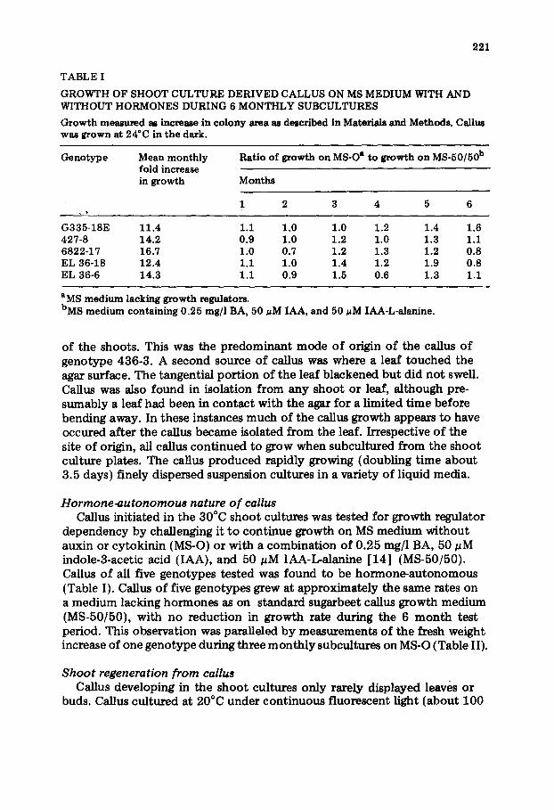
TABLE I
GROWTH OF SHOOT CULTURE DERIVED CALLUS ON MS MEDIUM WITH AND WITHOUT HORMONES DURING 6 MONTHLY SUBCULTURES
Growth measured as increase in colony area as described in Materials and Methods. Callus was grown at 24°C in the dark.
Genotype Mean monthly fold increase in growth
Ratio of growth on MS-O a to growth on MS-50/50 b
Months
1 2 3 4 5 6
G335-18E 11.4 1.1 1.0 1.0 1.2 1.4 1.6 427-8 14.2 0.9 1.0 1.2 1.0 1.3 1.1 6822-17 16.7 1.0 0.7 1.2 1.3 1.2 0.8 EL36-18 12.4 1.1 1.0 1.4 1.2 1.9 0.8 EL36-6 14.3 1.1 0.9 1.5 0.6 1.3 1.1
aMS medium lacking growth regulators. bMS medium containing 0.25 mg/l BA, 50 #M IAA, and 50 ~M IAA-L-alanine.
of the shoots. This was the predominant mode of origin o f the callus o f genotype 436-3. A second source of callus was where a leaf touched the agar surface. The tangential por t ion of the leaf blackened but did no t swell. Callus was also found in isolation from any shoot or leaf, al though pre- sumably a leaf had been in con tac t with the agar for a limited t ime before bending away. In these instances much of the callus growth appears to have occured after the callus became isolated from the leaf. Irrespective of the site of origin, all callus cont inued to grow when subcultured from the shoot culture plates. The callus produced rapidly growing (doubling t ime about 3.5 days) finely dispersed suspension cultures in a variety o f liquid media.
Hormone-autonomous nature o f callus Callus initiated in the 30°C shoot cultures was tested for growth regulator
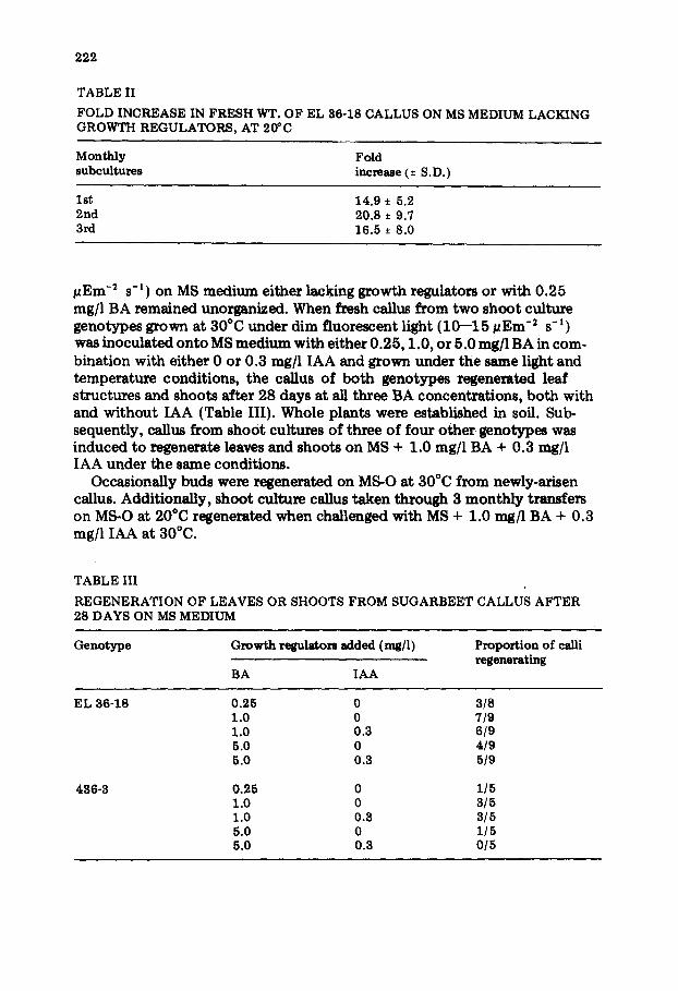
dependency by challenging it t o cont inue growth on MS medium without auxin or cytokinin (MS-O) or with a combinat ion of 0.25 rag/1 BA, 50 ~M indole-3-acetic acid (IAA), and 50 #M IAA-L-alanine [14] (MS-50/50). Callus o f all five genotypes tested was found to be hormone-au tonomous (Table I). Callus of five genotypes grew at approximately the same rates on a medium lacking hormones as on standard sugarbeet callus growth medium (MS-50/50), with no reduct ion in growth rate during the 6 mon th test period. This observation was paralleled by measurements of the fresh weight increase of one genotype during three month ly subcultures on MS-O (Table II).
Shoot regeneration from callus Callus developing in the shoot cultures only rarely displayed leaves or
buds. Callus cul tured at 20°C under cont inuous f luorescent light (about 100
222
TABLE II
FOLD INCREASE IN FRESH WT. OF EL 36-18 CALLUS ON MS MEDIUM LACKING GROWTH REGULATORS, AT 20~C
Monthly Fold subcultures increase (± S.D.)
1st 14.9 ± 5.2 2nd 20.8 ± 9.7 3rd 16.5 ± 8.0
g e m -2 s -1) on MS medium either lacking growth regulators or with 0.25 mg/l BA remained unorganized. When fresh callus from two shoot culture genotypes grown at 30°C under dim fluorescent light (10--15 ~Em -2 s -1) was inoculated onto MS medium with either 0.25, 1.0, or 5.0 rag/1BA in com- bination with either 0 or 0.3 rag/1 IAA and grown under the same light and temperature conditions, the callus of both genotypes regenerated leaf structures and shoots after 28 days at all three BA concentrations, both with and wi thout IAA (Table III). Whole plants were established in soil. Sub~ sequently, callus from shoot cultures of three of four other genotypes was induced to regenerate leaves and shoots on MS + 1.0 rag/1 BA + 0.3 rag/1 IAA under the same conditions.
Occasionally buds were regenerated on MS-O at 30°C from newly-arisen callus. Additionally, shoot culture callus taken through 3 month ly transfers on MS-O at 20°C regenerated when challenged with MS + 1.0 mgfl BA + 0.3 mg/l IAA at 30°C.
TABLE III
REGENERATION OF LEAVES OR SHOOTS FROM SUGARBEET 28 DAYS ON MS MEDIUM
CALLUS AFTER
Genotype Growth re~latom added (r~/l)
BA IAA
Proportion of calli regenerating
EL 36-18
486-3
0.25 0 3/8 1.0 0 "//9 1.0 0.3 6/9 5.0 0 4/9 5.0 0.3 519
0.25 0 1/5 1.0 0 3/5 1.0 0.3 3/5 5.0 0 1/5 5.0 0.3 0/5
223
DISCUSSION
The hormone-au tonomous sugarbeet callus described in our repor t differs in several ways from that described by DeGreef and Jacobs [10] . The callus studied in our laboratory is repeatedly obtainable, has been induced from numerous genotypes, and regenerates shoots only occasionally on media lacking cytokinins. The DeGreef-Jacobs habi tuated line arose in a single culture and is capable o f cont inued shoot regeneration in the absence o f growth regulators. Fur thermore, the DeGreef~lacobs line is apparent ly partially differentiated, whereas our callus is white, friable and appears un- organized. Other work has established that several different cytokinins are effective in inducing this callus from isolated shoot cul ture petioles and stimulating shoot regeneration from callus [ 15 ].
ACKNOWLEDGEMENTS
The authors wish to thank W.D. Taylor and C.R. Santerre.
REFERENCES
1 J. Margara, CR Acad. Sci. Paris, 270 (1970) 698. 2 A.T. Ata~_~ov, Z. Pflanzenzuecht., 84 (1980) 23. 3 J. Rogozinska and M. Goska, Bull. Acad. Pol. Sci. (Biol), 26 (1978) 343. 4 G. Hussey and A. Hepher, Ann. Bot., 42 (1978) 477. 5 J.W. Saunders, Crop Sci., 22 (1982) 1102. 6 J. Rogozinska, M. Goska and A. Kuzdowicz, Acta Bot. Soc. Pol., 46 (1977) 471. 7 R.G. Butenko, A. Atanassov and W. Un~nantsera, Phytomorphology, 22 (1972) 140. 8 A.M.S. Mohammad and H.S. CoUin, New Phytol., 82 (1979) 293. 9 J. Margara, CR Acad. Sci. Paris, 285 (1977) 1041.
10 W. DeGreef and M. Jacobs, Plant Sci. Lett., 17 (1979) 55. 11 P. Miederna, P.J. Groot and J.H.M. Zuidgeest, Euphytica, 29 (1980) 425. 12 J.W. Saunders and M.D. Mahoney, Euphytica, 31 (1982) 801. 13 T. Murashige and F. Skoog, Physiol. Plant, 15 (1962) 473. 14 R.P. Hangarter, M.D. Peterson and N.E. Good, Plant Physiol., 65 (1980) 761. 15 J.W. Saunders, Proc. 5th Int. ~ongr. Plant Tissue and Cell Culture, Tokyo, 1982,
p. 153.


![Research Article Chlorophytum borivilianum L.) Callus ... · the callus cultures of Safed musli, the method explained by Nakasha et al. [23] was followed. Briefly, shoot buds of](https://img.dokumen.tips/doc/110x75/60a3586207e8a759535f44b1/research-article-chlorophytum-borivilianum-l-callus-the-callus-cultures-of.jpg)