Embed Size (px)
Citation preview
163
© 2015 The Korean Society of Pathologists/The Korean Society for CytopathologyThis is an Open Access article distributed under the terms of the Creative Commons Attribution Non-Commercial License (http://creativecommons.org/licenses/by-nc/3.0) which permits unrestricted non-commercial use, distribution, and reproduction in any medium, provided the original work is properly cited.
pISSN 2383-7837eISSN 2383-7845
Collision tumors are best considered as separate primary neo-plasms. These tumors have been reported in various organs, such as the esophagus, stomach, liver, thyroid gland, ovary, and lung, but they are extremely rare in the ovaries.1 The majority of these tumors are a collision between carcinomas and sarcomas or lymphomas, and rarely between two types of carcinoma.2 The most common histological combination of collision tumor in the ovary is the coexistence of teratoma with mucinous tu-mors (mucinous cystadenoma or carcinoma).1 Here we report a very unusual combination of fibrothecoma and serous cystade-noma in the left ovary of an elderly woman who presented with an abdominal lump and ascites.
CASE REPORT
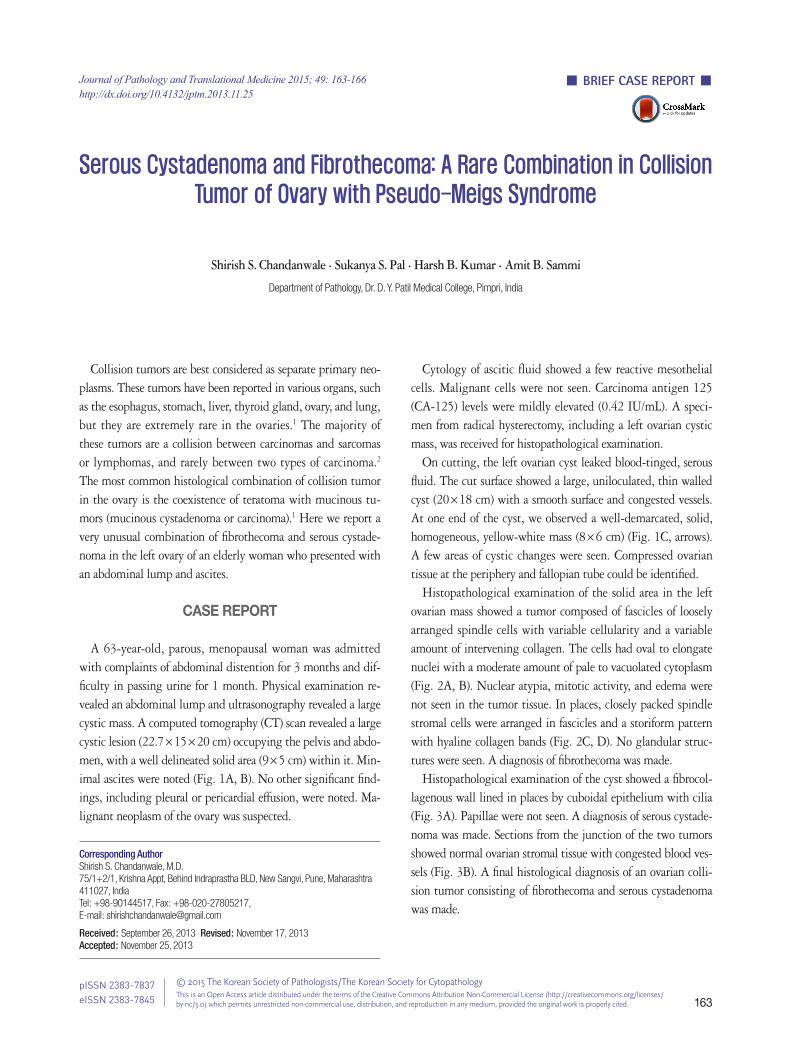
A 63-year-old, parous, menopausal woman was admitted with complaints of abdominal distention for 3 months and dif-ficulty in passing urine for 1 month. Physical examination re-vealed an abdominal lump and ultrasonography revealed a large cystic mass. A computed tomography (CT) scan revealed a large cystic lesion (22.7×15×20 cm) occupying the pelvis and abdo-men, with a well delineated solid area (9×5 cm) within it. Min-imal ascites were noted (Fig. 1A, B). No other significant find-ings, including pleural or pericardial effusion, were noted. Ma-lignant neoplasm of the ovary was suspected.
Cytology of ascitic fluid showed a few reactive mesothelial cells. Malignant cells were not seen. Carcinoma antigen 125 (CA-125) levels were mildly elevated (0.42 IU/mL). A speci-men from radical hysterectomy, including a left ovarian cystic mass, was received for histopathological examination.
On cutting, the left ovarian cyst leaked blood-tinged, serous fluid. The cut surface showed a large, uniloculated, thin walled cyst (20×18 cm) with a smooth surface and congested vessels. At one end of the cyst, we observed a well-demarcated, solid, homogeneous, yellow-white mass (8×6 cm) (Fig. 1C, arrows). A few areas of cystic changes were seen. Compressed ovarian tissue at the periphery and fallopian tube could be identified.
Histopathological examination of the solid area in the left ovarian mass showed a tumor composed of fascicles of loosely arranged spindle cells with variable cellularity and a variable amount of intervening collagen. The cells had oval to elongate nuclei with a moderate amount of pale to vacuolated cytoplasm (Fig. 2A, B). Nuclear atypia, mitotic activity, and edema were not seen in the tumor tissue. In places, closely packed spindle stromal cells were arranged in fascicles and a storiform pattern with hyaline collagen bands (Fig. 2C, D). No glandular struc-tures were seen. A diagnosis of fibrothecoma was made.
Histopathological examination of the cyst showed a fibrocol-lagenous wall lined in places by cuboidal epithelium with cilia (Fig. 3A). Papillae were not seen. A diagnosis of serous cystade-noma was made. Sections from the junction of the two tumors showed normal ovarian stromal tissue with congested blood ves-sels (Fig. 3B). A final histological diagnosis of an ovarian colli-sion tumor consisting of fibrothecoma and serous cystadenoma was made.
Journal of Pathology and Translational Medicine 2015; 49: 163-166http://dx.doi.org/10.4132/jptm.2013.11.25
▒ BRIEF CASE REPORT ▒
Corresponding AuthorShirish S. Chandanwale, M.D. 75/1+2/1, Krishna Appt, Behind Indraprastha BLD, New Sangvi, Pune, Maharashtra 411027, IndiaTel: +98-90144517, Fax: +98-020-27805217, E-mail: [email protected]
Received: September 26, 2013 Revised: November 17, 2013 Accepted: November 25, 2013
Serous Cystadenoma and Fibrothecoma: A Rare Combination in Collision
Tumor of Ovary with Pseudo-Meigs Syndrome
Shirish S. Chandanwale · Sukanya S. Pal · Harsh B. Kumar · Amit B. Sammi
Department of Pathology, Dr. D. Y. Patil Medical College, Pimpri, India
http://jpatholtm.org/ http://dx.doi.org/10.4132/jptm.2013.11.25
164 • Chandanwale SS, et al.
A
B C
Fig. 1. (A, B) Computed tomography scan showing a large cystic lesion occupying the pelvis and abdomen (solid area), and minimal ascites. (C) Specimen of left ovarian cyst with well-demarcated solid yellow tumor mass (arrows).
DISCUSSION
Collision tumors are defined as two adjacent but histological-ly distinct tumors, without admixture, in the same tissue or or-gan.1 These tumors are rare clinical entities.
The majority of these tumors are a collision between carcino-mas and sarcomas or lymphomas and have been reported in vari-ous organs. Collision tumors involving ovaries are extremely rare. Many hypotheses have been put forward to explain the rare phenomenon of collision tumors: coincidental occurrence; carci-nogenic agents of a primary tumor; oncogenic growth factors produced by a metastatic tumor; and alterations in the micro-environment.1 In this case, the finding was coincidental. Very few reports of collision tumors involving ovaries have been re-ported in the literature.1,3,4
The pathology of collision tumors reveal two different types of coexisting neoplastic tissues, with a sharp demarcation be-tween the two and without a substantial admixture of histology
at the interface. The most common combination of collision tu-mor in the ovary involves teratoma with mucinous tumors (cyst-adenoma and carcinoma).1
Thecoma and fibroma often merge, therefore the term fibroth-ecoma is appropriate. Pure thecomas are typically associated with estrogenic manifestations, which were not seen in our case. Ovar-ian fibrothecomas often clinically present as a solid adnexal mass and can mimic malignant ovarian tumors.
Ultrasonography features of fibrothecoma are usually nonspe-cific, and magnetic resonance imaging (MRI) is often needed for further differentiation from other solid ovarian masses.5 MRI was not done in our case. Ovarian fibromas and fibrothecomas can be associated with ascites, sometimes in combination with pleural effusion, which may lead to a mistaken impression of inoperable ovarian neoplasm.5
In 1937, Meigs described seven cases of combined pleural ef-fusion, ascites, and ovarian fibroma and named it Meigs syn-drome.6 In 1954, he limited the syndrome to cases where tu-

http://jpatholtm.org/http://dx.doi.org/10.4132/jptm.2013.11.25
A Rare Ovarian Collision Tumor • 165
A B
C D
Fig. 2. Microscopy of solid tumor showing areas of thecoma (A, B), fibroma (C), and mixture of fibroma and thecoma (D).
A
B
Fig. 3. (A) Microscopy of cyst wall showing lining of cuboidal epi-thelium with cilia. (B) Microscopy from the junction of solid and cys-tic tumors showing congested ovarian stroma.
mor removal cures the disease.6 Pseudo-Meigs is a variant, not possessing the original tumor cell types described by Meigs.7
Proposed mechanisms for the ascites in Meigs syndrome are
production of ascitic fluid by the tumor; lymphatic obstruction; hormonal stimulation; release of inflammatory mediators; and tumor torsion. Pleural effusion is thought to be caused by the migration of fluid and protein—perhaps by lymphatic chan-nels— across the diaphragm.6 In the present case, pleural effu-sion was not present, which was possibly due to the minimal amount of ascitic fluid. Elevated serum CA-125 levels, which are seen in Meigs or atypical Meigs syndrome, were seen in this case.8
To the best of our knowledge, we are presenting the first case in the English literature of collision of fibrothecoma and serous cystadenoma in an ovary with Pseudo-Meigs syndrome. For correct diagnosis, these tumors need to be differentiated from fibrothecoma with massive cystic changes and serous cystadeno-fibroma. The presence of cuboidal lining epithelium with cilia in the cyst wall ruled out massive cystic changes in fibrotheco-ma and a follicular cyst. The absence of glandular structures in fibrothecoma ruled out serous cystadenofibroma. Follow up ex-amination of the patient postoperatively showed the disappear-

http://jpatholtm.org/ http://dx.doi.org/10.4132/jptm.2013.11.25
166 • Chandanwale SS, et al.
ance of ascitic fluid on a CT scan. We conclude that a diagnosis of collision tumor involving an
ovary is challenging and often made postoperatively. The colli-sion of fibrothecoma and serous cystadenoma is a rare combina-tion and can cause Meigs or Pseudo-Meigs syndrome. Elevated serum CA-125 levels alone cannot differentiate between benign and malignant ovarian masses.
Conflicts of InterestNo potential conflict of interest relevant to this article was
reported.
REFERENCES
1. Bige O, Demir A, Koyuncuoglu M, Secil M, Ulukus C, Saygili U. Collision tumor: serous cystadenocarcinoma and dermoid cyst in the same ovary. Arch Gynecol Obstet 2009; 279: 767-70.
2. Murthaiah P, Truskinovsky AM, Shah S, Dudek AZ. Collision tu-mor versus multiphenotypic differentiation: a case of carcinoma
with features of colonic and lung primary tumors. Anticancer Res 2009; 29: 1495-7.
3. Papaziogas B, Souparis A, Grigoriou M, et al. A rare triple coexis-tence of a collision tumor, a benign mature cystic teratoma and a hemorrhagic follicular cyst of the ovaries. Internet J Surg 2008; 14: 19-24.
4. Moid FY, Jones RV. Granulosa cell tumor and mucinous cystadeno-ma arising in a mature cystic teratoma of the ovary: a unique case report and review of literature. Ann Diagn Pathol 2004; 8: 96-101.
5.KitajimaK,KajiY,SugimuraK.UsualandunusualMRIfindingsofovarianfibroma:correlationwithpathologicfindings.MagnRe-son Med Sci 2008; 7: 43-8.
6.ThakerDA,StridePJ,DettrickA.Apseudocaseofatypicalpseudo-Meigs syndrome. Indian J Cancer 2011; 48: 364-6.
7. Kazanov L, Ander DS, Enriquez E, Jaggi FM. Pseudo-Meigs’ syn-drome. Am J Emerg Med 1998; 16: 404-5.
8. Renaud MC, Plante M, Roy M. Ovarian thecoma associated with a large quantity of ascites and elevated serum CA 125 and CA 15-3. J Obstet Gynaecol Can 2002; 24: 963-5.


![Mucinous Neoplasm: A Case Report A Rare Case of Low-grade ... · cell adenocarcinoma, or neuroendocrine carcinoma [3]. Mucinous adenocarcinoma accounts for Mucinous adenocarcinoma](https://img.dokumen.tips/doc/110x75/5d66f73588c993283a8b59a1/mucinous-neoplasm-a-case-report-a-rare-case-of-low-grade-cell-adenocarcinoma.jpg)


![Mucinous Cystadenoma of the Ectopic Pancreas with …...Jul 04, 2015 · JOP. Journal of the Pancreas - - Vol. 16 No. 4 Jul 2015. [ISSN 1590-8577] 392 OP. Pancreas (Online) 21 ul](https://img.dokumen.tips/doc/110x75/5e9e333824cd1d57d126ffb5/mucinous-cystadenoma-of-the-ectopic-pancreas-with-jul-04-2015-jop-journal.jpg)