Embed Size (px)
Citation preview

SEMT One Day MeetingWednesday 12th December 2007
at The School of PharmacyUniversity of London
Society of Electron Microscope Technology
Keeping you informed of the latest technology & techniques

Prospective members can be added to our Members List by contacting the Hon.SecretaryMr. David McCarthy, School of Pharmacy, Brunswick Sq. London WC1N 1AXT 44 (0) 20 7753 5806; E [email protected]. The annual subscription is currently FREE.Our web site : www.semt.org.uk
Committee members are listed below and are available for further information.
Officers
Chair Mrs. Heather Davies
Secretary Mr. David McCarthy
Treasurer Mrs. Nicola Mordan
Committee Ms. Ann DewarMr. David RobertsonMr. Terry CooperMr. Chris JonesMr. Barry DowsettMiss. Anne Drewe Mrs. Trish Lovell Mr. Derrick LovellMrs. Hannah ArmerDr. Tony Brain
Honorary Advisors Mrs. Pauline BarberMr. Chris Walker
Honorary Archivist Dr. Jill Lewis
The Society of Electron Microscope Technology Page 2
Acknowledgments The SEMT wishes to express special thanks to The School of Pharmacy as host and to the following companies for supporting the Trade Exhibition :
Agar Scientific Ltd. Brady ScientificDeben UK Ltd.Edax UK Ltd.FEI UK Ltd.Gatan.Hitachi High-Technologies EuropeI.S.S. Group Services.Leica Microsystems UK Ltd. Jeol UK Ltd.Obducat Camscan Ltd.Quorum Technologies Ltd.Taab Laboratories Ltd.Zeiss SMT
2008 ProgrammeODM/AGM 10th December 2008

The Society of Electron Microscope TechnologyPage 3
09.15 am Registration, Tea/Coffee, Trade Exhibition.
09.55 a.m Introduction: Chair, Heather Davies.
10.00 a.m Cryo-imaging; its role, application and future at NIBSC Roland Fleck, National Institute of Biological Standards & Control. South Mimms.
10.35 am Imaging for Television and Publishing David Spears, Clouds Hill Imaging.
11.10 am Tea, Coffee.
11.30 am Multiple Immunofluorescence Labelling of Formalin Fixed Paraffin Embedded (FFPE)Tissues. David Robertson, Breakthrough, ICR, London.
12.05 pm Investigations using correlative confocal laser scanning microscopy and immunoTEM Susan Anderson, Nottingham University. (Joint Winner of the Don Claugher Bursary)
12.15 pm Buffet Lunch, Trade Exhibition.
14.00 pm Electron Microscopy in Paediatric Pathology Glenn Anderson, Histopath. Dept. Great Ormond Street Hospital for Children. 14.35 pm Is there a Future for Electron Microscopy in Healthcare? Tim Ryder, EM unit, Charing Cross Hospital.
15.10 pm Tea, Coffee.
15.30.pm Tissue engineering to repair the nervous system and model it in vitro James Phillips, Biological Sciences, Open University.
16.05 pm In search of nanoparticles: EM in the quest for ideal nano-components Prof. Kostas Kostarelos, School of Pharmacy, London.
16.40 pm Malaria and the red blood cell: mysteries of entrances and exits. Prof. Lawrie Bannister, Department of Anatomy and Human Sciences, Guy’s Campus, King’s College London. 17.15 pm AGM - Wine Reception.
19.00 pm Conference Dinner.
Trade Exhibitors Agar Scientific Ltd., Brady Scientific, Deben UK Ltd., Edax UK Ltd., FEI UK Ltd., Gatan, Hitachi High-Technologies Europe. I.S.S. Group Services, Jeol UK Ltd., Leica Microsystems UK Ltd., Obducat Camscan Ltd., Quorum Technologies Ltd., Taab Laboratories Ltd. & Zeiss SMT.
The Society of Electron Microscope Technology Page 4
Cryo-imaging; its role, application and future at NIBSC
Roland A. Fleck1*
1 National Institute for Biological Standards and Control, Blanche Lane,South Mimms, Potters Bar, EN6 3QG.*Correspondence: [email protected]
NIBSC routinely employs imaging, including; electron microscopy (EM) and epifluorescent microscopy in the testing and evaluation of biological medicines. However, as biological medicines develop so do the challenges of ensuring their safety. Today’s emerging and postulated therapies present an ever moving target with greater demands on basic research, control and standardisation. Recently, NIBSC has replaced much of its imaging facility with a new, high resolution imaging facility focused around cryo-techniques. This new capability presents a number of operational challenges. However, the largely artefact free, high resolution information threshold provided by cryo-techniques is core to the development of imaging at NIBSC over the coming years. At NIBSC, imaging techniques including; confocal, freeze drying, and electron-microscopy allow detailed studies of broader biological medicines to be performed, often
with the added value of immuno-specific labelling (e.g., conjugated antibody labelling of stem cell cultures pre- and post-differentiation, reference sera standardisation by immunogold labelling, cellular processing of fluorescent conjugated-vaccine components). Historically and currently, traditional fixation, dehydration and embedding have been employed to the study of vaccines and biological medicines. However, the accessibility of cryo-imaging has enhanced the imaging sections ability to respond to the needs of the institute and presents exciting opportunities for new and novel studies of biological medicines. Cryo-light microscopy
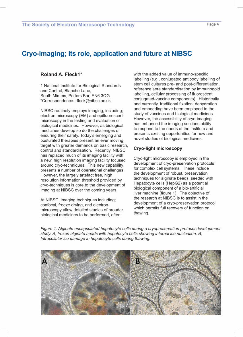
Cryo-light microscopy is employed in the development of cryo-preservation protocols for complex cell systems. These include the development of robust, preservation techniques for alginate beads, seeded with Hepatocyte cells (HepG2) as a potential biological component of a bio-artificial liver machine (figure 1). The objective of the research at NIBSC is to assist in the development of a cryo-preservation protocol which permits full recovery of function on thawing.
Figure 1. Alginate encapsulated hepatocyte cells during a cryopreservation protocol development study. A, frozen alginate beads with hepatocyte cells showing internal ice nucleation. B, Intracellular ice damage in hepatocyte cells during thawing.

Technique development and optimisation of the cryo-stage (Bal-tec VCT-100/cryo-stage) and sample processing (Bal-tec HPM-010, BAF-060) have allowed stable robust imaging of a wide range of biological materials (virus particles, unicellular algae, bacteria and nano-particles) (figure 3).
Figure 3. Influenza particles by cryo-TEM showing spikes and RNA
Operation of the TEM (JEOL, JEM-2100) under cryogenic conditions has improved our understanding of the stability of influenza vaccine candidates and allowed simple rapid observation of fully hydrated tissues. Process optimisation includes the use of plasma cleaning (Fischione 1020), in preference to less controlled glow discharge, to change the surface properties of carbon films as well as cleaning and controlled thinning of films. These techniques are beginning to play a routine part in vaccine safety/efficacy testing; present studies address Meningococcal group B and C vaccine programmes as well as H5N1 influenza vaccine development.
The Society of Electron Microscope TechnologyPage 5
Freeze drying-light microscopy
Freeze drying-light microscopy is employed in the development and assessment of freeze drying protocols for biological reference reagents prepared and maintained at NIBSC. The technique allows visualisation of freezing and primary drying of biological materials under conditions simulating those within a commercial freeze drier. Variation of vacuum, temperature and warming protocol can be assessed, on small volumes (10µl) of these valuable samples, with data on drying rate and collapse point being determined. This information is invaluable in the development of robust, successful, freeze drying protocols for biological standards.Cryo-EM has been recently added to the technical capability at NIBSC. This permits more detailed studies, arguably with less processing artefact, of biological materials than was possible with the previous, traditional EM instrumentation. Notable preliminary studies include, observation of membrane complexes of Neisseria meningtidis by cryo-FEGSEM (JEOL JSM-7401F) (figure 2).
Figure 2. Neisseria meningtidis by cryo-FEGSEM showing membrane proteins and capsule.

The Society of Electron Microscope Technology Page 6
Imaging for Television and Publication
David Spears B.Sc. MIET.
Although many images produced by the scientific community are well composed and technically of a very high standard, they are often very difficult for the lay person to understand. What is being portrayed can easily be misinterpreted and consequently skipped. As most images are designed to accompany scientific papers and presentations designed for a specific audience familiar with the kind of images presented, there is usually no problem. The delivery of scientific concepts to a lay audience whether in television programmes or in books or the press, inevitably uses a proportionally greater amount of visual material. Great care has to be taken that the images are relevant, clear and understandable. Failure to produce images and sequences with these characteristics often leads to the disappointing decision of a producer or editor to use graphics and animation instead of images of real phenomena.
The common woodlouse.
Ovum with sperm

The Society of Electron Microscope TechnologyPage 7
Multiple Immunofluorescence Labelling of Formalin Fixed Paraffin Embedded (FFPE) Tissues
David RobertsonBreakthrough, ICR, London
Presented here is a method that allows multiple immunofluorescence labelling of formalin fixed paraffin embedded (FFPE) tissues. It is achieved by the combination of antigen retrieval, indirect immunofluorescence and confocal laser scanning microscopy. This approach has the potential to unlock a large in vivo database for immunofluorescent investigations and has the advantage over peroxidase staining of the ability to label three antigens simultaneously plus nuclear counterstain.

The Society of Electron Microscope Technology Page 8
Investigating using correlative confocal laser scanning microscopy and immunoTEM.
Susan Anderson Ebling FJP, Joint Winner of the Don Claugher Bursary 2006University of Nottingham School of Biomedical Sciences.
The timing of puberty and reproductive function is closely linked to energy balance in mammals, but the neuroanatomical links between hypothalamic systems involved in metabolic physiology and the GnRH secretory system controlling the pituitary-gonadal axis are poorly understood. We have studied interactions between peptidergic neuronal phenotypes using confocal laser scanning microscopy (CLSM) in a transgenic mouse in which GnRH neurons express a green fluorescent protein (GFP) reporter. We have evidence of close appositions on gonadotropin-releasing hormone (GnRH) neurons of NPY, MCH, βendorphin and CART immunoreactive boutons (Leslie et al, Neuroscience Lett 314: 111-114) but the precise nature of these close contacts is unknown. Increased resolution is required to further address the interactions of these neurons, but because the number of GnRH neurons in the brain is small (c1000) correlative electron and confocal microscopy is the only realistic way to proceed to the ultrastructural level. The aim is to combine the speed and relative ease of confocal microscopy with the precision and resolution of
TEM and to develop a correlative CLSM-TEM method to further investigate appositions of peptidergic neurons upon GnRH neurons.Vibratome sections (60micron) of perfusion-fixed, mouse brain were prepared and peptidergic neurons labelled using fluoronanogold (FNG) to enable visualisation of each section by both light and electron microscopy. CLSM was used to map the fluorescence labelling of peptidergic neurons within each tissue slice. Tissue slices with a good number of labelled peptidergic neurons were subsequently processed, flat embedded and sectioned for TEM so that the area of interest could be located and examined. A variety of fixation, processing and labelling methods were used to develop a protocol to maintain adequate ultrastructural preservation and retain antigenicity of the tissue. Eventually transgenic animals with GFP-GnRH will be used as the dual label for CLSM. This will be subsequently labelled with anti-GFP antibodies conjugated to immunogold probes for TEM evaluation of the ultrastructure of these interactions. This project aims to contribute to the understanding of the method of interaction of specific neuronal subtypes in the brain. It aims to establish the opportunities and limitations of correlative CLSM-TEM for this application and seeks to develop appropriate protocols which can then be applied to a diverse range of neuronal ultrastructural studies
The authors express thanks to the SEMT and The Don Claugher Bursary for their generous support

The Society of Electron Microscope TechnologyPage 9
Electron Microscopy in Paediatric Pathology
Glenn AndersonGreat Ormond Street Hospital for Children
In conjunction with appropriate clinical information and other laboratory investigations, transmission electron microscopy is an invaluable tool in the diagnosis of many paediatric disorders. As in adult conditions, ultrastructural examination of tissue samples can provide important information in the diagnosis of renal, muscle and some neoplastic diseases. In addition there are many specific examples in childhood where electron microscopy is often the main or only diagnostic procedure to confirm the disease process. Lysosomal storage diseases are primarily diagnosed by biochemical enzymology. The neuronal ceroid lipofuscinoses are the most common group of neurodegenerative diseases in children. The distinctive storage material, identified ultrastructurally, is used to classify the disorder. This is particularly important
in distinguishing the classic forms of the condition from the numerous variant types often attributed to different regional groups. In dermatology, blistering skin disorders such as epidermolysis bullosa, an autosomally inherited condition, can be usefully characterised by identifying the region of the defect (split) present in the dermis or epidermis. Babies with severe diarrhoea may exhibit abnormalities of their intestinal microvilli. In congenital microvillus inclusion disease this brush border may be seen as invoulted groups of villi in the apical cytoplasm of enterocytes in the small intestine. The accurate identification of these rare disorders can provide useful information for genetic counselling and may also be effective for prenatal diagnosis.
A storage cell from a case of Gaucher’s disease. Orig.mag. x16,000

The Society of Electron Microscope Technology Page 10
Is there a future for diagnostic electron microscopy in healthcare?
Tim RyderCharing Cross Hospital, LondonChairman of the Association of Clinical Electron Microscopists
The potential use of EM in medicine has been recognised from its very beginning in the 1930’s. Advances in the techniques for the preparation of biological samples in the 1950’s, led to an increasing use of electron microscopy in medical research. The introduction of a new generation of reliable, easy to operate, microscopes in the early 1970’s encouraged widespread use of diagnostic EM in the fields of histopathology and virology. EM became the “must have” instrument of Pathology laboratories all over the country.
of the techniques favoured by our managerial masters, the so called “SWOT” analysis (an analysis of the Strengths, Weaknesses, Opportunities and Threats). Considering first the current “Strengths” of EM, compared to present and emerging alternative diagnostic technologies. There is, of course, EM’s superior resolution compared to light microscopy. EM also has greater sensitivity in certain crucial situations. It is also a “catch-all” method of diagnosis, a property of real value when faced with complex or new medical problems.Against that, one must recognise the real or perceived “Weaknesses” of EM. The apparent high cost; the reliance on a highly trained, and hence expensive, workforce; the low throughput of EM specimens compared to the often highly automated alternatives. This coupled with the fact that, hitherto, there has been little or no co-ordinated planning for the provision of EM services across the country. The size, type and distribution of the existing facilities has arisen almost by accident rather than by design!Future “Threats” to EM include the emergence of newer molecular techniques in the post human genome era, for example genomics and proteomics. The current aging workforce coupled with the lack of a structured career pathway and training opportunities will mean that, unless something is done soon, there won’t be any staff left with which to run a diagnostic EM service.There are “Opportunities”, however, that will provide a future for EM. We must develop a national strategy that will maximise and rationalise the use of existing facilities. We must decide upon, and implement, a robust career pathway, underpinned by appropriate training that will encourage “fresh blood” into the profession, and we must do this before the existing expertise is lost! So to come back to the original question, “Is there a future for diagnostic EM in Healthcare?” The answer must be a qualified “yes”, but only if we seize the opportunities we have now, before it’s too late!
Figure 1: With the EM we are able to examine structures of diagnostic significance that are well beyond the resolution of the light microscope, in this instance the components of the sperm tail axoneme in a case of asthenozoospermia.
Since the 1970’s, however, there has been a gradual decline in the medical use of EM and of the facilities in which it is carried out. This has mainly been brought about by the emergence of alternative technologies for diagnosis, for example immunocytochemistry in histopathology and enzyme linked immune absorbent assays in virology. The question is, will this decline continue to extinction? One way to find out is to use one

The Society of Electron Microscope TechnologyPage 11
Figure 2: In some situations EM is more sensitive than alternative methods, for example in this case of renal membranous glomerulonephritis where the immune complexes can clearly be seen by EM but the immunocytochemistry at the light microscope level was apparently negative (image courtesy of J.Moss)
Figure 3: A threat to the future of EM in healthcare is the age profile of the staff of UK diagnostic EM units that shows more than two thirds are over the age of 50 and approaching retirement.

The Society of Electron Microscope Technology Page 12
Tissue engineering to repair the nervous system and model it in vitro
Dr James PhillipsDepartment of Life Sciences, The Open University
Nervous system damage as a result of trauma or disease can result in severe loss of sensory and motor function and current clinical repair approaches are inadequate in terms of restoring function. Tissue engineering approaches are being developed in order to improve surgical repair of peripheral nerve and spinal cord injuries, in which biomimetic scaffolds can be implanted at the site of damage and used to deliver cells and factors to create an environment conducive to neuronal regeneration.
Peripheral nerves have the ability to regenerate following damage and some function can be restored if neurites are guided through the damaged area. Tissue engineered implantable devices were developed using collagen gels which can be seeded with Schwann cells and aligned under endogenous tension to form a guidance conduit. In addition to providing support and guidance following repair, this system also provides a 3-dimensional (3D) cell culture model of the peripheral nerve repair environment in which potential therapies can be tested in the laboratory (fig 1). Restoring the mechanical anatomy of peripheral nerves is also critical for successful surgical repair, so studies have been undertaken using electron microscopy (fig 2) and biomechanical testing to investigate the structural features of peripheral nerves that enable them to bend and stretch without their function being compromised. The damaged spinal cord does not support neuronal growth in the same way as a damaged peripheral nerve. Tissue engineered implantable devices have shown some promise, supporting axon growth through areas of damage, but growth of axons out of the graft environment in order to form functional connections with the undamaged CNS tissue beyond is elusive. In order to improve these approaches a better understanding of the cell biology underlying spinal cord damage and repair is required. Cell
Figure 2. Scanning electron micrograph showing the interface between core and sheath of a peripheral nerve (scale bar 100 µm). Understanding the mechanical features of tissue structure is useful in the design of tissue engineered repair devices.
Figure 1. Confocal micrograph showing new growth of peripheral neurons through aligned

The Society of Electron Microscope TechnologyPage 13
culture models have therefore been developed using CNS glia in 3D collagen gels which recapitulate some of the key responses that occur following damage in vivo in systems that can be analysed with a range of microscopy (fig 3) and molecular tools. This model provides an insight into the development of the inhibitory environment that prevents neuronal growth following CNS damage (the ‘glial scar’) and forms a powerful test-bed for developing the next generation of tissue engineered spinal cord repair devices.
Figure 3. Confocal micrograph showing astrocytes responding to the presence of dorsal root ganglia in 3D culture.
Wendy Tynan Joint Winner of the Don Claugher Bursary 2006Pharmacology Dept. Oxford.
Individual electrophysiologically characterised neurons were filled with neurobiotin, reacted with an avidin biotin peroxidise complex and stained with DAB. The brain was sectioned sagitally and flat embedded in araldite. Axons are traced through the tissue to assess synaptic contacts made enroute to the target areas. Morphology alone is useful but insufficient to identify the cell type of these contacts conclusively, which requires immunochemical labelling. The 70 micron sagital sections are too thick for successful penetration of antibodies using pre-embedding methods. Accordingly post embedding
immunolabelling on thin sections is being tried. There are two challenges, the moderate strength of fixative used to prevent diffusion of the neurobiotin and the removal or penetration of the resin. Trials to reveal the epitopes by published methods are in progress, so far without success. These are still ongoing as the retrieval of additional information from this and other archival material would be of great value.
Some of the neurobiotin filled cells were embedded in other resins, including LRW. Post-embedding labelling of the LRWhite sections is being tried first. In the event that the antibodies of interest cannot be revealed in the araldite material it is likely that protocols using accelerator polymerised LRWhite will be tested for use in future experiments
Post-embedding immunogold staining on tissue containing an existing chomagen ( A short progress report )
The Society of Electron Microscope Technology Page 14
In search of nanoparticles: EM in the quest for ideal nano-components
Kostas KostarelosNanomedicine Lab, Centre for Drug Delivery Research, The School of Pharmacy, University of London,
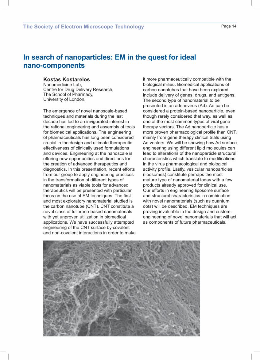
The emergence of novel nanoscale-based techniques and materials during the last decade has led to an invigorated interest in the rational engineering and assembly of tools for biomedical applications. The engineering of pharmaceuticals has long been considered crucial in the design and ultimate therapeutic effectiveness of clinically used formulations and devices. Engineering at the nanoscale is offering new opportunities and directions for the creation of advanced therapeutics and diagnostics. In this presentation, recent efforts from our group to apply engineering practices in the transformation of different types of nanomaterials as viable tools for advanced therapeutics will be presented with particular focus on the use of EM techniques. The first and most exploratory nanomaterial studied is the carbon nanotube (CNT). CNT constitute a novel class of fullerene-based nanomaterials with yet unproven utilization in biomedical applications. We have successfully attempted engineering of the CNT surface by covalent and non-covalent interactions in order to make
it more pharmaceutically compatible with the biological milieu. Biomedical applications of carbon nanotubes that have been explored include delivery of genes, drugs, and antigens. The second type of nanomaterial to be presented is an adenovirus (Ad). Ad can be considered a protein-based nanoparticle, even though rarely considered that way, as well as one of the most common types of viral gene therapy vectors. The Ad nanoparticle has a more proven pharmacological profile than CNT, mainly from gene therapy clinical trials using Ad vectors. We will be showing how Ad surface engineering using different lipid molecules can lead to alterations of the nanoparticle structural characteristics which translate to modifications in the virus pharmacological and biological activity profile. Lastly, vesicular nanoparticles (liposomes) constitute perhaps the most mature type of nanomaterial today with a few products already approved for clinical use. Our efforts in engineering liposome surface and structural characteristics in combination with novel nanomaterials (such as quantum dots) will be described. EM techniques are proving invaluable in the design and custom-engineering of novel nanomaterials that will act as components of future pharmaceuticals.
The Society of Electron Microscope TechnologyPage 15
Malaria and the red blood cell: mysteries of entrances and exits
Lawrie BannisterDepartment of Anatomy and Human Sciences, Guy’s Campus, King’s College London.
Malaria is one of the world’s most serious infectious diseases, with a major impact on life expectancy and general health of many millions globally as well as on the economies of many developing nations. It is caused by single celled organisms of the genus Plasmodium, transmitted by mosquito bite via the peripheral circulation to the liver,thence to red blood cells, and finally back to feeding mosquitoes.As the parasites have developed resistance to many antimalarial drugs, and mosquitoes have become resistance to insecticides, malaria remains a huge global problem. The minute parasites are at the resolution limit of light microscopy, and therefore the electron microscope has been ivital to malaria biology research. Among many unsolved problems are exactly how the parasite invades red blood
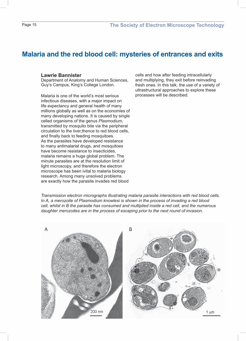
Transmission electron micrographs illustrating malaria parasite interactions with red blood cells. In A, a merozoite of Plasmodium knowlesi is shown in the process of invading a red blood cell, whilst in B the parasite has consumed and multiplied inside a red cell, and the numerous daughter merozoites are in the process of escaping prior to the next round of invasion.
cells and how after feeding intracellularly and multiplying, they exit before reinvading fresh ones. In this talk, the use of a variety of ultrastructural approaches to explore these processes will be described.


![Resolucion SUNAT Instructivo SEMT Grandes Compradores 1[1]](https://img.dokumen.tips/doc/110x75/557211aa497959fc0b8f51f1/resolucion-sunat-instructivo-semt-grandes-compradores-11.jpg)