Embed Size (px)
Citation preview
sEMG Signal and Hill Model based Continuous Prediction
for Hand Grasping Motion
Muye Pang*1
Shuxiang Guo*2,*3
Zhibin Song*2
and Songyuan Zhang*1
*1
Graduate School of Engineering *3
College of Automation *2 Dept. of Intelligent Mechanical Systems Eng’g, Kagawa University Harbin Engineering University
Hayashi-cho, Takamatsu, 761-0396, Japan 145 Nantong Street, Harbin, Heilongjiang, China
{s12d505,s11g528}@stmail.eng.kagawa-u.ac.jp {guo,song}@eng.kagawa-u.ac.jp
Abstract - This paper is aimed at the continuous hand
grasping motion prediction during all fingers flexion and
extension. Only sEMG signals recorded from flexor digitorum
superficialis and extensor digitorum of forearm are used to
predict the flexion and extension motion. In order to find the
relation between sEMG signals and hand grasping motion, a Hill
model is used to represent the force value of the muscles. Some
assumptions are also made for simplicity in calculating the
association. A simple and efficient motion recording system using
flex sensor, Mtx sensor and a glove is designed for the purpose of
recording fingers motion. The motions are voluntary finger
flexion and extension with no load. Acceptable results are
achieved. The purpose of this paper is to provide a method for
continuous hand grasping motion prediction based on sEMG
signals. Although some assumptions are made to simplify the
problem and indeed these assumptions brought prediction errors
in the experiment, the method shows itself an alternative way to
use sEMG signals for hand motion prediction.
Index Terms – Surface Electromyography (sEMG) signal, Hill
model, hand grasping motion, continuous prediction
I. INTRODUCTION
With the development of robot technology, the biorobots
are compelling during recent years [1]-[4]. After the discovery
of electromyographic (EMG) signal, it has been widely used
in biorobots control and some other fields such as
rehabilitation, human body motion detection and athlete
training. The nature feature of the EMG signal, which directly
represent for the activation potentials of skeleton muscle,
makes it very convenient and direct in representing status of
muscles. Many researchers are working on EMG-based
devices design, such as the EMG-driven exoskeleton hand
robotic training device, which is mounted on patient’s
impaired hand and detected sEMG signals are used as the
driven signals [5]; the EMG-driven musculoskeletal model of
the ankle, which combines the Hill-model and sEMG
signals to estimate the forces of the triceps surae muscle
and Achilles tendon [6]. Also some others are using EMG
signal to predict human body motions, such as Artemiadis et al
[7] used a switching regime model to predict the motion of
arm based on 11 channels of EMG signals.
There are mainly two kinds of EMG signals measurement:
the surface EMG signals detection method using non-invasive
surface electrodes and the invasive EMG signals detection
method using fine wire electrodes. The surface EMG signals
measurement is widely used in researches because of its non-
invasive characteristic. But the low signal to noise ratio (SNR)
is an unavoidable problem, at least under present
electromyography technology in this measurement. Many
factors result in this issue, such as the condition of the skin,
the thickness of the fat tissue under the skin, the crosstalk
between muscles, and the different recruitment level in motor
unit action potentials. Because of the low SNR and crosstalk
in muscles, many researchers are focusing on the method to
increase the recognition accuracy, such as Tang et al [8] used
two developed methods to extract features of EMG and
designed a novel cascaded-structure classifier to achieve hand
pattern recognition. Although some method can achieve high
recognition accuracy for special motions, it is far beyond
enough in whole human motion, especially in hand motion
detection. The crosstalk between EMG signals and the
coordination of small muscles on the forearm make the
recognition extremely complicated. Also many of these
studies separate motions into different patterns and deal with
problem as pattern recognition, thus the continuous prediction
is also a main issue.
In this paper, sEMG and Hill-model based continuous
hand grasping motion prediction method is represented. sEMG
signals recorded from flexor digitorum superficialis and
extensor digitorum of forearm are used to predict the fingers
flexion and extension motions. The Hill model is used to
represent the force value of the muscles. Three assumptions
are made to simplify the situation. Acceptable results are
achieved after experiments on three subjects.
II. DESIGN OF CONTINUOUS PREDICTION METHOD FOR HAND
GRASPING MOTION
In this paper, the grasping movement is flexing and
extending of all fingers at the same time. EMG signals are
recorded from flexor digitorum superficialis and extensor
digitorum of forearm. Then the Hill model is used to calculate
the force value of muscles according to activation level of
EMG. Under three assumptions, a prediction is calculated
according to the force value of muscles.
A. EMG Recorded from Forearm
The crosstalk and coordination between the small muscles
of forearm is one of the reasons making the prediction of hand
motion extremely complicated. It is very ideal to find each
muscle involved in the flexion and extension for each digit
separately but from the point of view in anatomy, there may
not be a separate muscle or a group of muscles affording the

a). Electrode placement on front side b). Electrode placement on back side
of forearm of forearm
Figure 1. Electrode placement on forearm
flexion or extension force for each finger because of the
phenomenon of fingers coordination. It is very difficult to
move each finger freely without involving other fingers. Also
in experiment, it is not easy or maybe impossible to find a
proper placement to detect EMG signals presented single
finger movement.
In this paper, flexor digitorum superficialis and extensor
digitorum of forearm are selected to detect EMG signals from.
The placement of the electrodes around the forearm is shown
in Fig.1, where electrodes 1, 2 and 5 are placed on three
different surfaces upon flexor digitorum superficialis belly,
which takes charge with flexion of middle phalanges at
proximal interphalangeal joints of medial four digits. After
compared the effects of the EMG signals, electrode 2 is
selected for experiment. And electrode 4 is placed on the
surface of extensor digitorum, which takes charge with
extension of medial four digits at metacarpophalangeal joints.
Electrode 3 isn’t included in this time.
Although the crosstalk between these muscles can be
easily observed from the experimental results, the
performance of these muscles is of coincidence of defined
finger motions.
B. Hill Model
The Hill model simulating the biomechanics of muscle is
one of the conventional and classical models to predict the
muscle behaviour. The model (Fig.2) contains a pair of
elements arranged in series: the passive serial element (SE)
and the active contractile element (CE); and a passive element
(PE) arranged in parallel to the previous two. The Hill model
calculates the force of muscle using the activation level,
muscle length and shortening velocity. Some researcher
calculated the muscle length and moment arm given the joint
angles and limb kinematics. The equations [9]-[10] used to
calculate the force are shown as follows:
[
] [
(
) ] (1)
{
( (
)
)
( (
))
( )
(2)
(3)
( ) ( ) ( ) (4)
Fig. 2 Schematic of Hill-model with CE, SE and PE elements
where is the change in length of the element with respect
to the slack length, is a shape parameter, is the
maximum force exerted by the element for the maximum
change in length , and is the passive force
generated by the PE or the SE element depending on the set of
parameters used. is the total force exerted by the muscle.
( ) is the activation level of a muscle.
The parameters such as length of muscle fiber and fiber
length change velocity are difficult to record through
conventional measurement. Some researchers used indirect
method such as change in joint angles to calculate the change
in muscle fiber length, or used the data from other project to
represent for a mean value. To simplify this situation, three
assumptions are made in the following section.
C. Assumptions for Simplicity
In this paper, subjects performed the experiments with
their hand holding nothing, in other words, there is no extra
force exerted by the muscle to conquer the external load.
Thus, only voluntary isotonic contraction is considered in this
time. So in first assumption, the force exerted by muscle is
considered to be only coordinated with the joint angle of
fingers. The Hill model calculates force exerted from muscle
mainly by muscle activation level, muscle length and
shortening velocity. Because of considering isotonic
contraction,
or
is defined as proportional to the
activation level of muscle in second assumption. And the third
assumption is that the velocity of CE element is considered to
be zero, because experimentation is performed at a very slow
speed, subjects performed the entire process in about 10
seconds.
D. Motion Detection Device
Two flex sensors (Spectrasymbol com.) are mounted on
proximal interpharangeal joint (PIP) and metacarpophalangeal
joint (MCP) of a rubber glove, as shown in Fig.3. The electric
resistance of the flex sensor is changing with the shape
bending. A simple OP circus is used to record the data of the
sensor. A 3DoF inertial orientation tracker (MTx sensor,
Xsens Technologies B.V.) is applied to calculate the
association between the change of resistance and the degree of
the shape bending. In the experiment, the MTx sensor is
sticked on the flex sensor and both the data from the MTx

sensor and flex sensor are recorded through data acquisition
system (USB4716, Advantech Co. Ltd.) at the same time,
while operator bended the flex sensor from to , as
shown in Fig.4. Then a curve fitting toolbox (MathWorks Co.
USA) is used to generate a function ( ), where is the
change of resistance and output y is the degree of the joint
mounted with the flex sensor.
E. Experimental Protocol
Three healthy volunteers aged from 22-26 years, all male,
one left-handed and two right-handed, participated in the
experiment. Before electrodes are placed that are aligned
parallel to muscle fibers over the belly of the muscle and
positioned following recommendations, the skin is shaved and
cleaned with alcohol in order to reduce skin impedance. The
subjects are asked to wear on the glove and keep their forearm
relaxed vertically. The EMG signals from this position are
observed in order to guarantee no extra movement interfering
the hand motion. Because the motion velocity must be low
enough, the subjects are asked to practice the flexion and
extension movement several times before experiment. The
entire process took about 10 seconds and subjects had a 30
seconds rest after one process.
Fig. 3 Experimental setup detecting motions of fingers. a) rubber glove with
flex sensor; b) flex sensor attached on MTx sensor; c) AD board
Fig. 4 Experiment to calculate function between changes in voltage and joint angle
a). sEMG Filter box b). Electrode
Figure 5. sEMG recording devices
F. EMG Recording Setup
sEMG signals are collected using bipolar surface
electrodes 12mm long, located 18mm apart, as shown in Fig.5.
The sampling rate is 3000Hz with differentially amplified
(gain 1000) and common mode rejection (104dB). This
sampling rate is sufficient because the most frequency power
of EMG signals is between 20 to 400Hz. Sampling data are
pre-processed with a commercial filter box (Oisaka Electronic
Device Ltd. Japan.) before being recorded in the control
program through an AD board.
III. EXPERIMENTAL RESULTS
A. Recording of Finger Motions
The sampling rate of recording the data from flex sensor
and MTx sensor is 1KHz. The data from flex sensor is voltage
and from MTx sensor is pitch degree. The experimental result
is shown in Fig.6
Fig. 6 Flex sensor compared with MTx sensor
Fig. 7 Curve fitting process of flex sensor
a. rubber glove
b. MTx sensor
c. AD board
Voltage of flex sensor / V
MTx sensor
Flex sensor
Flex sensor

The quadratic function is implemented to represent the
coordination between voltage and degree in curve fitting
method. The function is written in Eq.5, and the fitting process
is shown in Fig.7, where the dots represent the original data,
the green line is calculated by smooth method, and the red line
is the fitting curve. Coefficient of determination is 0.97, which
is sufficient for finger motion record.
( ) (5)
B. EMG Signals Processing Results
The EMG signals are filtered with band passed (20-
450Hz), 5 order Butterworth filter, full wave rectified.
Following shows one experiment result from one subject,
where the above two are the EMG signals from extensor
digitorum and flexor digitorum superficialis separately, and
the two below are degree changes of MCP and PIP joints. It
can be indicated that the flexor digitorum superficialis
activated in flexion movement, and the activation level
increased with MCP joint degree increasing. The extensor
digitorum activated in two periods: first at the extension
period (from 0 to 1000 and 4000 to 5000 in Fig. 8), and
second at the flexion period (from 1500 to 3000 in Fig. 8). The
movement of finger is from the entire extension to entire
flexion and back to the entire extension. Thus the extensor
digitorum activated at the beginning and at the end of the
movement. Although it is natural that the extensor digitorum
activated at the finger extension period, activation at flexion
period is considered as coordination at this time and the
contribution of this part is eliminated.
The processed EMG signals are calculated using root
mean square (RMS) via a 50ms window. The value of RMS is
then used as input of (4) to get the activation level of a muscle
and the activation level is put into (1) and (2) to calculate the
force of the muscle, which is considered proportional to the
degree of finger joint. Fig.9 shows the result of calculation
using Hill model, and the PIP and MCP data are resampled via
a 50ms time window. The RMS value of extensor digitorum is
ignored when the activation level of flexor digitorum
superficialis is exceeded 10% of maximum because this part is
considered as coordination at this time.
Fig.8 Recorded EMG and flex sensor data from one subject in one experiment
Fig. 9 Predicted forces compared with angles of joints
The non-linear relationship between the predicted force
and the recorded angles of joints can be indicated from Fig.9.
At the beginning part ( from 0 to 15 ) and the end part ( from
90 to 110 ) the value of force level keeps upon a threshold
indicating an un-movement motion, and the muscle status at
these two parts is more like isometric contraction. A curve
fitting method is implemented to find a preliminary function to
predict the motion of finger via force value calculated by Hill
model. The MCP and PIP joints are predicted separately. The
functions are described as follows:
(6)
(7)
( )
(8)
( )
(9)
Where and
are used to predict the flexion and
extension motion of MCP joint and and
are used for
PIP joint. Especially, and
are used when flexor
digitorum superficialis is activated and and
are used
when extensor digitorum is activated. Coefficients of
determination for and
are 0.61 and 0.65 separately.
And towards and
, they are 0.80 and 0.75 separately.
Fig.10 and Fig.11 show the prediction results compared with
the records from flex sensors where the solid blue lines are the
prediction results and dot lines are records from flex sensors.
Fig. 10 EMG based MCP joint motion prediction
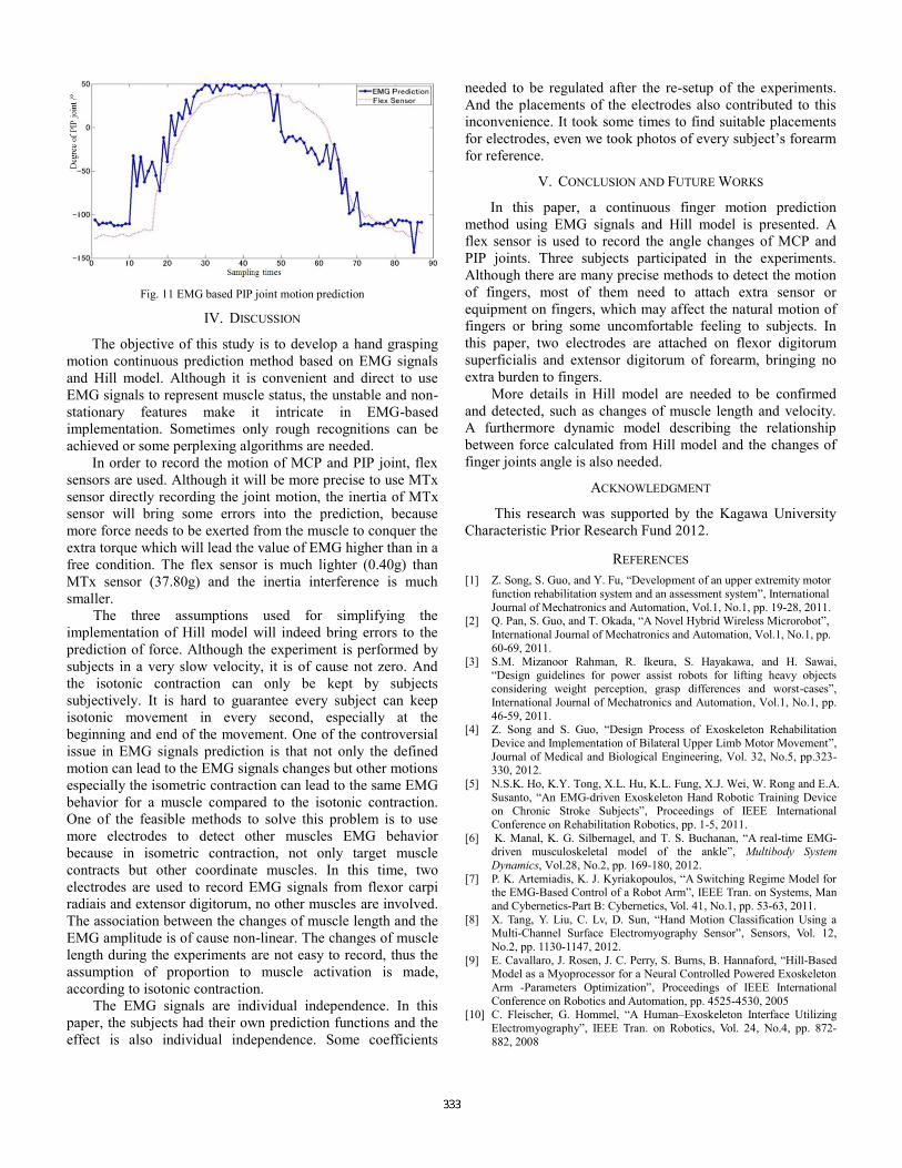
Fig. 11 EMG based PIP joint motion prediction
IV. DISCUSSION
The objective of this study is to develop a hand grasping
motion continuous prediction method based on EMG signals
and Hill model. Although it is convenient and direct to use
EMG signals to represent muscle status, the unstable and non-
stationary features make it intricate in EMG-based
implementation. Sometimes only rough recognitions can be
achieved or some perplexing algorithms are needed.
In order to record the motion of MCP and PIP joint, flex
sensors are used. Although it will be more precise to use MTx
sensor directly recording the joint motion, the inertia of MTx
sensor will bring some errors into the prediction, because
more force needs to be exerted from the muscle to conquer the
extra torque which will lead the value of EMG higher than in a
free condition. The flex sensor is much lighter (0.40g) than
MTx sensor (37.80g) and the inertia interference is much
smaller.
The three assumptions used for simplifying the
implementation of Hill model will indeed bring errors to the
prediction of force. Although the experiment is performed by
subjects in a very slow velocity, it is of cause not zero. And
the isotonic contraction can only be kept by subjects
subjectively. It is hard to guarantee every subject can keep
isotonic movement in every second, especially at the
beginning and end of the movement. One of the controversial
issue in EMG signals prediction is that not only the defined
motion can lead to the EMG signals changes but other motions
especially the isometric contraction can lead to the same EMG
behavior for a muscle compared to the isotonic contraction.
One of the feasible methods to solve this problem is to use
more electrodes to detect other muscles EMG behavior
because in isometric contraction, not only target muscle
contracts but other coordinate muscles. In this time, two
electrodes are used to record EMG signals from flexor carpi
radiais and extensor digitorum, no other muscles are involved.
The association between the changes of muscle length and the
EMG amplitude is of cause non-linear. The changes of muscle
length during the experiments are not easy to record, thus the
assumption of proportion to muscle activation is made,
according to isotonic contraction.
The EMG signals are individual independence. In this
paper, the subjects had their own prediction functions and the
effect is also individual independence. Some coefficients
needed to be regulated after the re-setup of the experiments.
And the placements of the electrodes also contributed to this
inconvenience. It took some times to find suitable placements
for electrodes, even we took photos of every subject’s forearm
for reference.
V. CONCLUSION AND FUTURE WORKS
In this paper, a continuous finger motion prediction
method using EMG signals and Hill model is presented. A
flex sensor is used to record the angle changes of MCP and
PIP joints. Three subjects participated in the experiments.
Although there are many precise methods to detect the motion
of fingers, most of them need to attach extra sensor or
equipment on fingers, which may affect the natural motion of
fingers or bring some uncomfortable feeling to subjects. In
this paper, two electrodes are attached on flexor digitorum
superficialis and extensor digitorum of forearm, bringing no
extra burden to fingers.
More details in Hill model are needed to be confirmed
and detected, such as changes of muscle length and velocity.
A furthermore dynamic model describing the relationship
between force calculated from Hill model and the changes of
finger joints angle is also needed.
ACKNOWLEDGMENT
This research was supported by the Kagawa University
Characteristic Prior Research Fund 2012.
REFERENCES
[1] Z. Song, S. Guo, and Y. Fu, “Development of an upper extremity motor
function rehabilitation system and an assessment system”, International
Journal of Mechatronics and Automation, Vol.1, No.1, pp. 19-28, 2011. [2] Q. Pan, S. Guo, and T. Okada, “A Novel Hybrid Wireless Microrobot”,
International Journal of Mechatronics and Automation, Vol.1, No.1, pp.
60-69, 2011. [3] S.M. Mizanoor Rahman, R. Ikeura, S. Hayakawa, and H. Sawai,
“Design guidelines for power assist robots for lifting heavy objects considering weight perception, grasp differences and worst-cases”,
International Journal of Mechatronics and Automation, Vol.1, No.1, pp.
46-59, 2011. [4] Z. Song and S. Guo, “Design Process of Exoskeleton Rehabilitation
Device and Implementation of Bilateral Upper Limb Motor Movement”,
Journal of Medical and Biological Engineering, Vol. 32, No.5, pp.323-330, 2012.
[5] N.S.K. Ho, K.Y. Tong, X.L. Hu, K.L. Fung, X.J. Wei, W. Rong and E.A.
Susanto, “An EMG-driven Exoskeleton Hand Robotic Training Device on Chronic Stroke Subjects”, Proceedings of IEEE International
Conference on Rehabilitation Robotics, pp. 1-5, 2011.
[6] K. Manal, K. G. Silbernagel, and T. S. Buchanan, “A real-time EMG-driven musculoskeletal model of the ankle”, Multibody System
Dynamics, Vol.28, No.2, pp. 169-180, 2012.
[7] P. K. Artemiadis, K. J. Kyriakopoulos, “A Switching Regime Model for the EMG-Based Control of a Robot Arm”, IEEE Tran. on Systems, Man
and Cybernetics-Part B: Cybernetics, Vol. 41, No.1, pp. 53-63, 2011.
[8] X. Tang, Y. Liu, C. Lv, D. Sun, “Hand Motion Classification Using a Multi-Channel Surface Electromyography Sensor”, Sensors, Vol. 12,
No.2, pp. 1130-1147, 2012.
[9] E. Cavallaro, J. Rosen, J. C. Perry, S. Burns, B. Hannaford, “Hill-Based Model as a Myoprocessor for a Neural Controlled Powered Exoskeleton
Arm -Parameters Optimization”, Proceedings of IEEE International
Conference on Robotics and Automation, pp. 4525-4530, 2005 [10] C. Fleischer, G. Hommel, “A Human–Exoskeleton Interface Utilizing
Electromyography”, IEEE Tran. on Robotics, Vol. 24, No.4, pp. 872-
882, 2008