Embed Size (px)
Citation preview

Glycobiology vol. 6 no. 3 pp. 367-376, 1996
Selective ganglioside desialylation in the plasma membrane ofhuman neuroblastoma cells*
Jiirgen Kopitz, Carolina von Reitzenstein, Karin Sinzand Michael Cantz1
Institute of Pathochemistry and General Neurochemistry, Universityof Heidelberg, Im Neuenheimer Feld 220, D-69120 Heidelberg,Germany'To whom correspondence should be addressed•Dedicated to Professor Glinter Quadbeck on the occasion of his 80thbirthday
Gangliosides of the plasma membrane are important mod-ulators of cellular functions. Previous work from our labo-ratory had suggested that a plasma membrane sialidase wasinvolved in growth control and differentiation in culturedhuman neuroblastoma cells (SK-N-MC), but its substrateshad remained obscure. We now performed sialidase speci-ficity studies in subcellular fractions and found gangliosideGM3 desialylating activity in presence of Triton X-100 tobe associated with the plasma membrane, but absent inlysosomes. This Triton-activated plasma membrane en-zyme desialylated also gangliosides GDla, GDlb, andGTlb, thereby forming GM1; cleavage of GM1 and GM2,however, was not observed. Sialidase activity towards theglycoprotein fetuin with modified C-7 sialic acids and to-wards 4-methylumbeUiferyl neuraminate was solely foundin lysosomal, but not in plasma membrane fractions.The role of the plasma membrane sialidase in gangliosidedesialylation of living cells was examined by following thefate of [JHlgalactose-labelled individual gangliosides inpulse-chase experiments in absence and presence of theextracellular sialidase inhibitor 2-deoxy-2,3-dehydro-N-acetylneuraminic acid. When the plasma membrane siali-dase was inhibited, radioactivity of all gangliosides chasedat the same rate. In the absence of inhibitor, GM3, GDla,GDlb, GD2, GD3 and GTlb were degraded at a consider-ably faster rate in confluent cultures, whereas the GM1-pool seemed to be filled by the desialylation of highergangliosides. The results thus suggest that the plasma mem-brane sialidase causes selective ganglioside desialylation,and that such surface glycolipid modification triggersgrowth control and differentiation in human neuro-blastoma cells.
Key words: ganglioside/neuroblastoma cells/plasma mem-brane/sialidase
Introduction
Gangliosides are sialylated glycosphingolipids that areubiquitous in the plasma membrane of vertebrate cells. Inneurons, they comprise approximately two thirds of theglycoconjugate sialic acid content, thereby making an im-portant contribution to the glycocalix surrounding the neu-
ral membrane (Yu and Saito, 1989; Ledeen, 1989). Gangli-osides are involved in various processes of the nervoussystem, including cell proliferation, cell differentiation,neural repair, cell-cell recognition and cell adhesion (Led-een, 1989; Zeller and Marchase, 1992; Hakomori, 1993;Tettamanti and Riboni, 1993).
Specific regulatory effects of gangliosides on cell func-tions require tight control of their amounts in the plasmamembrane. Ganglioside synthesis occurs in the Golgi, andthe level of specific glycosyltransferases determines theganglioside pattern (van Echten and Sandhoff, 1993; Ruanand Lloyd, 1992). On the other hand, ganglioside degrada-tion also appears to influence the ganglioside pattern ofthe plasma membrane (Schengrund and Repman, 1982;Riboni etal., 1990). Enzymes of ganglioside catabolism arelocated in the lysosomes with the exception of sialidasewhich occurs in both lysosomal and nonlysosomal compart-ments (e.g., Miyagi et al, 1990a and 1990b; Pitto et al,1992). In cultured fibroblasts and tissues from patients withsialidosis there is a genetic deficiency of a lysosomal siali-dase that degrades sialyloligosaccharides, sialoglycopro-teins, and gangJiosides, whereas a plasma membrane gan-glioside sialidase is unaffected (Thomas and Beaudet,1995).
As already shown in earlier studies, ganglioside sialidaseactivity was found to be present at the surface of a variety ofcells and to be involved in, or modulated by, fundamentalprocesses such as proliferation, cell contact, or phase ofthe cell cycle. Thus, cell surface ganglioside sialidase activ-ity was clearly detected in transformed, but not in non-transformed, hamster cells (Schengrund et al, 1973). Celldensity-dependent changes of surface ganglioside sialidasewere studied in mouse fibroblasts, and the activity wasfound to be greatly enhanced at early stages of contact,but decreased when cultures were confluent (Yogeeswaranand Hakomori, 1975). In cultured murine neuroblastomaor cerebellar granule cells, on the other hand, gangliosidesialidase activity increased with cell density and was there-fore considered to be involved in cell differentiation(Schengrund and Repman, 1982; Riboni et al, 1990; Pittoet al, 1992).
In previous work we had shown that cultured humanneuroblastoma cells (SK-N-MC) contained two sialidaseactivities that degrade gangliosides: a plasma membrane-bound activity that was specifically activated by TritonX-100, was readily inactivated by preincubating intactcells in the presence of millimolar concentrations ofCu++, and whose specific activity increased strikinglyduring cell growth, and a lysosomal activity stimulatedby glycodeoxycholate that was protected from inactiva-tion upon exposure of the cells to Cu++ and showed noactivity increase during cell proliferation (Kopitz et al,1994). When the specific sialidase inhibitor 2-deoxy-2,3-
© Oxford University Press 367
Dow
nloaded from https://academ
ic.oup.com/glycob/article/6/3/367/629405 by guest on 17 January 2022
J.Kopitz et aL
8 10
Fraction
12 14 16 18
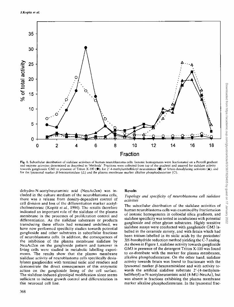
Fig. 1. Subcellular distribution of sialidase activities of human neuroblastoma cells. Isotonic homogenates were fractionated on a Percoll gradientand enzyme activities determined as described in 'Methods' Fractions were collected from top of the gradient and assayed for sialidase activitytowards ganglioside GM3 in presence of Triton X-100 (•) , for 2'-4-methylumbelliferyl neuraminate (•) or fetuin-desialylating activities (A), andfor the lysosomal marker /3-hexosaminidase (A) and the plasma membrane marker alkaline phosphodiesterase (O).
dehydro-N-acetylneuraminic acid (NeuAc2en) was in-cluded in the culture medium of the neuroblastoma cells,there was a release from density-dependent control ofcell division and loss of the differentiation marker acetyl-cholinesterase (Kopitz et ai, 1994). The results thereforeindicated an important role of the sialidase of the plasmamembrane in the processes of proliferation control anddifferentiation. As the sialidase substrates or productstransducing these effects had remained undefined, wehave now performed specificity studies towards potentialganglioside and other substrates in subcellular fractionsof neuroblastoma cells. In addition, the consequences ofthe inhibition of the plasma membrane sialidase byNeuAc2en on the ganglioside pattern and turnover inliving cells were studied in metabolic labelling experi-ments. The results show that the plasma membranesialidase activity of neuroblastoma cells specifically desia-lylates gangliosides with terminal sialic acid residues anddemonstrate the direct consequences of this enzymaticaction on the ganglioside lining of the cell surface.The sialidase-induced glycolipid modification alone seemssufficient to induce growth control and differentiation inthis neuronal cell line.
ResultsTopology and specificity of neuroblastoma cell sialidaseactivitiesThe subcellular distribution of the sialidase activities ofhuman neuroblastoma cells was examined by fractionationof isotonic homogenates in colloidal silica gradients, andsialidase specificity was tested in incubations with potentialganglioside and other glycan substrates. Highly sensitivesialidase assays were conducted with ganglioside GM3 la-belled in the ceramide moiety, and with fetuin which hadbeen tritium-labelled in its sialic acids by the periodate/3H-borohydride reduction method yielding the C-7 analog.As shown in Figure 1, sialidase activity towards gangliosideGM3 in presence of the detergent Triton X-100 was foundto co-distribute with the marker for plasma membranes,alkaline phosphodiesterase. On the other hand, sialidaseactivity towards fetuin was found to fractionate with thelysosomal marker /3-hexosaminidase and with activity to-wards the artificial sialidase substrate 2'-(4-methylum-belliferyl)-a-N-acetylneuraminic acid (4-MU-NeuAc), butwas absent in fractions exhibiting the plasma membranemarker alkaline phosphodiesterase. In the lysosomal frac-
368
Dow
nloaded from https://academ
ic.oup.com/glycob/article/6/3/367/629405 by guest on 17 January 2022

Ganglioside desialylalion in plasma membrane
Table I. Specific activities of sialidases and
Tnton-activated GM3 sialidaseAlkaline phosphodiesteraseNa+/K+-ATPaseAcetylcholinesteraseLeucine aminopeptidase5'-NucleotidaseGDC-activated GM3 Sialidase4-MU-NeuAc sialidase/9-N-AcetylhexosaminidaseGalactosyltransferaseSuccinate dehydrogenaseLactate dehydrogenaseDNA
marker enzymes in plasma membrane
Specific activity
A Homogenate
30 /xU/mg protein3.72 U/mg protein2.21 mU/mg protein0.224 mU/mg protein29.6 U/mg protein2.85 mU/mg protein91 /tU/mg protein295 /xU/mg protein28.22 mU/mg protein49 fiVlmg protem285 U/mg protein3325 U/mg protein64.6 fig/mg protein
preparation and homogenate of neuroblastoma cells
B Plasma membranes
487 /iU/mg protein64.4 U/mg protein44.1 mU/mg protein3.87 mU/mg protein487 U/mg protein46.1 mU/mg protein3.3 fiXI/mg protein15 /iU/mg protein21.54 mU/mg protein137 /xU/mg protein357 U/mg protein1069 U/mg protein8.31 figlmg protein
B/A ratio
16.217.32017316.416.20.040.050.762.81.250.320.13
GM1
GD13
GOibGTib
1
Fig. 2. Thin-layer chromatography of the products of gangliosideGTib degradation by the Triton-activated sialidase. Ganglioside GTlbwas incubated in presence of Triton X-100 and neuroblastoma cellhomogenate and the reaction products separated by thin-layerchromatography as desribed under 'Methods'. Bands were visualizedwith orcinol spray. Lane 1: Standards, 50nmol of each ganglioside;lane 2: incubation stopped immediately after mixing by freezing inliquid nitrogen; lane 3: incubation stopped after 8h; lane 4: gangliosidewas omitted from the reaction mixture; lane 5; homogenate fromCu++-pretreated cells was used; lane 6: lmM NeuAc2en was includedin the reaction mixture; lane 7: imM NeuAc2en was included buthomogenate protein and GTlb were omitted from the reactionmixture.
tions, sialidase activity towards ganglioside GM3 could bedetected in presence of the detergent glycodeoxycholate,but was rather unstable (results not shown).
The topology of the Triton X-100-activated gangliosidesialidase activity was further tested in a plasma membranepreparation purified by sucessive sucrose and Percoll den-sity gradient centrifugations. As shown in Table I, thespecific activity of the Triton-activated ganglioside GM3
sialidase of the plasma membrane preparation was in-creased over the homogenate by a factor of 16, a similarincrease as observed for the plasma membrane markersouabain-sensitive Na+/K+-ATPase, acetylcholinesterase,5'-nucleotidase, alkaline phosphodiesterase, and leucineaminopeptidase. The markers for microsomes and mito-chondria, UDP-galactosyltransferase and succinate dehy-drogenase, showed relative increases of 2.8 and 1.3, respec-tively. The specific activity ratio of the lysosomal marker/3-N-acetylhexosaminidase, on the other hand, was de-creased to a value of about 0.8, and those for the glycodeox-ycholate-activated ganglioside GM3 and the 4-MU-NeuAcsialidases were decreased even further, presumably dueto inactivation. The ratios of the cytosolic marker lactatedehydrogenase and the marker for nuclei, DNA, were alsomarkedly diminished.
In order to test the most abundant gangliosides of neuralcells as potential substrates of the plasma membrane siali-dase, homogenates of neuroblastoma cells were incubatedwith the gangliosides GM1, GM2, GM3, GDla, GDlb andGTlb in the presence of Triton X-100. Qualitative analysisof the reaction products was achieved by thin layer chroma-tography. An example is given in Figure 2, where ganglio-side GTlb was tested as substrate. GM1 appeared as theonly ganglioside product of the sialidase reaction, and nodisialoganglioside was detected (lane 3). A control incuba-tion without addition of the substrate proved that GM1 isindeed the reaction product of GTlb-desialylation and isnot derived from the homogenate (lane 4). Cu++ pretreat-ment of intact cells specifically inactivated enzymes on theexternal surface of the plasma membrane (Kopitz et ai,1994) and consequently no sialidase reaction was observed(lane 5). Likewise, in the presence of the sialidase inhibitorNeuAc2en, no reaction product was detected (lane 6).Applying this procedure to the other gangliosides revealedthat also GDla and GDlb were desialylated by the plasmamembrane sialidase, yielding GM1; gangliosides GM1 andGM2, on the other hand, were not degraded (data notshown).
Specific activity of the Triton-activated plasma mem-brane sialidase activity of neuroblastoma cells towards gan-gliosides GM1, GM2, GDla, GDlb and GTlb was quanti-tated by FPLC-analysis of the reaction products andcompared to ganglioside GM3-desialylation (Table II).
369
Dow
nloaded from https://academ
ic.oup.com/glycob/article/6/3/367/629405 by guest on 17 January 2022

J.Kopitz el al
Table IL Specificity towards gangliosides of the plasma membranesialidase in homogenates of neuroblastoma cells
Substrate
Ganglioside GM3Ganglioside GDlaGanglioside GDlbGanglioside GTlbGanglioside GM1Ganglioside GM2
Triton-activated sialidase activity(ftU/mg protein)
73.828.721.818.5<0.1< 0.1
Homogenates of neuroblastoma cells (0.65 mg protein) were incubatedwith 25 nmol ganglioside in the presence of 0.04% Triton X-100 for 8h. Reaction products were separated by FPLC ion exchangechromatography on a Mono Q column (HR 5/5) and quantitated withresorcinol.For gangliosides GM1 and GM2 radiometric assays with tritium-labelled gangliosides were used additionally, in order to achieve lowerdetection limits.
When the rate of desialylation of ganglioside GM3 was setto 100 per cent, the rates for gangliosides GDla, GDlb,and GTlb were 39,30, and 25 per cent, respectively; gangli-osides GM1 and GM2, on the other hand, were not at-tacked at all. Degradation of gangliosides GM1 and GM2was not observed either when very sensitive radiometricassays with a detection limit of about 0.1/u.U/mg proteinwere applied.
Taken together, the results show that human neuro-blastoma cells contain at least two different sialidase activi-ties. One is associated with the plasma membrane and isactive on gangliosides GM3, GDla, GDlb and GTlb, butinactive towards gangliosides GM1, GM2, the artificial sub-strate 4-MU-NeuAc and the glycoprotein fetuin with modi-fied C-7 sialic acids. The other one is associated with lyso-somes, and it desialylates 4-MU-NeuAc, as well as fetuin.
Pattern and catabolism of metabolically labelledgangliosides of neuroblastoma cellsTo find out which gangliosides are desialylated by theplasma membrane sialidase in living neuroblastoma cells,cultures were grown in the presence of PH]galactose,chased with unlabelled medium, and the ganglioside pat-tern during chase compared to cultures where the plasmamembrane sialidase was inhibited by NeuAc2en. For anal-ysis, the gangliosides were extracted from the cells andtheir labelling pattern determined by HPLC on Lichro-sorb-NH2. Figure 3 shows the ganglioside pattern afterseven days of labelling, which consisted of the major spe-cies GM3, GM1, GDla and GM2, whereas GDlb, GTlb,GD2 and GD3 were minor components.
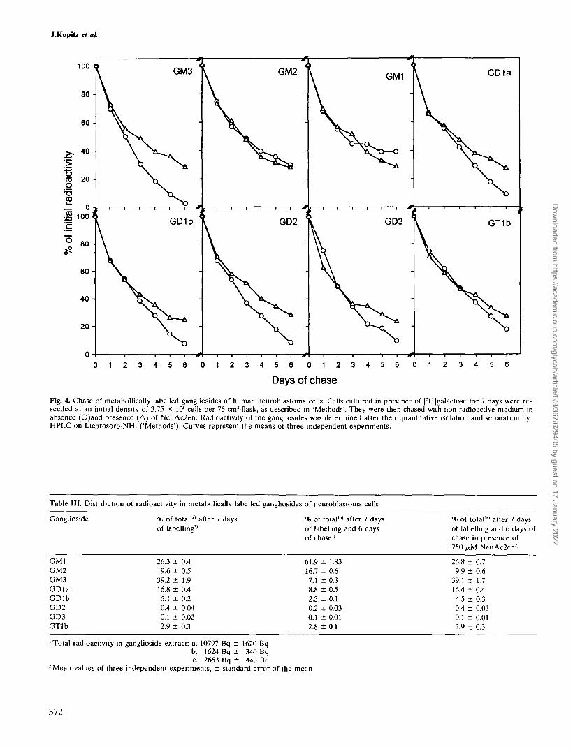
For the determination of ganglioside desialylation dur-ing growth of neuroblastoma cells, metabolically labelledcells were trypsinized, seeded at low density in cultureflasks and chased for 6 days with unlabelled medium inabsence or presence of 250/AM NeuAc2en. Because of thenegative charge on NeuAc2en, uptake of the inhibitor bythe cells should be low ( Hirschberg et al, 1976) and itseffect directed predominantly towards the plasma mem-brane sialidase. Determination of the radioactivity of eachof the gangliosides at different points of the chase allowedan estimate of their catabolism. In the absence of NeuA-
c2en the decrease in ganglioside-associated radioactivityshould be a function of all cellular ganglioside sialidases,whereas in the presence of the inhibitor, it should mainlydepend on lysosomal degradation. The difference betweenthe kinetics in presence or absence of the inhibitor shouldthus be an approximate measure of the activity of theplasma membrane-bound sialidase. As shown in Figure4, radioactivity in all gangliosides except GM1 and GM2decreased faster in the absence than in the presence ofNeuAc2en. The difference was greatest with gangliosideGM3, again indicating that it seems the preferred substrateof the plasma membrane sialidase, but was also markedwith the other gangliosides having terminal sialic acid resi-dues. The lack of an effect of the inhibitor on the degrada-tion of gangliosides GM1 and GM2 was not unexpectedin view of the fact that these were no substrates of theplasma membrane sialidase in the above-mentioned speci-ficity studies. The effects of the plasma membrane sialidaseinhibition were not detectable during the initial days ofchase but only appeared with increasing cell density. Inthe presence of the sialidase inhibitor, the degradativeprofiles of all gangliosides appeared virtually identical witha half-life of about 2.5 days (Figure 4), indicating a commonmechanism of membrane internalization followed by lyso-somal degradation.
When the relative amounts of radioactively labelled gan-gliosides at the beginning and at the end of the chase werecompared, there was an important change of the labellingpattern in the absence of the sialidase inhibitor. As shownin Table III, the proportion of GM1 more than doubled,whereas that of GM3 decreased to about one fifth and thatof GDla, GDlb and GD2 was also found to be diminished.There was a relative increase of GM2 that is attributedto higher desialylation rates of other gangliosides due toplasma membrane sialidase action. This effect also contri-butes, besides the filling of the GM1 pool by degradationof higher gangliosides, to the relative increase in GM1label. In the presence of the inhibitor, on the other hand,the pattern of labelled gangliosides essentially remainedunchanged, indicating their common and non-selective en-docytosis and catabolism in lysosomes.
Discussion
In a previous investigation, evidence had been presentedthat in human neuroblastoma cells a plasma membrane-bound sialidase is involved in the processes of proliferationcontrol and differentiation, but the identity of the sialidasesubstrates had remained unclear (Kopitz et al., 1994). Thepresent study was therefore aimed at elucidating the sub-strate specificity of the enzyme as a key to an understandingof its function, and to clearly differentiate it from othercellular sialidases.
In subcellular fractionation studies using colloidal silicagradients it could now be shown that human neuro-blastoma cells contain two sialidase activities: one that co-distributed with the marker for plasma membrane anddesialylated ganglioside GM3 in presence of Triton X-100,and another rather labile one that was associated withlysosomes and degraded the glycoprotein fetuin and thesynthetic compound 4-MU-NeuAc. Further evidence forthe plasma membrane localization of the Triton-activatedganglioside sialidase was obtained by comparing its specific
370
Dow
nloaded from https://academ
ic.oup.com/glycob/article/6/3/367/629405 by guest on 17 January 2022

Ganglioside desialylation in plasma membrane
0 20 40
Fraction
60 80
Fig. 3. Pattern of metabollically labelled gangliosides of human neuroblastoma cells. Cells were grown in the presence of fHJgalactose for 7 daysand gangliosides extracted quantitatively as described in 'Methods'. One ml of the extract containing 5.5 x 10⁵ d.p.m. was separated on aLichrosorb-NH2 column, 250 x 4mm I.D., using an acetonitrile-phosphate buffer system (Gazzoti et aL, 1985). Fractions of lml were collected andcounted for radioactivity. The column was calibrated with authentic ganglioside standards that were detected with resorcinol.
activities in the homogenate and a plasma membrane frac-tion prepared by density gradient centrifugation. The spe-cific activity of the ganglioside sialidase was increased bya factor of 16 in the plasma membrane fraction, similar tothe increases observed for five plasma membrane markerenzymes. The specific activities of the lysosomal markerand of the 4-MU-NeuAc and glycodeoxycholate-activatedganglioside GM3 sialidase activities, on the other hand,were found to be decreased in the plasma membrane prep-aration. The results thus corroborated our earlier findingsin cultured human fibroblasts and human neuroblastomacells of a Triton-activated ganglioside sialidase that wasselectively inactivated upon incubation of intact cells inthe presence of millimolar concentrations of Cu2+ and wastherefore considered to reside on the outer leaflet of theplasma membrane, and a lysosomal sialidase that degraded4-MU-NeuAc and ganglioside substrates and was pro-tected from Cu2+ in the above inactivation assay (Schnei-der-Jakob and Cantz, 1991; Kopitz etal., 1994). The presentsubcellular fractionation experiment also showed that siali-dase activity towards the synthetic substrate 4-MU-NeuAcand the glycoprotein fetuin that co-distributed with thelysosomal marker was absent in the plasma membranefractions. As the sensitivity of the fetuin sialidase assay wascomparable to that using ganglioside GM3 as substrate,the results could either mean that the plasma membranesialidase is specific only for ganglioside, but not for the
sialoglycoprotein, or that the modification of the sialic acidresidues produced by the tritium-labelling procedurecaused the glycoprotein not to be recognized as a substrateanymore. The latter possibility seems unlikely, however,as the Triton-activated ganglioside sialidase activity thathad been purified 1300-fold from human brain tissue failedto desialylate fetuin with unmodified sialic acid residues(unpublished observation). As fetuin contains both a2-3-and a2-6-linked sialic acids (in a 2 to 1 ratio; Baenzigerand Fiete, 1979), it also appears unlikely that activity ofthe plasma membrane sialidase towards the glycoproteinwould have been missed due to restricted linkage specific-ity. When the activity of the plasma membrane sialidasewas tested towards potential ganglioside substrates otherthan GM3, GDla, GDlb and GTlb were found to beefficiently desialylated to yield GM1. Gangliosides GM1and GM2, however, were not attacked at all. Taken to-gether, the results suggest that the Triton X-100-activatedsialidase activity of the plasma membrane cleaves fromgangliosides sialic acid residues that are in a terminal butnot in a branching position. On the other hand, the plasmamembrane sialidase appears to be inactive towards termi-nal sialic acid residues in the sialoglycoprotein fetuin andtowards 4-MU-NeuAc. A somewhat peculiar finding foran enzyme directed towards the extracellular space is itsacidic pH optimum of about 4.5. Although there is at pres-ent no direct explanation it must be borne in mind that
371
Dow
nloaded from https://academ
ic.oup.com/glycob/article/6/3/367/629405 by guest on 17 January 2022
J.Kopilz el al
100
80
60
radi
oact
iv
3
O
initi
al o o
c
° 80-
6 0 -
40 •
20 -
0 -
t GM3 '
\
V
t GD1b
\
x!
X{ GM2
V•
( GD2
V\ S . •
J
4
\
-
GD3
V\v^ -
\ GD1a
\
\
I GT1b
V
0 1 2 3 4 5 6 0 1 2 3 4 5 6 0 1 2 3 4 5 6 0 1 2 3 4 5 6
Days of chase
Fig. 4. Chase of metabollically labelled gangliosides of human neuroblastoma cells. Cells cultured in presence of [3H]galactose for 7 days were re-seeded at an initial density of 3.75 X 10⁶ cells per 75 cm2-f1ask, as described in 'Methods'. They were then chased with non-radioactive medium inabsence (O)and presence (A) of NeuAc2en. Radioactivity of the gangliosides was determined after their quantitative isolation and separation byHPLC on Lichrosorb-NH2 ('Methods'). Curves represent the means of three independent experiments.
Table III. Distnbution of radioactivity in metabolically labelled gangliosides of neuroblastoma cells
Ganglioside % of total"' after 7 daysof labelling2'
% of total"" after 7 daysof labelling and 6 daysof chase2'
% of total1'1 after 7 daysof labelling and 6 days ofchase in presence of250 nM NeuAc2en2)
GMIGM2GM3GDlaGDlbGD2GD3GTlb
26.3 ± 0.49.6 ± 0.5
39.2 ± 1.916.8 ± 0.45.1 ± 0.20.4 ± 0 040.1 ± 0.022.9 ± 0.3
61.916.77.18.82.30.20.12.8
: 1.83: 0.6: 0.3: 0.5: 0.1: 0.03: 0.01
01
26.8 ± 0.79.9 ± 0.6
39.1 ± 1.716.4 ± 0.44.5 ± 0.30.4 ± 0.030.1 ± 0.012.9 ± 0.3
"Total radioactivity in ganglioside extract: a, 10797 Bq ± 1620 Bqb. 1624 Bq ± 340 Bqc, 2653 Bq ± 443 Bq
2|Mean values of three independent experiments, ± standard error of the mean
372
Dow
nloaded from https://academ
ic.oup.com/glycob/article/6/3/367/629405 by guest on 17 January 2022
Ganglioside desialylation in plasma membrane
the assay conditions in a homogenate are of course vastlydifferent from the situation of the intact cell where, forinstance, the substrate is not presented as a ganglioside-detergent mixed micelle, but is embedded in the membraneof the same or an adjacent cell.
The membrane association and subcellular distributionof ganglioside sialidase has been studied by a number ofworkers in various cells and tissues. In bovine brain, gangli-oside sialidase was found to be highly enriched in isolatedsynaptosomes (Schengrund and Rosenberg, 1970). In mu-rine neuroblastoma cells (S20Y), ganglioside sialidase ac-tivity towards endogenous and exogenous substrates wasmembrane-bound and increased when the cultures becameconfluent (Schengrund and Repman, 1982). Cultured hu-man fibroblasts exhibited two ganglioside sialidases, anactivity that was plasma membrane-bound and was acti-vated by Triton X-100, and another rather labile activitythat was associated with lysosomes, needed activation bydeoxycholate and was genetically deficient in cells frompatients with sialidosis; the lysosomal sialidase, in contrastto the plasma membrane enzyme, exhibited broad specifitytowards sialyloligosaccharides, glycoproteins and ganglio-sides, and both sialidases could be distiguished by theirdifferent sensitivities to inhibitors (Lieser et ai, 1989;Zeigler et ai 1989; Schneider-Jakob and Cantz, 1991) .Miyagi et ai (1990a, 1990b) characterized intralysosomal,cytosolic, and two membrane sialidases present in rat tis-sues; of the membrane-bound ganglioside-hydrolyzing en-zymes, one was associated with brain synaptosomes,whereas the other had multi-substrate specificity (ganglio-sides, glycoproteins) and was detected in synaptosomal andlysosomal membrane fractions. A subcellular fractionationstudy of cultured rat cerebellar granule cells found evi-dence for the existence of possibly two plasma membrane-ganglioside sialidases, differing in pH-optima, in additionto a lysosomal sialidase (Pitto et ai, 1992). Our presentresults in cultured human neuroblastoma cells are in sup-port of a dual subcellular localization of ganglioside-de-grading sialidase activities, that is, in the plasma membraneand in lysosomes. Regarding the substrate specificity ofthe sialidases it has again to be remembered that the assayconditions are highly artificial, and that in vivo so-calledactivator proteins may replace the detergents that wererequired for the in-vitro activity determinations; indeed,it was shown that degradation of gangliosides by the lyso-somal sialidase requires such an activator (Fingerhut et ai,1992). Nevertheless, our present results in human neuro-blastoma cells are in general agreement with the publishedwork cited above and indicate that the plasma membranesialidase activity has a more restricted specificity towardsgangliosides, whereas the lysosomal activity is able to at-tack various classes of substrates such as glycoproteinsand gangliosides.
The question as to which gangliosides are actually desia-lylated by the sialidase in the plasma membrane in growingand differentiating neuroblastoma cells was studied by fol-lowing the fate of metabolically labelled gangliosides inpulse-chase experiments and in absence and presence ofthe sialidase inhibitor NeuAc2en. The combination of aprocedure for quantitative extraction of gangliosides fromcells with an HPLC-method for the analysis of complexganglioside mixtures allowed exact determination ofchanges in the labelling pattern. After a 7-day pulse in
presence of 3H-galactose, the cells were harvested, re-seeded under conditions of logarithmic growth and chasedwith unlabelled medium for 6 days. Specific activity of theplasma membrane sialidase increased about 15-fold duringthe logarithmic growth phase under these conditions (Kop-itz et ai, 1994). Inclusion of the sialidase inhibitor NeuA-c2en in the culture medium was used to specifically blockthe sialidase on the external surface of the plasma mem-brane. Although an inhibition of the lysosomal sialidaseunder the conditions employed cannot be rigorously ex-cluded, it should be minimal as the uptake of free sialicacid, and hence NeuAc2en, by cultured cells was reportedto be very inefficient (Hirschberg et ai, 1976). Any differ-ences in desialylation kinetics of labelled gangliosides inpresence and absence of the sialidase inhibitor shouldtherefore be mainly due to the action of the plasma mem-brane-bound sialidase. Indeed, extracellular NeuAc2enhad a marked effect on the decrease of the radioactivelabel in gangliosides GM3, GDla, GDlb, GD2, GD3 andGTlb. This effect was hardly detectable during the initialproliferative phase of the cells, but became prominent dur-ing logarithmic growth, simultaneously with confluencyand with the induction of plasma membrane sialidase activ-ity. Again, ganglioside GM3 was found to be the preferen-tial substrate, but also a considerable share of GDla,GDlb, GT1 b and of the minor gangliosides GD2 and GD3was desialylated in the plasma membrane. For gangliosideGM2, loss of radioactivity during chase was identical inpresence and absence of the inhibitor, confirming thatGM2 was not attacked by the plasma membrane enzyme.Radioactive label in ganglioside GM1 decreased faster inpresence than in absence of the inhibitor. Obviously, gan-glioside GM1 is not a substrate of the plasma membranesialidase, but the GMl-pool is filled by the desialylationof the higher gangliosides GDla, GDlb and GTlb, andthis 'filling reaction' is prevented by the inhibitor. In pres-ence of NeuAc2en, the rate of disappearance of radioactiv-ity during the chase was the same for all gangliosides andthe labelling pattern remained unchanged. This probablymeans that under these conditions, ganglioside degrada-tion occurs after membrane internalization and transportto lysosomes, the transit time being rate-limiting. The find-ing of an identical rate of disappearance of radioactivityfrom all of the gangliosides in presence of NeuAc2en alsosuggests that lysosomal ganglioside degradation proceededrelatively unimpaired, because an effect of the inhibitoron the lysosomal sialidase, an exohydrolase, would haveled to diminished catabolism of gangliosides with terminalsialic acids such as GM3, GDla, GDlb, and GTlb, butwould have spared gangliosides GM2 and GM1 with 'inter-nal' sialic acids. As cultured cells endocytose about halftheir plasma membrane per h (Steinman et ai, 1983), ourobservation of a half-time of approximately 60 h for intra-cellular ganglioside degradation may indicate that themembrane is recycled many times before its gangliosidesreach the lysosomes for degradation.
The results of the metabolic labelling experiment thusclearly show that certain gangliosides of the plasma mem-brane are selectively desialylated by the plasma membranesialidase. Specifically, the action of the enzyme causes ashift from higher gangliosides to GM1, and a strong de-crease of GM3 with production of lactosylceramide. Thisglycolipid change alone seems necessary and sufficient to
373
Dow
nloaded from https://academ
ic.oup.com/glycob/article/6/3/367/629405 by guest on 17 January 2022

J.KopHz et al
cause neuroblastoma cells to engage in cell density-depen-dent inhibition of growth and neuronal differentiation, asthe presence in the medium of the sialidase inhibitor Neu-Ac2en prevented these events (Kopitz et al, 1994). Theaction of a sialidase that would be secreted into the me-dium, as described for fibroblasts (Usuki et al, 1988) orChinese hamster ovary cells (Warner et al, 1993), seemsto be excluded here as no activity was detected in theconditioned medium of SK-N-MC cells using highly sensi-tive sialidase assays (unpublished results). Desialylation ofexogenously added gangliosides GDla and GDlb to GM1by membrane-bound sialidase had already been demon-strated in primary cultures of cerebellar neurons (Ribonietal, 1991).
Both our results on the substrate specificity of the plasmamembrane-bound ganglioside sialidase activity and on theaction of the enzyme in living cells thus are in support ofeach other. We therefore conclude that a selective desialy-lation of gangliosides with terminal sialic acid residuesoccurs in the plasma membrane of neuroblastoma cellsand that the previously observed effects of the plasmamembrane-bound sialidase activity on cellular processesare most likely transduced by such in-situ glycolipid modi-fication. Further knowledge on the complex functions ofglycolipids in neural cells is necessary to reveal whetherchanges of a single ganglioside or global effects on glyco-lipid composition of the plasma membrane are involvedin the plasma membrane sialidase's action on growth anddifferentiation of neuroblastoma cells.
Materials and methodsGangliosides were obtained from Dr. Pallmann GmbH (Munich, Ger-many). Fetuin (from fetal calf serum), V. cholerae neuraminidase and 2-deoxy-23-dehydro-N-acetylneuraminic acid were purchased from Boeh-ringer Mannheim (Germany).2'-4-Methylumbelliferyl-a-N-acetylneur-aminic acid and orcwo! monohydrate were from Sigma (Munich). PH]-sodium borohydride (2.5TBq / mmol) and D-[4,5-3H(N)]-galactose (2TBq / mmol) were from DuPont de Nemours (Bad Homburg, Germany).InstaGel from Canberra-Packard (Frankfurt, Germany) was applied forliquid scintillation counting. Percoll was from Pharmacia (Freiburg, Ger-many). HPTLQo silica gel plates were obtained from Merck (Darmstadt,Germany). Other reagents were from different suppliers and of the high-est available purity.
Neuroblastoma cell culture
Neuroblastoma cells (strain SK-N-MC) were obtained from the AmericanType Culture Collection and cultured in Eagle's minimal essential me-dium supplemented with 10% fetal calf serum (Boehringer Mannheim,Germany), penicillin (100 IU / ml), streptomycin (100 /xg / ml) and non-essential aminoacids in an atmosphere of 5% (v/v) COi in air.
For enzyme and other assays, the cells were trypsinized (0.5% (w /v)trypsin (Boehringer Mannheim, Germany) in 0.15M NaCl washed twicewith 0.15M NaCl and homogenized in 5mM HEPES, pH 7.4 / 0.5mMEDTA. Pretreatment of intact neuroblastoma cells with 5mM CuSO<to inactivate the plasma membrane sialidase was exactly as describedpreviously (Kopitz et al, 1994).
Subcellular fractionation
Neuroblastoma cells from 3 flasks (Nunclon Triple Flask™; 500cm2) wereharvested by trypsinization, suspended in 5ml 5mM HEPES, pH 7.6 /0.25M sucrose / 0.2mM EDTA, and disrupted by nitrogen cavitation (lOmin, 20bar) in a nitrogen cavitation bomb (Vetter LaborgerSte GmbH,Germany) and the resulting homogenate (100mg protein) was centrifuged(lOOOg / 10mm). The resulting postnuclear supernatant (5ml) was fraction-ated on 30ml of a serf-generating gradient of 30% (v/v) Percoll in 5mMHEPES, pH 7.6 / 0.25M Sucrose / 0.2mM EDTA in a Sorvall rotor T-
865 (310OOr.p.m., 40min) at 4°C. Fractions (2ml) were collected from topof the gradient.
Purification of plasma membranes
Neuroblastoma cells were homogenized by nitrogen cavitation exactlyas described before. The homogenate, 5ml, was layered on 3.5ml 18%(w/v) sucrose / 5mM HEPES pH 7.4 and centrifuged for 17 min at 200g.The resulting supernatant, 7.5ml, was diluted with 1.5ml 5mM HEPESpH 7.4, loaded over 27ml of 1M sucrose / 5mM HEPES pH 7.4 and spunat lOOOOOg for 20min in a Sorvall AH 269 rotor. The first 10ml werecollected from top, layered on 27ml 30% Percoll and centrifuged atlOOOOOg for 30min in a Sorvall T-865-rotor. Plasma membranes werecollected from a band (3ml / (p = 1,05-1,06 g/ml determined with densitymarker beads) approximately 10 -13ml from top of the tube. This fractionwas diluted 1:11 with 5mM HEPES pH 7.4 and the membranes werepelleted by centrifugation (Sorvall rotor T-865 / lOOOOOg / 60 min). Thepellet was resuspended in lml 5mM HEPES pH 7.4. The whole procedurewas carried out at 4°C.
Radioactive labelling of gangliosides and fetuin
Gangliosides GM1, GM2 and GM3 were [3H]-labelled in their ceramidemoiety using 370MBq PH]-sodium borohydride per fimo\ of gangliosideand palladium as catalyst according to the method of Schwarzmann(1978). Specific radioactivities were 28,2MBq/mg for GM1, 36MBq/mgfor GM2, and 10,9MBq/mg for GM3.
Fetuin was tritium-labelled by a modification of the penodate /PH]]borohydride reduction method (Van Lenten and Ashwell, 1971). 1mg of fetuin was disolved in 300^1 0.1M sodium acetate, 0.15M NaCl,pH 5.6, and 300/il of 5mM sodium periodate were added. After lOminon ice the oxidation was stopped by the addition of 400 y\ ethyleneglycol. Ultrafiltration in Centricon 10™ (Amicon) was applied for removalof excess ethylene glycol and for changing solvent to 500/xl 0.1M sodiumphosphate, 0.15M NaCl, pH 7.5. Then 370MBq PH]-sodium borohydridewas added and the reaction mixture was shaken on a Vortex-mixer inan atmosphere of argon at room temperature for 30 min. Reduction wascompleted by a further incubation for 30 min with 2 mM 'cold' sodiumborohydride. The labelled product was purified by gel filtration on acolumn (5X1 cm) of Sephadex G-25 M and concentrated in Centricon10™. Enzymatic degradation of the product with V. cholerae neuramini-dase (Suttajit and Winzler, 1971) released 81% of the radioactivity.
Enzyme assays
Plasma membrane sialidase activity towards tritium-labelled gangliosideswas assayed after specific activation with Triton X-100 (Kopitz et al,1994): 10 - 30jig homogenate protein were incubated with 0.04% TritonX-100, 6/iM[3H]ganglioside and lOOmM sodium acetate (pH 4.5) at 37°Cfor 15min in a final volume of 50/il. Reaction was stopped by the additionof lml ice-cold methanol. Reaction products were separated from unde-graded ganglioside by ion-exchange chromatography on DEAE Sepha-roseCL-6B minicolumns (Lieser et al, 1989).
For measurement of sialidase activity towards glycoprotein samples,aliquots from Percoll fractions (100/xl) were incubated with 3J3KBq of[3H]fetuin in the presence of lOOmM sodium acetate (pH 4.5) withoutdetergent in a final volume of 200/tl for 2 h at 37°C. The reaction wasstopped by the addition of an equal volume of ice-cold 10% trichloraceticacid. After centrifugation (14000g, 4°C, 15min) the supernatant wascounted for radioactivity, representing liberated C7-neuraminic acid.Sialidase activity towards 2'-4-methylumbelliferyl-a-N-acetylneuraminicand was determined without detergent according to Harzer et al (1986).
Alkaline phosphodiesterase was assayed with the substrate thymidine5'monophosphate p-nitrophenylester (Storrie and Madden, 1990) and/3-hexosaminidase was measured fluorimetrically with the substrate 4-methylumbelUferyl-2-acetamido-2-deoxy-/3-D-glucopyranoside (Storrieand Madden, 1990).
Recoveries of enzyme activities in Percoll gradient were 62% for Triton-activated sialidase towards ganglioside GM3, 91% for sialidase activitytowards fetuin, 85 % for activity towards 2'-4-methylumbelliferyl-a-N-acetylneuraminic acid, 92% for ^-hexosaminidase and 95% for alkalinephosphodiesterase.
Ouabain-sensitive Na+/K+-ATPase, leucine aminopeptidase, UDP-ga-lactosyltransferase and succinate dehydrogenase were determined ac-cording to Graham (1993), lactate dehydrogenase acording to Storrie andMadden (1990), acetylcholinesterase acording to Blume elal (1970), and5'-nucleotidase according to Chatterjee et al. (1979).
374
Dow
nloaded from https://academ
ic.oup.com/glycob/article/6/3/367/629405 by guest on 17 January 2022

Ganglioside desialylation in plasma membrane
In all enzyme assays 1 U corresponds to the turnover of 1 /nmol substrateper min.
Specificity of the plasma membrane sialidase activity towardsgangliosides
Neuroblastoma homogenate protein, 0.65mg, was incubated at 37°C for8h with 100/xmol ammonium acetate, pH4.5, 0.04% (w/v) Triton X-100and 25nmol ganglioside (GM1, GM2, GM3, GDla, GDlb or GTlb) ina final volume of lml. Reaction was stopped by freezing the samples mliquid nitrogen. After lyophilization gangliosides were disolved in lOOftlmethanol and separated on HPTLC plates (silica gel 60; 20 x 20 cm)using chloroform / methanol / 0.02% CaCl2 (55:45:10; v / v/ v) as runningsolvent Bands were visualized with orcinol spray (0.2% orcinol monohy-drate in 75% sulphuric acid).
Quantitative determination of the plasma membrane sialidase activitytowards gangliosides was done by separating the reaction products byFPLC ion exchange chromatography (Pharmacia): the methanolic ex-tracts of the incubations were applied on a Mono Q column (HR 5/5)equilibrated with methanol and eluted with a linear gradient of 0 - 500mMammonium acetate in methanol (34ml) at a flow rate of lml / min and lmlfractions were collected. Ganglioside-containing fractions were quantifiedwith resorcinol according to Miettinen and Takki-Luukkainen (1959).
Pulse-chase-labelling of neuroblastoma cells
Neuroblastoma cells were seeded in tissue culture flasks (75cm2) at aninitial cell number of 3.75 x 10s cells and 18.5 MBq [3H]galactose wasincluded in the culture medium. This medium was renewed 4 days afterseeding. After 7 days of labelling, cells were trypsinized and their ganglio-side pattern analyzed, or the prelabelled cells reseeded at a density of3.75 X 106 cells per flask in nonradioactive medium. After allowing thecells 24h for attachment, they were chased for various time periods inthe presence or absence of 250 t̂M NeuAc2en.
Isolation and quantitation of metabolically labelled gangliosides
Cells were trypsinized, washed 3 times with 10ml 0.9% NaCl, and totallipids isolated by Folch extraction (Folch et aL, 1957). The methanolextracts (lml) were dried under a stream of nitrogen and labelled ganglio-sides were quantitatively isolated from the residue by the procedure ofSvennerholm and Fredman (1980). Salts and other nonlipid contaminantswere removed using Sep-Pak C18 cartridges (Williams and McCluer,1980) and the resulting pure ganglioside fraction was lyophilized. Thegangliosides were dissolved in 50/xl of acetonitrile / 5 mM phosphatebuffer, pH 5.6 (83:17; v/v) and analytical high-performance liquid chroma-tography on a Lichrosorb-NH2 column (250 X 4 mm I.D.; Merck, Ger-many) using the solvent system acetonitrile-phosphate buffer at differentvolume ratios and iomc strengths was applied for separation of the gangli-oside mixture (Gazzotti etaL, 1985). The resulting 80 fractions (lml each)were counted for radioactivity. Calibration of the column was achievedby chromatographing pure ganglioside standards and a mixture of allstandards, which were detetected with resorcinol (Miettinen and Takki-Luukkainen, 1959). Ganglioside recovery of the whole procedure wasbetween 80 and 90 % and there was no selective loss of any one gangliosideas checked by the addition of an internal standard (mixture of gangliosidestandards) during Folch extraction.
Other assays
In samples containing Percoll protein was measured according to Vincentand Nadeau (1983), in all other cases the Lowry procedure was used(Lowry et aL, 1951). DNA was determined according to Bashford andHarris (1987).
AcknowledgementsWe thank Barbara Zflller and Cornelia Lehmann for expert technicalassistance. Thanks are also due to Dr. Reinhard Brossmer, Universityof Heidelberg, for his critical reading of the manuscript.
This work was supported by grant Ca 76/7-2 from Deutsche Forschung-sgemeinschaft.
AbbreviationsNeuAc2en,2-Deoxy-2,3-<lehydro-N-acetylneuraminic acid; 4-MU-NeuAc, 2'-4-Methylumbelliferyl-a-N-acetylneuraminic acid; HEPES, (N-[2-Hydroxyethyl]piperazine-N'-[2-ethanesulfomc acid]; EDTA, Ethyl-
enediaminetetraacetic acid; GDC, Glycodeoxycholate; Gangliosides areabbreviated according to the recommendations of the IUPAC - IUBCommision on Biochemical Nomenclature (1977): GM3, II3NeuAc-Lac-Cer, GM2, iPNeuAc-GgOsej-Cer; GM1, iPNeuAc-GgOse^Cer, GDla,lV>NeuAc,II3NeuAc-GgOse4-Cer; GDlb, II3NeuAcrGgOse4-Cer; GD2,iPNeuAcrGgOsej-Cer, GD3, IPNeuAcj-Lac-Cer; GTlb, iVNeuAclPNeuAc2-GgOse4-Cer,
EnzymesSialidase (neuramimdase), EC 3.2.1.18; /9-Hexosaminidase, EC 3.2.1.30;
Alkaline phosphodiesterase, EC 3.1.4.1; Na+/K+-ATPase,EC 3.6.1.3;Acetylcholinesterase, EC 3.1.1.7; UDP-galactosyltransferase, EC 2.4.1.38;5'-Nucleotidase, EC 3.1.3.5; Succinate dehydrogenase,EC 13.99.1; Lac-tate dehydrogenase, EC 1.1.1.27;Leucine aminopeptidase, EC 3.4.1.1.
ReferencesBaenzigerJ.U. and Fiete,D. (1979) Structure of the complex oligosaccha-
rides of fetuin. J. Biol. Chem., 254, 789-795.Blume,A., Gilbert^., FarberJ., Rosenberg.R. and Nirenberg.M. (1970)
Regulation of acetylcholinesterase in neuroblastoma cells. Proc Natl.Acad. ScL USA 67, 786-792
Chatterjee,S.K., Bhattacharya.M. and BarlowJJ. (1979) A simple, spe-cific assay for 5'-nucleotidase. AnaL Biochem. 95, 497-506.
Fingerhut.R., van der Horst,G.TJ., Verheijen^F.W. and Conzelmann, E.(1992) Degradation of gangliosides by the lysosomal sialidase requiresan activator protein. Eur. J. Biochem., 208, 623-629.
FolchJ.M., Lees,M. and Sloane-Stanley,G.H. (1957) A simple methodfor the isolation and purification of total lipids from animal tissues. /.Biol Chem., 226, 497-509.
Gazzotti.G., Sonnino,S. and Ghidoni.R. (1985) Normal-phase high-per-formance liquid chromatographic separation of non-derivatized gangli-oside mixtures. /. Chromatogr., 348, 371-378.
GrahamJ.M. (1993) Identification of subcellular fractions. GrahamJ.M.and HigginsJ.A. (eds), Methods in Molecular Biology. Human press,Totowa, pp. 1-18.
Hakomori,S. (1993) Structure and function of sphingoglycolipids in trans-membrane signalling and cell-cell interactions. Biochem. Soc. Trans.,21, 583-595.
Harris,D.A. (1987) Spectrophotometric assays. Bashford.C.L. and Har-ris.D.A. (eds), Spectrophotometry and Spectrofluorimetry. IRL Press,Oxford, pp. 49-89.
Harzer.K., Cantzjvl, Sewell.A.C, Dhareshwar.S.S., Roggendorf,W.,Heckl.R.W., Schofer.O., Thumler.R., PfeifferJ. and Schlote,W. (1986)Normomorphic sialidosis in two female adults with severe neurologicdisease and without sialyl oligosacchariduria. Hum. Genet., 74,209-214.
Hirschberg,C.B., Goodman,S.R. and Green,C. (1976) Sialic acid uptakeby fibroblasts. Biochemistry 15, 3591-3599.
IUPAC-IUB Commission on Biochemical Nomenclature (1977) The no-menclature of lipids. Eur. J. Biochem., 79, 11-21.
KopitzJ., von Reitzenstein.C, MUhl.C. and Cantz,M. (1994) Role ofplasma membrane ganglioside sialidase of human neuroblastoma cellsin growth control and differentiation. Biochem. Biophys. Res. Com-mun., 199, 1188-1193.
Ledeen,R.W. (1989) Biosythesis, metabolism, and biological effects ofgangliosides. In Margolis,R.U. and Margolis,R.K. (eds.), Neurobiologyof Glycoconjugates. Plenum Press, New York, pp. 43-84.
Lieser.M., Harms.E., Kern,H., Bach.G. and Cantz,M. (1989) GangliosideGM3 sialidase activity in fibroblasts of normal individuals and of pa-tients with sialidosis and mucolipidosis IV. Biochem. J., 260, 69-74.
Lowry,O.H., Rosebrough,NJ., Farr.A.L. and Randall.RJ. (1951) Proteinmeasurement with the Folin phenol reagent. /. BioL Chem., 193,265-275.
Miettinen.T. and Takki-Luukkainen,L.T. (1959) Use of butyl acetate indetermination of sialic acid. Acta Chem. Scand., 13, 856-858.
Miyagi.T., SagawaJ., Konno,K., Handa.S and Tsuiki.S. (1990a) Biochemi-cal and immunological studies on two distinct ganglioside-hydrolyzingsialidases from the paniculate fraction of rat brain. J. Biochem. Tokyo,107, 787-793.
Miyagi.T., SagawaJ., Konno.K. and Tsuiki,S. (1990b) Immunological dis-crimination of intralysosomal, cytosolic, and two membrane sialidasespresent in rat tissues. J. Biochem. Tokyo., 107, 794-798.
Pitto,M., Giglioni,A. and Tettamanti.G. (1992) Dual subcellular localiza-tion of sialidase in cultured granule cells differentiated in culture. Neu-rochem. Int., 21, 367-374.
375
Dow
nloaded from https://academ
ic.oup.com/glycob/article/6/3/367/629405 by guest on 17 January 2022

J.Kophz et al.
Riboni.L., Prinetti,A., Pitto,M. and Tettamanti.G. (1990) Patterns of en-dogenous gangliosides and metabolic processing of exogenous ganglio-sides in cerebellar granule cells during differentiation in culture. Neuro-chem. Res., 15, 1175-1183.
Riboni.L., Prinetti,A., Bassi.R. and Tettamanti.G. (1991) Cerebellar gran-ule cells in culture exhibit a ganglioside-sialidase presumably linkedto the plasma membrane. FEES Lett., 2S1, 42-46.
Ruan,S. and Lloyd,K.O. (1992) Glycosylation pathways in the biosynthe-sis of gangliosides in melanoma and neuroblastoma cells - Relativeglycosyltransferase levels determine ganglioside patterns. Cancer Res.,52,5725-5731.
Schengrund.C.L., Lausch.R.N. and Rosenberg,A. (1973) Sialidase activityin transformed cells. J Biol. Chem., 248, 4424^428.
Schengrund,C.L. and Repman.M.A. (1982) Density dependent changesin gangliosides and sialidase activity of murine neuroblastoma cells. J.Neurochem., 39, 940-947.
Schengrund,C.L. and Rosenberg.A. (1970): Intracellular location andproperties of bovine brain sialidase. J.Biol.Chem., 245, 6196-6200.
Schneider-Jakob,H.R. and Cantz.M. (1991) Lysosomal and plasma mem-brane ganglioside GM3 sialidases of cultured human fibroblasts Biol.Chem. Hoppe-Seyler, 372, 443^50.
Schwarzmann,G. (1978) A simple and novel method for tritium labelingof gangliosides and other sphingolipids. Biochim. Biophvs. Ada,529,106-114.
Steinman.R.M., Mellman.I.S., Muller.W.A. and Cohn.Z.A. (1983) Endo-cytosis and the recycling of plasma membrane. J. Cell Biol., 96, 1-27.
Storne.B. and Madden,E.A. (1990) Isolation of subcellular organelles.Methods Enzymoi, 182, 203-225.
Suttajit,M. and Winzler,R.J. (1971) Effect of modification of N-acetyl-neuraminic acid on the binding of glycoproteins to influenca vimsand on susceptibility to cleavage by neuraminidase. J. Biol. Chem.,246, 3398-3404.
Svennerholm,L. and Fredman.P. (1980) A procedure for the quantitativeisolation of brain gangliosides. Biochim. Biophys. Ada, 617, 97-109.
Tettamanti,G. and Riboni.L. (1993) Gangliosides and modulation of thefunction of neural cells. Adv. Lipid Res., 25, 235-267.
Thomas,G.H. and Beaudet.A.L. (1995): Disorders of glycoprotein degra-dation and structure: a-mannosidosis, /3-mannosidosis, fucosidosis, sia-lidosis, aspartylglucosaminuria, and carbohydrate-deficient glycopro-tein syndrome. In Scriver,C.R., Beaudet.A.L., Sly,W.S., Valle,D. (eds.),The Metabolic Basb of Inherited Disease. McGraw Hill, Inc., NewYork, pp. 2529-2562.
Usuki.S., Lyu,S.C. and Sweeley.C.C. (1988) Sialidase activities of culturedhuman fibroblasts and the metabolism of GM3 ganglioside. J. Biol.Chem., 263, 6847-6853.
van Echten,G. and Sandhoff,K. (1993) Ganglioside metabolism - enzy-mology, topology, and regulation. /. Biol. Chem., 268, 5341 -5344.
van Lenten,L. and Ashwell,G. (1971) Studies on the chemical and enzy-matic modification of glycoproteins. A general method for the tritiationof sialic acid-containing glycoproteins. J. BioL Chem , 246,1889-1894.
Vincent,R. and Nadeau,D. (1983) A micromethod for the quantitationof cellular proteins in Percoll with the Coomassie Brilliant Blue dye-binding assay. Anal. Biochem., 135, 355-362.
Warner.T.G., ChangJ., FerrariJ., Harris,R., Mcnemey.T., Bennett,G.,BurnierJ. and Sliwkowski,M.B. (1993) Isolation and properties of asoluble sialidase from the culture fluid of Chinese hamster ovary cells.Glycobiology, 3, 455—463.
Williams.M.A. and McCluer.R.H. (1980) The use of SepPak C18 car-tridges during the isolation of gangliosides. J. Neurochem., 35,266—269.
Wu,G., Vaswani.K.K., Lu.Z.H., and Ledeen.R.W. (1990) Gangliosidesstimulate calcium flux in Neuro-2A cells and require exogenous calciumfor neuritogenesis. J. Neurochem., 55, 484-491.
Yogeeswaran.G. and Hakomori,S. (1975) Cell contact-dependent gangli-oside changes in mouse 3T3 fibroblasts and a suppressed sialidaseactivity on cell contact. Biochemistry, 14, 2151-2156.
Yu,R.K. and Saito.M. (1989) Structure and localization of gangliosides.In Margolis.R.U. and Margolis,R.K. (eds.), Neurobiology ofGlycocon-jugates. Plenum Press, New York, pp. 1-42.
Zeigler,M., Sury,V. and Bach.G. (1989) The identification of lysosomalganglioside sialidase in human cells. Eur J. Biochem., 183, 455^(58.
Zeller.C.B. and Marchase.R.B. (1992) Gangliosides as modulators of cellfunction. Am. J. Physiol., 262, C1341-C1355.
376
Dow
nloaded from https://academ
ic.oup.com/glycob/article/6/3/367/629405 by guest on 17 January 2022