Embed Size (px)
Citation preview

© 2014 Pearson Education, Inc.
Select topics from Chapter 15

© 2014 Pearson Education, Inc.
Overview: Differential Expression of Genes
§ Prokaryotes and eukaryotes alter gene expression in response to their changing environment
§ Multicellular eukaryotes also develop and maintain multiple cell types
§ Gene expression is often regulated at the transcription stage, but control at other stages is important, too

© 2014 Pearson Education, Inc.
Concept 15.1: Bacteria often respond to environmental change by regulating transcription
§ Natural selection has favored bacteria that produce only the gene products needed by the cell
§ A cell can regulate the production of enzymes by feedback inhibition or by gene regulation
§ Gene expression in bacteria is controlled by a mechanism described as the operon model

© 2014 Pearson Education, Inc.
Figure 15.2
Regulation of gene expression
Precursor
trpE gene
(a) Regulation of enzyme activity
Feedback inhibition
Enzyme 1
Enzyme 2
Enzyme 3
Tryptophan
(b) Regulation of enzyme production
trpD gene
trpC gene
trpB gene
trpA gene

© 2014 Pearson Education, Inc.
Operons: The Basic Concept
§ A group of functionally related genes can be coordinately controlled by a single “on-off switch”
§ The regulatory “switch” is a segment of DNA called an operator usually positioned within the promoter
§ An operon is the entire stretch of DNA that includes the operator, the promoter, and the genes that they control

© 2014 Pearson Education, Inc.
§ The operon can be switched off by a protein repressor
§ The repressor prevents gene transcription by binding to the operator and blocking RNA polymerase
§ The repressor is the product of a separate regulatory gene

© 2014 Pearson Education, Inc.
§ The repressor can be in an active or inactive form, depending on the presence of other molecules
§ A corepressor is a molecule that cooperates with a repressor protein to switch an operon off
§ For example, E. coli can synthesize the amino acid tryptophan

© 2014 Pearson Education, Inc.
§ By default the trp operon is on and the genes for tryptophan synthesis are transcribed
§ When tryptophan is present, it binds to the trp repressor protein, which then turns the operon off
§ The repressor is active only in the presence of its corepressor tryptophan; thus the trp operon is turned off (repressed) if tryptophan levels are high

© 2014 Pearson Education, Inc.
Figure 15.3
Promoter Promoter
trp operon
Genes of operon DNA
Regulatory gene
mRNA
Protein
RNA polymerase
Inactive repressor
Operator Start codon Stop codon
mRNA 5ʹ
trpE trpD trpC trpB trpA
E D C B A
(a) Tryptophan absent, repressor inactive, operon on
DNA
mRNA
Protein Active repressor
No RNA made
Tryptophan (corepressor)
(b) Tryptophan present, repressor active, operon off
Polypeptide subunits that make up enzymes for tryptophan synthesis
5ʹ
3ʹ
trpR

© 2014 Pearson Education, Inc.
Figure 15.3a
(a) Tryptophan absent, repressor inactive, operon on
Polypeptide subunits that make up enzymes for tryptophan synthesis
Protein Inactive repressor
mRNA
5ʹ
3ʹ
E D C B A
Promoter
DNA Regulatory gene
RNA polymerase
Promoter
trp operon
Genes of operon
Operator Start codon Stop codon
mRNA 5ʹ
trpE trpD trpC trpB trpA trpR

© 2014 Pearson Education, Inc.
Figure 15.3b
DNA
mRNA
Protein Active repressor
No RNA made
Tryptophan (corepressor)
(b) Tryptophan present, repressor active, operon off
© 2014 Pearson Education, Inc.
Repressible and Inducible Operons: Two Types of Negative Gene Regulation
§ A repressible operon is one that is usually on; binding of a repressor to the operator shuts off transcription
§ The trp operon is a repressible operon
§ An inducible operon is one that is usually off; a molecule called an inducer inactivates the repressor and turns on transcription
© 2014 Pearson Education, Inc.
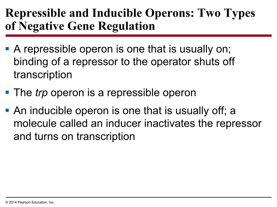
§ The lac operon is an inducible operon and contains genes that code for enzymes used in the hydrolysis and metabolism of lactose
§ By itself, the lac repressor is active and switches the lac operon off
§ A molecule called an inducer inactivates the repressor to turn the lac operon on

© 2014 Pearson Education, Inc.
§ For the lac operon, the inducer is allolactose, formed from lactose that enters the cell
§ Enzymes of the lactose pathway are called inducible enzymes
§ Analogously, the enzymes for tryptophan synthesis are said to be repressible enzymes
Video: lac Repressor Model

© 2014 Pearson Education, Inc.
Figure 15.4
DNA
mRNA
Promoter Operator
Regulatory gene
No RNA made
RNA polymerase
3ʹ
5ʹ
IacZ
Active repressor Protein
(a) Lactose absent, repressor active, operon off
IacZ IacY IacA IacI DNA
mRNA
Protein
RNA polymerase
Inactive repressor
Allolactose (inducer)
(b) Lactose present, repressor inactive, operon on
mRNA 5ʹ 3ʹ
5ʹ
lac operon
lacI
Permease Transacetylase β-Galactosidase

© 2014 Pearson Education, Inc.
Figure 15.4a
DNA
Promoter Operator
Regulatory gene
No RNA made
IacZ lacI
mRNA RNA polymerase
3ʹ
5ʹ
Active repressor Protein
(a) Lactose absent, repressor active, operon off

© 2014 Pearson Education, Inc.
Figure 15.4b
IacZ IacY IacA IacI
DNA lac operon
Permease Transacetylase β-Galactosidase
mRNA
Protein
RNA polymerase
mRNA 5ʹ 3ʹ
5ʹ
Inactive repressor
Allolactose (inducer)
(b) Lactose present, repressor inactive, operon on

© 2014 Pearson Education, Inc.
§ Inducible enzymes usually function in catabolic pathways; their synthesis is induced by a chemical signal
§ Repressible enzymes usually function in anabolic pathways; their synthesis is repressed by high levels of the end product
§ Regulation of the trp and lac operons involves negative control of genes because operons are switched off by the active form of the repressor

© 2014 Pearson Education, Inc.
Positive Gene Regulation
§ E. coli will preferentially use glucose when it is present in the environment
§ When glucose is scarce, CAP (catabolite activator protein) acts as an activator of transcription
§ CAP is activated by binding with cyclic AMP (cAMP)
§ Activated CAP attaches to the promoter of the lac operon and increases the affinity of RNA polymerase, thus accelerating transcription

© 2014 Pearson Education, Inc.
§ When glucose levels increase, CAP detaches from the lac operon, and transcription proceeds at a very low rate, even if lactose is present
§ CAP helps regulate other operons that encode enzymes used in catabolic pathways

© 2014 Pearson Education, Inc.
Figure 15.5
DNA
Promoter
Operator IacZ lacI
CAP-binding site
cAMP
Inactive CAP
Active CAP
RNA polymerase binds and transcribes
Allolactose
Inactive lac repressor
(a) Lactose present, glucose scarce (cAMP level high):
Promoter
abundant lac mRNA synthesized
DNA
Operator IacZ lacI
CAP-binding site RNA polymerase less likely to bind
(b) Lactose present, glucose present (cAMP level low): little lac mRNA synthesized
Inactive CAP
Inactive lac repressor

© 2014 Pearson Education, Inc.
Figure 15.5a
DNA
Promoter
IacZ lacI Operator RNA
polymerase binds and transcribes
Active CAP
Inactive CAP
Allolactose
Inactive lac repressor
CAP-binding site
cAMP
(a) Lactose present, glucose scarce (cAMP level high): abundant lac mRNA synthesized

© 2014 Pearson Education, Inc.
Figure 15.5b
Promoter
DNA Operator
IacZ lacI
CAP-binding site
Inactive CAP Inactive lac
repressor
RNA polymerase less likely to bind
little lac mRNA synthesized (b) Lactose present, glucose present (cAMP level low):

© 2014 Pearson Education, Inc.
Concept 15.2: Eukaryotic gene expression is regulated at many stages
§ All organisms must regulate which genes are expressed at any given time
§ In multicellular organisms regulation of gene expression is essential for cell specialization

© 2014 Pearson Education, Inc.
Differential Gene Expression
§ Almost all the cells in an organism are genetically identical
§ Differences between cell types result from differential gene expression, the expression of different genes by cells with the same genome
§ Abnormalities in gene expression can lead to diseases, including cancer
§ Gene expression is regulated at many stages

© 2014 Pearson Education, Inc.
Regulation of Transcription Initiation
§ Chromatin-modifying enzymes provide initial control of gene expression by making a region of DNA either more or less able to bind the transcription machinery

© 2014 Pearson Education, Inc.
§ DNA methylation is the addition of methyl groups to certain bases in DNA, usually cytosine
§ Individual genes are usually more heavily methylated in cells where they are not expressed
§ Once methylated, genes usually remain so through successive cell divisions
§ After replication, enzymes methylate the correct daughter strand so that the methylation pattern is inherited
DNA Methylation

© 2014 Pearson Education, Inc.

© 2014 Pearson Education, Inc.
Organization of a Typical Eukaryotic Gene
§ Associated with most eukaryotic genes are multiple control elements, segments of noncoding DNA that serve as binding sites for transcription factors that help regulate transcription
§ Control elements and the transcription factors they bind are critical for the precise regulation of gene expression in different cell types

© 2014 Pearson Education, Inc.
The Roles of Transcription Factors
§ To initiate transcription, eukaryotic RNA polymerase requires the assistance of proteins called transcription factors
§ General transcription factors are essential for the transcription of all protein-coding genes
§ In eukaryotes, high levels of transcription of particular genes depend on interaction between control elements and specific transcription factors

© 2014 Pearson Education, Inc.
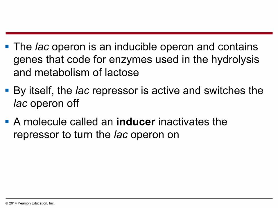
§ Proximal control elements are located close to the promoter
§ Distal control elements, groupings of which are called enhancers, may be far away from a gene or even located in an intron
Enhancers and Specific Transcription Factors

© 2014 Pearson Education, Inc.
§ An activator is a protein that binds to an enhancer and stimulates transcription of a gene
§ Activators have two domains, one that binds DNA and a second that activates transcription
§ Bound activators facilitate a sequence of protein-protein interactions that result in transcription of a given gene
© 2014 Pearson Education, Inc.
Figure 15.10-3
DNA
Enhancer Distal control element
Activators Promoter Gene
TATA box
DNA- bending protein
Group of mediator proteins
General transcription factors
RNA polymerase II
RNA polymerase II
RNA synthesis Transcription initiation complex

© 2014 Pearson Education, Inc.
Mechanisms of Post-Transcriptional Regulation
§ Transcription alone does not account for gene expression
§ Regulatory mechanisms can operate at various stages after transcription
§ Such mechanisms allow a cell to fine-tune gene expression rapidly in response to environmental changes

© 2014 Pearson Education, Inc.
RNA Processing
§ In alternative RNA splicing, different mRNA molecules are produced from the same primary transcript, depending on which RNA segments are treated as exons and which as introns

© 2014 Pearson Education, Inc.
Figure 15.12
DNA
Primary RNA transcript
mRNA or
Exons
Troponin T gene
RNA splicing
1 2 3 4 5
1 2 3 5 1 2 4 5
1 2 3 4 5

© 2014 Pearson Education, Inc.
Select topics from Chapter 16

© 2014 Pearson Education, Inc.
Different genes are expressed in different cells…
§ A fertilized egg gives rise to many different cell types
§ Cell types are organized successively into tissues, organs, organ systems, and the whole organism
§ Gene expression orchestrates the developmental programs of animals

© 2014 Pearson Education, Inc.
A Genetic Program for Embryonic Development
§ The transformation from zygote to adult results from cell division, cell differentiation, and morphogenesis
© 2014 Pearson Education, Inc.
§ Cell differentiation is the process by which cells become specialized in structure and function
§ The physical processes that give an organism its shape constitute morphogenesis
§ Differential gene expression results from genes being regulated differently in each cell type
§ Materials in the egg can set up gene regulation that is carried out as cells divide

© 2014 Pearson Education, Inc.
Cytoplasmic Determinants and Inductive Signals
§ An egg’s cytoplasm contains RNA, proteins, and other substances that are distributed unevenly in the unfertilized egg
§ Cytoplasmic determinants are maternal substances in the egg that influence early development
§ As the zygote divides by mitosis, the resulting cells contain different cytoplasmic determinants, which lead to different gene expression

© 2014 Pearson Education, Inc.
Figure 16.3
(a) Cytoplasmic determinants in the egg (b) Induction by nearby cells Unfertilized egg Early embryo
(32 cells) Sperm
Fertilization
Nucleus
Two-celled embryo
Mitotic cell division
Zygote (fertilized egg)
Molecules of two different cytoplasmic determinants Signal
transduction pathway
Signaling molecule (inducer)
Signal receptor
NUCLEUS

© 2014 Pearson Education, Inc.
§ The other major source of developmental information is the environment around the cell, especially signals from nearby embryonic cells
§ In the process called induction, signal molecules from embryonic cells cause transcriptional changes in nearby target cells
§ Thus, interactions between cells induce differentiation of specialized cell types

© 2014 Pearson Education, Inc.
Sequential Regulation of Gene Expression During Cellular Differentiation
§ Determination commits a cell irreversibly to its final fate
§ Determination precedes differentiation

© 2014 Pearson Education, Inc.
Differentiation of Cell Types
§ Today, determination is understood in terms of molecular changes, the expression of genes for tissue-specific proteins
§ The first evidence of differentiation is the production of mRNAs for these proteins
§ Eventually, differentiation is observed as changes in cellular structure

© 2014 Pearson Education, Inc.
Pattern Formation: Setting Up the Body Plan
§ Pattern formation is the development of a spatial organization of tissues and organs
§ In animals, pattern formation begins with the establishment of the major axes
§ Positional information, the molecular cues that control pattern formation, tells a cell its location relative to the body axes and to neighboring cells

© 2014 Pearson Education, Inc.
§ Pattern formation has been extensively studied in the fruit fly Drosophila melanogaster
§ Combining anatomical, genetic, and biochemical approaches, researchers have discovered developmental principles common to many other species, including humans

© 2014 Pearson Education, Inc.
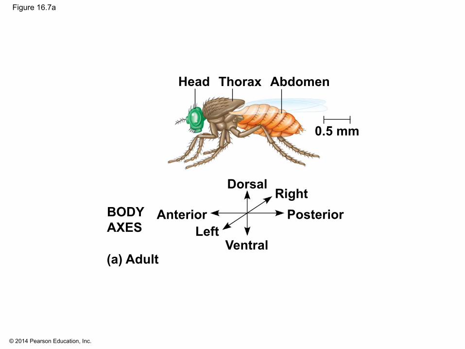
The Life Cycle of Drosophila
§ Fruit flies and other arthropods have a modular structure, composed of an ordered series of segments
§ In Drosophila, cytoplasmic determinants in the unfertilized egg determine the axes before fertilization
© 2014 Pearson Education, Inc.
Figure 16.7a
0.5 mm
Head Thorax Abdomen
Dorsal
Ventral
Posterior Anterior Right
Left
(a) Adult
BODY AXES

© 2014 Pearson Education, Inc.
§ The Drosophila eggs develop in the female’s ovary, surrounded by ovarian cells called nurse cells and follicle cells
§ After fertilization, embryonic development results in a segmented larva, which goes through three stages
§ Eventually the larva forms a cocoon within which it metamorphoses into an adult fly

© 2014 Pearson Education, Inc.
Figure 16.7b-5
Larval stage
(b) Development from egg to larva
Segmented embryo
Body segments
Fertilized egg
Unfertilized egg
Egg developing within ovarian follicle
Hatching 0.1 mm
Embryonic development
Egg shell Depleted
nurse cells Fertilization Laying of egg
Egg
Nurse cell
Follicle cell Nucleus 1
2
3
4
5

© 2014 Pearson Education, Inc.
Genetic Analysis of Early Development: Scientific Inquiry
§ Edward B. Lewis, Christiane Nüsslein-Volhard, and Eric Wieschaus won a Nobel Prize in 1995 for decoding pattern formation in Drosophila
§ Lewis discovered the homeotic genes, which control pattern formation in late embryo, larva, and adult stages

© 2014 Pearson Education, Inc.
Figure 16.8
Wild type
Eye
Mutant

© 2014 Pearson Education, Inc.
§ Nüsslein-Volhard and Wieschaus studied segment formation
§ They created mutants, conducted breeding experiments, and looked for the corresponding genes
§ Many of the identified mutations were embryonic lethals, causing death during embryogenesis
§ They found 120 genes essential for normal segmentation

© 2014 Pearson Education, Inc.
Axis Establishment
§ Maternal effect genes encode cytoplasmic determinants that initially establish the axes of the body of Drosophila
§ These maternal effect genes are also called egg-polarity genes because they control orientation of the egg and consequently the fly

© 2014 Pearson Education, Inc.
§ One maternal effect gene, the bicoid gene, affects the front half of the body
§ An embryo whose mother has no functional bicoid gene lacks the front half of its body and has duplicate posterior structures at both ends
Bicoid: A Morphogen Determining Head Structures

© 2014 Pearson Education, Inc.
Figure 16.9
Head Tail
Tail Tail
Wild-type larva
Mutant larva (bicoid)
T1 T2 T3 A2 A3 A1 A4
A8
A5 A6 A7
A8 A6 A7 A8 A7
250 µm

© 2014 Pearson Education, Inc.
§ This phenotype suggested that the product of the mother’s bicoid gene is concentrated at the future anterior end and is required for setting up the anterior end of the fly
§ This hypothesis is an example of the morphogen gradient hypothesis; gradients of substances called morphogens establish an embryo’s axes and other features

© 2014 Pearson Education, Inc.
§ The bicoid mRNA is highly concentrated at the anterior end of the embryo
§ After the egg is fertilized, the mRNA is translated into Bicoid protein, which diffuses from the anterior end
§ The result is a gradient of Bicoid protein
§ Injection of bicoid mRNA into various regions of an embryo results in the formation of anterior structures at the site of injection

© 2014 Pearson Education, Inc.
Figure 16.10
Anterior end 100 µm
Bicoid protein in early embryo
Results
Bicoid protein in early embryo
Bicoid mRNA in mature unfertilized egg
Bicoid mRNA in mature unfertilized egg
Fertilization, translation of bicoid mRNA

© 2014 Pearson Education, Inc.
§ The bicoid research is important for three reasons § It identified a specific protein required for some early
steps in pattern formation
§ It increased understanding of the mother’s role in embryo development
§ It demonstrated a key developmental principle that a gradient of molecules can determine polarity and position in the embryo

© 2014 Pearson Education, Inc.
§ In organismal cloning one or more organisms develop from a single cell without meiosis or fertilization
§ The cloned individuals are genetically identical to the “parent” that donated the single cell
§ The current interest in organismal cloning arises mainly from its potential to generate stem cells
Concept 16.2: Cloning organisms showed that differentiated cells could be reprogrammed and ultimately led to the production of stem cells

© 2014 Pearson Education, Inc.
Cloning Plants and Animals
§ F. C. Steward and his students first cloned whole carrot plants in the 1950s
§ Single differentiated cells from the root incubated in culture medium were able to grow into complete adult plants
§ This work showed that differentiation is not necessarily irreversible
§ Cells that can give rise to all the specialized cell types in the organism are called totipotent

© 2014 Pearson Education, Inc.
§ In cloning of animals, the nucleus of an unfertilized egg cell or zygote is replaced with the nucleus of a differentiated cell, called nuclear transplantation
§ Experiments with frog embryos showed that a transplanted nucleus can often support normal development of the egg
§ The older the donor nucleus, the lower the percentage of normally developing tadpoles
§ John Gurdon concluded from this work that nuclear potential is restricted as development and differentiation proceeds

© 2014 Pearson Education, Inc.
Figure 16.11
Frog embryo
Less differ- entiated cell
Results
Enucleated egg cell
Donor nucleus trans- planted
Experiment Frog egg cell Frog tadpole
Donor nucleus trans- planted
Fully differ- entiated (intestinal) cell
Egg with donor nucleus activated to begin
development
Most develop into tadpoles.
Most stop developing before tadpole stage.
UV
© 2014 Pearson Education, Inc.
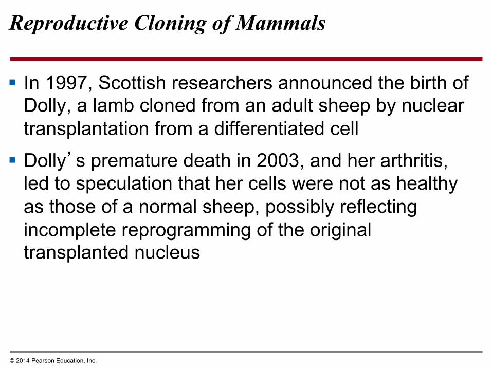
Reproductive Cloning of Mammals
§ In 1997, Scottish researchers announced the birth of Dolly, a lamb cloned from an adult sheep by nuclear transplantation from a differentiated cell
§ Dolly’s premature death in 2003, and her arthritis, led to speculation that her cells were not as healthy as those of a normal sheep, possibly reflecting incomplete reprogramming of the original transplanted nucleus

© 2014 Pearson Education, Inc.
Figure 16.12
Grown in culture
Results
Cell cycle arrested, causing cells to dedifferentiate
Implanted in uterus of a third sheep
Cultured mammary cells
Embryonic development
Technique Mammary cell donor
Egg cell donor
Egg cell from ovary
Nucleus removed
Nucleus from mammary cell
Surrogate mother
Cells fused
Early embryo
Lamb (“Dolly”) genetically identical to mammary cell donor
1 2
3
4
5
6

© 2014 Pearson Education, Inc.
Figure 16.12a
Cell cycle arrested, causing cells to dedifferentiate
Cultured mammary cells
Technique Mammary cell donor
Egg cell donor
Egg cell from ovary
Nucleus removed
Nucleus from mammary cell
Cells fused
1 2
3

© 2014 Pearson Education, Inc.
Figure 16.12b
Grown in culture
Results
Implanted in uterus of a third sheep
Embryonic development
Nucleus from mammary cell
Surrogate mother
Early embryo
Lamb (“Dolly”) genetically identical to mammary cell donor
4
5
6
Technique

© 2014 Pearson Education, Inc.
Faulty Gene Regulation in Cloned Animals
§ In most nuclear transplantation studies, only a small percentage of cloned embryos have developed normally to birth
§ Many cloned animals exhibit defects
§ Epigenetic changes must be reversed in the nucleus from a donor animal in order for genes to be expressed or repressed appropriately for early stages of development

© 2014 Pearson Education, Inc.
Stem Cells of Animals
§ A stem cell is a relatively unspecialized cell that can reproduce itself indefinitely and differentiate into specialized cells of one or more types
§ Stem cells isolated from early embryos at the blastocyst stage are called embryonic stem (ES) cells; these are able to differentiate into all cell types
§ The adult body also has stem cells, which replace nonreproducing specialized cells

© 2014 Pearson Education, Inc.
Figure 16.14
Stem cell
Stem cell Precursor cell
Fat cells
Cell division
and
or or Bone cells White blood cells

© 2014 Pearson Education, Inc.
Figure 16.15
Cultured stem cells
Embryonic stem cells
Liver cells Nerve cells Blood cells
Adult stem cells
Different culture conditions
Different types of differentiated cells
Cells that can generate all embryonic cell types
Cells that generate a limited number of cell types

© 2014 Pearson Education, Inc.
§ ES cells are pluripotent, capable of differentiating into many cell types
§ Researchers are able to reprogram fully differentiated cells to act like ES cells using retroviruses
§ Cells transformed this way are called iPS, or induced pluripotent stem cells

© 2014 Pearson Education, Inc.
§ Cells of patients suffering from certain diseases can be reprogrammed into iPS cells for use in testing potential treatments
§ In the field of regenerative medicine, a patient’s own cells might be reprogrammed into iPS cells to potentially replace the nonfunctional (diseased) cells

© 2014 Pearson Education, Inc.
Concept 16.3: Abnormal regulation of genes that affect the cell cycle can lead to cancer
§ The gene regulation systems that go wrong during cancer are the same systems involved in embryonic development

© 2014 Pearson Education, Inc.
Types of Genes Associated with Cancer
§ Cancer research led to the discovery of cancer-causing genes called oncogenes in certain types of viruses
§ The normal version of such genes, called proto-oncogenes, code for proteins that stimulate normal cell growth and division
§ An oncogene arises from a genetic change leading to either an increase in the amount or the activity of the protein product of the gene

© 2014 Pearson Education, Inc.
Figure 16.16
within a control element
Proto-oncogene
Gene amplification: multiple copies of the gene
Proto-oncogene Proto-oncogene
Point mutation:
within the gene
Translocation or transposition: gene moved to new locus, under new controls
Normal growth- stimulating protein in excess
New promoter
Oncogene Oncogene Oncogene
Normal growth- stimulating protein in excess
Normal growth- stimulating protein in excess
Hyperactive or degradation- resistant protein

© 2014 Pearson Education, Inc.
Figure 16.16a
Proto-oncogene
Translocation or transposition: gene moved to new locus, under new controls
Normal growth- stimulating protein in excess
New promoter
Oncogene

© 2014 Pearson Education, Inc.
Figure 16.16b
Gene amplification: multiple copies of the gene
Proto-oncogene
Normal growth- stimulating protein in excess

© 2014 Pearson Education, Inc.
Figure 16.16c
within a control element
Proto-oncogene
Point mutation:
within the gene
Oncogene Oncogene
Normal growth- stimulating protein in excess
Hyperactive or degradation- resistant protein

© 2014 Pearson Education, Inc.
§ Proto-oncogenes can be converted to oncogenes by § Movement of the oncogene to a position near an
active promoter, which may increase transcription
§ Amplification, increasing the number of copies of a proto-oncogene
§ Point mutations in the proto-oncogene or its control elements, causing an increase in gene expression

© 2014 Pearson Education, Inc.
§ Tumor-suppressor genes encode proteins that help prevent uncontrolled cell growth
§ Mutations that decrease protein products of tumor-suppressor genes may contribute to cancer onset
§ Tumor-suppressor proteins
§ Repair damaged DNA
§ Control cell adhesion § Inhibit the cell cycle

© 2014 Pearson Education, Inc.
Interference with Cell-Signaling Pathways
§ Mutations in the ras proto-oncogene and p53 tumor-suppressor gene are common in human cancers
§ Mutations in the ras gene can lead to production of a hyperactive Ras protein and increased cell division

© 2014 Pearson Education, Inc.
Figure 16.17
Growth factor
G protein
Receptor Protein kinases
NUCLEUS Transcription factor (activator)
Protein that stimulates the cell cycle
NUCLEUS Transcription factor (activator)
Overexpression of protein
MUTATION
Ras protein active with or without growth factor.
GTP
Ras
GTP
Ras
1
2
3
4
5 6

© 2014 Pearson Education, Inc.
§ Suppression of the cell cycle can be important in the case of damage to a cell’s DNA; p53 prevents a cell from passing on mutations due to DNA damage
§ Mutations in the p53 gene prevent suppression of the cell cycle

© 2014 Pearson Education, Inc.
Figure 16.18
Protein kinases
NUCLEUS
DNA damage in genome
Defective or missing transcription factor
MUTATION UV light
1
2
3
Inhibitory protein absent
DNA damage in genome
UV light
Active form of p53
Protein that inhibits the cell cycle

© 2014 Pearson Education, Inc.
The Multistep Model of Cancer Development
§ Multiple somatic mutations are generally needed for full-fledged cancer; thus the incidence increases with age
§ The multistep path to cancer is well supported by studies of human colorectal cancer, one of the best-understood types of cancer
§ The first sign of colorectal cancer is often a polyp, a small benign growth in the colon lining

© 2014 Pearson Education, Inc.
§ About half a dozen changes must occur at the DNA level for a cell to become fully cancerous
§ These changes generally include at least one active oncogene and the mutation or loss of several tumor-suppressor genes

© 2014 Pearson Education, Inc.
Figure 16.19
Colon
Loss of tumor- suppressor gene APC (or other)
Loss of tumor-suppressor gene p53
Activation of ras oncogene
Colon wall Normal colon epithelial cells
Small benign growth (polyp)
Loss of tumor- suppressor gene DCC
Malignant tumor (carcinoma)
Larger benign growth (adenoma)
Additional mutations
1 2
3
4
5

© 2014 Pearson Education, Inc.
Figure 16.19a
Loss of tumor- suppressor gene APC (or other)
Loss of tumor-suppressor gene p53
Activation of ras oncogene
Colon wall Normal colon epithelial cells
Small benign growth (polyp)
Loss of tumor-suppressor gene DCC Malignant tumor
(carcinoma) Larger benign growth (adenoma)
Additional mutations
1
2
3
4
5

© 2014 Pearson Education, Inc.
Inherited Predisposition and Other Factors Contributing to Cancer
§ Individuals can inherit oncogenes or mutant alleles of tumor-suppressor genes
§ Inherited mutations in the tumor-suppressor gene adenomatous polyposis coli (APC) are common in individuals with colorectal cancer
§ Mutations in the BRCA1 or BRCA2 gene are found in at least half of inherited breast cancers, and tests using DNA sequencing can detect these mutations

© 2014 Pearson Education, Inc.
§ DNA breakage can contribute to cancer, thus the risk of cancer can be lowered by minimizing exposure to agents that damage DNA, such as ultraviolet radiation and chemicals found in cigarette smoke
§ Also, viruses play a role in about 15% of human cancers by donating an oncogene to a cell, disrupting a tumor-suppressor gene, or converting a proto-oncogene into an oncogene