Embed Size (px)
Citation preview
Section G Gene manipulation
ContentContent
G1-DNA CLONING: AN OVERVIEWG1-DNA CLONING: AN OVERVIEW
G2-PREPARATION OF PLASMID DNAG2-PREPARATION OF PLASMID DNA
G3-RESTRICTION ENZYMES AND G3-RESTRICTION ENZYMES AND
ELECTROPHORESISELECTROPHORESIS
G4-LIGATION, TRANSFORMATION AND G4-LIGATION, TRANSFORMATION AND ANALYSIS OF RECOMBINANTSANALYSIS OF RECOMBINANTS
G1-1 DNA cloning
G1-2 Hosts and vectors
G1-3 Subcloning
G1-4 DNA libraries
G1-5 Screening libraries
G1-6 Analysis of a clone
G1 DNA cloning: an overview

G1-1 DNA cloning
The transfer of a DNA fragment of interest from one
organism to a self-replicating genetic element such as
a bacterial plasmid. The DNA of interest can then be
propagated in a foreign host cell. This technology has
been around since the 1970s, and it has become a
common practice in molecular biology labs today.
G1-2 Hosts and vectors
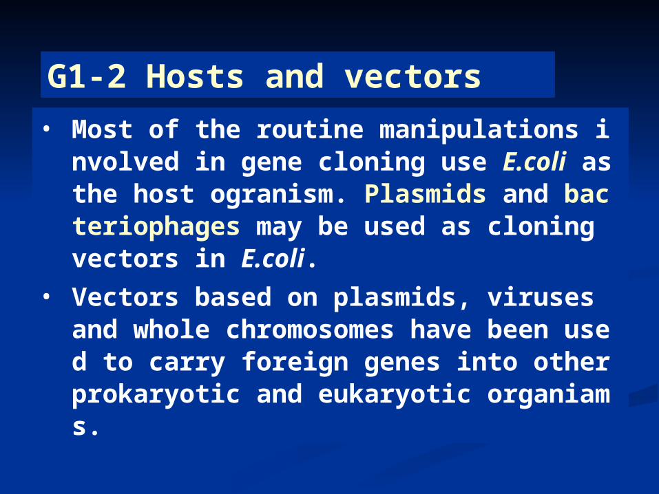
• Most of the routine manipulations involved in gene cloning use E.coli as the host ogranism. Plasmids and bacteriophages may be used as cloning vectors in E.coli.
• Vectors based on plasmids, viruses and whole chromosomes have been used to carry foreign genes into other prokaryotic and eukaryotic organiams.
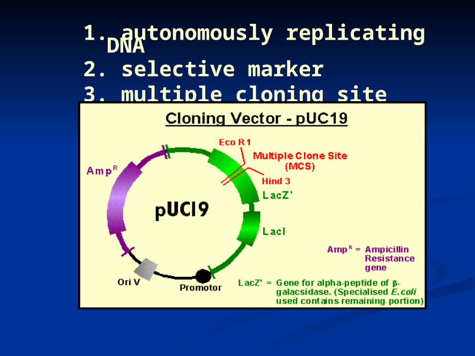
1. autonomously replicating DNA2. selective marker3. multiple cloning site (MCS)
Types of vectors
A-Cloning vectors
B-Expression vectors
C-Integration vectors
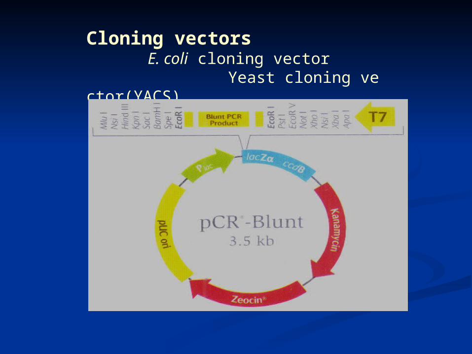
Cloning vectors E. coli cloning vector Yeast cloning vector(YACS)
MCS
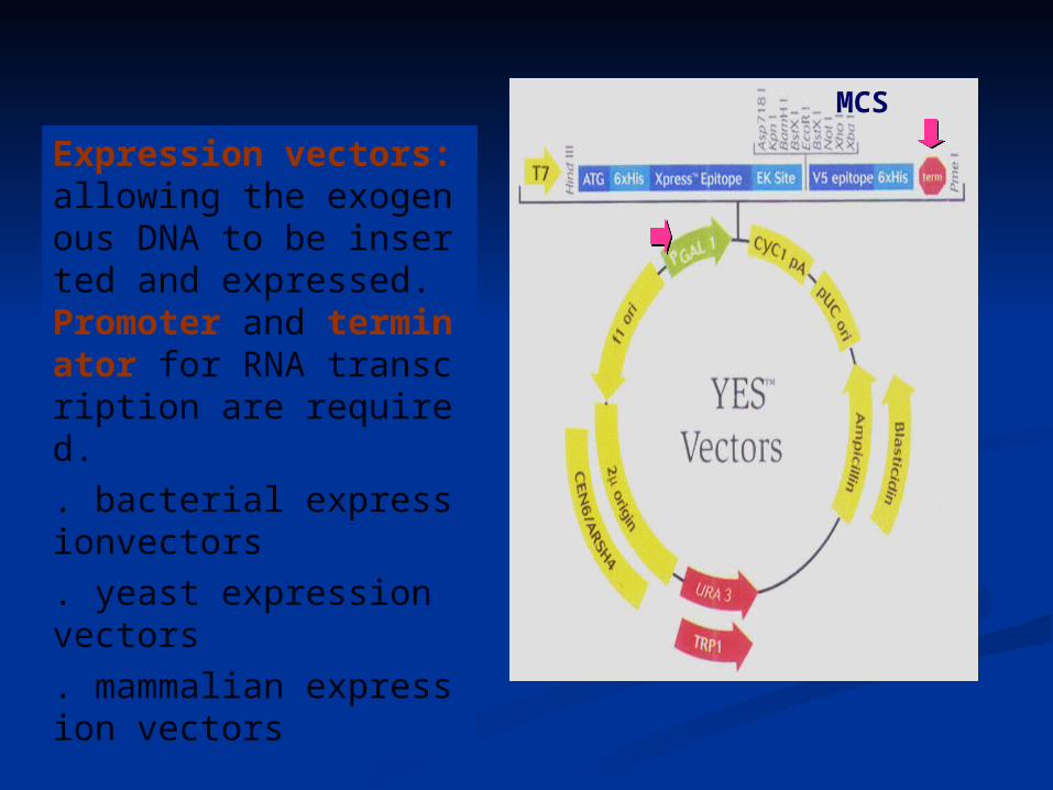
Expression vectors: allowing the exogenous DNA to be inserted and expressed. Promoter and terminator for RNA transcription are required. . bacterial expressionvectors. yeast expression vectors. mammalian expression vectors
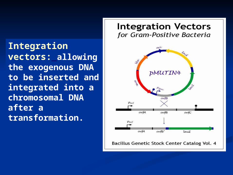
Integration vectors: allowing the exogenous DNA to be inserted and integrated into a chromosomal DNA after a transformation.
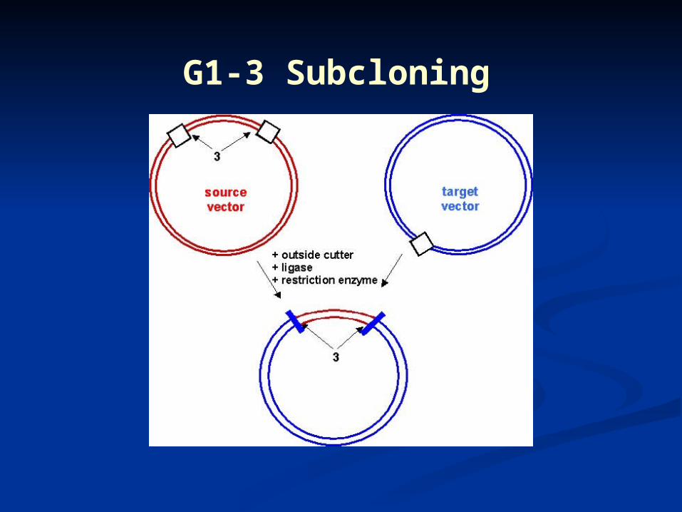
G1-3 Subcloning
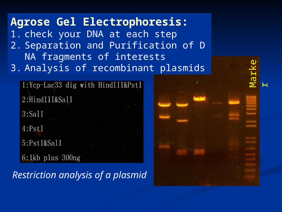
Agrose Gel Electrophoresis: 1. check your DNA at each step2. Separation and Purification of DNA fragments o
f interests3. Analysis of recombinant plasmids
Mar
ker
Restriction analysis of a plasmid
G1-4 DNA libraries Genomic librariesprepared form random fragments of genomic DNA, which may be inefficient to find a gene because of the huge abundance of the non-coding DNA
cDNA libraries DNA copies (cDNA) synthesized from the mRNA by reverse transcription are inserted into a vector to form a cDNA library. Much more efficient in identifying a gene, but do not contain DNA coding functional RNA or noncoding sequence.
G1-5 Screening libraries
Libraries are screened for the presence of a gene sequence by hybridization with a sequence derived from its protein product or a rlated gene, or through the screening of the protein products of the cloned fragments.
Searching the genes of interest in a DNA library
G1-6 Analysis of a clone
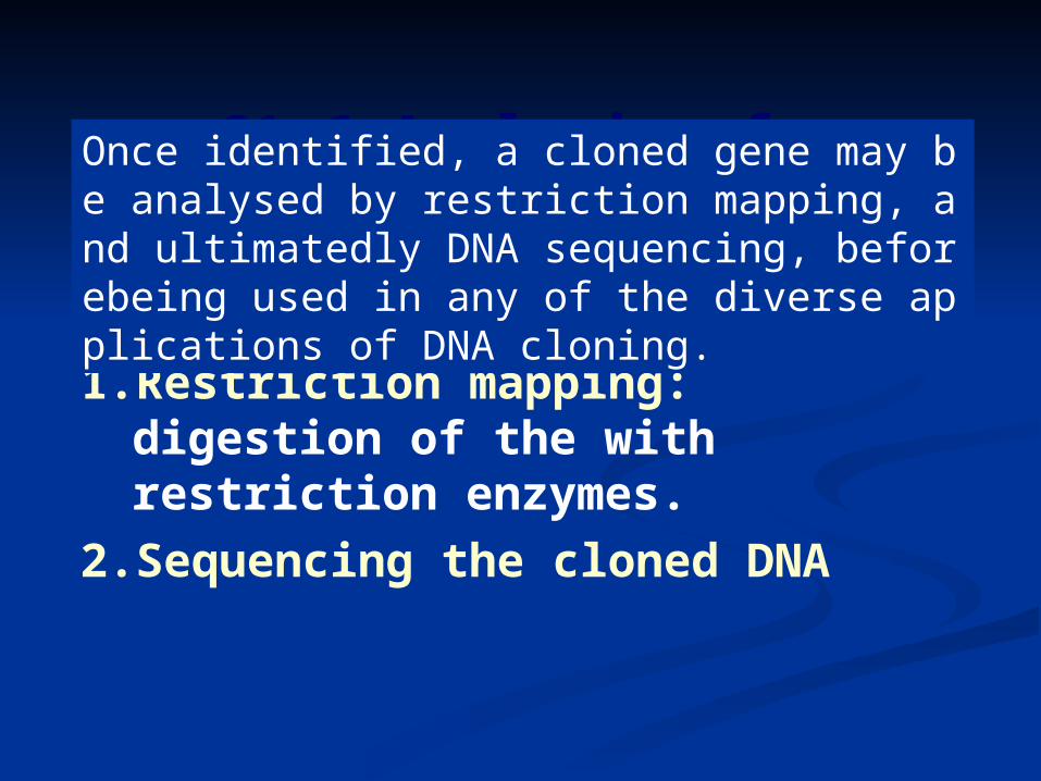
1. Restriction mapping: digestion of the with restriction enzymes.
2. Sequencing the cloned DNA
Once identified, a cloned gene may be analysed by restriction mapping, and ultimatedly DNA sequencing, beforebeing used in any of the diverse applications of DNA cloning.
G2 Preparation of plasmid DNA
. Plasmid as vector
. Plasmid minipreparation
. Alkaline lysis
. Phenol extraction
. Ethanol precipitation
. Cesium chloride gradient (purification)
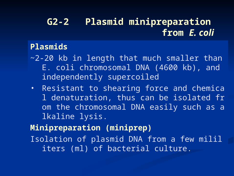
G2-2 Plasmid minipreparation from E. coli
Plasmids
~2-20 kb in length that much smaller than E. coli chromosomal DNA (4600 kb), and independently supercoiled
• Resistant to shearing force and chemical denaturation, thus can be isolated from the chromosomal DNA easily such as alkaline lysis.
Minipreparation (miniprep)
Isolation of plasmid DNA from a few mililiters (ml) of bacterial culture.
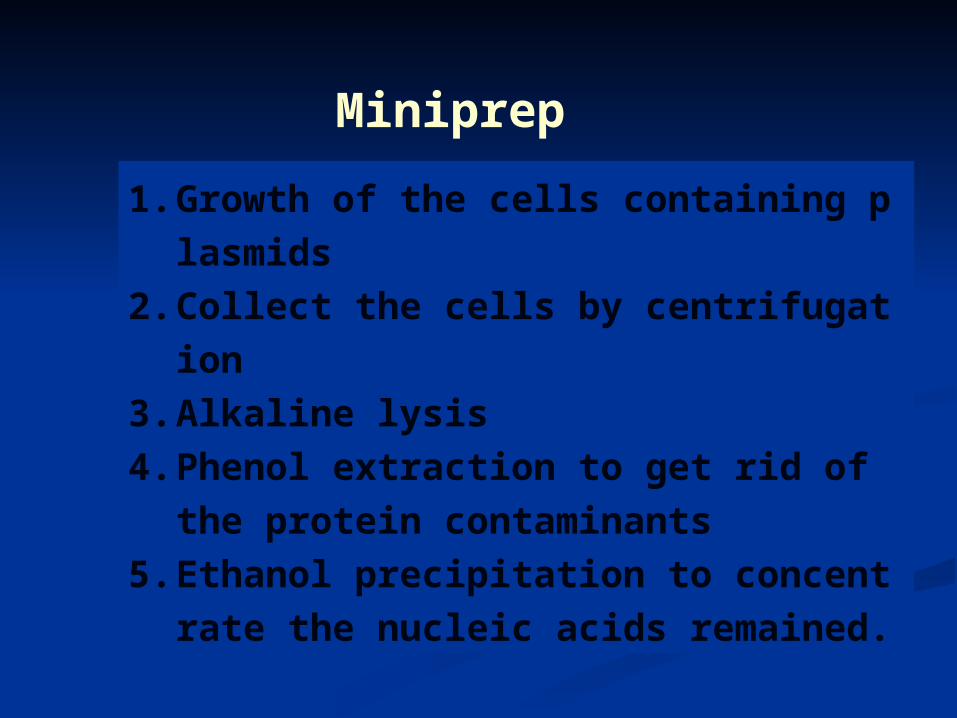
Miniprep
1. Growth of the cells containing plasmids
2. Collect the cells by centrifugation
3. Alkaline lysis
4. Phenol extraction to get rid of the protein con
taminants
5. Ethanol precipitation to concentrate the nucl
eic acids remained.
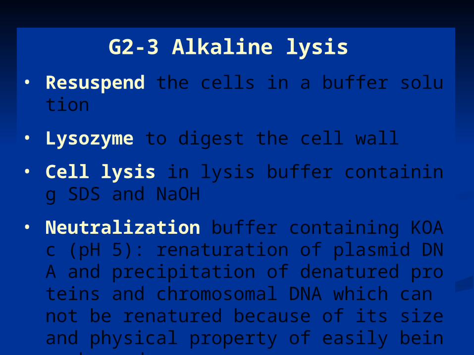
G2-3 Alkaline lysis • Resuspend the cells in a buffer solution
• Lysozyme to digest the cell wall
• Cell lysis in lysis buffer containing SDS and NaOH
• Neutralization buffer containing KOAc (pH 5): renaturation of plasmid DNA and precipitation of denatured proteins and chromosomal DNA which can not be renatured because of its size and physical property of easily being sheared.
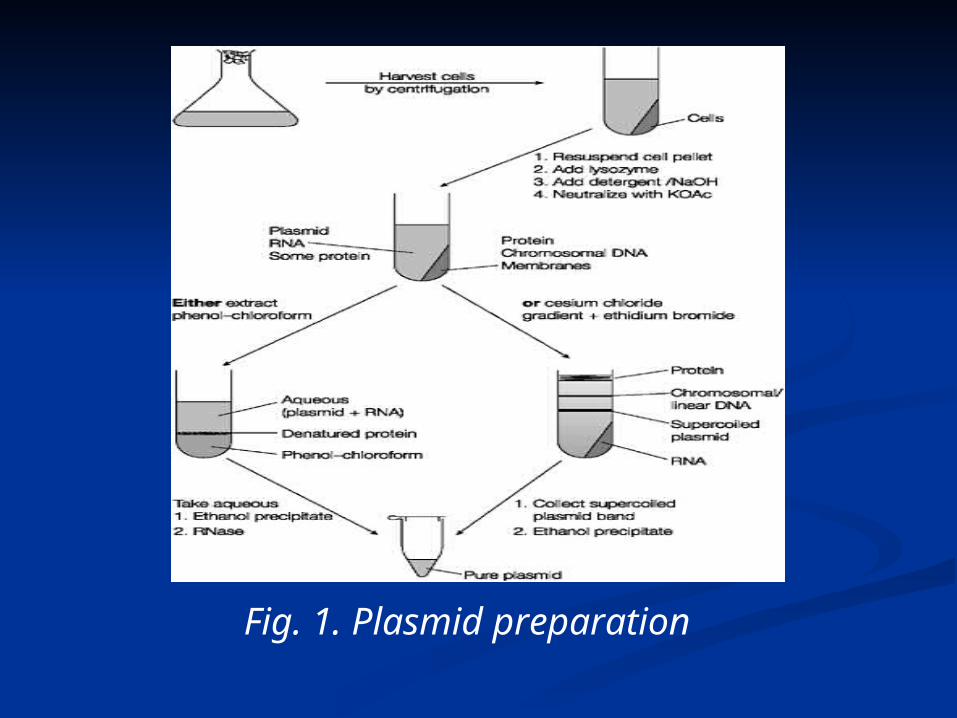
Fig. 1. Plasmid preparation
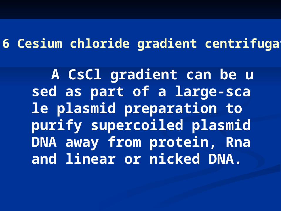
G2- 6 Cesium chloride gradient centrifugation
A CsCl gradient can be used as part of a large-scale plasmid preparation to purify supercoiled plasmid DNA away from protein, Rna and linear or nicked DNA.
G3 Restriction Enzymes and electrophoresis
. Restriction endonuclease
. Recognition sequences
. Cohesive ends
. Restriction digests
. Agarose gel electrophoresis
. Isolation of fragments

G3-1 Restriction endonuclease
Restriction enonucleases are bacterial enzymes which cut(hydrolyze) DNA into defined and reproducible fragments.
In bacteria, they form part of the restriction-modification defense mechanism against foreign DNA.
They are the basic tools of gene cloning.

G3-2&3 Restriction sequences&Cohesive ends
Fig. 1. (a) The action of restriction endonucleases at their recognition sequences; (b) the annealing of cohesive ends.
Recognition sequences
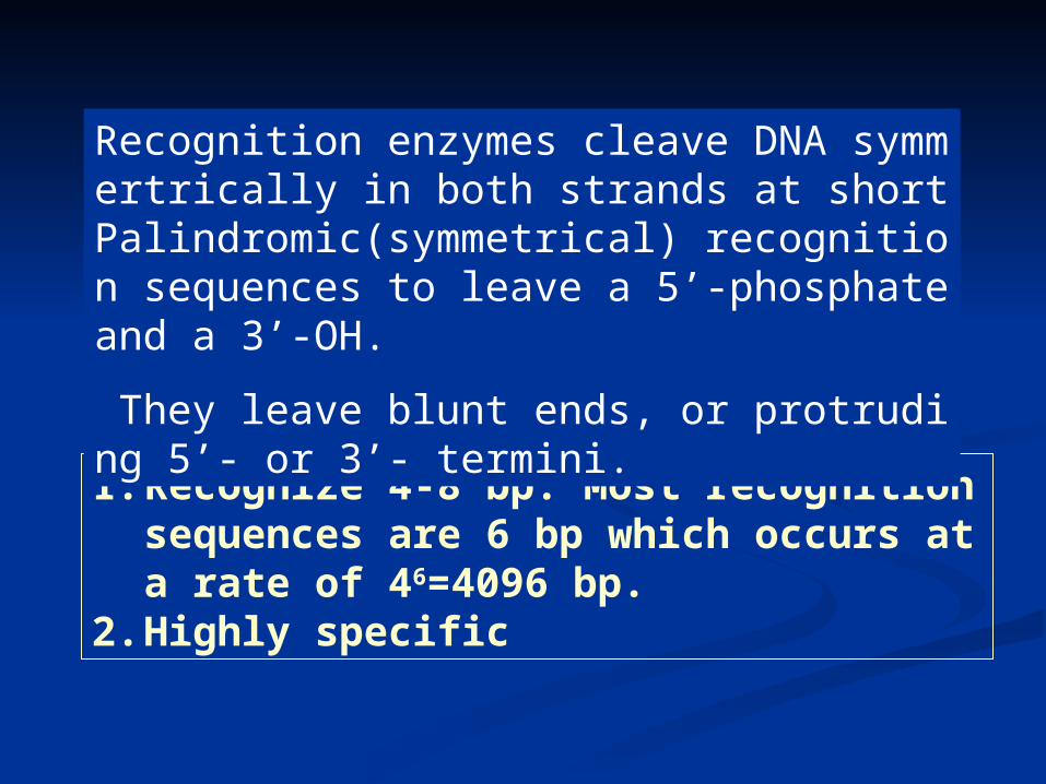
1. Recognize 4-8 bp. Most recognition sequences are 6 bp which occurs at a rate of 46=4096 bp.
2. Highly specific
Recognition enzymes cleave DNA symmertrically in both strands at short Palindromic(symmetrical) recognition sequences to leave a 5’-phosphate and a 3’-OH.
They leave blunt ends, or protruding 5’- or 3’- termini.
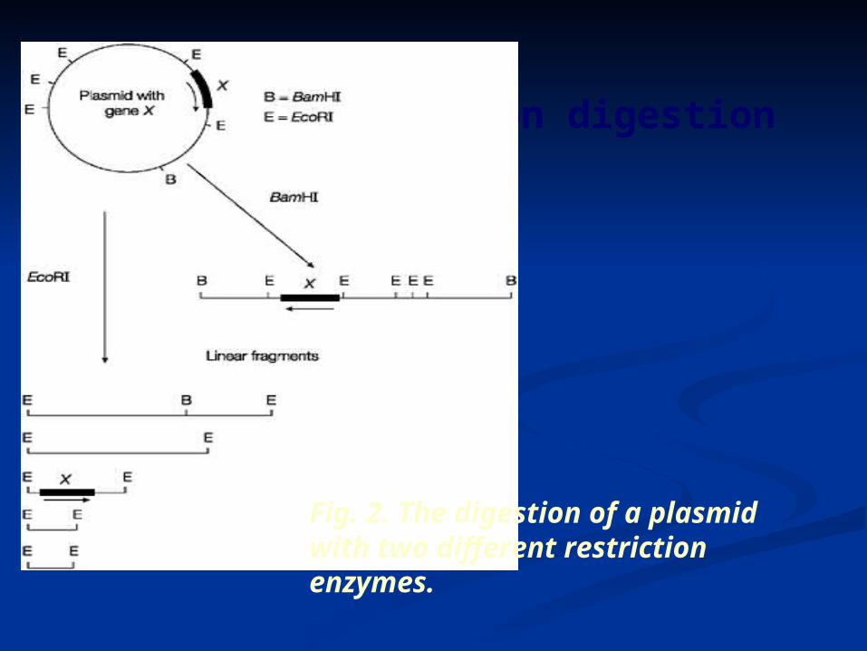
G3-4 Restriction digestion
Fig. 2. The digestion of a plasmid with two different restriction enzymes.
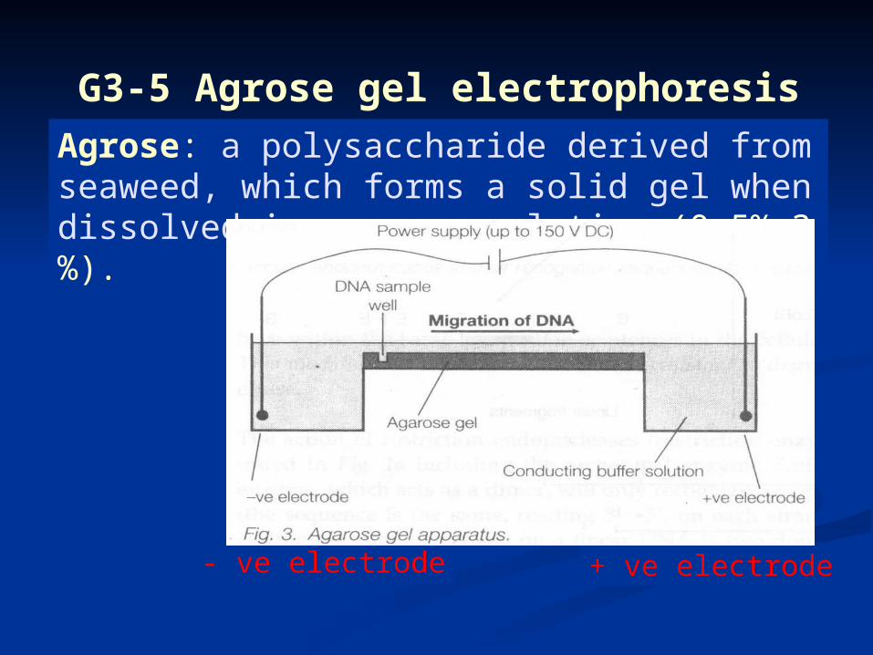
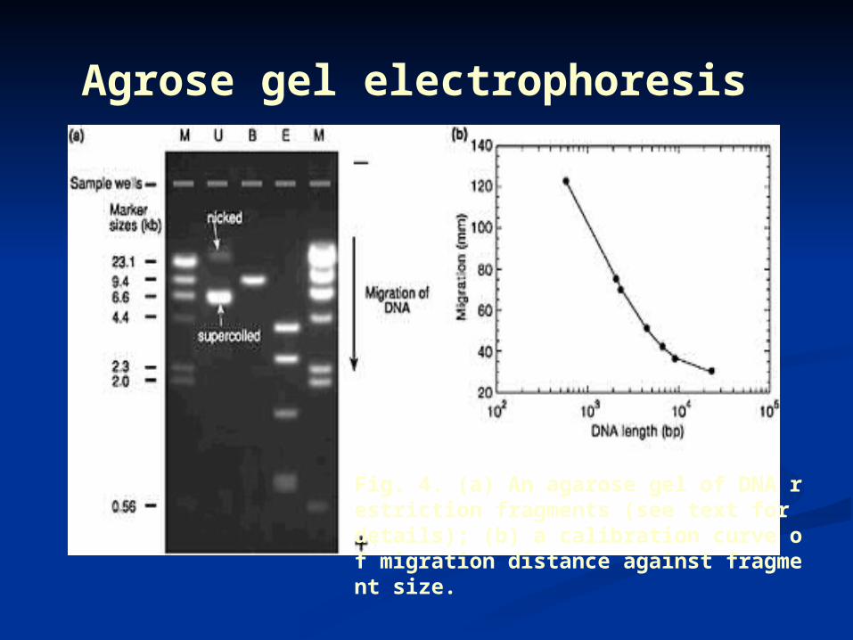
G3-5 Agrose gel electrophoresis
Agrose: a polysaccharide derived from seaweed, which forms a solid gel when dissolved in aqueous solution (0.5%-3%).
- ve electrode + ve electrode
Negatively charged DNA
Agrose gel electrophoresis
Fig. 4. (a) An agarose gel of DNA restriction fragments (see text for details); (b) a calibration curve of migration distance against fragment size.
G4 Ligation, transformation and analysis of G4 Ligation, transformation and analysis of recombinants recombinants
G4.1 Alkaline phophatse
G4.2-3 DNA ligation & recombinant DNA molecules
G4.4-5 Transformation & selection
G4.6 Transformation efficiency
G4.7 Screening transformants
G4.8 Growth and storage of transformants
G4.9 Gel analysis
G4.10 Fragment orientation
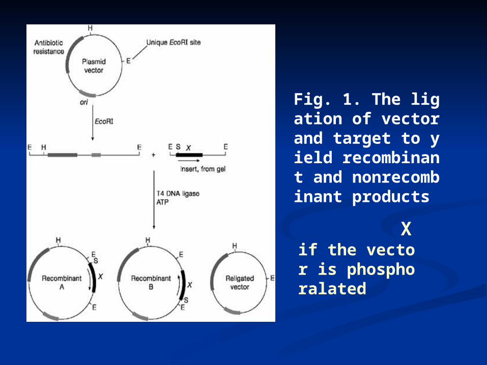
X if the vector is phosphoralated
Fig. 1. The ligation of vector and target to yield recombinant and nonrecombinant products
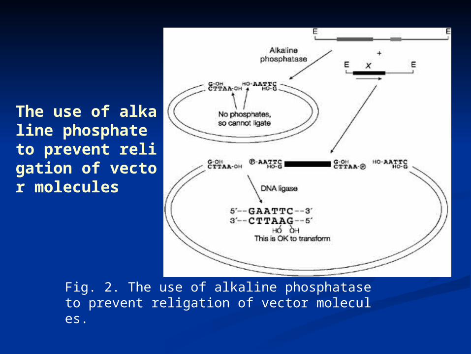
The use of alkaline phosphate to prevent religation of vector molecules
Fig. 2. The use of alkaline phosphatase to prevent religation of vector molecules.
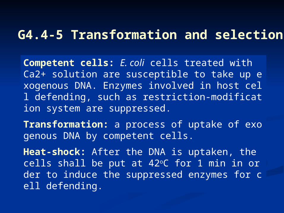
G4.4-5 Transformation and selection
Competent cells: E. coli cells treated with Ca2+ solution are susceptible to take up exogenous DNA. Enzymes involved in host cell defending, such as restriction-modification system are suppressed.
Transformation: a process of uptake of exogenous DNA by competent cells.
Heat-shock: After the DNA is uptaken, the cells shall be put at 42oC for 1 min in order to induce the suppressed enzymes for cell defending.
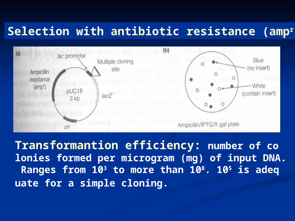
Selection with antibiotic resistance (ampr)
Transformantion efficiency: number of colonies formed per microgram (mg) of input DNA. Ranges from 103 to more than 108. 105 is adequate for a simple cloning.
Transformantion efficiency- number of colon
ies formed per microgram (mg) of input DNA. Ra
nges from 103 to more than 108. 105 is adequate fo
r a simple cloning.
G4-6 Transformation efficiency
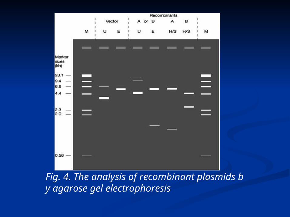
Fig. 4. The analysis of recombinant plasmids by agarose gel electrophoresis