Embed Size (px)
Citation preview

J Clin Exp Dent. 2018;10(1):e1-6. Universal adhesive in fissure sealant
e1
Journal section: Community and Preventive Dentistry Publication Types: Research
Sealing effectiveness of fissure sealant bonded with universal adhesive systems on saliva-contaminated and noncontaminated enamel
Mahtab Memarpour 1, Fereshteh Shafiei 2, Mehran Zarean 3, Faranak Razmjoei 4
1 DMD, MScD, Professor, Oral and Dental Disease Research Center, Department of Pediatric Dentistry, School of Dentistry, Shiraz University of Medical Sciences, Shiraz, Iran2 DMD, MScD, Professor, Oral and Dental Disease Research Center, Department of Operative Dentistry, School of Dentistry, Shi-raz University of Medical Sciences, Shiraz, Iran3 DDS, Dentist, Department of Pediatric Dentistry, School of Dentistry, Azad Shiraz University of Medical Sciences, Shiraz, Iran4 DMD, MScD, Assistant Professor, Oral and Dental Disease Research Center, Department of Pediatric Dentistry, School of Den-tistry, Shiraz University of Medical Sciences, Shiraz, Iran
Correspondence:Oral and Dental Disease Research CenterSchool of Dentistry Shiraz University of Medical Sciences Shiraz, Iran [email protected]
Received: 27/10/2017Accepted: 18/11/2017
Abstract Background: The effectiveness of sealants is dependent upon their adhesion to enamel surface. The aim of the study was to evaluate the sealing ability of a pit and fissure sealant used with a universal adhesive (etch-and-rinse vs. self-etch modes) when the site is contaminated with saliva. Adhesive properties were evaluated as microleakage and scanning electron microscopic (SEM) characteristics. Material and Methods: A total of 72 mandibular third molars were randomly divided into 6 groups (n=12). Occlusal pits and fissures were sealed with an unfilled resin fissure sealant (FS) material with or without saliva contami-nation. The groups included: 1) phosphoric acid etching + FS (control), 2) phosphoric acid etching + Scotchbond Universal (etch-and-rinse) + FS, 3) phosphoric acid etching + saliva + Scotchbond Universal (etch-and-rinse) + FS, 4) Scotchbond Universal (self-etching) + FS,5) Scotchbond Universal (self-etching) + saliva + FS, and 6) Scotch-bond Universal (self-etching) + saliva + Scotchbond Universal + FS. After thermocycling, the teeth were placed in 0.5% fuchsin, sectioned, and evaluated by digital microscopy. Two samples from each group were also observed by SEM. The data were analyzed with Kruskal-Wallis and Mann-Whitney tests for a significance of p<0.05.Results: There were significant differences among groups. Groups 1,2 and 4 showed the least microleakage, with no significant differences among groups. Saliva contamination led to increased microleakage and gap formation in SEM images in groups 3, 5 and 6. Conclusions: The fissure sealing ability of the universal adhesive in etch-and-rinse or self-etch modes was similar to that of conventional acid etching. Saliva contamination had a negative effect on sealant adhesion to pretreated enamel.
Key words: Pit and fissure sealant, Universal adhesive, Saliva.
doi:10.4317/jced.54471http://dx.doi.org/10.4317/jced.54471
Article Number: 54471 http://www.medicinaoral.com/odo/indice.htm© Medicina Oral S. L. C.I.F. B 96689336 - eISSN: 1989-5488eMail: [email protected] in:
PubmedPubmed Central® (PMC)ScopusDOI® System
Memarpour M, Shafiei F, Zarean M, Razmjoei F. Sealing effective-. Sealing effective-ness of fissure sealant bonded with universal adhesive systems on saliva-contaminated and noncontaminated enamel. J Clin Exp Dent. 2018;10(1):e1-6.http://www.medicinaoral.com/odo/volumenes/v10i1/jcedv10i1p1.pdf
J Clin Exp Dent. 2018;10(1):e1-6. Universal adhesive in fissure sealant
e2
IntroductionPit and fissure sealing is a widely accepted method to prevent dental caries on occlusal surfaces, especially in newly erupted permanent teeth in children (1). However, sealant application in young patients is a challenge for dentists due to difficult access to the teeth, the possibility of saliva contamination, and sometimes lack of coopera-tion by the patient (1,2).Conventional methods of sealant therapy include ena-mel acid etching (followed by a rinsing step to remove dissolved minerals) to create microporosities that faci-litate penetration of the sealant material in the enamel (3). Because the formulation of resin sealant is similar to composite resin, some studies reported that adding an adhesive layer beneath the fissure sealant led to in-creased retention and reduced mircoleakge through the sealant and enamel surface (4,5). However, other studies found that the addition of this step had no advantages (6,7). One study reported that applying an adhesive re-duced the negative effect of saliva contamination on sea-lant bond strength to the enamel surface (8).In general, adhesives are categorized as total-etch (two- or three-step adhesives) or self-etch (one- or two-bottle adhesives). Recently a new generation of one-bottle adhesives called “universal”, “multi-mode” or “multi-purpose” adhesives was developed. Universal adhesives (UA) have advantages compared to previous genera-tions: they can be used in two modes, i.e., either as an etch-and-rinse adhesive with selective acid etching be-fore application of the adhesive, or as a one-bottle self-etching adhesive without additional etching. Moreover, UAs are simpler, more user-friendly and less technique-sensitive, and reduce patient chair time – all of which are advantages in pediatric dentistry (9-12). Some stu-dies showed that acid etch techniques had advantages over self-etch adhesives before sealant therapy (13,14). However, other studies found no significant differences in terms of microleakage between conventional acid et-
ching techniques and self-etch adhesives before sealant application (15,16). One study showed that UA in etch-and-rinse mode yielded sealing outcomes similar to the self-etch mode of fissure sealant application (17). Because few data are available on the use of UAs in sealant therapy, the purpose of this in vitro study was to determine the sealing effectiveness of a new one-step self-etch adhesive. The adhesive was applied with two different methods: selective pre-etching (etch-and-rinse mode) and self-etching mode, with or without saliva contamination. The results were compared by measuring microleakage and observing morphological characteris-tics of the tooth–sealant interface with scanning electron microscopy (SEM).
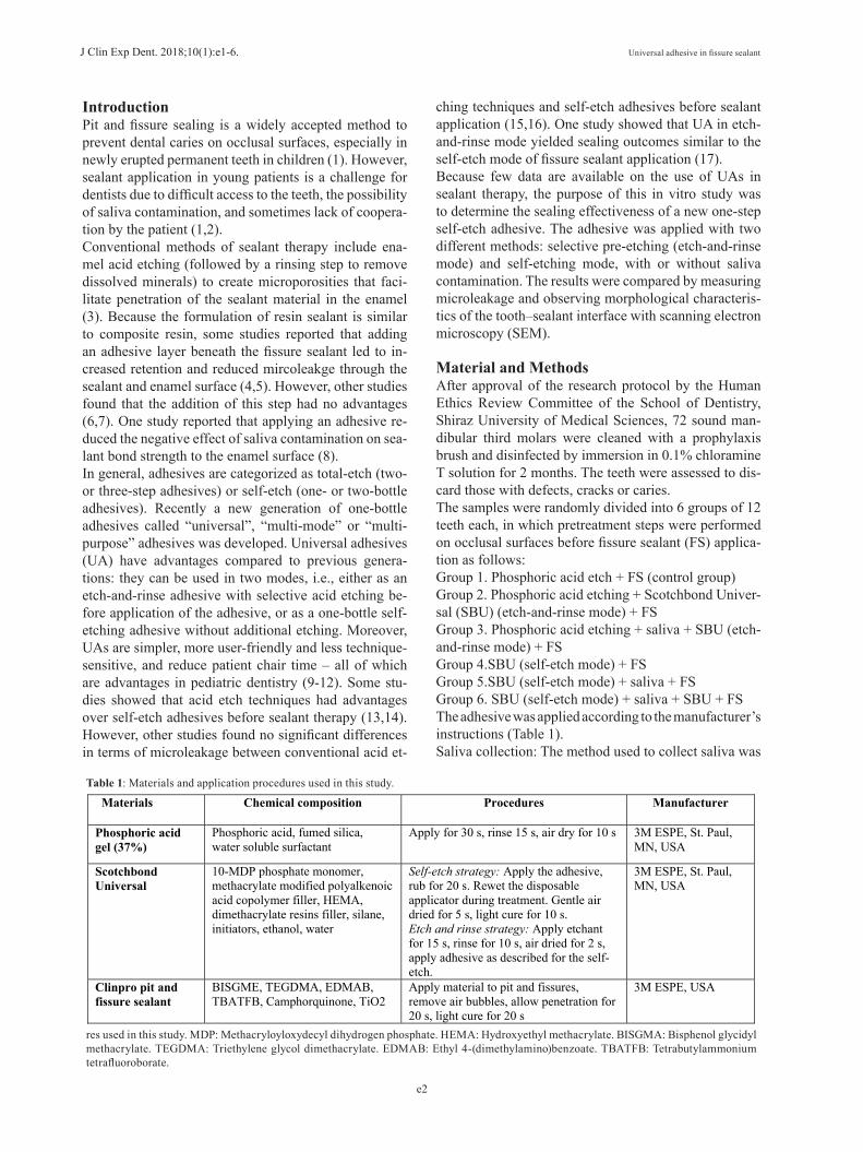
Material and MethodsAfter approval of the research protocol by the Human Ethics Review Committee of the School of Dentistry, Shiraz University of Medical Sciences, 72 sound man-dibular third molars were cleaned with a prophylaxis brush and disinfected by immersion in 0.1% chloramine T solution for 2 months. The teeth were assessed to dis-card those with defects, cracks or caries. The samples were randomly divided into 6 groups of 12 teeth each, in which pretreatment steps were performed on occlusal surfaces before fissure sealant (FS) applica-tion as follows: Group 1. Phosphoric acid etch + FS (control group) Group 2. Phosphoric acid etching + Scotchbond Univer-sal (SBU) (etch-and-rinse mode) + FS Group 3. Phosphoric acid etching + saliva + SBU (etch-and-rinse mode) + FSGroup 4.SBU (self-etch mode) + FSGroup 5.SBU (self-etch mode) + saliva + FSGroup 6. SBU (self-etch mode) + saliva + SBU + FSThe adhesive was applied according to the manufacturer’s instructions (Table 1).Saliva collection: The method used to collect saliva was
Materials Chemical composition Procedures Manufacturer
Phosphoric acid gel (37%)
Phosphoric acid, fumed silica, water soluble surfactant
Apply for 30 s, rinse 15 s, air dry for 10 s 3M ESPE, St. Paul, MN, USA
Scotchbond Universal
10-MDP phosphate monomer, methacrylate modified polyalkenoic acid copolymer filler, HEMA, dimethacrylate resins filler, silane, initiators, ethanol, water
Self-etch strategy: Apply the adhesive, rub for 20 s. Rewet the disposable applicator during treatment. Gentle air dried for 5 s, light cure for 10 s.Etch and rinse strategy: Apply etchant for 15 s, rinse for 10 s, air dried for 2 s, apply adhesive as described for the self-etch.
3M ESPE, St. Paul, MN, USA
Clinpro pit and fissure sealant
BISGME, TEGDMA, EDMAB, TBATFB, Camphorquinone, TiO2
Apply material to pit and fissures, remove air bubbles, allow penetration for 20 s, light cure for 20 s
3M ESPE, USA
Table 1: Materials and application procedures used in this study.
res used in this study. MDP: Methacryloyloxydecyl dihydrogen phosphate. HEMA: Hydroxyethyl methacrylate. BISGMA: Bisphenol glycidyl methacrylate. TEGDMA: Triethylene glycol dimethacrylate. EDMAB: Ethyl 4-(dimethylamino)benzoate. TBATFB: Tetrabutylammonium tetrafluoroborate.

J Clin Exp Dent. 2018;10(1):e1-6. Universal adhesive in fissure sealant
e3
based on a previous study (18). Unstimulated human saliva was collected from a 7-year-old child1hour after food or drink consumption. “The sample was centrifu-ged and stored at −80 °C. The saliva was thawed at room temperature and about 6 µL of saliva was applied on the tooth surface with a micropipette, left undisturbed for 10 s, and air dried for 5 s”. Then the adhesive and FS were applied.Fissure sealant application: The occlusal surfaces were sealed with unfilled fissure sealant (Clinpro, 3M ESPE, St. Paul, MN, USA) and light-cured for 20 s with a ha-logen light curing unit (Coltolux, Coltene, Whaledent, Altstaetten, Switzerland) at a power density of 550 mW/cm2. The specimens were then aged in a thermocycling bath at temperatures between 5 °C and 55 °C for 1000 cycles. The root apices were sealed with sticky wax, and the en-tire the tooth surface except for 1mm around the margins of each fissure sealant was covered with two layers of nail polish. Then the specimens were immersed in 0.5% basic fuchsin dye (Merck, Darmstadt, Germany) solu-tion for 24 h to observe microleakage. Next the samples were rinsed and sectioned faciolingually across the cen-ter of the sealant with a diamond saw (Mecatome, Presi, Eybens, France) under continuous water irrigation.Two dentists observed the sectioned teeth under a digital microscope (Dino Lite, Taipei, Taiwan) at 50× magnifi-cationto score linear dye penetration in millimeters from the margin of the fissure sealant through the interface between the tooth and sealant. The proportion of micro-leakage was calculated by dividing the total distance of dye penetration by the total length of the enamel sea-lant interface (19). The microscope was calibrated befo-re evaluations. To ensure inter observer agreement, the examiners evaluated 10 sectioned teeth before actual test samples were evaluated. These 10 teeth were not inclu-ded in the sample used for analysis.SEM observation: Two samples in each experimental group were selected for SEM evaluation. The specimens were sectioned perpendicular to the adhesive interface and polished with 400, 600, 1000 and 2000 grit silicon carbide paper under water cooling. Then the tooth surfa-ce was treated with 37% phosphoric acid for 10 s, rinsed for 30 s, and immersed in 5% NaOCl for 2 min. After rinsing, the teeth were dehydrated in a series of 70%, 80%, 90% and 99% ethanol. Next the samples were coa-ted with gold in a vacuum evaporator, and the interfaces were examined in a SEM (KYKY-EM3200, Beijing, China) at 1000×magnification.Statistical analysis: The data are reported as the median, mean rank and interquartile range (IQR). The assump-tion of normality was tested with the Shapiro–Wilk test. Kruskal–Wallis H and Dunnpost-hoc tests were used to compare microleakage among groups. Statistical analy-ses were done with IBM SPSS version 22.0 statistics
software (IBM, New York, NY, USA). For all compari-sons, p values less than 0.05 were considered statistica-lly significant.
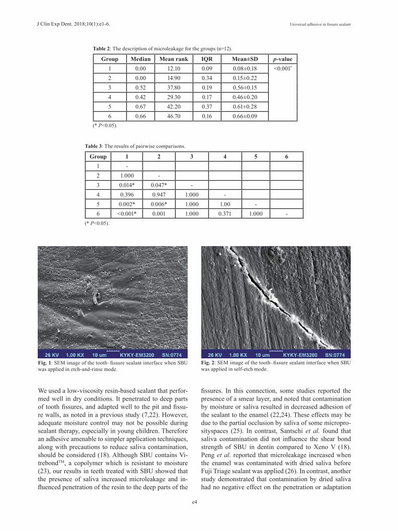
ResultsShapiro–Wilk tests indicated that the data were not nor-mally distributed in most groups. Table 2 summarizes microleakage for all groups. Overall, we found signifi-cant differences among groups (p<0.001). The results of pairwise comparisons (Table 3) indicated that micro-leakage was significantly lower in groups 1 (median=0, IQR=0.09) and 2 (median=0, IQR=0.34) compared to groups 3 (median=0.52, IQR=0.19), 5 (median=0.67, IQR=0.37), and 6 (median=0.66, IQR=0.16) (all p<0.05). However, there were no significant differences between other groups.The SEM images of all groups were assessed at the FS and enamel interfaces. Group 1 and 2 interfaces showed resin penetration into the enamel in the deepest points and lateral walls (Fig. 1). The application of SBU in self-etch mode (group 2) resulted in less demineraliza-tion and slight infiltration compared to SBU in etch-and-rinse mode (group 1) (Fig. 2). Saliva contamination pre-vented interlocking along the enamel–sealant interfaces, and the formation of gaps along the fissure sealant and lateral walls of the tooth surfaces. In addition, pooling of the saliva in the deepest part of the fissure led to frank gap formation in groups 3,5,6 (Fig. 3).
DiscussionThis study compared microleakage and SEM images of the FS–tooth interface in permanent teeth after the appli-cation of UA in etch-and-rinse and self-etch modes with or without saliva contamination. As in previous studies, all experimental groups in the present study showed some degree of microleakage (7,16). We found no signi-ficant differences in the extent of microleakage between the use of UA in etch-and-rinse or self-etch modes, or between either of these groups and the control group. This finding is consistent with a previous study that re-ported no difference in the sealing ability of SBU used in self-etch or etch-and-rinse mode (17). For pediatric patients, the self-etch technique as potential advantages especially in sealant therapy, because clinical procedures are simpler and technique sensitivity is lower (11,12).The bond strength of an adhesive is related to its ena-mel etching capacity as well as its mechanical properties (20). Scotchbond Universal, used in the present study, contains the acidic monomer10-methacryloyloxydecyl (10-MDP), which interacts chemically with calcium in the hydroxyapatite present in enamel. However, some reports have noted that UA is less acidic than phosphoric acid (10-12). Some researchers have therefore recom-mended selective pre-etching to increase bond strength between the composite and enamel (21).
J Clin Exp Dent. 2018;10(1):e1-6. Universal adhesive in fissure sealant
e4
Group Median Mean rank IQR Mean±SD p-value1 0.00 12.10 0.09 0.08±0.18 <0.001*
2 0.00 14.90 0.34 0.15±0.223 0.52 37.80 0.19 0.56±0.154 0.42 29.30 0.17 0.46±0.205 0.67 42.20 0.37 0.61±0.286 0.66 46.70 0.16 0.66±0.09
Table 2: The description of microleakage for the groups (n=12).
(* P<0.05).
Group 1 2 3 4 5 61 -2 1.000 -3 0.014* 0.047* -4 0.396 0.947 1.000 -5 0.002* 0.006* 1.000 1.00 -6 <0.001* 0.001 1.000 0.371 1.000 -
Table 3: The results of pairwise comparisons.
(* P<0.05).
Fig. 1: SEM image of the tooth–fissure sealant interface when SBU was applied in etch-and-rinse mode.
Fig. 2: SEM image of the tooth–fissure sealant interface when SBU was applied in self-etch mode.
We used a low-viscosity resin-based sealant that perfor-med well in dry conditions. It penetrated to deep parts of tooth fissures, and adapted well to the pit and fissu-re walls, as noted in a previous study (7,22). However, adequate moisture control may not be possible during sealant therapy, especially in young children. Therefore an adhesive amenable to simpler application techniques, along with precautions to reduce saliva contamination, should be considered (18). Although SBU contains Vi-trebondTM, a copolymer which is resistant to moisture (23), our results in teeth treated with SBU showed that the presence of saliva increased microleakage and in-fluenced penetration of the resin to the deep parts of the
fissures. In this connection, some studies reported the presence of a smear layer, and noted that contamination by moisture or saliva resulted in decreased adhesion of the sealant to the enamel (22,24). These effects may be due to the partial occlusion by saliva of some micropro-sityspaces (25). In contrast, Santschi et al. found that saliva contamination did not influence the shear bond strength of SBU in dentin compared to Xeno V (18). Peng et al. reported that microleakage increased when the enamel was contaminated with dried saliva before Fuji Triage sealant was applied (26). In contrast, another study demonstrated that contamination by dried saliva had no negative effect on the penetration or adaptation
J Clin Exp Dent. 2018;10(1):e1-6. Universal adhesive in fissure sealant
e5
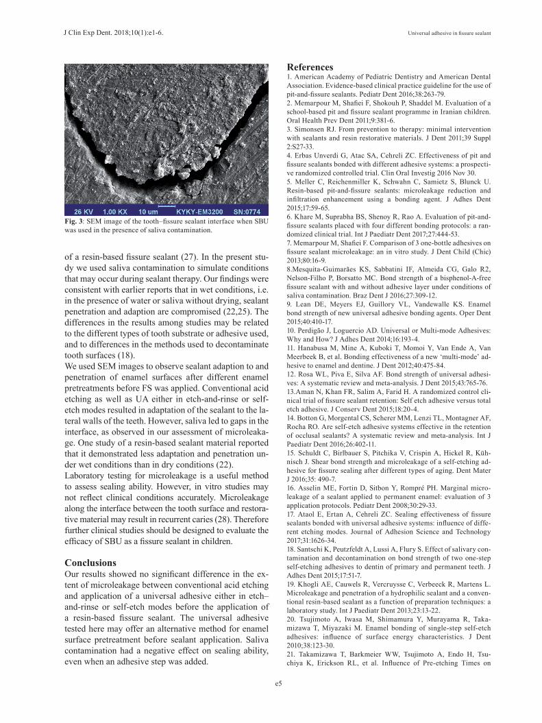
Fig. 3: SEM image of the tooth–fissure sealant interface when SBU was used in the presence of saliva contamination.
of a resin-based fissure sealant (27). In the present stu-dy we used saliva contamination to simulate conditions that may occur during sealant therapy. Our findings were consistent with earlier reports that in wet conditions, i.e. in the presence of water or saliva without drying, sealant penetration and adaption are compromised (22,25). The differences in the results among studies may be related to the different types of tooth substrate or adhesive used, and to differences in the methods used to decontaminate tooth surfaces (18).We used SEM images to observe sealant adaption to and penetration of enamel surfaces after different enamel pretreatments before FS was applied. Conventional acid etching as well as UA either in etch-and-rinse or self-etch modes resulted in adaptation of the sealant to the la-teral walls of the teeth. However, saliva led to gaps in the interface, as observed in our assessment of microleaka-ge. One study of a resin-based sealant material reported that it demonstrated less adaptation and penetration un-der wet conditions than in dry conditions (22).Laboratory testing for microleakage is a useful method to assess sealing ability. However, in vitro studies may not reflect clinical conditions accurately. Microleakage along the interface between the tooth surface and restora-tive material may result in recurrent caries (28). Therefore further clinical studies should be designed to evaluate the efficacy of SBU as a fissure sealant in children.
ConclusionsOur results showed no significant difference in the ex-tent of microleakage between conventional acid etching and application of a universal adhesive either in etch–and-rinse or self-etch modes before the application of a resin-based fissure sealant. The universal adhesive tested here may offer an alternative method for enamel surface pretreatment before sealant application. Saliva contamination had a negative effect on sealing ability, even when an adhesive step was added.
References 1. American Academy of Pediatric Dentistry and American Dental Association. Evidence-based clinical practice guideline for the use of pit-and-fissure sealants. Pediatr Dent 2016;38:263-79.2. Memarpour M, Shafiei F, Shokouh P, Shaddel M. Evaluation of a school-based pit and fissure sealant programme in Iranian children. Oral Health Prev Dent 2011;9:381-6.3. Simonsen RJ. From prevention to therapy: minimal intervention with sealants and resin restorative materials. J Dent 2011;39 Suppl 2:S27-33.4. Erbas Unverdi G, Atac SA, Cehreli ZC. Effectiveness of pit and fissure sealants bonded with different adhesive systems: a prospecti-ve randomized controlled trial. Clin Oral Investig 2016 Nov 30. 5. Meller C, Reichenmiller K, Schwahn C, Samietz S, Blunck U. Resin-based pit-and-fissure sealants: microleakage reduction and infiltration enhancement using a bonding agent. J Adhes Dent 2015;17:59-65.6. Khare M, Suprabha BS, Shenoy R, Rao A. Evaluation of pit-and-fissure sealants placed with four different bonding protocols: a ran-domized clinical trial. Int J Paediatr Dent 2017;27:444-53.7. Memarpour M, Shafiei F. Comparison of 3 one-bottle adhesives on fissure sealant microleakage: an in vitro study. J Dent Child (Chic) 2013;80:16-9.8.Mesquita-Guimarães KS, Sabbatini IF, Almeida CG, Galo R2, Nelson-Filho P, Borsatto MC. Bond strength of a bisphenol-A-free fissure sealant with and without adhesive layer under conditions of saliva contamination. Braz Dent J 2016;27:309-12.9. Lean DE, Meyers EJ, Guillory VL, Vandewalle KS. Enamel bond strength of new universal adhesive bonding agents. Oper Dent 2015;40:410-17.10. Perdigão J, Loguercio AD. Universal or Multi-mode Adhesives: Why and How? J Adhes Dent 2014;16:193-4.11. Hanabusa M, Mine A, Kuboki T, Momoi Y, Van Ende A, Van Meerbeek B, et al. Bonding effectiveness of a new ‘multi-mode’ ad-hesive to enamel and dentine. J Dent 2012;40:475-84.12. Rosa WL, Piva E, Silva AF. Bond strength of universal adhesi-ves: A systematic review and meta-analysis. J Dent 2015;43:765-76.13.Aman N, Khan FR, Salim A, Farid H. A randomized control cli-nical trial of fissure sealant retention: Self etch adhesive versus total etch adhesive. J Conserv Dent 2015;18:20-4.14. Botton G, Morgental CS, Scherer MM, Lenzi TL, Montagner AF, Rocha RO. Are self-etch adhesive systems effective in the retention of occlusal sealants? A systematic review and meta-analysis. Int J Paediatr Dent 2016;26:402-11.15. Schuldt C, Birlbauer S, Pitchika V, Crispin A, Hickel R, Küh-nisch J. Shear bond strength and microleakage of a self-etching ad-hesive for fissure sealing after different types of aging. Dent Mater J 2016;35: 490-7.16. Asselin ME, Fortin D, Sitbon Y, Rompré PH. Marginal micro-leakage of a sealant applied to permanent enamel: evaluation of 3 application protocols. Pediatr Dent 2008;30:29-33.17. Ataol E, Ertan A, Cehreli ZC. Sealing effectiveness of fissure sealants bonded with universal adhesive systems: influence of diffe-rent etching modes. Journal of Adhesion Science and Technology 2017;31:1626-34.18. Santschi K, Peutzfeldt A, Lussi A, Flury S. Effect of salivary con-tamination and decontamination on bond strength of two one-step self-etching adhesives to dentin of primary and permanent teeth. J Adhes Dent 2015;17:51-7.19. Khogli AE, Cauwels R, Vercruysse C, Verbeeck R, Martens L. Microleakage and penetration of a hydrophilic sealant and a conven-tional resin-based sealant as a function of preparation techniques: a laboratory study. Int J Paediatr Dent 2013;23:13-22.20. Tsujimoto A, Iwasa M, Shimamura Y, Murayama R, Taka-mizawa T, Miyazaki M. Enamel bonding of single-step self-etch adhesives: influence of surface energy characteristics. J Dent 2010;38:123-30. 21. Takamizawa T, Barkmeier WW, Tsujimoto A, Endo H, Tsu-chiya K, Erickson RL, et al. Influence of Pre-etching Times on

J Clin Exp Dent. 2018;10(1):e1-6. Universal adhesive in fissure sealant
e6
Fatigue Strength of Self-etch Adhesives to Enamel. J Adhes Dent 2016;18:501-11.22. Al-Jobair A. Scanning electron microscope analysis of sealant penetration and adaptation in contaminated fissures. J Indian Soc Pedod Prev Dent 2013;31:169- 74.23. Mitra SB, Lee CY, Bui HT, Tantbirojn D, Rusin RP. Long-term adhesion and mechanism of bonding of a paste-liquid resin-modified glass-ionomer. Dent Mater 2009;25:459-66.24. de Goes MF, Shinohara MS, Freitas MS. Performance of a new one-step multi-mode adhesive on etched vs non-etched enamel on bond strength and interfacial morphology. J Adhes Dent 2014;16:243-50.25. Silverstone LM, Hicks MJ, Featherstone MJ. Oral fluid contami-nation of etched enamel surfaces: An SEM study. J Am Dent Assoc 1985;110:329-32.26. Peng Y, Stark PC, Rich A Jr, Loo CY. Marginal microleakage of triage sealant under different moisture contamination. Pediatr Dent 2011;33:203-6.27. Duangthip D, Lussi A. Microleakage and penetration ability of resin sealant versus bonding system when applied following conta-mination. PediatrDent 2003;25:505-11.28. Kantovitz KR, Pascon FM, Alonso RC, Nobre-dos-Santos M, RontaniRM. Marginal adaptation of pit and fissure sealants after thermal and chemical stress. A SEM study. Am J Dent 2008;21:377-82.
Acknowledgements This work was supported by the Vice-Chancellory of Research of Shiraz University of Medical Science, Shiraz, Iran, (Grant No. 99-10090). We thank Dr. M. Vossoughi of the Center for Improvement, Shiraz Dental School, for the statistical analysis, and K. Shashok (AuthorAID in the Eastern Mediterranean) for improving the use of English in the manuscript.
Conflicts of InterestThe authors state that they have no conflicts of interest.