Embed Size (px)
Citation preview

Schemes of xonation in the mandibular condyle
W. A. Beresford, M.A., D.Phil. Morgantown, W. Vu.
A histologic study of the growth cartilage of the rat’s penile bone revealed many similarities to the mandibular condylar cartilage, including a distinctive pattern of layers. The literature on the growing mandibu1a.r condyle offers no generally accepted scheme for the recognition, numbering, and naming of its layers. The existing schemes are reviewed and tabulated here in the hope of arriving at one that holds true for the mandible and has an external validity in that it fits another growth cartilage.
In the growing condyle there is a complex sequence of cell proliferation, differentiation, and maturation, accompanied by partial destruction of the most mature cartilaginous elements and their replacement by bony trabeculae. At any one time all these events are taking place in different regions through the growth area. To a limited extent, one can infer from the morphology of the cells and surrounding matrix what is occurring at a particular place. Attempts to do this across the whole growth area have resulted in the various schemes for dividing the cartilage into named zones.
There are some general limitations to such schemes. First, names, as opposed to numbers, carry a heavy burden of implication. Having the “zone of interstitial growth” next to the “zone of maturing and mature cartilage” suggests that interstitial growth is not occurring in the maturing zone. The name for a zone should express one idea and not lend itself to other interpretations. Second, the boundaries between zones are at times restricted to a strip two or three cells wide; at other times the transitions are gradual and extend over areas many cells wide; boundaries within such transitional regions may be impossible to define. Third, as Schaffer13 pointed out, the zones are more easily distinguished at a certain time during growth and an additional layer may then be discerned. Should a layering scheme be based on such a period or on what is to be seen at all times during growth?
The growing mandibular condyle has been examined histologically by many
From the Department of Anatomy, West Virginia University.
189

Table I. Five- and six-zoned schemes for the mandibular condyle
1.
2. Chondrogenic 3. Small-celled cartilage 4. Large-celled cartilage
Apposition:\1 growth liesting cartilage l’,mbryonic. lntwstitial groxvtll Proliferating cartilage frrtermediabe
Hydropic tlt~grner:ttiorr Maturing cartilage Vwiculnr
5. Calcified cartilage Matrix dcifiwtion Calcifying cartilage *:rosion 6. Erosion and bone formation Matxia remov:d and bone Developing bone
apposit,ion By description Labeled photomicrograph Description Labeled
photomierograph
“Levy10 recognized the zones of Collins et al. as holding for the mouse.
t Symons acknowledged that Pritchard’sl? term was somewhat inappropriate for the condyle.
investigators working with primates, rodents, and other mammals. Some have published photomicrographs labeled to show the names and boundaries of their zones; others have described or tabulated the zones in the text of their results. The various schemes divide themselves into two classes: those favoring five cartilaginous and precartilaginons layers, and others describing only four layers. Table I gives the zonations of workers using five layers; Table II shows the schemes embracing four layers. The schemes of investigators employing additional layers and of Meikle,ll with even fewer, arc included in these tabula- tions in order to show that there is a general correspondence between schemes. A broken line indicates that the authors did not describe that layer specifically but its existence was implicit. The authors’ illustrations show some age differ- ences but no significant variations between species. The fifth column of Table II relates the system of layering suggested in this article to those already in existence.
Within each class there is good agreement in terminology, reflecting what the authors think is happening, and in the plac.ing of lines of demarcation for the first four or three layers. The fact that Table T includes some schemes with six and Table II includes one with five layers immediat,cly points to a difficulty at the eroding face of the cartilage. All authors distinguish between maturing or hypertrophic cartilage and calcified caartilage, but the zone of calcifying cartilage is sometimes termed that of erosion and/or osteogenesis and elsewhere is identified by others as a zone in it,s own right to bc followed by a fifth or sixth zone of erosion or bone formation.
A smoothly curved line can be drawn through the apparent points of deepest penetration by the vessels and chondroclasts, but one should remember t,hat, with such a seemingly haphazard pattern of rrosion and single sections, it is imposGble to be certain that, a particular lacuna dots not run deeper. Above that line there will be a narrow zone of calcified cart,ilage i for example, in Fig. 14 of Durkin” and Figs. 6. 9, and 15 of Durkin, Heelp, and Irving,’ who offer a subdivision of the hppertrophic zone into mineralized and nonmineralizcd

Volume 68 Number 2 S&emes of zonation. in man&b&w coadyle 191
C~unat et al.4
I
symons~~
I
Duterloos Rat Rat Rat I
Kmmuse et al.9 Monkey
- Perichondrialt Articular Articular fibroblasts
Mesodermal cells Prechondroblasts Proliferating tntermediate Transition Chondroblasts Transitional Differentiation and interstitial growth Swollen chondrocytes Hypertrophic Cartilage Maturing and mature cartilage
chondrocytes Calcified matrix - Erosion Erosion and new bone formation Destruction Cartilaginous
trabeculae Description Labeled Labeled Labeled photomicrograph
photomicrograph photomicrograph
Table II. Four- and five-zoned schemes for the mandibular condyle
Ilzlrkin et al.7 Prommer~ Mm and rodents Mouse
Rlackwoo@ Rat
Meiklel f I
Beresford Rat Rat
1. Resting or surface - Articular Articular Fibrous articular
2. Transitional or Chondrogenic Intermediate Proliferative Chondrogenic proliferative
3. Hypertrophic Cartilaginous Hypertrophic Condylar cartilage Cartilaginous 4. Erosion Osteogenic Resorption Condylar cartilage Erosive 5. Subchondral bone
formation By description Labeled Description Labeled Labeled
photomicrograph photomicrograph pl~otomicrograpl~
portions). The six-zoned schemes of Schaffer,13 Schour,14 and Seipp15 arrive at a sixth zone by recognizing such a distinction within the hypertrophic cartilage. This subdivision cannot be identified with certainty unless the mineral is seen with the electron microscope as crystals or preserved and stained for specifically. Below the line of deepest erosion is a region of many events. Erosion of calcified cartilage continues, sparing only very small islands of matrix upon which new bone is deposited, some of which is promptly destroyed. Also present are growing vascular and hematopoietic elements. The defects of the various names for this zone below the line are these: “Calcified cartilage” overlaps in a seriously misleading way with the region of intact, calcified, hypertrophic cartilage above the eroded lacunae. ‘(Resorption” and “erosion” do not convey either that the resorption is partial or that, since bone is also being destroyed, an “eroding” zone extends without limit into the subjacent bone. “Osteogenic” does not admit that resorption precedes bone formation, although the lag in distance is very small and, with the irregular penetration, there is considerable overlap in activities; even within the same small eroded cavity, resorption of cartilage may
192 Beresford
Fig. 1. A low-power view of the mandibular condyle of a 21-day-old rat stained with
hematoxylin and eosin. Layers are discernible although unlabeled. (Magnification, xl 0.)
be level with osteogenesis on its other side. Like the one t,ewmed ‘Lresorption,” an “osteogenic” zone knows no limit, in the trabecular bone.
The more practical of the last two choices is “erosion,” since a line is more easily visualized through the ends of eroded lacunae than the tapered ends of the most recently formed seams of boric. All that has been done is to define a line serving as one kind of limit to the cartilage and marking the start of a zone lacking its other border.
Leaving the problems of the zone of erosion, the essential difference between the five- and four-layered schemes hinges on whrther or not the chondrogenic layer ran be divided into one of less differentiated cells and another of cells more like chondrocptcs. The problem seems to be this. Yes, at certain times one can readily recognize cells nearer the hgpertrophic zone as looking more chondrocytic than those closer to the articulation. Howevclr, since the transitional forms are so numerous, varied, and dispersed that a precise line of demarcation cannot bc drawn ant1 defined, such a name as “chondroblastic” or “transitional” has a value to match the vagueness of its territoyv. Although Kanouse, Ramfjord, and Xasjlet? worked with five layers, Blackwood, also using a radioactive isotope to study cell proliferation and needing clearly defined zones in which to count labeled (‘ells, employed four layers and even thtln remarked on the difficulty of delineating his intermediate and hypertrophic zones.
Another drawback to the five-laycred schemes is that the extra lapcr and its name are again a potential source of great confusion. For example, the inter- mediate layer of Kanouse and associates” is not the same as the layer given that name by Collins and colleagues” and differs ?-et, from the intcrmediatc of

Schemes of xowtion in mclndibulnr coadyle 193
Fig. 2. A low-power view of the growth cartilage of the penile bone of a 21-day-old rat stained with hematoxylin and eosin. Above the cartilage is erectile tissue; below it are
bone trabeculae. (Magnification, xl 0.)
BlackwoodY four layers. A future technique may reveal clear-cut and generally acceptable dividing lines in the region between the undifferentiated cells and the chondrocytes. Meanwhile, the scheme that offers least confusion, a brief terminology with the fewest unsubstantiated implications, and boundaries that all investigators can recognize and agree upon with the usual histologic methods, merits consideration. The one proposed here has four layers and a hybrid terminology owing most to Frommer .* Thus, as illustrated in Fig. 3, zone 1 would be fibrous, zone 2 would be chondrogenic, zone 3 would be cartilaginous, and zone 4 would be erosive. For the reasons outlined earlier, the zone of erosion is not shown as having a lower limit, and the cartilaginous zone includes un- stained the band of calcified but uneroded matrix.
The close resemblance of the mandibular condylar cartilage (Figs. 1 and 3) to the growth cartilage of the penile bone (Figs. 2 and 4) in the rat is apparent in the convex shape, layering, disorderly distribution of the chondrocytes, irregular pattern of erosion, and haphazard arrangement of the underlying bone trabeculae. When one bears in mind that the penile cartilage is smaller and its fibrous perichondrial layer merges with the connective tissue of the erectile corpus fibrosum, the four zones chosen to subdivide the condyle (Fig. 3) fit well the appearance and relative width of the layers in the penile cartilage (Fig. 4). Other similarities and differences between the structures will be presented later. Baumel has spoken of the unique nature of the mandib- ular condyle. The rat deploys endochondral cartilages in its penile bone and mandible in ways sufficiently similar to call into question the point of emphasiz- ing the undoubted uniqueness of either structure.
194 Keyesford ..l?JL. J. Ortkod. August 1975
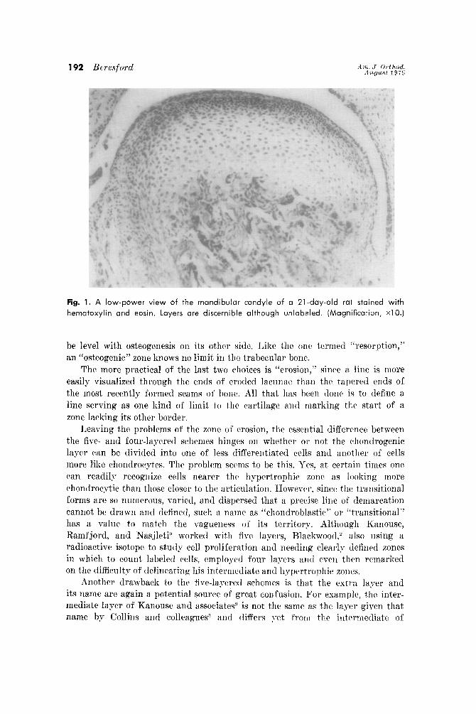
Fig. 3. A view of roughly one quarter of the mandibular condyle of a 21-day-old rat
stained with hematoxylin and eosin. From above, the four layers marked are fibrous,
chondrogenic, cartilaginous, and erosive. (Magnification, x25.1
Fig. 4. A view of roughly one third of the growth cartilage on the penile bone of a 19-
day-old rat stained with hematoxylin and eosin. From above, the four layers marked are
fibrous, chondrogenic, cartilaginous, and erosive. The fibrous layer merges with the con-
nective tissue of the erectile corpus tibrosum. (Magnification, x25.1
K. Ellifritz, M. Kent, B. Bragg, K. Smith, and C. Roberts are thanked for technical and secretarial help. West Virginia Universit,y Schools of Dentistry and Medicine provided the funds for this study.
REFERENCES 1. Baume, L. J.: Differential response of condylar, epiphyseal, synchondrotic, and articular
cartilages of the rat to varying levels of vitamin A, A&r. J. ORTHOIL 58: 537-551, 1970. 2. Blackwood, H. J. J.: Growth of the nyandibular condyle of the rat studied wit,h tritiated
thymidine, Arch. Oral Biol. 11: 493-500, 1966. 3. Collins, D. A., Recks, H., Simpson, M. E., and Evans, H. M.: Growth and transformation
of the mandibular joint in the rat. I. Normal female rats, AM. J. ORTEIOD. 32: 431-442,
1946.
4. Cunat, J. J., Bhaskar, S. N., and Weinmann, J. I’.: Development of the squamoso- mandibular articulation in the rat, J. Dent. Res. 35: 533-546, 1956.
5. Duterloo, H. S.: In vivo transplantation of the mandibular condyle of the rat, Ph.D. thesis, University of Nymegen, Netherlands, 1967.
6. Durkin, J. F. : Secondary cartilage: A misnomer? AM. J. OW~HOD. 62: 15-41, 1972.
i. Durkin, J. F., Heeley, J. I)., and Irving, J. T.: The cartilage of the mandilmlnr condyle, Oral Rci. Rev. 2: 29-99, 1973.
8. Frommer, J.: Prenatal development of the mandibular joint in mice, hat. Rec. 150: 449-462 1964.
9. Kanousi, M. C., Ramfjord, S. P., and Sasjleti, C. E.: Contlylar growth in rhesus monkeys, J. Dent. Res. 48: llil-1176, 1969.
10. Levy, B. AI.: Growth of mandibular joint in normal mice, J. Am. Dent. Assoc. 36: 177. 182, 1948.
11. Rleikle, RI. C.: In viro tranxplautatiou of the mandilmlnr joint of the rat; an nutorxdio- graphic investigation into cellular changes at the condyle, Arch. Oral Biol. 18: 1011-1020, 1973.
12. Pritchard, J. J.: A cytological and l~istocl~emicnl study of bone and cartilage formation in the rat, J. hat. 86: 259-277, 1952.
13. Schaffer, J. : Die VerknGeherung des Unterkiefers und die Metaplasiefragc, Arch. Mikrosk. hat. 32: 266-377, 1888.
14. Scliour, I.: Soyes’ oral histology and embryology, ed. 8, Philadelphia, 1960, Lea & Febiger.
15. Seipp, J. H., Jr.: The temporomandibular joint. 111, Provenzn, D. V. (editor) : Oral histology, Philadelphia, 1964, J. B. Lippincott Company.
16. Symons, N. B. B.: A l~istocl~emieal study of the secondary cart,ilage of the mandibular condyle in the rat, Arch. Oral Biol. 10: 5i9-584, 1965.


![Current Advances in Mandibular Condyle Reconstruction · The LIPUS is considered the preferred method of mechanical stimulation, also known as “preferred bioreactor” [25]. 5](https://img.dokumen.tips/doc/110x75/5e96a9d67ba2de640562addd/current-advances-in-mandibular-condyle-reconstruction-the-lipus-is-considered-the.jpg)