Embed Size (px)
Citation preview

Sara E. Ortiz-Romero, MD
Pediatric Radiologist
OAK LAWN

Disclosures
Nothing to disclose

Outline
Clinical diagnosis of acute appendicitis.
Imaging modalities and their utility in the diagnosis of acute appendicitis
Ultrasound (US)
Computed Tomography (CT)
Magnetic Resonance Imaging (MRI)
Imaging for complications of acute appendicitis

Acute Appendicitis
Most common condition that requires acute surgical intervention in the pediatric population.
Continues to cause significant morbidity despite improvement in its diagnosis and treatment.
Complicated appendicitis can be prevented by earlier diagnosis.

Epidemiology
Approximately 70,000 – 80,000 children are diagnosed annually in the Unites States
1/1000 children per year
Lifetime risk
9% for boys
7% for girls
Peak incidence between 11 and 12
years of age
Uncommon in children under 2 years

Presentation
Classic Symptoms of Acute Appendicitis
Crampy periumbilical pain that migrates to
Right Lower Quadrant (RLQ)
Nausea and vomiting
Anorexia
Fever
Point tenderness is RLQ
Leukocytosis with left shift

Alvarado and Pediatric Appendicitis Scores

Diagnostic dilemma
Clinical diagnosis of acute appendicitis is not always straightforward.
Abdominal pain is a common complaint in the pediatric population:
4% of doctor visits
Most times is a self-limited condition
○ Viral syndrome, gastroenteritis, constipation, pharyngitis, among others.

Diagnostic Dilemma
What about the classical symptoms?? Only one third of patients present with classical clinical
symptoms.
Scores: A large number of patients fall in the equivocal range.
Patients younger than 5 years cannot describe symptoms clearly.
False-negative appendectomy rate: 11.83% from 1998-2007* Declining
○ 14.7% in 1998
○ 8.47% in 2007
* Seetahal SA et al. Negative appendectomy: a 10-year review of a nationally representative sample. Am J Surg. 2011 Apr;201(4):433-7.
Morbidity and Mortality
Delayed diagnosis can lead to perforation
Complications: abscess, sepsis, infertility, bowel
obstruction and death.
Perforation rate in the pediatric population
is approximately 30%*
Higher in preschool children
Death from appendicitis in general
population is 1%
*Barrett ML, et al. Trends in Rates of Perforated Appendix, 2001-2010: Statistical Brief #159. Healthcare Cost and Utilization Project, July2013

Why imaging?
Help diagnose appendicitis earlier, therefore
preventing complications.
Reduce negative appendectomy rate
Reduce length of stay and cost of treatment

Ultrasound

US for Pediatric Appendicitis
Graded-compression US has been used since mid 1980’s in the diagnosis of appendicitis
Diagnostic accuracy has varied widely
Pooled Sensitivity: 88%; Pooled Specificity: 94%*
In experienced hands and with the appropriate patient, it is a great imaging modality
*Doria AS, et al. US or CT for Diagnosis of Appendicitis in Children and Adults? A Meta-Analysis. Radiology. 2006 Oct;241(1):83-94.

Advantages of US
Good in pediatric population because of small patient size.
Fast and inexpensive modality.
Portable
No radiation
Assess the appendix for compression
Assess patients clinically

Protocol of US in Appendicitis
Order: US Appendix
Ask patient to point with one finger where it hurts the
most and image there
Compress Right Lower Quadrant (RLQ) attempting to
decompress air filled bowel
○ Psoas muscle and iliac vessels are visualized.
Identify ascending colon move transducer inferiorly to
identify terminal ileum
The appendix should be 1-2 cm below terminal ileum

Normal findings
Smaller than 6 mm
Compressible
No free fluid
Normal hypoechoic muscular layer and echogenic mucosa

Normal findings
Smaller than 6 mm
Compressible
No free fluid
Normal hypoechoic muscular layer and echogenic mucosa

Acute appendicitis
Larger than 6 mm
Non-compressible
Hypervascular
Appendicolith
Associated findings:
Periappendiceal fat
Free fluid: adjacent to appendix and RLQ
Abscess
Point tenderness over appendix

Acute appendicitis
Larger than 6 mm
Non-compressible
Hypervascular
Appendicolith
Associated findings:
Periappendiceal fat
Free fluid: adjacent to appendix and RLQ
Abscess
Point tenderness over appendix

Acute appendicitis
Larger than 6 mm
Non-compressible
Hypervascular
Appendicolith
Associated findings:
Periappendiceal fat
Free fluid: adjacent to appendix and RLQ
Abscess
Point tenderness over appendix

Most cases: Not Visualized

Disadvantages
Very limited in obese and gassy patients
Limited in the setting of severe pain preventing adequate compression
Operator dependent:
Gives up too fast.
Different location of appendix

Not that simple
Appendix can be in
different locations
Most common is
retrocecal
Air in cecum
obscures its
visualization
http://www.anatomyatlases.org/AnatomicVariants/OrganSystem/Images/08.shtml

Different location

Disadvantages
Very limited in obese and gassy patients
Limited in the setting of severe pain preventing adequate compression
Operator dependent:
Gives up too fast.
Different location of appendix
Looking too superficial in the RLQ

Looking too superficial


The diagnosis of appendicitis in children continues to be a challenging endeavor, despite advances in laboratory and imaging diagnosis. There is increasing concern for life-time radiation-induced malignancy risk associated with the use of computed tomography (CT). The study by Mittal et al provides both good and bad news about the use of US as the primary imaging modality for the diagnosis of suspected appendicitis. The good news in this multicenter observational study is that US had a specificity rate of >96% across all centers studied. The bad news is that the sensitivity was only 77% at the clinical sites with the highest utilization, and as low as 35% in those sites with the lowest use. This study makes clear that, regarding US for appendicitis, practice makes “better,” but not “perfect.” Thus, increasing a center’s experience with US will only go so far in improving diagnosis. Fortunately, there are several studies showing that US followed by CT in patients with nondiagnostic US studies is an efficient and effective approach.1 Used together with validated decision support rules, the high specificity of US for appendicitis eliminates the need for many CT scans while preserving overall diagnostic accuracy in the clinical environment.2 Early studies also point to a potential role for MRI as a substitute for CT in diagnostic protocols.

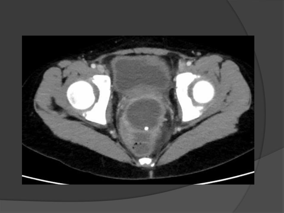
Computed Tomography

Computed Tomography
Excellent modality for the evaluation of
acute appendicitis
Diagnostic accuracy better than US
Pooled Sensitivity: 94%; Pooled Specificity: 95%*
Not operator dependent
Identifies other pathology easier than US *Doria AS, et al. US or CT for Diagnosis of Appendicitis in Children and Adults? A Meta-Analysis. Radiology. 2006 Oct;241(1):83-94.

Computed Tomography
Although a great test, it is not perfect.
Maximize the protocol to visualize appendix.
Visualization of appendix is directly related to
amount of peritoneal fat*
Little peritoneal fat: seen in 36% of cases
Moderate to marked fat: seen in 69% of cases
*Grayson DE, et al. Appendiceal CT in pediatric patients: relationship of visualization to amount of peritoneal fat. Am J Roentgenol. 2001 Feb;176(2):497-500.


Computed Tomography
Different ways to order
Abdomen/Pelvis versus Pelvis only


Computed Tomography
Different ways to order
Abdomen and Pelvis versus Pelvis only
Intravenous contrast
Enteric contrast:
○ Oral versus rectal contrast
Appendicitis Protocol at our hospital
Pelvis Only with IV and rectal contrast

Computed tomography
Normal appendix: tubular and blind ending
structure arising from cecum
Air/contrast filled
7 mm or less in children
The appendix can be larger and normal as long as
there is no surrounding inflammation
No RLQ inflammatory changes
Another cause for pain


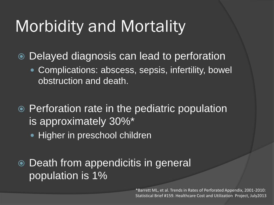
CT- Acute appendicitis
Abnormal appendix Thick measuring greater than 7 mm in diameter
from outer to outer wall
Fluid filled
Hyperenhancement of wall
Wall discontinuity in perforation
Appendicolith
Surrounding inflammation and free fluid
Reactive lymph nodes
Complications


Main disadvantage of CT
Radiation exposure Children are more radiosensitive than adults
5 mSv with correct age-adjusted parameters*
○ Low but not negligible as it is not known how this
will increase cancer risk
*Strouse PJ. Pediatric appendicitis: an argument for US. Radiology. 2010 Apr;255(1):8-13.

Radiation reduction
Dedicated pediatric CT parameters
Which can adjust dose to age and size
Limiting the area of imaging
Combining sonography and/or
appendicitis scoring techniques with CT


What else can be done to reduce
radiation exposure??

Magnetic Resonance Imaging

Magnetic Resonance Imaging
Advantages
No radiation
Not operator dependent like CT
CT like images
Disadvantages
Long scan times
Limited availability
Highly sensitive to motion
Scary for children: noise and small space
Expensive

Evidence for MRI

Evidence for MRI
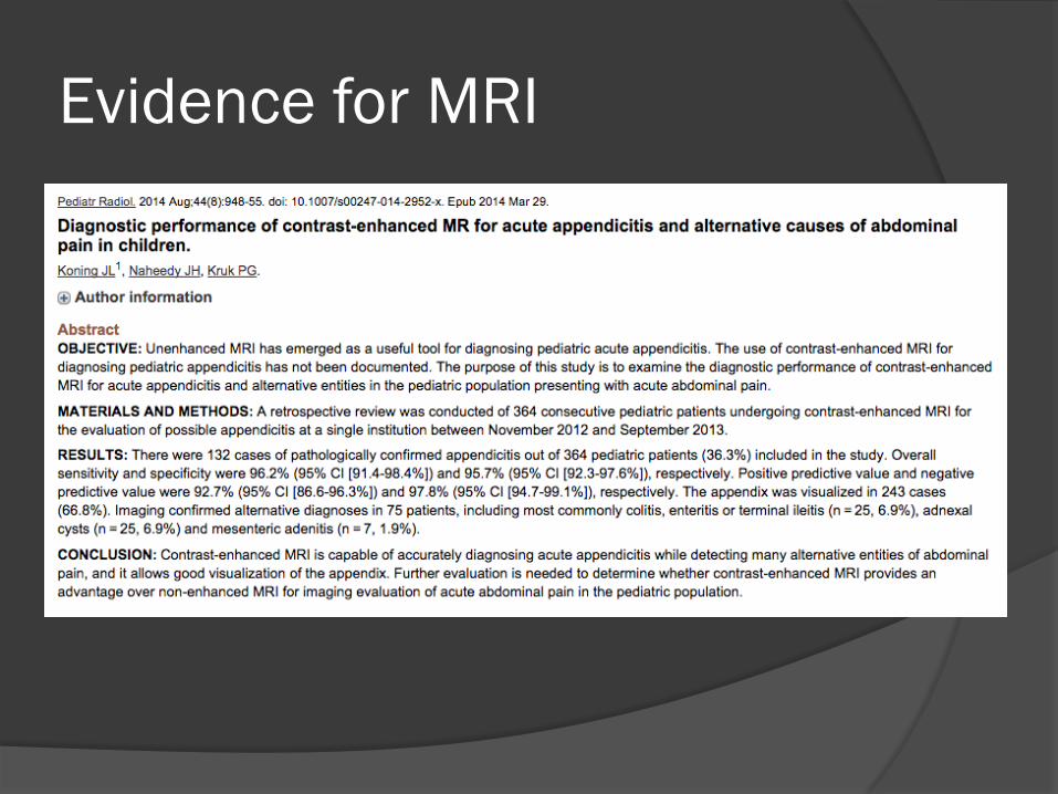
Evidence for MRI
Magnetic Resonance Imaging
Given advantages and mounting
evidence for MRI in the diagnosis of
acute appendicitis, we decided to start
an Appy MRI program in our ED which
started on 2/9/15.

Our MRI Program
Done in conjunction with the ED, pediatric
surgery, pediatrics and child life department.
Did not require IRB approval
M-F, exams ordered through ED 8AM – 5PM
Age guidelines: 7 years and older
Limited length of study (goal < 20 min.)
Goal: Consistently move the needle towards or
away from appendicitis, limiting the use of CT
Support Image Gently and ALARA principles

Our current MRI protocol
Axial SS FSE T2 FS (breath hold)
Coronal SS FSE (breath hold)
Axial pre-contrast
Axial and Coronal post (LAVA)

MRI in Appendicitis
Negative
Normal appendix less
than 7 mm thick
No inflammation or
large amount of free
fluid
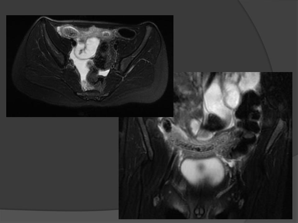
Positive Fluid-filled and thick
appendix measuring
>7mm
Wall hyperenhancement
Appendicolith
Inflammatory changes
Free fluid
Abscess
MRI in Appendicitis
Normal appendix can be difficult to
visualize in MRI
Like in CT the lack of secondary findings
is highly suggestive of a normal study*
Try to place results in terms of:
Positive
Equivocal positive
Indeterminate
Equivocal negative
Negative *Moore MM, et al. MRI for clinically suspected pediatric appendicitis: case interpretation. Pediatr Radiol. 2014 May;44(5):605-12.


MRI in Appendicitis
Alternative diagnosis and unexpected findings:
Most negative cases have no clear alternate
diagnosis
Hydronephrosis
Musculoskeletal abnormality
Horseshoe kidney
Ovarian cyst

Right Hydronephrosis

Horseshoe Kidney

Right ovarian hemorrhgic cyst
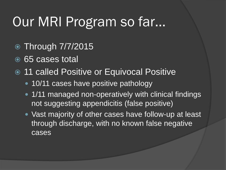
Our MRI Program so far…
Through 7/7/2015
65 cases total
11 called Positive or Equivocal Positive
10/11 cases have positive pathology
1/11 managed non-operatively with clinical findings
not suggesting appendicitis (false positive)
Vast majority of other cases have follow-up at least
through discharge, with no known false negative
cases

Dealing with MRI disadvantages
Long Scan Times
Time to image patients (scan time goal is
average of 20 minutes or less):
○ Time from start to finish imaging
(April/May/June):
Average: 21 minutes
Median: 19 minutes

Dealing with MRI disadvantages
Limited availability
Time to get patients on the table (goal is
average of 120 minutes or less):
○ Time from exam order to start imaging
(April/May/June):
Average: 97 minutes
Median: 78 minutes
Using Pediatric Appendicitis Score to guide
which cases get MRI
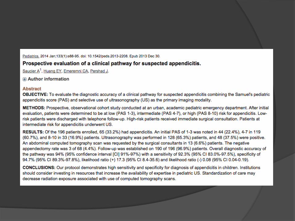

PAS in Article
PAS Score
0-3: Low likelihood
4-7: Intermediate
likelihood
8-10: Elevated likelihood
In the article:
196 patients followed
65 positive appendicitis cases
0/65 positive cases had low
PAS
31% of intermediate cases were
positive
85% of elevated cases were
positive
1 of 3 negative appendectomies
had a low PAS

Dealing with MRI disadvantages
Motion and Scary for children
No sedation has been used
Patients over 7 years of age
Child life involved in program preparing
patients for the MRI
Short scan time

Dealing with MRI disadvantages
Cost
We have been tracking payment
Difficult to accurately gauge cost
Comparing MRI alone to US +/- CT or MRI
An area of future research

What we have learned so far?
MRI is excellent imaging modality for acute
appendicitis without ionizing radiation


Presentation and Symptoms
The longer the symptoms the more likely
to have complications
More common in children younger than
5 years
Generalized abdominal pain
Very elevated fever and white count

Imaging Findings
Very dilated appendix which
Wall discontinuity
Large amount of free fluid
Pneumoperitoneum
Extraluminal appendicolith
Abscess

Importance
Although different opinions on how to treat,
some surgeons prefer non-operative
treatment of complicated appendicitis
Treat abscess, antibiotics and then do interval
appendectomy
Hostile abdominal environment for surgery
Decreases further complications
Earlier recovery

What imaging modality?
Ultrasound or now with MRI to start
Abscess: IR consult for percutaneous drainage
Vast majority of cases a CT will be ordered
Evaluate for additional collections
Better delineate anatomy
IR will drain the abscess
With CT or US Guidance


Complications after Appendectomy
Patients with persistent fever and
abdominal pain despite appendectomy
When to image?
Immediately post op patients will have
heterogeneous free fluid
Post operative abscess takes at least 5 days
to develop

Journal of Surgical Research

Complications after Appendectomy
What imaging modality?
Start with ultrasound to look for fluid on at
least day 5 post appendectomy
A CT may be needed to confirm presence of
abscess and for drainage planning


Conclusions Acute appendicitis, although common, remains a diagnostic
dilemma and can cause significant morbidity.
CT remains the gold standard in the imaging of acute
appendicitis but utilizes ionizing radiation
MRI is an excellent modality for the diagnosis of acute
appendicitis but is not always feasible
CT is still widely used, especially in the setting of complicated
appendicitis. Therefore, radiation reducing techniques are
critically important.