Embed Size (px)
Citation preview
Pediatric Head Trauma Imaging Do’s & Don’ts
Rachel Pevsner, D.O.
Pediatric Radiologist
Special thanks to Ricardo Restrepo, MD & Luisa Cervantes, MD
Objectives
• Understand mechanisms of head injury: ACCIDENTAL VS NON-ACCIDENTAL
• Ordering appropriate imaging
• CT vs Radiography for trauma
• Image gently: radiation and imaging
• How to report child abuse (see appendix)
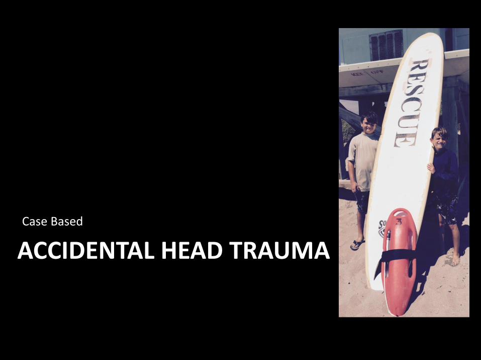
ACCIDENTAL HEAD TRAUMA Case Based
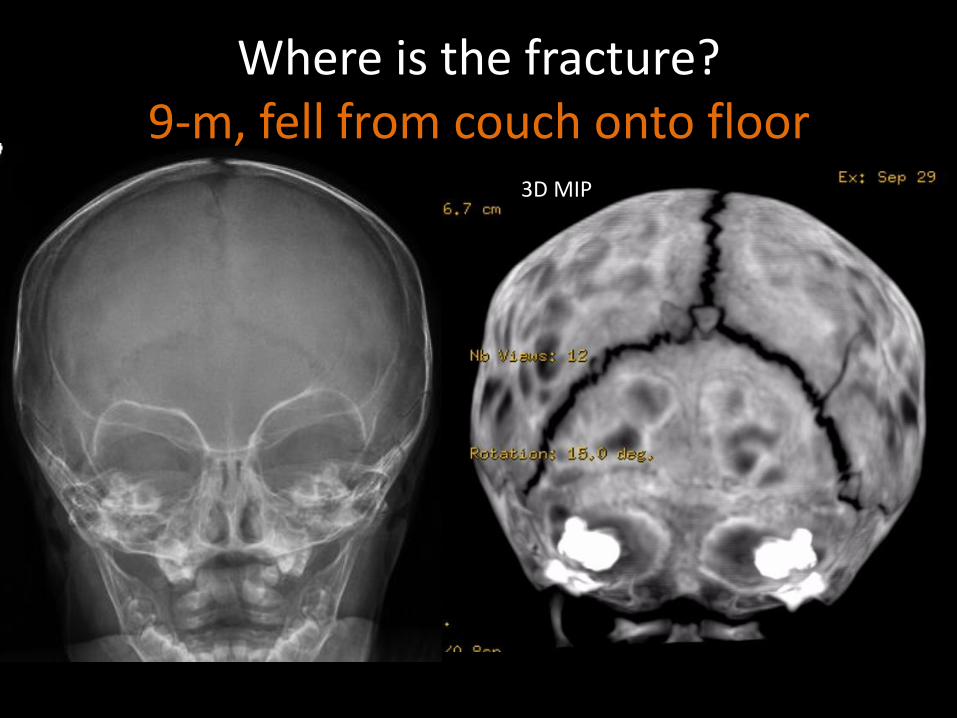
3D MIP
Where is the fracture? 9-m, fell from couch onto floor
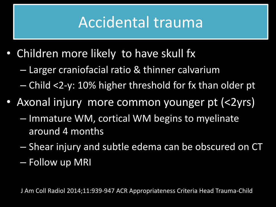
Accidental trauma
• Children more likely to have skull fx
– Larger craniofacial ratio & thinner calvarium
– Child <2-y: 10% higher threshold for fx than older pt
• Axonal injury more common younger pt (<2yrs)
– Immature WM, cortical WM begins to myelinate around 4 months
– Shear injury and subtle edema can be obscured on CT
– Follow up MRI
J Am Coll Radiol 2014;11:939-947 ACR Appropriateness Criteria Head Trauma-Child
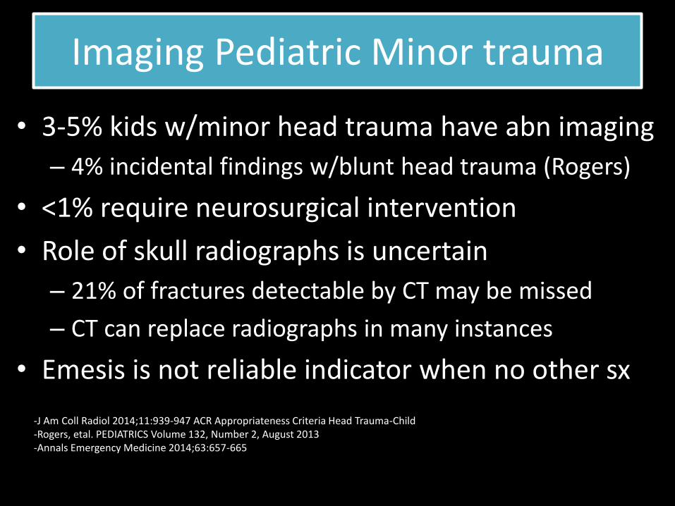
Imaging Pediatric Minor trauma
• 3-5% kids w/minor head trauma have abn imaging
– 4% incidental findings w/blunt head trauma (Rogers)
• <1% require neurosurgical intervention
• Role of skull radiographs is uncertain
– 21% of fractures detectable by CT may be missed
– CT can replace radiographs in many instances
• Emesis is not reliable indicator when no other sx
-J Am Coll Radiol 2014;11:939-947 ACR Appropriateness Criteria Head Trauma-Child -Rogers, etal. PEDIATRICS Volume 132, Number 2, August 2013 -Annals Emergency Medicine 2014;63:657-665

When to Image? • Imaging not routine in minor trauma
– normal post event mental status. No neuro signs or sx.
• Imaging when clinical exam is indeterminate – Clinical eval can be difficult in children, preverbal.
• PECARN (pediatric emergency care applied research network) prediction rule – predicts need for imaging
– largest Prospective cohort study 42,412 <18yrs
– out performed CHALICE & CATCH on validation studies
– High sensitivity • 100% <2yrs & 98% >2yrs
Lancet 2009; 374: 1160–70 Arch Dis Child 2014;99:427–431
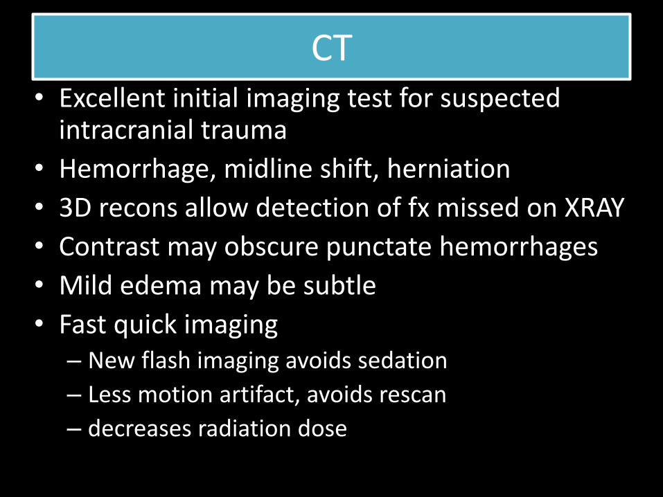
CT • Excellent initial imaging test for suspected
intracranial trauma
• Hemorrhage, midline shift, herniation
• 3D recons allow detection of fx missed on XRAY
• Contrast may obscure punctate hemorrhages
• Mild edema may be subtle
• Fast quick imaging – New flash imaging avoids sedation
– Less motion artifact, avoids rescan
– decreases radiation dose
CT: WHAT ABOUT THE RADIATION?
Radiation sickness or acute radiation syndrome is an immediate or acute reaction of the human body to radiation when the dose exceeds a specific
level or threshold
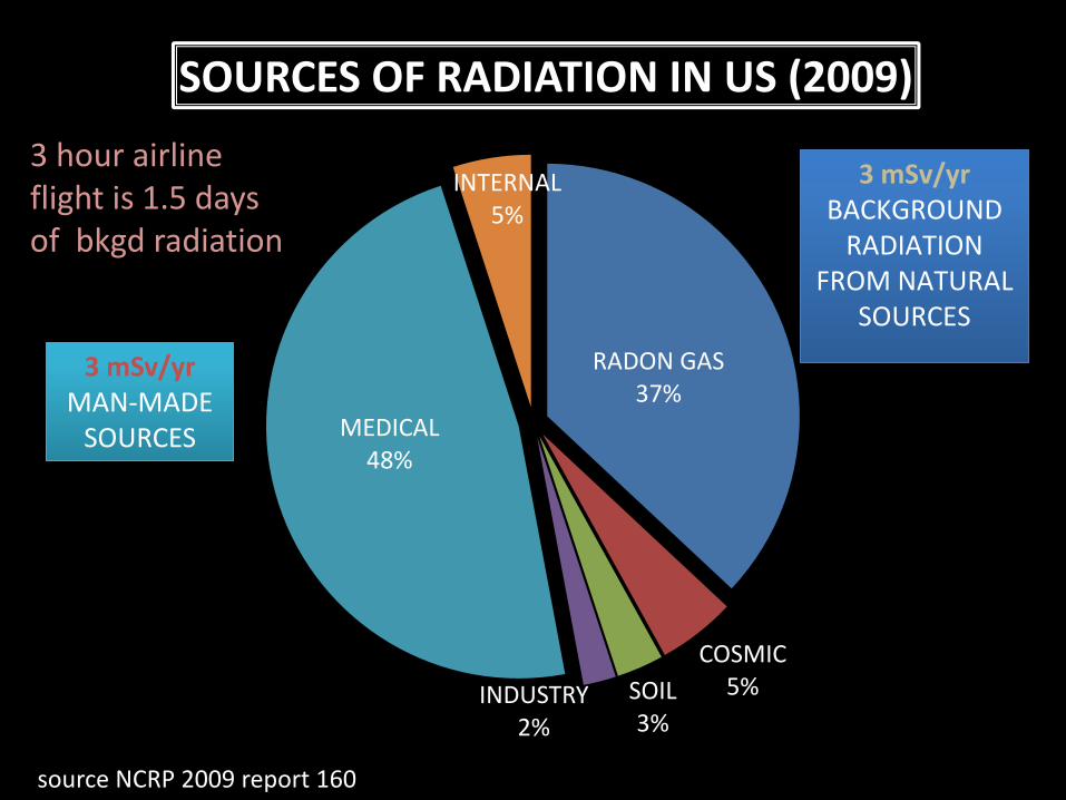
RADON GAS 37%
COSMIC 5% SOIL
3% INDUSTRY
2%
MEDICAL 48%
INTERNAL 5%
SOURCES OF RADIATION IN US (2009)
3 mSv/yr BACKGROUND
RADIATION FROM NATURAL
SOURCES
source NCRP 2009 report 160
3 mSv/yr MAN-MADE
SOURCES
3 hour airline flight is 1.5 days of bkgd radiation
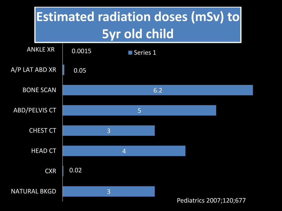
3
0.02
4
3
5
6.2
0.05
0.0015
NATURAL BKGD
CXR
HEAD CT
CHEST CT
ABD/PELVIS CT
BONE SCAN
A/P LAT ABD XR
ANKLE XR
Estimated radiation doses (mSv) to 5yr old child
Series 1
Pediatrics 2007;120;677
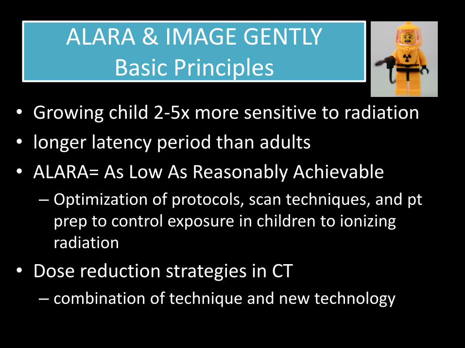
ALARA & IMAGE GENTLY Basic Principles
• Growing child 2-5x more sensitive to radiation
• longer latency period than adults
• ALARA= As Low As Reasonably Achievable
– Optimization of protocols, scan techniques, and pt prep to control exposure in children to ionizing radiation
• Dose reduction strategies in CT
– combination of technique and new technology
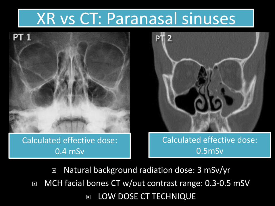
Calculated effective dose: 0.5mSv
Calculated effective dose: 0.4 mSv
Natural background radiation dose: 3 mSv/yr
MCH facial bones CT w/out contrast range: 0.3-0.5 mSV
LOW DOSE CT TECHNIQUE
XR vs CT: Paranasal sinuses
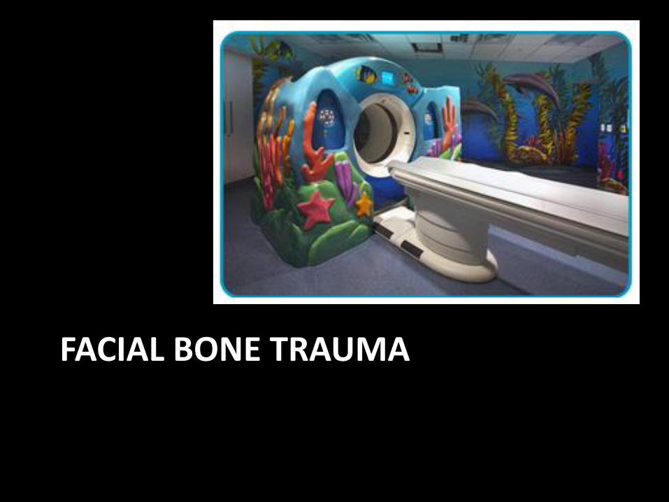
FACIAL BONE TRAUMA
Facial Bone Fractures
• Interpretation pedi facial XR is challenging
– Particularly midfacial and condylar fractures
• CT is best modality
• Facial fx lowest infants and inc with age
• Usually associated severe trauma-MVA & sports
• Nasal bone fx most common followed by mandibular fx
RadioGraphics 2008; 28:441–461
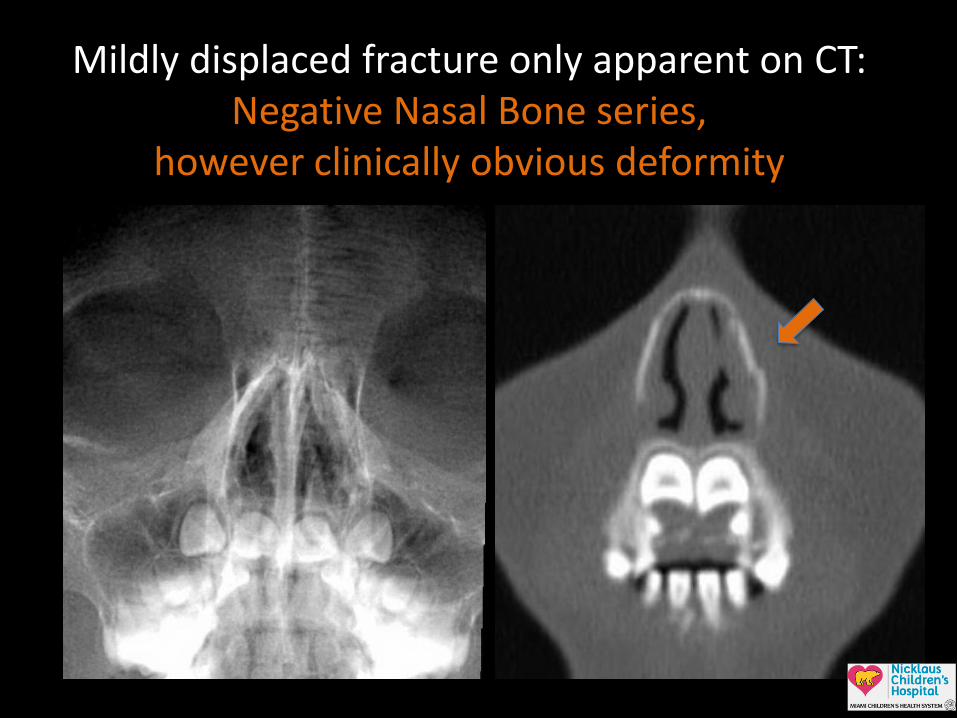
Mildly displaced fracture only apparent on CT: Negative Nasal Bone series,
however clinically obvious deformity
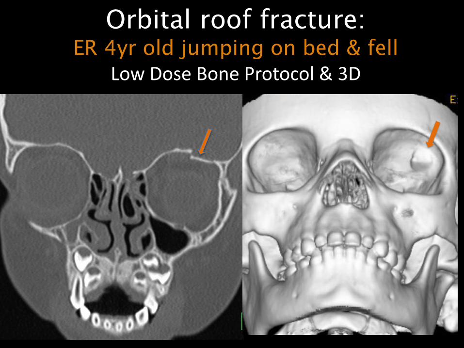
Orbital roof fracture: ER 4yr old jumping on bed & fell
Low Dose Bone Protocol & 3D
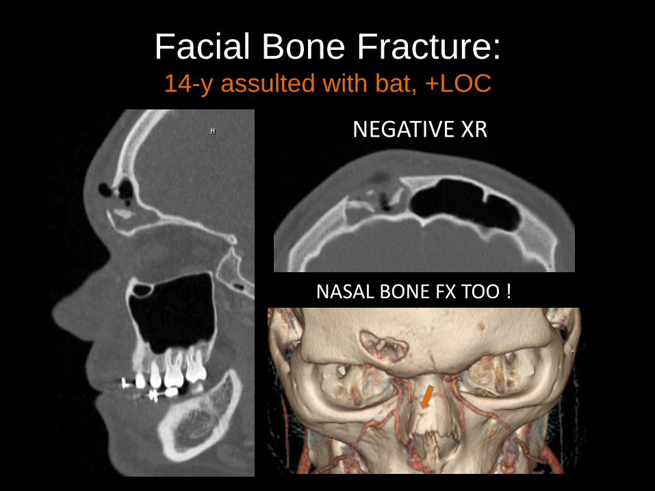
NEGATIVE XR
Facial Bone Fracture: 14-y assulted with bat, +LOC
NASAL BONE FX TOO !
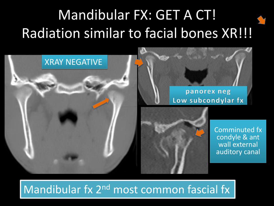
Mandibular FX: GET A CT! Radiation similar to facial bones XR!!!
Mandibular fx 2nd most common fascial fx
XRAY NEGATIVE
Comminuted fx condyle & ant wall external
auditory canal
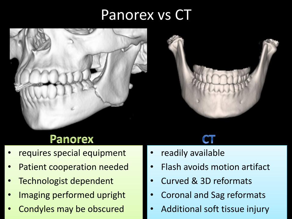
Panorex vs CT
• requires special equipment
• Patient cooperation needed
• Technologist dependent
• Imaging performed upright
• Condyles may be obscured
• readily available
• Flash avoids motion artifact
• Curved & 3D reformats
• Coronal and Sag reformats
• Additional soft tissue injury
SKULL FX
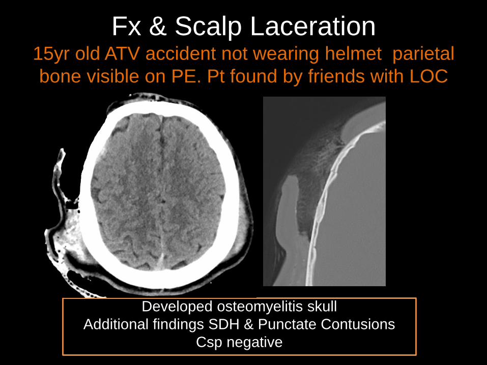
Fx & Scalp Laceration 15yr old ATV accident not wearing helmet parietal
bone visible on PE. Pt found by friends with LOC
Developed osteomyelitis skull
Additional findings SDH & Punctate Contusions
Csp negative
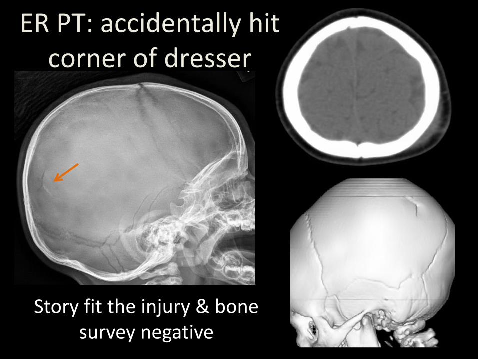
Story fit the injury & bone survey negative
ER PT: accidentally hit corner of dresser
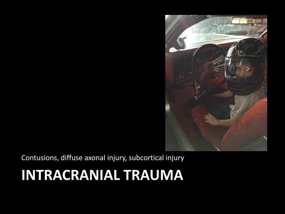
INTRACRANIAL TRAUMA Contusions, diffuse axonal injury, subcortical injury
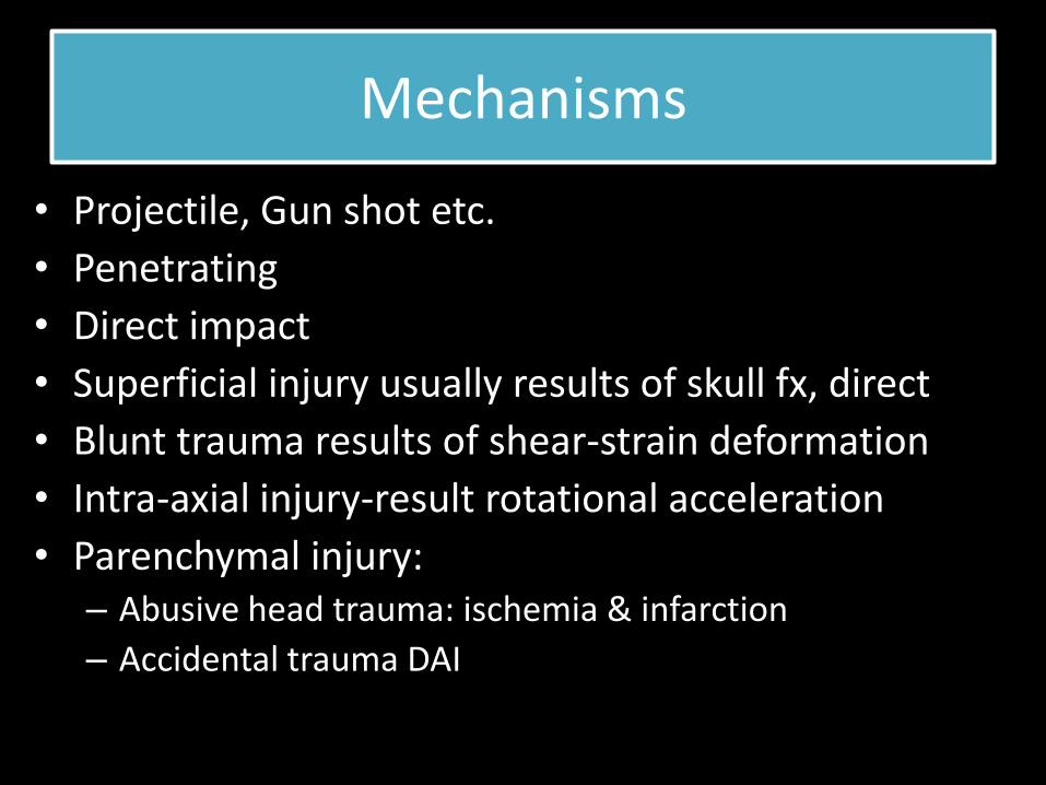
Mechanisms
• Projectile, Gun shot etc.
• Penetrating
• Direct impact
• Superficial injury usually results of skull fx, direct
• Blunt trauma results of shear-strain deformation
• Intra-axial injury-result rotational acceleration
• Parenchymal injury: – Abusive head trauma: ischemia & infarction
– Accidental trauma DAI
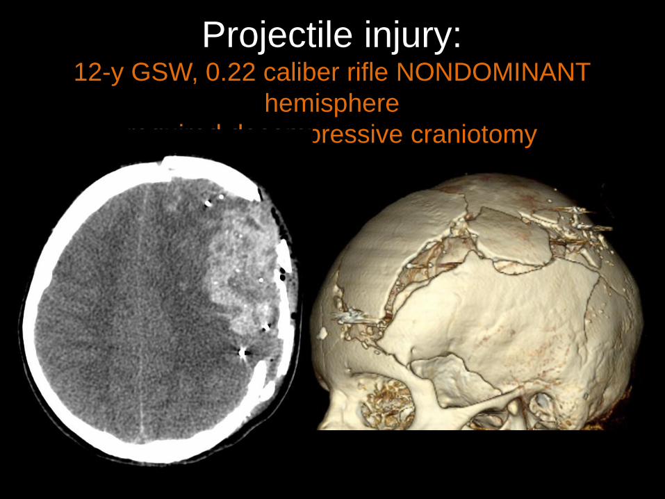
Projectile injury: 12-y GSW, 0.22 caliber rifle NONDOMINANT
hemisphere
required decompressive craniotomy
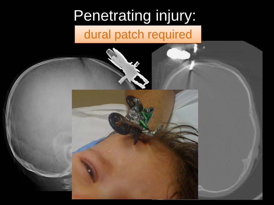
Penetrating injury:
What is it? dural patch required
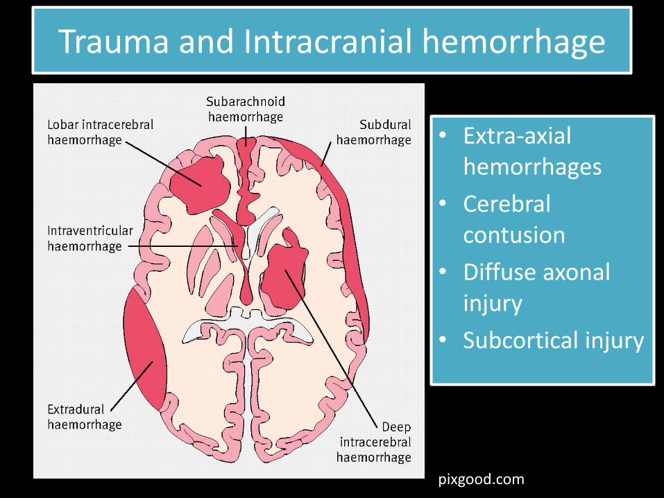
Trauma and Intracranial hemorrhage
• Extra-axial hemorrhages
• Cerebral contusion
• Diffuse axonal injury
• Subcortical injury
pixgood.com
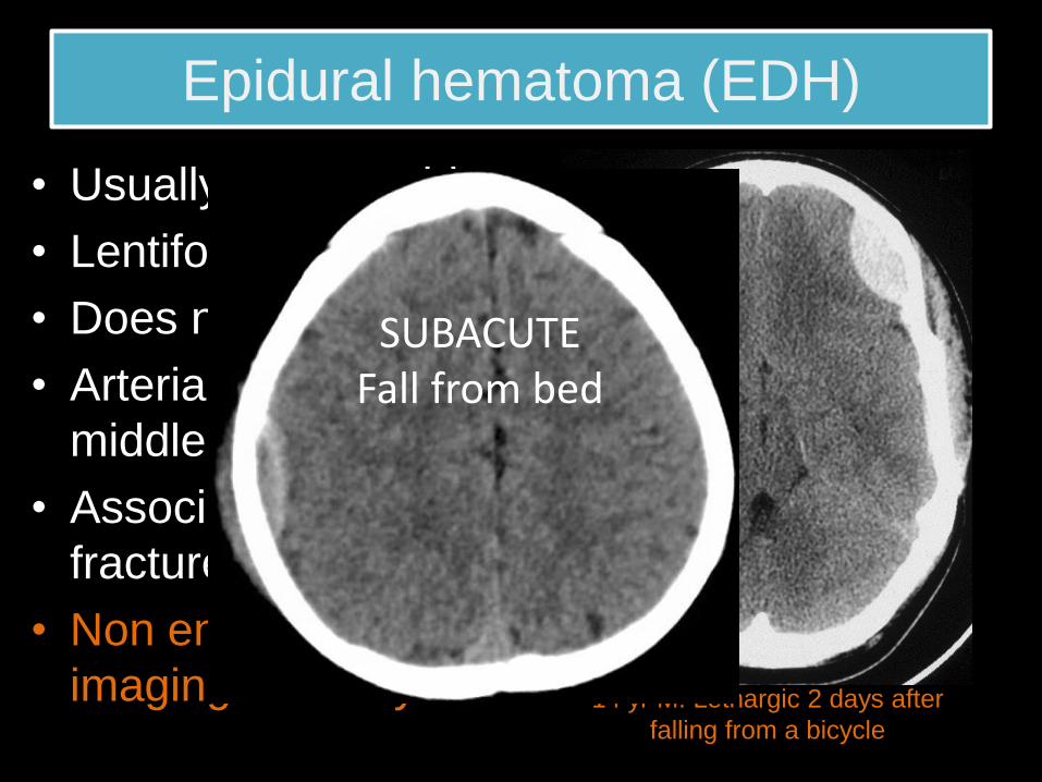
Epidural hematoma (EDH)
• Usually at coup side
• Lentiform shape on CT
• Does not cross sutures
• Arterial bleed usually
middle meningeal a.
• Associated with skull
fracture, 50-60%
• Non enhanced CT best
imaging modality 14 yr M: Lethargic 2 days after
falling from a bicycle
SUBACUTE Fall from bed
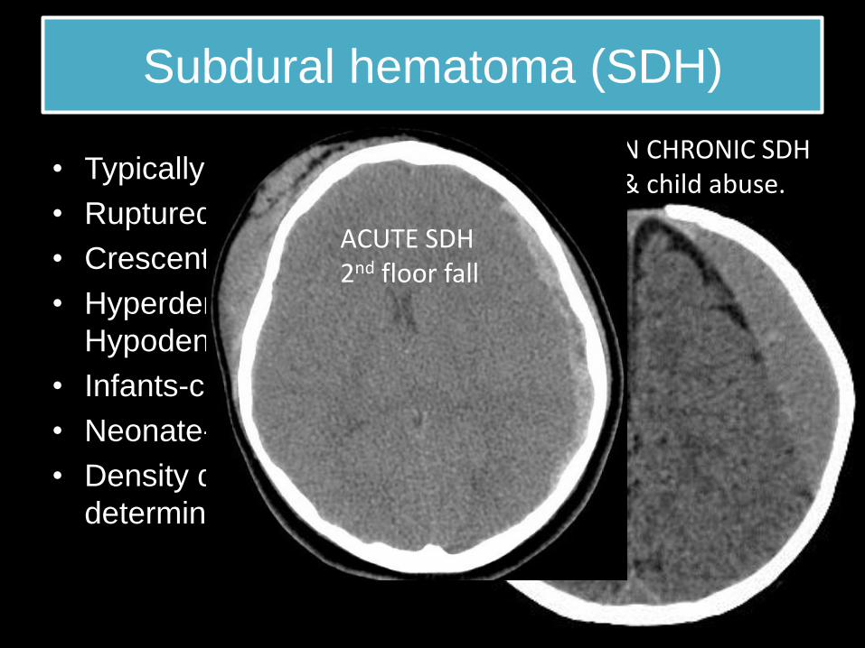
Subdural hematoma (SDH)
• Typically contrecoup side
• Ruptured bridging veins
• Crescent shape
• Hyperdense, isodense,
Hypodense
• Infants-child abuse
• Neonate-perinatal injury
• Density does not
determine age of trauma
SUBACUTE ON CHRONIC SDH ex-premie & child abuse.
ACUTE SDH 2nd floor fall
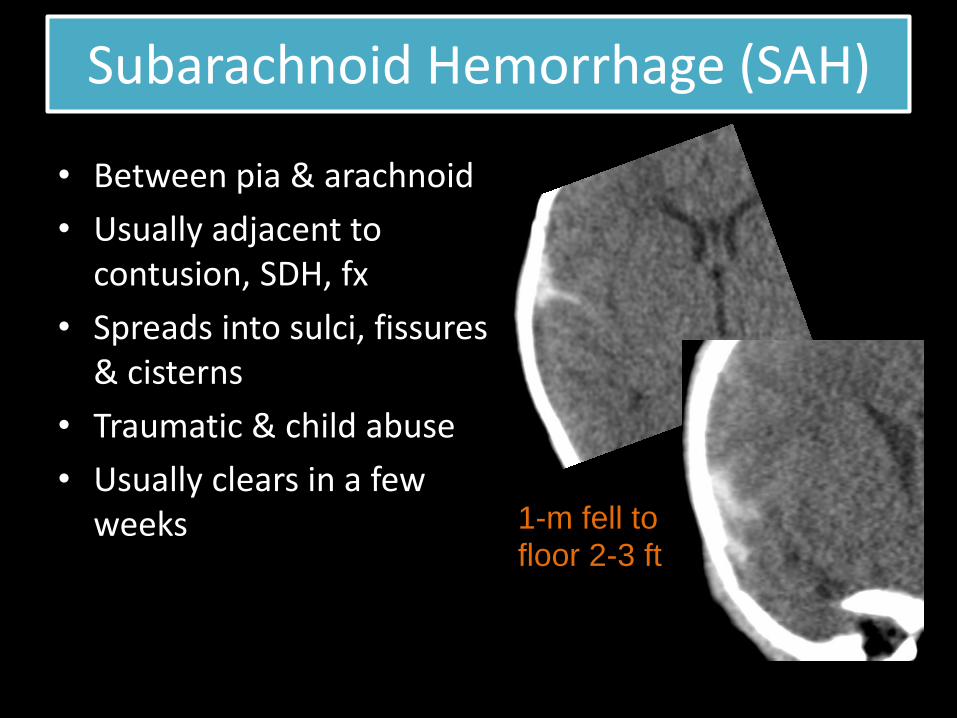
Subarachnoid Hemorrhage (SAH)
• Between pia & arachnoid
• Usually adjacent to contusion, SDH, fx
• Spreads into sulci, fissures & cisterns
• Traumatic & child abuse
• Usually clears in a few weeks
1-m fell to floor 2-3 ft
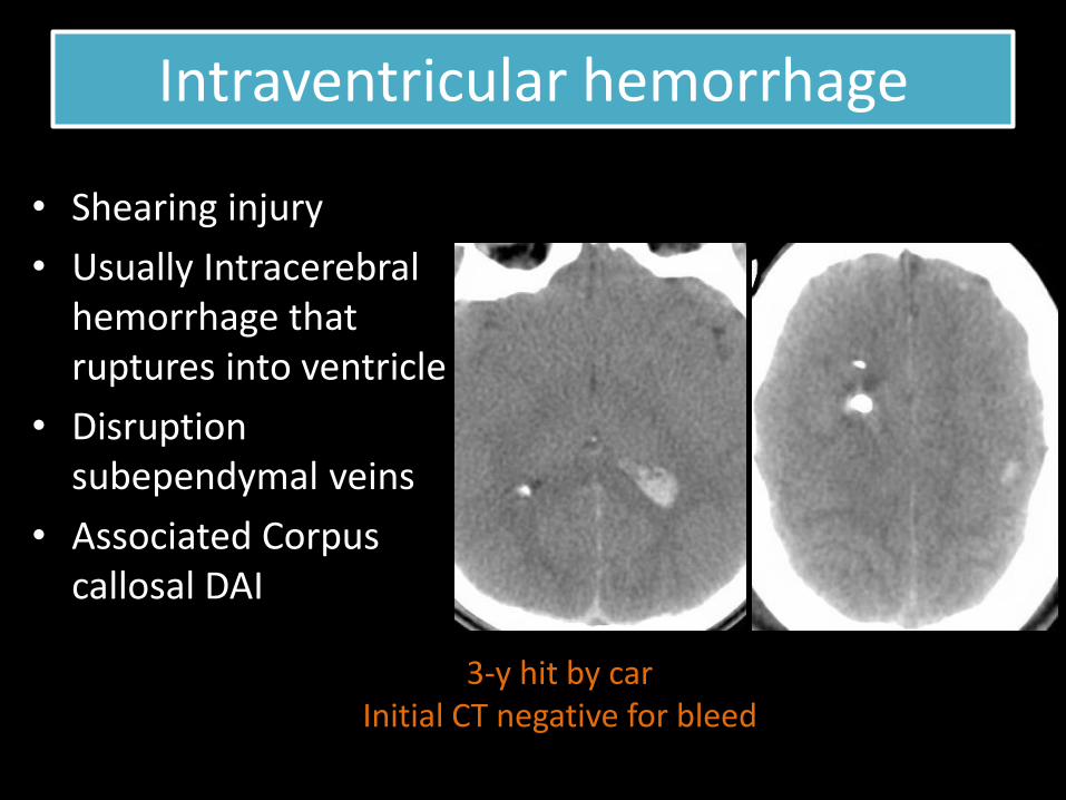
Intraventricular hemorrhage
• Shearing injury
• Usually Intracerebral hemorrhage that ruptures into ventricle
• Disruption subependymal veins
• Associated Corpus callosal DAI
3-y hit by car
Initial CT negative for bleed
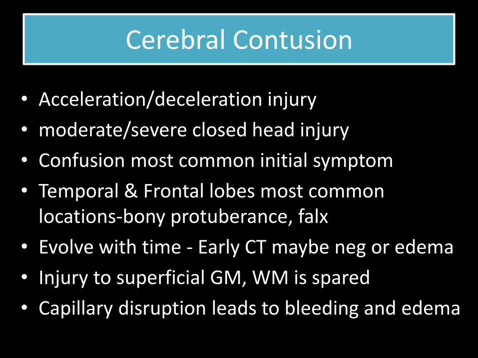
Cerebral Contusion
• Acceleration/deceleration injury
• moderate/severe closed head injury
• Confusion most common initial symptom
• Temporal & Frontal lobes most common locations-bony protuberance, falx
• Evolve with time - Early CT maybe neg or edema
• Injury to superficial GM, WM is spared
• Capillary disruption leads to bleeding and edema
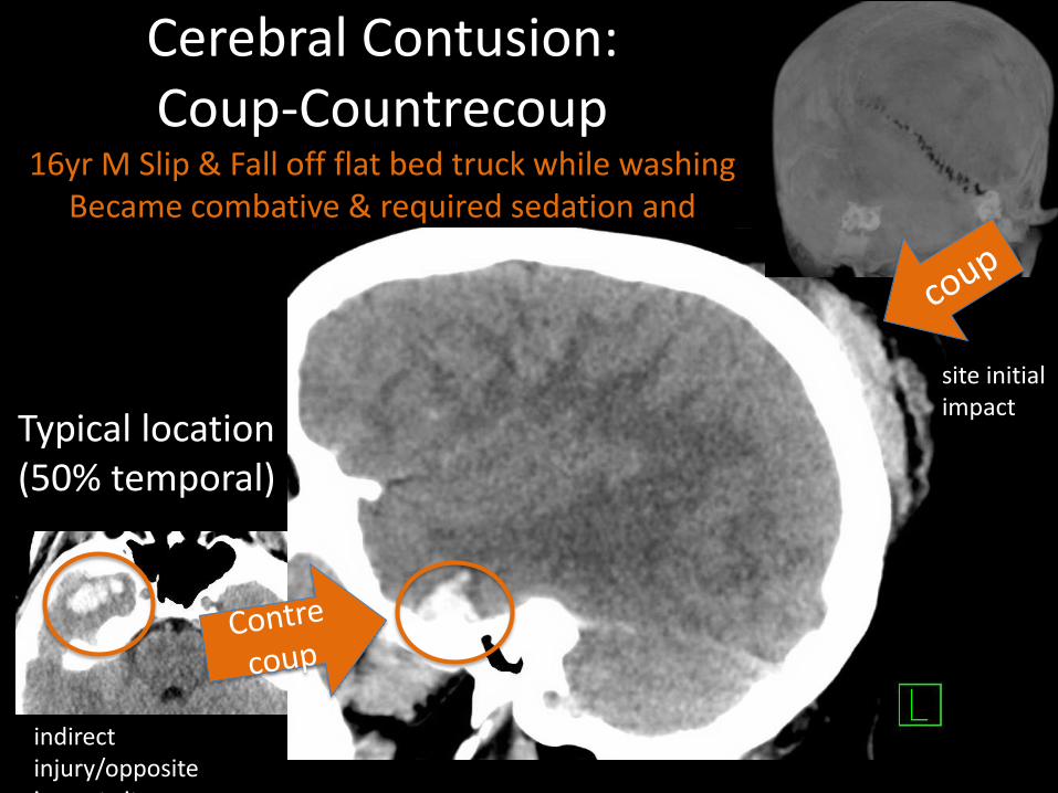
Cerebral Contusion: Coup-Countrecoup
16yr M Slip & Fall off flat bed truck while washing Became combative & required sedation and
intubation
Typical location (50% temporal)
indirect injury/opposite impact site
site initial impact
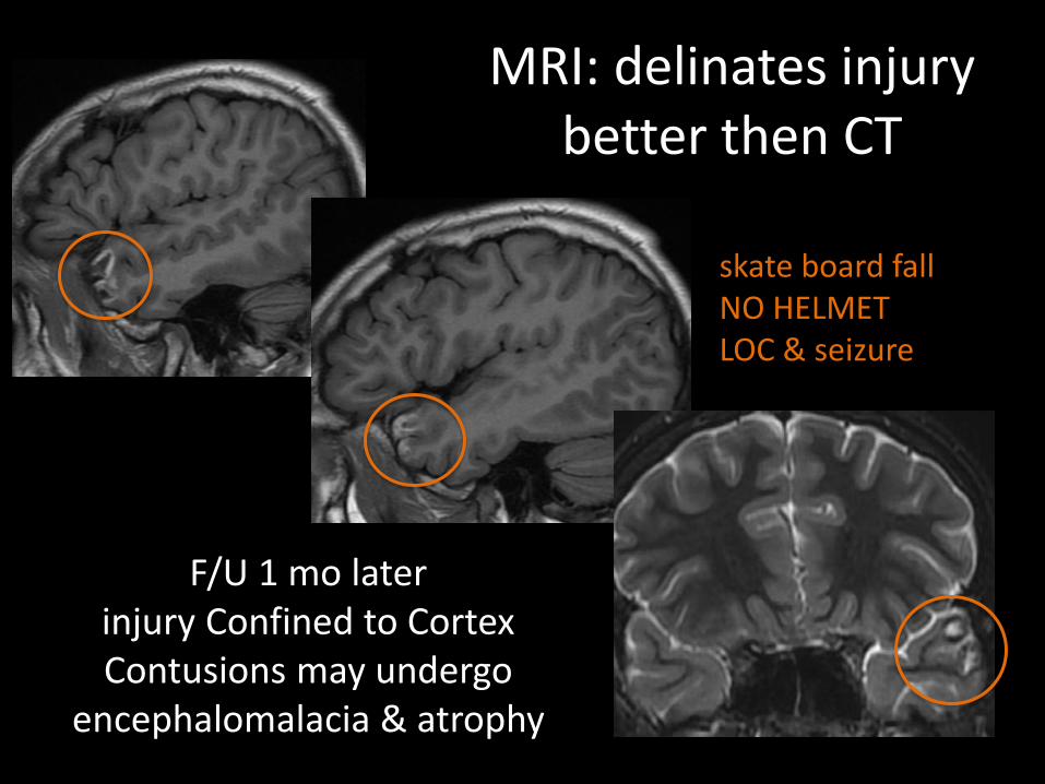
F/U 1 mo later injury Confined to Cortex Contusions may undergo
encephalomalacia & atrophy
MRI: delinates injury better then CT
skate board fall NO HELMET LOC & seizure
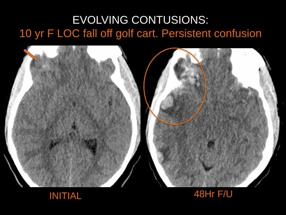
EVOLVING CONTUSIONS:
10 yr F LOC fall off golf cart. Persistent confusion
INITIAL 48Hr F/U
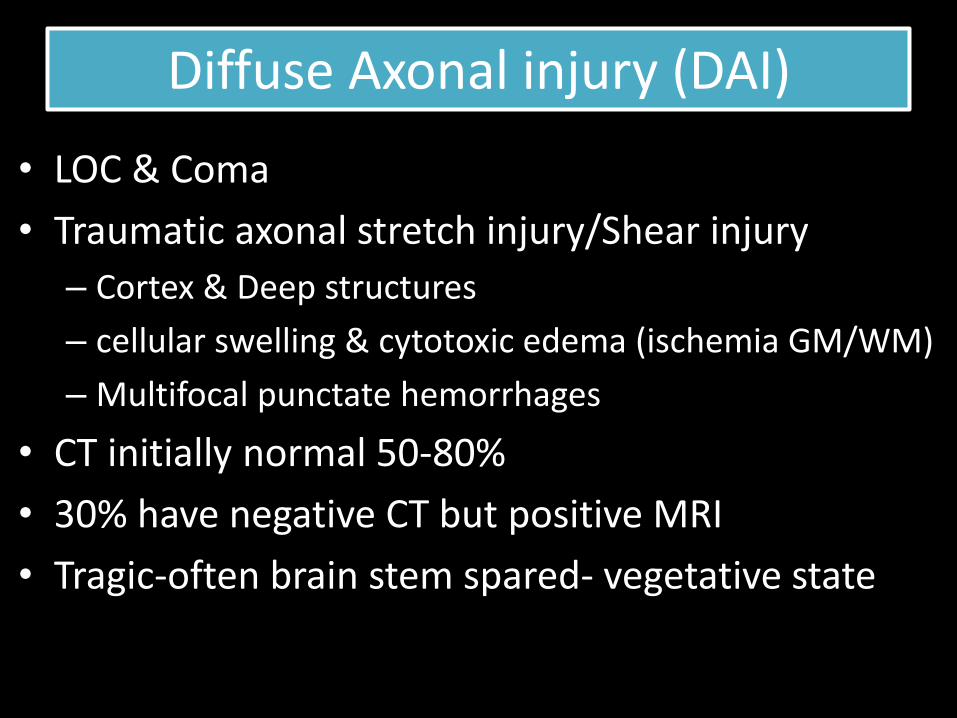
Diffuse Axonal injury (DAI)
• LOC & Coma
• Traumatic axonal stretch injury/Shear injury
– Cortex & Deep structures
– cellular swelling & cytotoxic edema (ischemia GM/WM)
– Multifocal punctate hemorrhages
• CT initially normal 50-80%
• 30% have negative CT but positive MRI
• Tragic-often brain stem spared- vegetative state

• MRI is best modality: SWI & DWI • Symptoms disproportionate to imaging findings
• Multifocal punctate foci at GM/WM jct. • Temporal & frontal location common
DAI: DWI, Edema & Ischemia Special sequences
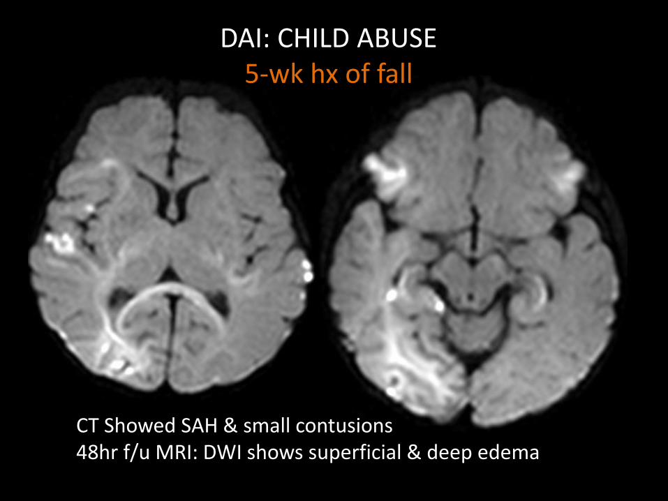
DAI: CHILD ABUSE 5-wk hx of fall
CT Showed SAH & small contusions 48hr f/u MRI: DWI shows superficial & deep edema
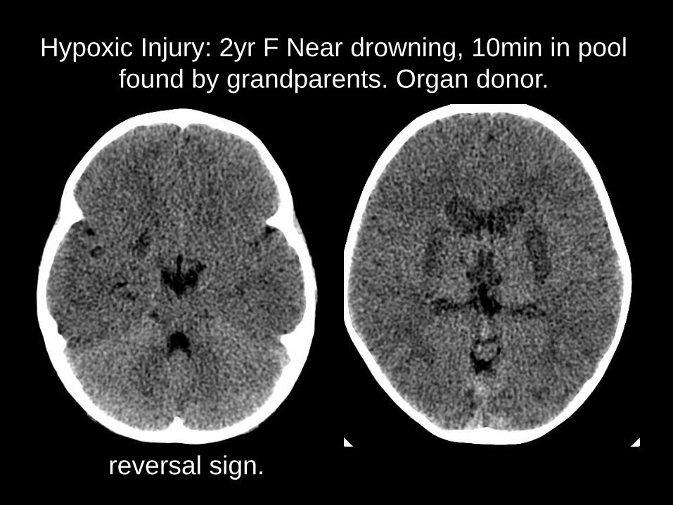
Hypoxic Injury: 2yr F Near drowning, 10min in pool
found by grandparents. Organ donor.
reversal sign.
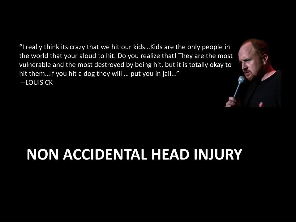
NON ACCIDENTAL HEAD INJURY
“I really think its crazy that we hit our kids…Kids are the only people in the world that your aloud to hit. Do you realize that! They are the most vulnerable and the most destroyed by being hit, but it is totally okay to hit them…If you hit a dog they will … put you in jail...” --LOUIS CK
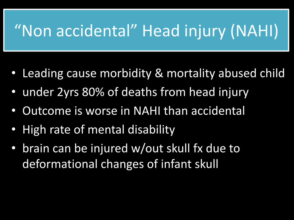
“Non accidental” Head injury (NAHI)
• Leading cause morbidity & mortality abused child
• under 2yrs 80% of deaths from head injury
• Outcome is worse in NAHI than accidental
• High rate of mental disability
• brain can be injured w/out skull fx due to deformational changes of infant skull
Published in: Gael J. Lonergan; Andrew M. Baker; Mitchel K. Morey; Steven C. Boos; RadioGraphics 2003, 23, 811-845. DOI: 10.1148/rg.234035030
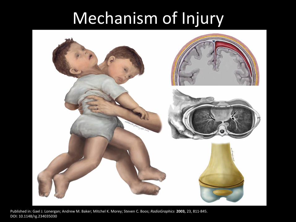
Mechanism of Injury
Head trauma-shaken baby syndrome
• Clinical presentation: seizures, apnea coma, cardiopulmonary arrest
• Main relate Injuries: subdural hemorrhages, retinal hemorrhages, bone fx & spine injury
• Cervical injury < 6mo old due to undeveloped cervical musculature
• Hypoxic-ischemic injury
– brain stem injury causing cardiopulmonary arrest and apnea resulting in brain swelling and DAI
Pediatr Radiol (2014) 44 (Suppl 4):S565-S570
NAHI-what to order • Non-enhanced CT
– Initial work up
– 3D bone reconstructions • Excellent differentiate skull fractures from sutures
• MRI brain – neurologic symptoms
– other signs of abuse
– history of head trauma
– If abnormal CT add cervical spine
– Infarcts include MRA
• CT & MRI are complimentary
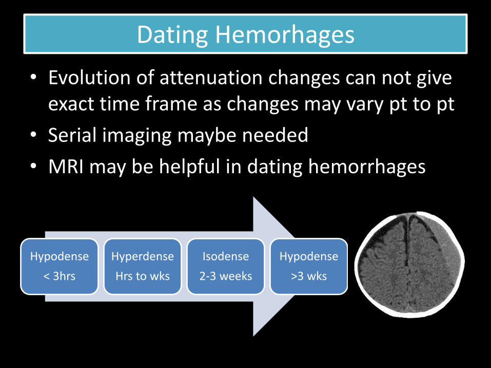
Dating Hemorhages
• Evolution of attenuation changes can not give exact time frame as changes may vary pt to pt
• Serial imaging maybe needed
• MRI may be helpful in dating hemorrhages
Hypodense
< 3hrs
Hyperdense
Hrs to wks
Isodense
2-3 weeks
Hypodense
>3 wks
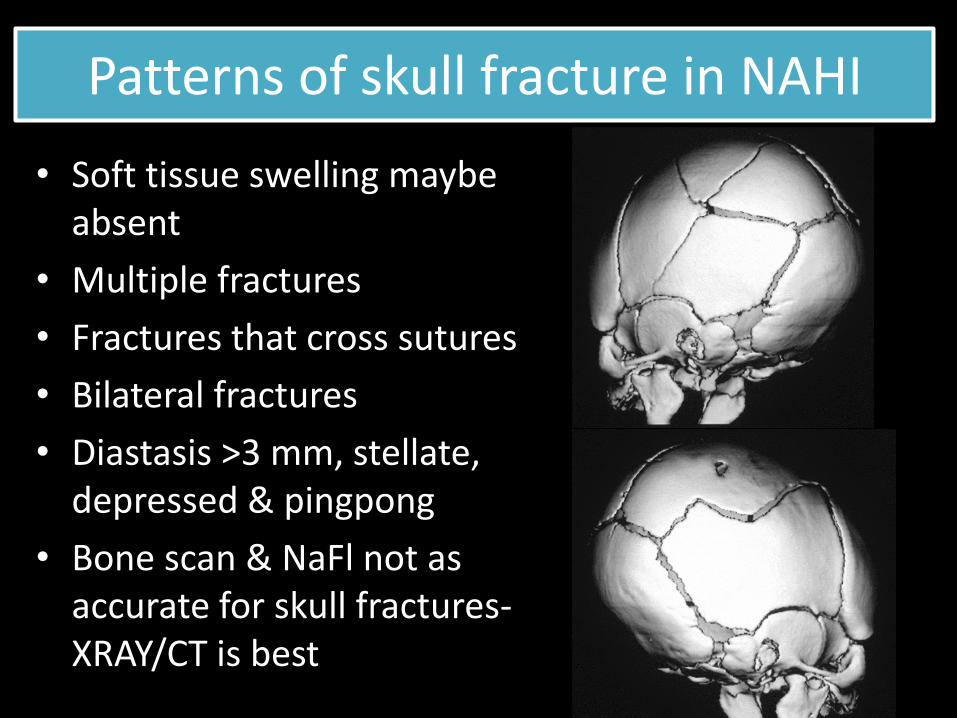
Patterns of skull fracture in NAHI
• Soft tissue swelling maybe absent
• Multiple fractures
• Fractures that cross sutures
• Bilateral fractures
• Diastasis >3 mm, stellate, depressed & pingpong
• Bone scan & NaFl not as accurate for skull fractures-XRAY/CT is best

Same Patient Diffuse edema & multiple hemorrhages
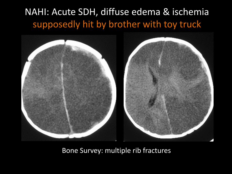
7-25-08 NAHI: Acute SDH, diffuse edema & ischemia supposedly hit by brother with toy truck
Bone Survey: multiple rib fractures
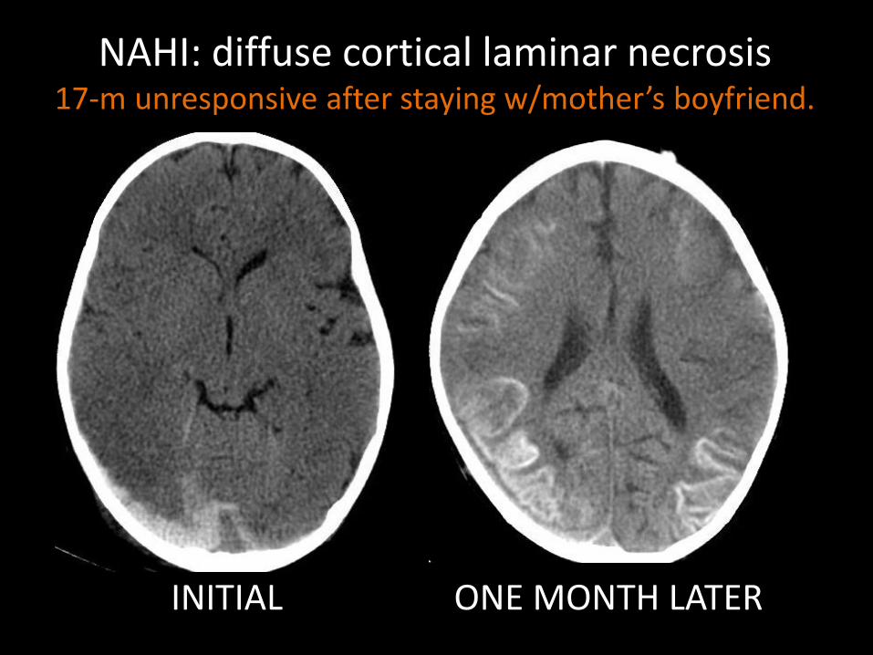
NAHI: diffuse cortical laminar necrosis 17-m unresponsive after staying w/mother’s boyfriend.
INITIAL ONE MONTH LATER
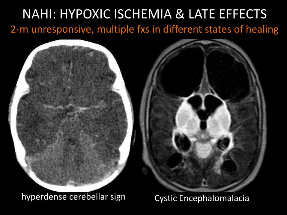
NAHI: HYPOXIC ISCHEMIA & LATE EFFECTS 2-m unresponsive, multiple fxs in different states of healing
hyperdense cerebellar sign Cystic Encephalomalacia
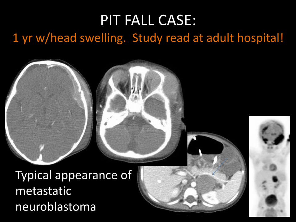
PIT FALL CASE: 1 yr w/head swelling. Study read at adult hospital!
Typical appearance of metastatic neuroblastoma
Stay on target: Take Home Points • CT is a valuable tool!
• Head, Sinus or Facial Bones CT have radiation similar to 1 yr bkgd radiation
• Use NECT 1st for emergent eval
• 3D recons especially if <2yrs
• IF CT avail preferable to Facial XR
• MRI if neurologic or worsening sx.
• Skeletal Survey REQUIRED in suspected child abuse! DO NOT MISS NON ACCIDENTAL TRAUMA!
THANK YOU!!!!
Comments & Questions [email protected]
Miami Children’s Hospital
Radiology Department
3100 sw 62 ave
(305) 666-6511
Ext. 4424 Ext. 8440
Please check Appendix in syllabus for references, medical legal issues
& reporting child abuse