Embed Size (px)
DESCRIPTION
Citation preview
Safe Use of Fluoroscopic Equipment
Intention:
This training course intends to provide physicians the necessary training in radiation safety specifically aimed at the safe use of fluoroscopic equipment. Although the safe use of radiation, the specific medical application, and medical judgment involved are not mutually exclusive, this training course does not attempt to treat the medical competency of the physician.
The Moses Cone Health Care System (MCHS) credentials committee requires proof of radiation safety training to qualify as a user of any radiation-producing equipment. The MCHS policy requires all physicians who operate or supervise the operation of x-ray producing equipment be prepared to show documentation that they meet one of the following requirements for safety training:
A. Board certified, or Board eligible in Radiology, Radiation Oncology, Nuclear Cardiology and Interventional Cardiology.
B. Successful completion of this in-house radiation safety training course.

To document your participation in this self-study, we have provided a short multiple choice test. A score of 90% or better must be achieved to receive credit for this safety training. You have the option of taking this test electronically or hard copy (paper).
Goals of this training course are:
• To minimize the likelihood of radiation-induced injuries to patients treated with fluoroscopically-guided medical procedures.
• To reduce the radiation dose of physicians and ancillary personnel who work around fluoroscopic equipment.
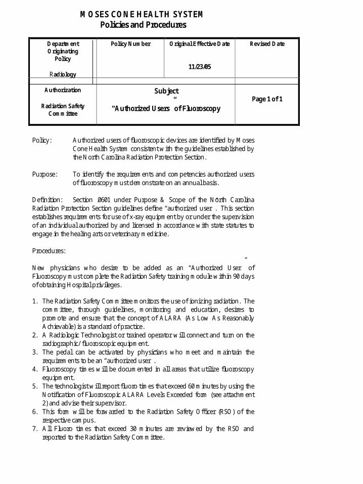
MOSES CONE HEALTH SYSTEMPolicies and Procedures
DepartmentOriginating
Policy
Radiology
Policy Number Original Effective Date
11/23/05
Revised Date
Authorization
Radiation SafetyCommittee
Subject
“Authorized Users” of FluoroscopyPage 1 of 1
Policy: Authorized users of fluoroscopic devices are identified by MosesCone Health System consistent with the guidelines established bythe North Carolina Radiation Protection Section.
Purpose: To identify the requirements and competencies authorized usersof fluoroscopy must demonstrate on an annual basis.
Definition: Section .0601 under Purpose & Scope of the North CarolinaRadiation Protection Section guidelines define “authorized user”. This sectionestablishes requirements for use of x-ray equipment by or under the supervisionof an individual authorized by and licensed in accordance with state statutes toengage in the healing arts or veterinary medicine.
Procedures:
New physicians who desire to be added as an “Authorized User” ofFluoroscopy must complete the Radiation Safety training module within 90 daysof obtaining Hospital privileges.
1. The Radiation Safety Committee monitors the use of ionizing radiation. Thecommittee, through guidelines, monitoring and education, desires topromote and ensure that the concept of ALARA (As Low As ReasonablyAchievable) is a standard of practice.
2. A Radiologic Technologist or trained operator will connect and turn on theradiographic/ fluoroscopic equipment.
3. The pedal can be activated by physicians who meet and maintain therequirements to be an “authorized user”.
4. Fluoroscopy times will be documented in all areas that utilize fluoroscopyequipment.
5. The technologist will report fluoro times that exceed 60 minutes by using theNotification of Fluoroscopic ALARA Levels Exceeded form (see attachment2) and advise their supervisor.
6. This form will be forwarded to the Radiation Safety Officer (RSO) of therespective campus.
7. All Fluoro times that exceed 30 minutes are reviewed by the RSO andreported to the Radiation Safety Committee.
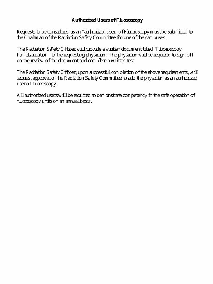
Authorized Users of Fluoroscopy
Requests to be considered as an “authorized user” of Fluoroscopy must be submitted tothe Chairman of the Radiation Safety Committee for one of the campuses.
The Radiation Safety Officer will provide a written document titled “FluoroscopyFamiliarization” to the requesting physician. The physician will be required to sign-offon the review of the document and complete a written test.
The Radiation Safety Officer, upon successful completion of the above requirements, willrequest approval of the Radiation Safety Committee to add the physician as an authorizeduser of fluoroscopy.
All authorized users will be required to demonstrate competency in the safe operation offluoroscopy units on an annual basis.
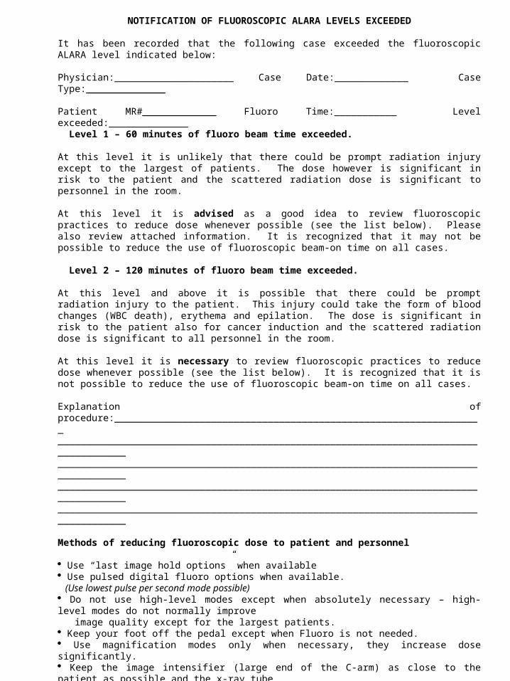
NOTIFICATION OF FLUOROSCOPIC ALARA LEVELS EXCEEDED
It has been recorded that the following case exceeded the fluoroscopic ALARA level indicated below:
Physician:_____________________ Case Date:_____________ Case Type:______________
Patient MR#_____________ Fluoro Time:___________ Level exceeded:______________
� Level 1 – 60 minutes of fluoro beam time exceeded.
At this level it is unlikely that there could be prompt radiation injury except to the largest of patients. The dose however is significant in risk to the patient and the scattered radiation dose is significant to personnel in the room.
At this level it is advised as a good idea to review fluoroscopic practices to reduce dose whenever possible (see the list below). Please also review attached information. It is recognized that it may not be possible to reduce the use of fluoroscopic beam-on time on all cases.
� Level 2 – 120 minutes of fluoro beam time exceeded.
At this level and above it is possible that there could be prompt radiation injury to the patient. This injury could take the form of blood changes (WBC death), erythema and epilation. The dose is significant in risk to the patient also for cancer induction and the scattered radiation dose is significant to all personnel in the room.
At this level it is necessary to review fluoroscopic practices to reduce dose whenever possible (see the list below). It is recognized that it is not possible to reduce the use of fluoroscopic beam-on time on all cases.
Explanation of procedure:_________________________________________________________________________________________________________________________________________________________________________________________________________________________________________________________________________________________________________________________________________________________________________________________________________________________
Methods of reducing fluoroscopic dose to patient and personnel
Use “last image hold options” when available Use pulsed digital fluoro options when available. (Use lowest pulse per second mode possible) Do not use high-level modes except when absolutely necessary – high-level modes do not normally improve image quality except for the largest patients. Keep your foot off the pedal except when Fluoro is not needed. Use magnification modes only when necessary, they increase dose significantly. Keep the image intensifier (large end of the C-arm) as close to the patient as possible and the x-ray tube (small end of the C-arm) as far away from the patient as possible.
I have read and understand this notice:
____________________________________________________(Signature of MD)
Return to the Radiology Supervisor c/o Director of Imaging
Quality Management Program for High Dose FluoroscopyMay 2005
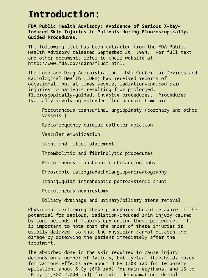
Introduction:FDA Public Health Advisory: Avoidance of Serious X-Ray-Induced Skin Injuries to Patients during Fluoroscopically-Guided Procedures.
The following text has been extracted from the FDA Public Health Advisory released September 30, 1994. For full text and other documents refer to their website at http://www.fda.gov/cdrh/fluor.html.
The Food and Drug Administration (FDA) Center for Devices and Radiological Health (CDRH) has received reports of occasional, but at times severe, radiation-induced skin injuries to patients resulting from prolonged, fluoroscopically-guided, invasive procedures. Procedures typically involving extended fluoroscopic time are:
Percutaneous transuminal angioplasty (coronary and other vessels.)
Radiofrequency cardiac catheter ablation
Vascular embolization
Stent and filter placement
Thrombolytic and fibrinolytic procedures
Percutaneous transhepatic cholangiography
Endoscopic retrogradecholangiopancreatography
Transjugular intrahepatic portosystemic shunt
Percutaneous nephrostomy
Biliary drainage and urinary/biliary stone removal.
Physicians performing these procedures should be aware of the potential for serious, radiation-induced skin injury caused by long periods of fluoroscopy during these procedures. It is important to note that the onset of these injuries is usually delayed, so that the physician cannot discern the damage by observing the patient immediately after the treatment.
The absorbed dose in the skin required to cause injury depends on a number of factors, but typical thresholds doses for various effects are about 3 Gy (300 rad for temporary epilation, about 6 Gy (600 rad) for main erythema, and 15 to 20 Gy (1,500-2,000 rad) for moist desquamation, dermal necrosis and secondary ulceration. The absorbed dose rate in the skin from the direct beam of a fluoroscopic x-ray system is typically between 0.02 Gy/min and 0.05 Gy/min (2 to 5 rad/min), but may be higher, depending on the mode in which the equipment is operated and the size of the patient. Even typical dose rates can result in skin injury with less than one hour of fluoroscopy.

FDA suggests that facilities performing fluoroscopically-guided procedures observe the following principles:
Establish standard operating procedures and clinical protocols for each specific type of procedure performed. The protocols should address all aspects of the procedure, such as patient selection, normal conduct of the procedure, actions in response to complications and consideration of limits on fluoroscopy exposure time.
Know the radiation dose rates for the specific fluoroscopic system and for each mode of operation used during the clinical protocol. These dose rates should be derived from measurements performed at the facility.
Assess the impact of each procedure’s protocol on the potential for radiation injury to the patient.
Modify the protocol, as appropriate, to limit the cumulative absorbed dose to any irradiated area of the skin to the minimum necessary for the clinical tasks, and particularly to avoid approaching cumulative doses that would induce unacceptable adverse effects.
Enlist a qualified medical physicist to assist in implementing these principles in such a manner so as not to adversely affect the clinical objectives of the procedure.
Physicians should know that radiation-induced injuries from fluoroscopy are not immediately apparent. Other than the mildest symptoms, such as transient erythema, the effects of the radiation may not appear until weeks following the exposure. Physicians performing these procedures may not be in direct contact with patients following the procedure and may not observe the symptoms when they occur. Missing the milder symptoms in some patients can lead to surprise at the magnitude of the absorbed doses delivered to the skin of other patients when more serious symptoms appear. For this reason, we recommend that information be recorded in the patient’s record that permits estimation of the absorbed dose to the skin. Patients should also be advised to report signs and/or symptoms of radiation induced injury to their attending physician.
The Safe Medical Devises Act of 1990 (SMDA) requires hospitals and other user facilities to report deaths, serious illnesses and injuries associated with the use of medical devices. Follow the procedures established by your facility for such mandatory reporting. Practitioners who become aware of any medical device related adverse event or product problem/malfunction should report to their Medical Device User Facility Reporting person.
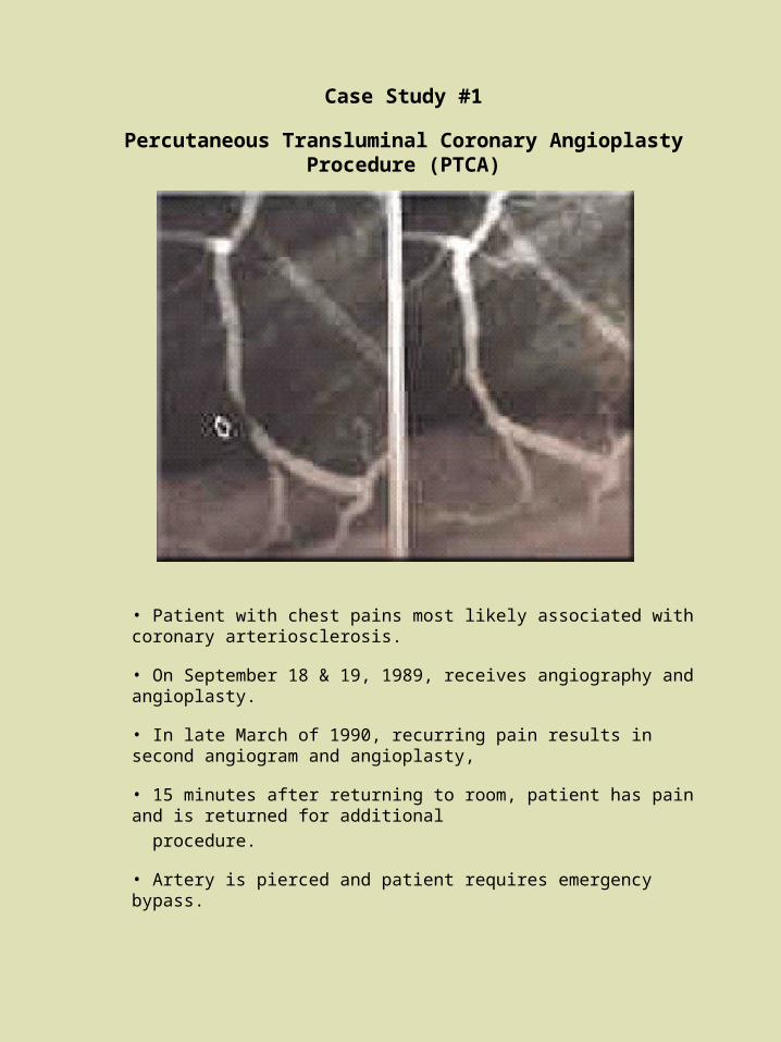
Case Study #1
Percutaneous Transluminal Coronary Angioplasty Procedure (PTCA)
• Patient with chest pains most likely associated with coronary arteriosclerosis.
• On September 18 & 19, 1989, receives angiography and angioplasty.
• In late March of 1990, recurring pain results in second angiogram and angioplasty,
• 15 minutes after returning to room, patient has pain and is returned for additional
procedure.
• Artery is pierced and patient requires emergency bypass.
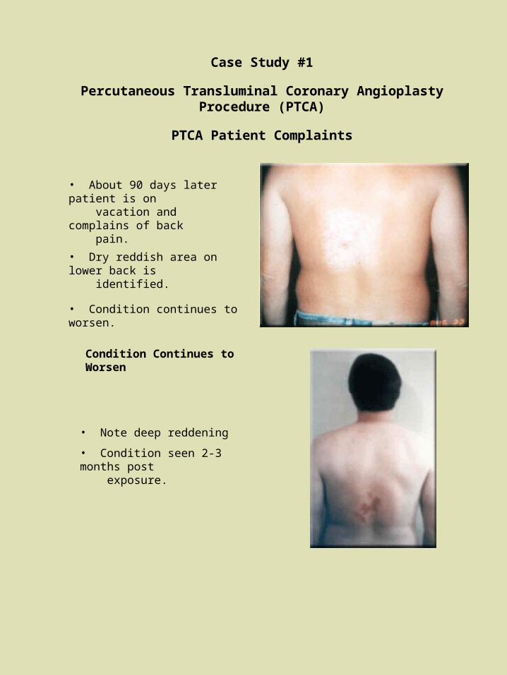
Case Study #1
Percutaneous Transluminal Coronary Angioplasty Procedure (PTCA)
PTCA Patient Complaints
• About 90 days later patient is on vacation and complains of back pain.
• Dry reddish area on lower back is identified.
• Condition continues to worsen.
• Note deep reddening
• Condition seen 2-3 months post exposure.
Condition Continues to Worsen
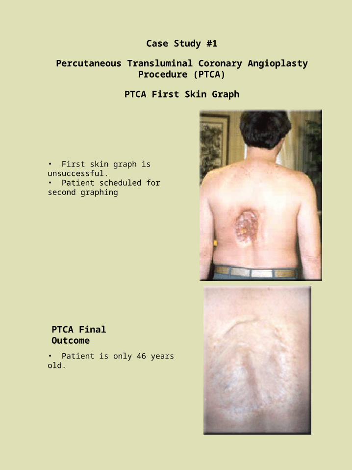
Case Study #1
Percutaneous Transluminal Coronary Angioplasty Procedure (PTCA)
PTCA First Skin Graph
• First skin graph is unsuccessful.• Patient scheduled for second graphing
• Patient is only 46 years old.
PTCA Final Outcome
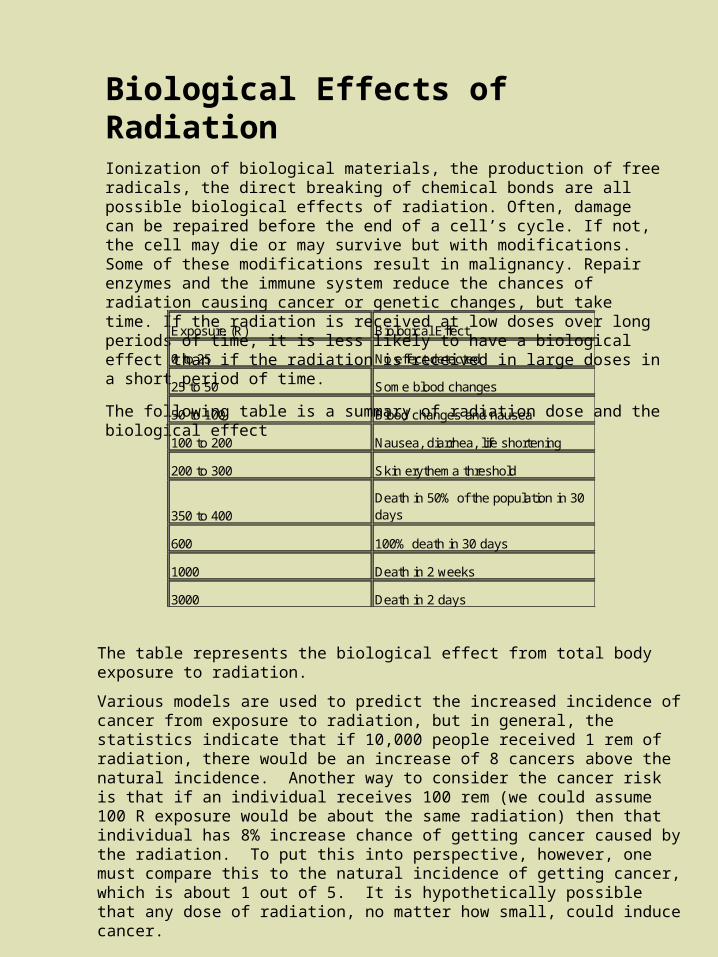
Biological Effects of Radiation Ionization of biological materials, the production of free radicals, the direct breaking of chemical bonds are all possible biological effects of radiation. Often, damage can be repaired before the end of a cell’s cycle. If not, the cell may die or may survive but with modifications. Some of these modifications result in malignancy. Repair enzymes and the immune system reduce the chances of radiation causing cancer or genetic changes, but take time. If the radiation is received at low doses over long periods of time, it is less likely to have a biological effect than if the radiation is received in large doses in a short period of time.
The following table is a summary of radiation dose and the biological effect
Exposure (R) Biological Effect
0 to 25 No effect detected
25 to 50 Some blood changes
50 to 100 Blood changes and nausea
100 to 200 Nausea, diarrhea, life shortening
200 to 300 Skin erythema threshold
350 to 400Death in 50% of the population in 30 days
600 100% death in 30 days
1000 Death in 2 weeks
3000 Death in 2 days
The table represents the biological effect from total body exposure to radiation.
Various models are used to predict the increased incidence of cancer from exposure to radiation, but in general, the statistics indicate that if 10,000 people received 1 rem of radiation, there would be an increase of 8 cancers above the natural incidence. Another way to consider the cancer risk is that if an individual receives 100 rem (we could assume 100 R exposure would be about the same radiation) then that individual has 8% increase chance of getting cancer caused by the radiation. To put this into perspective, however, one must compare this to the natural incidence of getting cancer, which is about 1 out of 5. It is hypothetically possible that any dose of radiation, no matter how small, could induce cancer.
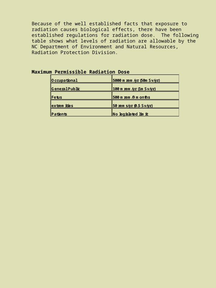
Because of the well established facts that exposure to radiation causes biological effects, there have been established regulations for radiation dose. The following table shows what levels of radiation are allowable by the NC Department of Environment and Natural Resources, Radiation Protection Division.
Maximum Permissible Radiation Dose
Occupational 5000 mrem/yr (50mSv/yr)
General Public 100 mrem/yr (1mSv/yr)
Fetus 500 mrem/9 months
extremities 50 rems/yr (0.5 Sv/yr)
Patients No legislated limit
Fluoroscopy Fluoroscopy differs from conventional x-ray imaging in that the x-ray image can be viewed in real time. Instead of film, the detector is an image intensifier fluorescent screen coupled to a video camera. The x-ray image can be viewed directly on the TV screen or can be captured in digital format and viewed/manipulated later. To maintain a constant image quality, an automatic brightness system (ABS) detects the x-ray intensity that is reaching the detector and adjusts the mA and/or kVp. So, for example, if the fluoroscope moves from a thick part of the body to a thin part of the body, the x-ray intensity is reduced to avoid flooding the detector and to reduce the radiation dose to the patient.
In some equipment there is an x-ray grid between the patient and detector. This is a flat plat with a series of lead strips that allow the primary radiation beam to pass through but stops the radiation scattered by the patient from reach the detector. This is a means of suppressing scatter and improving the contrast in the image. Some primary photons are absorbed by the grid which means that fewer photons reach the detector. So more photons have to be generated (increase mA or kVp) and the patient receives a higher radiation dose when using the grid.
Geometry
Several factors that can affect radiation dose to the patient and the quality of the image are geometric in nature. The size of the patient, the size of the viewing field (collimation), the distance from the x-ray tube to the patient, the distance of the detector (image intensifier) from the patient and the use of a grid.
1. The size of the patient is an uncontrollable variable. For a large patient or dense region, to obtain enough x-rays that get to the detector, a more intense x-ray beam is required. Hence an increase in mA or kVp is needed. This also increases the radiation dose to the patient.
• Radiation dose is greater for larger patients.
• Use as low mA as possible and as high a kVp as possible to obtain image quality and lowest dose.
2. Collimators are used to determine the size of the x-ray field. The larger the field size, the larger the amount of scatter which degrades the image quality. By tightly collimating the x-ray beam to the area of interest reduces the amount of scatter, reduces the volume of tissue exposed and improves the quality of the image.
• Use the tightest collimation possible
3. In the x-ray tube the x-rays emanate from the focal spot of the x-ray tube, typically 1 mm diameter. The intensity of the x-ray beam decreases as the square of the distance from this focal spot. Thus, the further away from the x-ray tube the patient is located, the less radiation per square meter and the less likely a radiation skin burn will occur. This is especially important in lateral or oblique views since the x- ray tube is usually much closer to the patient than in AP or PA views.
• The x-ray tube should be as far from the patient as possible.
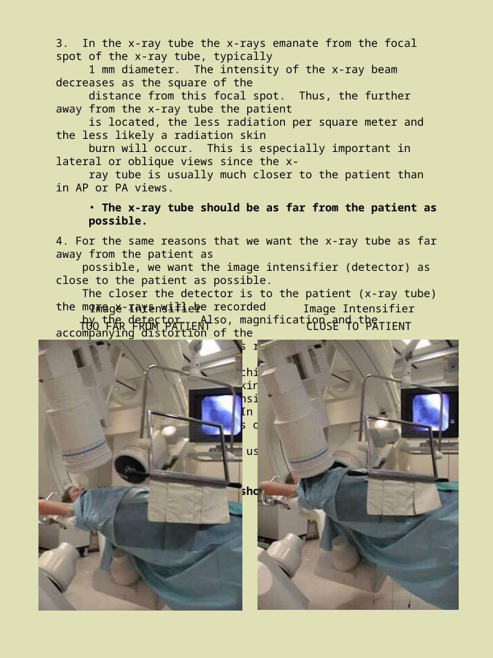
4. For the same reasons that we want the x-ray tube as far away from the patient as possible, we want the image intensifier (detector) as close to the patient as possible. The closer the detector is to the patient (x-ray tube) the more x-rays will be recorded by the detector. Also, magnification and the accompanying distortion of the anatomy and image blur is reduced. Thus, better image quality and less radiation dose to the patient is achieved. In some procedures an air gap is needed for working space and the image intensifier is placed at some distance from the patient. In these circumstances other means of reducing dose such as removing the grid, keeping exposure times short and using tight collimation become even more important.
•The image intensifier should be as close to the patient as possible.
Image Intensifier
TOO FAR FROM PATIENT
Image Intensifier
CLOSE TO PATIENT
5. The grid is a device with parallel strips of lead that intends to filter out scattered radiation when placed in the x-ray beam in front of the image intensifier. The grid also stops many non-scattered photons resulting in the need to increase the x-ray intensity. This also increases the radiation dose to the patient. If highest resolution images are critical to the procedure, then grids should be used. If image quality is adequate without the grid, the grid should be removed to keep the radiation dose at a minimum. In general, for pediatric cases and when the image intensifier cannot be moved closer than 25 cm to the patient, grids should not be used.
• The use of a grid increases radiation dose.
Radiation Dose From Fluoroscopy Fluoroscopy is a particularly high dose rate procedure. The fluoroscopist should be concerned about radiation dose to the patient, themselves, and co-workers in the vicinity of the fluoroscopic machine.
Radiation to the patient can be reduced by considering the following factors:
• Short periods of screening exposure.
• Digital image storage.
• Temporary removal of anti-scatter grid (less radiation needed for film exposure with sacrifice in image quality).
• Automatic brightness control compensates for different anatomy.
• 90 kV at 0.5 mA should be exposure level to deliver 1 R/min (10 mGy) at the tabletop.
• X-ray tube at maximal distance from patient.
• Image intensifier as close to patient as possible.
• Appropriate use of the magnification mode of operation consistent with the procedure.
• Collimate the beam to the smallest field.
• Keep beam-on time to a minimum.
Radiation to personnel can be reduced by the following:
• Use local shielding around equipment (example ceiling mounted lead-glass shields).• Wear protective clothing (lead aprons, thyroid shields).• Exposure timing devices with audible warning.• Display elapsed fluoroscopic time on monitor screen.
Personnel monitoring
This does not reduce radiation, but it is important to wear the monitors (badges) to be able to know what is the radiation dose received. It is also a requirement if you are employed by the Moses Cone Health System as a registered radiation worker. If assigned one badge, it should be worn on the outside of the “apron” at the collar level. If assigned two badges, one badge should be worn on the outside of the “apron” at the collar level and the other badge worn under the apron at the waist level. Badges should be returned at the end of the monitoring period.
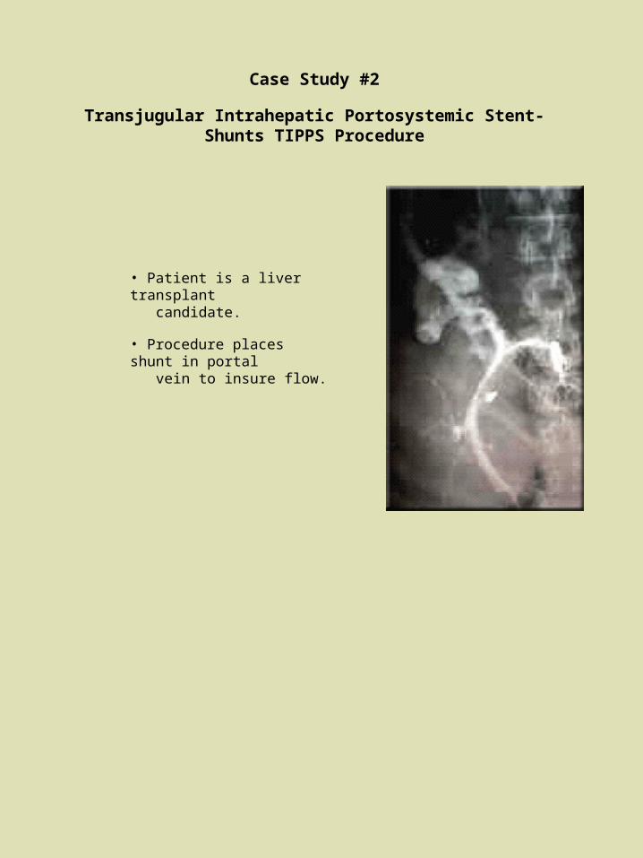
Case Study #2
Transjugular Intrahepatic Portosystemic Stent-Shunts TIPPS Procedure
• Patient is a liver transplant candidate.
• Procedure places shunt in portal vein to insure flow.
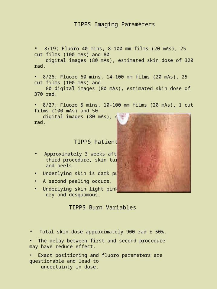
TIPPS Imaging Parameters
• 8/19; Fluoro 40 mins, 8-100 mm films (20 mAs), 25 cut films (100 mAs) and 80 digital images (80 mAs), estimated skin dose of 320 rad.
• 8/26; Fluoro 60 mins, 14-100 mm films (20 mAs), 25 cut films (100 mAs) and 80 digital images (80 mAs), estimated skin dose of 370 rad.
• 8/27; Fluoro 5 mins, 10-100 mm films (20 mAs), 1 cut films (100 mAs) and 50 digital images (80 mAs), estimated skin dose of 90 rad.
TIPPS Patient Complaints
• Approximately 3 weeks after third procedure, skin turns red and peels.
• Underlying skin is dark purple.
• A second peeling occurs.
• Underlying skin light pink, dry and desquamous.
TIPPS Burn Variables
• Total skin dose approximately 900 rad ± 50%.
• The delay between first and second procedure may have reduce effect.
• Exact positioning and fluoro parameters are questionable and lead to uncertainty in dose.
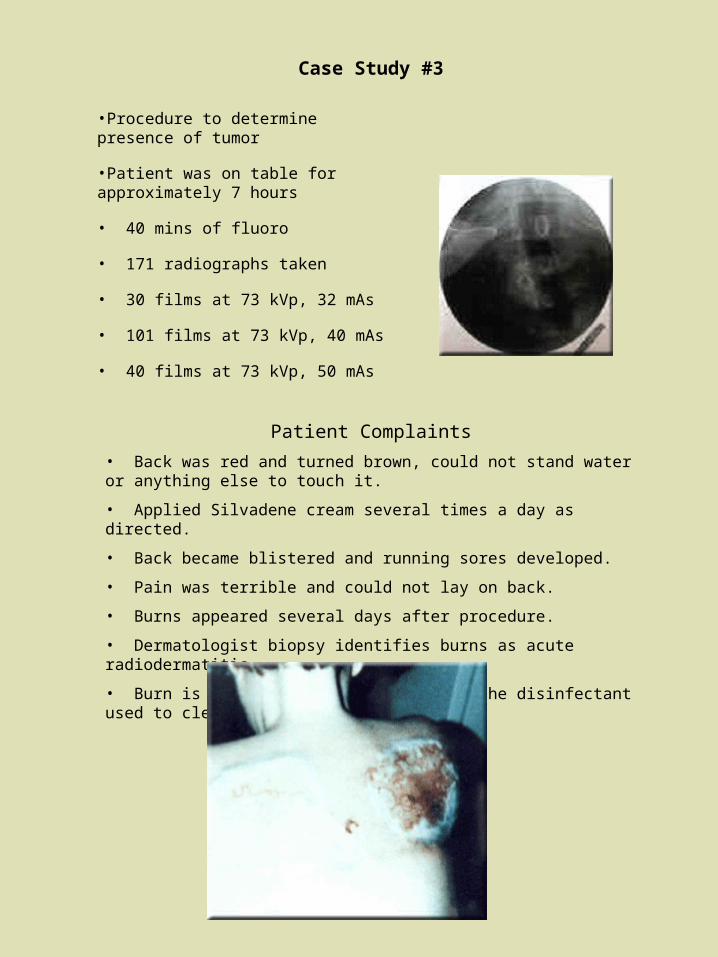
•Procedure to determine presence of tumor
•Patient was on table for approximately 7 hours
• 40 mins of fluoro
• 171 radiographs taken
• 30 films at 73 kVp, 32 mAs
• 101 films at 73 kVp, 40 mAs
• 40 films at 73 kVp, 50 mAs
Patient Complaints
• Back was red and turned brown, could not stand water or anything else to touch it.
• Applied Silvadene cream several times a day as directed.
• Back became blistered and running sores developed.
• Pain was terrible and could not lay on back.
• Burns appeared several days after procedure.
• Dermatologist biopsy identifies burns as acute radiodermatitis.
• Burn is actually a skin reaction to the disinfectant used to clean x-ray couch.
Case Study #3
Case Study #4
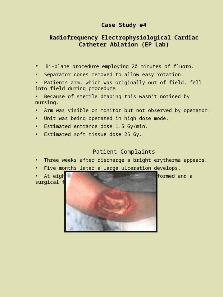
Radiofrequency Electrophysiological Cardiac Catheter Ablation (EP Lab)
• Bi-plane procedure employing 20 minutes of fluoro.
• Separator cones removed to allow easy rotation.
• Patients arm, which was originally out of field, fell into field during procedure.
• Because of sterile draping this wasn’t noticed by nursing.
• Arm was visible on monitor but not observed by operator.
• Unit was being operated in high dose mode.
• Estimated entrance dose 1.5 Gy/min.
• Estimated soft tissue dose 25 Gy.
Patient Complaints• Three weeks after discharge a bright erytherma appears.
• Five months later a large ulceration develops.
• At eight months, a debridement was performed and a surgical flap was put in place.
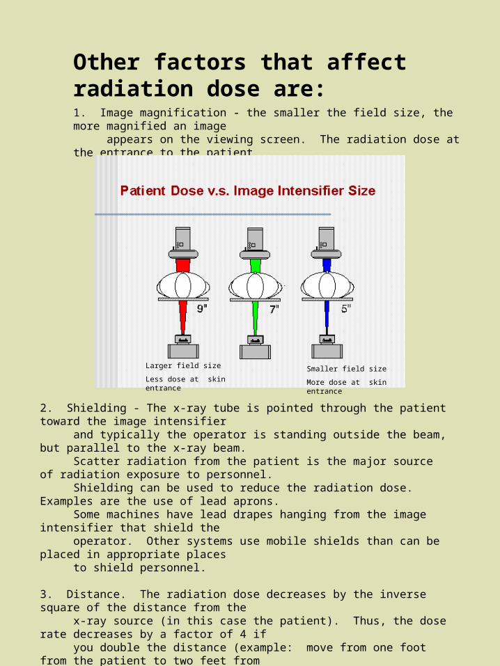
Other factors that affect radiation dose are: 1. Image magnification - the smaller the field size, the more magnified an image appears on the viewing screen. The radiation dose at the entrance to the patient generally increases with magnification.
2. Shielding - The x-ray tube is pointed through the patient toward the image intensifier and typically the operator is standing outside the beam, but parallel to the x-ray beam. Scatter radiation from the patient is the major source of radiation exposure to personnel. Shielding can be used to reduce the radiation dose. Examples are the use of lead aprons. Some machines have lead drapes hanging from the image intensifier that shield the operator. Other systems use mobile shields than can be placed in appropriate places to shield personnel.
3. Distance. The radiation dose decreases by the inverse square of the distance from the x-ray source (in this case the patient). Thus, the dose rate decreases by a factor of 4 if you double the distance (example: move from one foot from the patient to two feet from the patient.
4. Beam-On time. The single major control over the amount of radiation dose given to patient or personnel is the amount of time the x-ray machine is generating x-rays. Long durations of On Time, absentmindedly leaving the beam on, being distracted by doing other things while the beam is on, all contribute to unnecessary radiation dose and in fact it adds up very quickly.
Larger field size
Less dose at skin entranceSmaller field size
More dose at skin entrance
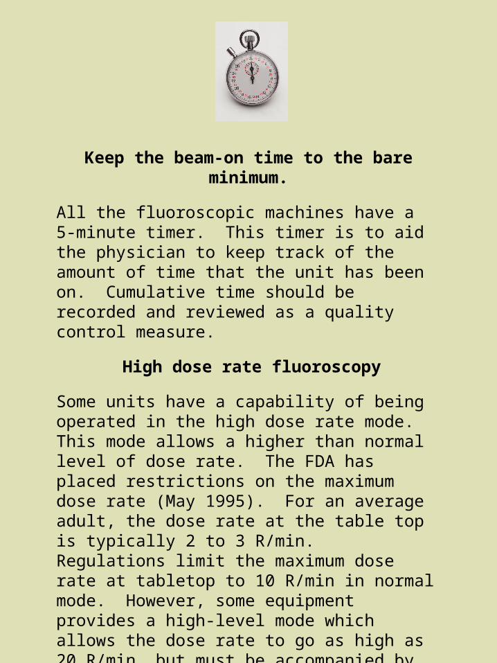
Keep the beam-on time to the bare minimum.
All the fluoroscopic machines have a 5-minute timer. This timer is to aid the physician to keep track of the amount of time that the unit has been on. Cumulative time should be recorded and reviewed as a quality control measure.
High dose rate fluoroscopy
Some units have a capability of being operated in the high dose rate mode. This mode allows a higher than normal level of dose rate. The FDA has placed restrictions on the maximum dose rate (May 1995). For an average adult, the dose rate at the table top is typically 2 to 3 R/min. Regulations limit the maximum dose rate at tabletop to 10 R/min in normal mode. However, some equipment provides a high-level mode which allows the dose rate to go as high as 20 R/min, but must be accompanied by an audible alarm signal. High-level mode should be used sparingly and with caution. Fifteen minutes of fluoroscopy at the maximum level can produce skin erythema.
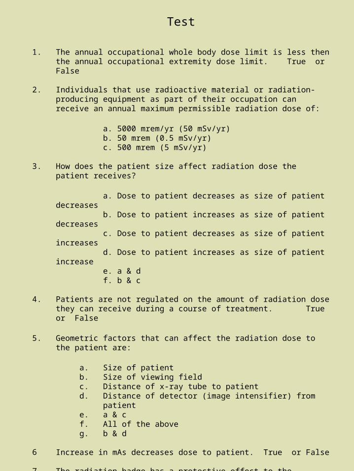
Test
1. The annual occupational whole body dose limit is less then the annual occupational extremity dose limit. True or False
2. Individuals that use radioactive material or radiation-producing equipment as part of their occupation can receive an annual maximum permissible radiation dose of:
a. 5000 mrem/yr (50 mSv/yr) b. 50 mrem (0.5 mSv/yr)c. 500 mrem (5 mSv/yr)
3. How does the patient size affect radiation dose the patient receives?
a. Dose to patient decreases as size of patient decreasesb. Dose to patient increases as size of patient decreasesc. Dose to patient decreases as size of patient increasesd. Dose to patient increases as size of patient increasee. a & df. b & c
4. Patients are not regulated on the amount of radiation dose they can receive during a course of treatment. True or False
5. Geometric factors that can affect the radiation dose to the patient are:
a. Size of patientb. Size of viewing fieldc. Distance of x-ray tube to patientd. Distance of detector (image intensifier) from patiente. a & cf. All of the aboveg. b & d
6 Increase in mAs decreases dose to patient. True or False
7 The radiation badge has a protective effect to the personnel wearing their badge. True or False
8. Personnel can reduce their radiation dose by:
a. wearing protective clothing such as lead aprons or thyroid shieldb. minimizing the amount of time they spend near a source of radiationc. increasing their distance to the source of radiationd. wearing their radiation badgee. all of the abovef. a,b,c only
9. The single major control over the amount of radiation dose received by patients and personnel is:
Choose the best answer
a. technique: mAs and kVpb. Beam-on timec. image magnification
10. The threshold dose for erythema isa. 6 Gy (600 rads)b. 20 Gy (2000 rads)c. 3 Gy (300 rads)
11. How many minutes of beam-on time would it take to deliver a threshold dose for temporary epilation of 3 Gy?
a. 5 – 15 minutesb. 15 – 60 minutesc. 60 – 150 minutes
12. Pregnant patients can receive as much radiation dose as necessary to complete the treatment without concern for the fetus? True or False
Bonus question: Who discovered x-rays?a. Madame Curieb. Niels Bohrc. Wilhelm Roentgen