Embed Size (px)
Citation preview

C I N D Y A . O W E N E D W A R D G . G R A N T
1-2-3 Step Ultrasound Education& Test Preparation
Step 1Review text
Step 2Mock examination
Step 3Q&A memory skills flashcard drill
AbdominalSonography Review
Abdomen A Q&A Review for the ARDMS Specialty Exam
DAVIESRegistry Reviews & Study Aids
L I F E I S A T E S T . . . P A S S I T !
Approved for 12 hours CME Credit
C o n t i n u i n g E d u c a t i o n A c t i v i t y

ISBN 0-941022-46-3
DAV
IESAbdominal Sonography Review
Test yourself before the ARDMS tests you! Abdominal Sonography Review illuminates the
facts and principles on which you will be tested, hones your test-taking skills, and reveals
your strengths and weaknesses by exam topic. Based on the abdomen specialty exam
outline published by ARDMS, this bestselling review by Cindy Owen and Dr. Edward
Grant contains 550 registry-like questions together with instructive illustrations, answers,
clear explanations, and quick references for further study. More than 50 image-based
cases prepare you to tackle the images on the exam. Coverage includes liver, biliary tree,
pancreas, urinary tract, abscesses, scrotum, prostate, spleen, retroperitoneum, abdom-
inal vasculature, GI tract, neck, superficial structures, and instrumentation—all in the
same proportion as in the exam itself. Why are our mock exams so popular and effec-
tive? Because they contain the same kinds of thought-provoking questions you will find
on the exam! Approved for 12 hours of continuing medical education credit. Davies
catalog #11023.
Ready to score? You can!
About the authors . . .
Cindy Owen, RT, RVT, RDMS, FSDMS, is the author of Ultrasound PhysicsReview: SPI Edition, ScoreCards for Vascular Technology, and the related CD-ROM Mock Exams. A former ARDMS and ICAVL board member, she is GlobalManager of Luminary & Research Programs at GE Healthcare and lectureswidely throughout the world. Cindy lives with her family in Memphis.
Edward Grant, MD, is Professor of Radiology and Chair of the Department ofRadiology at USC School of Medicine in Los Angeles. An internationally recog-nized lecturer, Dr. Grant has published widely, participated in the developmentof the ICAVL, AIUM, and ACR ultrasound accreditation programs, serves asexaminer for the radiology oral board examinations, and sits on the editorialboards of all major ultrasound and radiology journals.
www.DaviesPublishing.com
The New CD-ROM Mock Exams from Davies are the most effective andfeaturesome CDs available. Interactive, fun, and packed with continuously updated peer-reviewedcontent for SPI, Abdomen, Vascular, Ob/Gyn, Breast, and Fetal Echo. Work in Test Mode orStudy & Learn Mode and easily customize your Test and Study sessions to focus on specificexam topics. Hundreds of continuously variable questions and images in registry format sharpen your wits and ensure your knowledge of underlying principles and facts. Clear, simple explanations and current references illuminate answers. Expert tutorials cover key concepts in depth. Additional explanatory images and illustrations further clarify answers, anatomy, and pathology. Test timer keeps you on track. Instant results analysis scores and guides you topic by topic. Automatically review missed questions with one click. Available CME credit. A snap to use. Bonus feature: Contact directory of key organizations and societies, including ARDMS, SDMS, ARRT, CCI, ACR, and SVU. Order toll-free 1-877-792-0005 or download from our website.
Mock Exam_Abdomen Bk Cvr 9/25/09 9:52 AM Page 1

Abdominal Sonography Review
A REVIEW FOR THE ARDMS ABDOMEN SPECIALTY EXAM
2016
Cindy Owen, RT, RVT, RDMS, FSDMS
Edward G. Grant, MD Series Editor in Chief

Abdominal Sonography Review iii
v
Copyright © 1999 – 2016 by Davies Publishing, Inc.
All rights reserved. No part of this work may be reproduced, stored in a retrieval system, or transmitted in any form or by any means, electronic or mechanical, including photocopying,
scanning, and recording, without prior written permission from the publisher.
Davies Publishing, Inc. 32 South Raymond Avenue
Pasadena, California 91105-1935 Phone 626-792-3046
Facsimile 626-792-5308 e-mail [email protected]
www.daviespublishing.com
Printed and bound in the United States of America
ISBN 0-941022-46-3
Library of Congress Cataloging-in-Publication Data
Owen, Cindy. Abdominal sonography review : a review for the ARDMS
Abdomen specialty exam/ Cindy Owen; Edward G. Grant. p. ; cm.
Includes bibliographical references.

Abdominal Sonography Review ix
Contents
Preface
Taking and Passing Your Exam
1 The Liver 1
Anatomy
Technique
Laboratory Values
Indications
Parenchymal Disease
Masses
Cysts
Abscesses
Hematomas
2 The Biliary Tree 19
Anatomy
Technique
Laboratory Values
Indications
Dilitation
Masses
Cholelithiasis
Cholecystitis
3 The Pancreas 33
Anatomy
Technique
Laboratory Values
Indications
Parenchymal Disease (Pancreatitis)

Abdominal Sonography Review x
Masses
Cysts
4 The Urinary Tract 44
Anatomy
Technique
Laboratory Values
Indications
Renal Parenchymal Disease
Masses
Cysts
Abscesses
Hematomas
Calculi
Obstructive Disease
Infarctions
Anomalies
Transplants
Bladder
5 The Scrotum 60
Anatomy
Technique
Laboratory Values
Indications
Parenchymal Disease
Masses
Cysts and Fluid Collections
Inflammation
Hematoma

Abdominal Sonography Review xi
6 The Prostate 66
Anatomy
Technique
Laboratory Values
Indications
Parenchymal Disease and Benign Hypertrophy
Masses and Cancer
Cysts
Abscesses
7 The Spleen 71
Anatomy
Technique
Laboratory Values
Indications
Parenchymal Disease
Masses
Cysts
Abscesses
Hematomas
Infarctions
8 The Retroperitoneum 76
Anatomy
Technique
Laboratory Values
Indications
Masses and Adenopathy
Hematomas
Adrenal
Abdominal Sonography Review xii
9 The Abdominal Vasculature 82
Anatomy
Technique
Laboratory Values
Indications
Aneurysms
Thrombus
Arteriovenous Shunts
Doppler Waveforms
10 The GI Tract 96
Anatomy
Technique
Laboratory Values
Indications
Inflammatory Diseases
Masses
Obstruction
Hernia
Peritoneal Fluid
11 The Neck 102
Anatomy
Technique
Laboratory Values
Indications
Thyroid Parenchymal Disease
Thyroid Masses
Thyroid Cysts
Parathyroid Masses
Abscesses

Abdominal Sonography Review xiii
Lymph Nodes
Carotid Arteries and Jugular Veins
12 Superficial Structures 109
Anatomy
Technique
Laboratory Values
Indications
Masses
Cysts, Fluid Collections
Abscesses
Hematomas
Vessels
Breast
Noncardiac Chest
13 Instrumentation 114
Techniques
Transducers
Machine Settings
Image Recording
Artifacts
Quality Assurance
Invasive Procedures
14 Image Gallery 121 15 Answers, Explanations & References 138 16 Application for CME Credit 194 17 ARDMS Exam Outline 225
Why CME Credit Is Important inside back cover

Abdominal Sonography Review 1
PART 1
The Liver
Anatomy
Technique
Laboratory Values
Indications
Parenchymal Disease
Masses
Cysts
Abscesses
Hematomas
1. You are scanning a patient with a known mass in the left medial segment of the
liver. What anatomic landmark can you use to identify the left medial segment separate from the right anterior segment of the liver?
A. Left portal vein B. Ligamentum teres C. Ligamentum venosum D. Middle hepatic vein E. Left hepatic vein
2. You suspect enlargement of the caudate lobe in a patient with liver disease. What
structure located at the anterior border of the caudate lobe will help you to identify this lobe of the liver?
A. Left portal vein B. Fissure for the ligamentum venosum C. Inferior vena cava D. Fissure for the ligamentum teres E. Main lobar fissure
3. You are asked to rule out the presence of a recannalized paraumbilical. Which
anatomic structure is a useful landmark in location of this structure?

Abdominal Sonography Review 2
A. Ligamentum teres B. Ligamentum venosum C. Coronary ligament D. Hepatoduodenal ligament E. Glisson’s ligament
4. Which vessel courses within the main lobar fissure?
A. Main portal vein B. Left portal vein C. Middle Hepatic vein D. Proper hepatic artery E. Right hepatic vein
5. Oxygenated blood is supplied to the liver via the:
A. Portal vein and hepatic vein B. Hepatic vein and hepatic artery C. Hepatic vein and portal vein D. Portal vein and hepatic artery E. Hepatic artery only
6. You are performing a sonogram on a slender female and notice a long, thin
extension of the inferior aspect of the right lobe of the liver. This most likely represents:
A. Caudate lobe B. Quadrate lobe C. Reidel’s lobe D. Accessory liver E. Papillary projection of the caudate lobe
7. Which of the following forms the caudal border of the left portal vein?
A. Ligamentum venosum B. Hepatoduodenal ligament C. Main lobar fissure D. Coronary ligament E. Ligamentum teres
8. What ligament divides the left lobe of the liver into medial and lateral segments?
A. Ligamentum venosum B. Hepatoduodenal ligament C. Main lobar fissure D. Coronary ligament E. Ligamentum teres

Abdominal Sonography Review 3
9. You are asked to perform a Doppler study on the hepatic veins in the liver. What differentiates the hepatic veins from the portal veins?
A. The hepatic veins converge toward the porta hepatis. B. The hepatic veins have brightly echogenic walls. C. The portal veins are largest near the dome of the liver. D. The portal veins are accompanied by branches of the biliary tree and hepatic
artery. E. The portal veins normally exhibit a triphasic flow pattern.
10. You have detected a mass anterior and to the left of ligamentum venosum. This
mass is located in what lobe of the liver?
A. Left lobe B. Caudate lobe C. Reidel’s’ lobe D. Right lobe E. Quadrate lobe
11. The thin capsule surrounding the liver is known as:
A. Reidel’s capsule B. Glisson’s capsule C. Teres capsule D. Langerhans’ capsule E. Wirsung’s capsule
12. Which of the following course interlobar and intersegmental within the liver?
A. Bile ducts B. Portal veins C. Hepatic arteries D. Lymphatics E. Hepatic veins
13. What lobe of the liver does the letter A represent?
A. Left lobe B. Caudate lobe
Questions 13–15 refer to this sagittal image of the liver.

Abdominal Sonography Review 4
C. Posterior right lobe D. Anterior right lobe E. Quadrate lobe
14. What structure does the letter B represent?
A. Ligamentum teres B. Falciform ligament C. Hepatoduodenal ligament D. Ligamentum venosum E. Main lobar fissure
15. What lobe of the liver does the letter C represent?
A. Left lobe B. Caudate lobe C. Posterior right lobe D. Anterior right lobe E. Quadrate lobe
16. Identify the structure labeled A in this image of the liver.
A. Ligamentum venosum B. Ligamentum teres C. Hepatoduodenal ligament D. Coronary ligament E. Glisson’s capsule
Abdominal Sonography Review 5
A.
BC.
D.
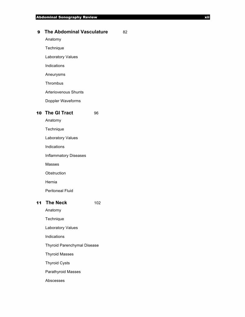
17. Identify the anatomy labeled A.
A. Main portal vein B. Right posterior portal vein branch C. Left portal vein D. Right hepatic vein E. Middle hepatic vein
18. Vessel A is located in what lobe of the liver?
A. Caudate lobe B. Medial segment left lobe C. Lateral segment left lobe D. Posterior segment right lobe E. Anterior segment right lobe
19. Identify the anatomy labeled C.
A. Main portal vein B. Right posterior portal vein C. Left portal vein D. Right hepatic vein E. Middle hepatic vein
20. The arrow labeled D is pointing to what lobe of the liver?
A. Medial segment left lobe B. Lateral segment left lobe C. Posterior segment right lobe D. Anterior segment right lobe E. Caudate lobe
Questions 17–20 refer to this image of the liver.
Abdominal Sonography Review 6
21. You are performing an ultrasound exam of the liver on a small patient with a 5 MHz curved linear array. Although you have increased the overall gain to its maximum setting, the posterior border of the liver and diaphragm are not visualized. What should you do?
A. Call the service representative to repair your equipment. B. Decrease the transmit power on the ultrasound unit. C. Move the focal zone into the near field. D. Rescan the liver with a higher frequency transducer E. Rescan the liver with a lower frequency transducer.
22. Which of the following correctly describes the probe placement and imaging plane
you would use to demonstrate the three hepatic veins and inferior vena cava in one view?
A. Subcostal oblique approach with the probe angled superiorly and to the patient’s
right B. Intercostal approach with the probe angled inferiorly and to the patient’s left C. Intercostal approach with the probe oriented in a coronal plane D. Subcostal oblique approach with the probe angled inferiorly and to the patient’s
left E. Sagittal subcostal approach with the probe just to the right of midline
23. You are performing a follow-up sonogram on a patient in which a 5-mm cyst was
previously identified at the anterior border of the left lobe of the liver. Although you are using a 3.5 MHz curved linear array probe, you do not see the cyst. Which of the following would be most helpful in improving visibility of this cyst?
A. Increase the overall gain B. Increase the dynamic range C. Increase the transmit power D. Rescan the left lobe with a higher frequency transducer E. Rescan the left lobe with a lower frequency linear array transducer
24. You are imaging a patient with a high liver. Subcostal images do not clearly
demonstrate the liver tissue. What should you do?
A. Scan the patient in deep inspiration B. Scan the patient in expiration C. Place the patient in a Trendelenburg position and rescan D. Have the patient drink 32 oz. of water and rescan E. Scan with the patient in quiet respiration
25. A patient is referred for ultrasound evaluation of a questionable mass in the dome of
the liver seen on a CAT scan. Which method below would improve visualization in this area of the liver?
A. Perform a subcostal scan with the probe angled superior and the patient in deep
inspiration.

Abdominal Sonography Review 7
B. Perform an intercostal scan with the probe in a coronal plane and the patient in expiration.
C. Perform a subcostal scan with the patient performing a Valsalva maneuver. D. Roll the patient into a right lateral decubitus position and scan from a subcostal
approach with the patient in expiration. E. The dome of the liver cannot be seen with ultrasound.
26. A patient is referred for a liver ultrasound with the clinical history of a raised serum
alpha-fetoprotein level. What should you look for?
A. Focal nodular hyperplasia B. Fatty liver C. Hepatocellular carcinoma D. Hydatid disease E. Increased alpha-fetoprotein levels are not associated with liver disease
27. You are reviewing lab work prior to performing an abdominal ultrasound exam.
Elevated lab values include Gamma-glutamyl transpeptidase (GGT) and alkaline phosphatase. Which statement below is true?
A. Elevation of both GGT and alkaline phosphatase suggests the source of elevated
alkaline phosphatase is due to metastatic bone cancer. B. Elevation of both GGT and alkaline phosphatase is a sensitive indicator of
pancreatitis. C. If both GGT and alkaline phosphatase are elevated, the lab work is invalid and
must be repeat D. Concomitant elevation of both GGT and alkaline phosphatase indicates the
source of the elevated alkaline phosphatase is the liver. E. Concomitant elevation of both of these lab values is highly specific for
hepatocellular carcinoma. 28. Which of the following lab tests is NOT used in evaluation of liver function?
A. Gamma-glutamyl transpeptidase (GGT) B. Aspartate aminotransferase (AST) C. Direct bilirubin D. Indirect bilirubin E. Lipase
29. A patient is referred with right upper quadrant tenderness and a history of oral
contraceptive use. A solid, hypoechoic mass is identified in the right lobe of the liver. Color Doppler reveals hypervascularity of the mass. Which of the following scenarios is most likely?
A. Hydatid liver disease B. Hepatic lipoma C. Hepatic abscess D. Hepatic adenoma E. Hepatocellular carcinoma

Abdominal Sonography Review 8
30. A liver ultrasound on a 49-year-old obese male demonstrates diffuse increased
echogenicity with a focal hypoechoic area anterior to the portal vein. This most likely represents:
A. Liver cirrhosis with hepatocellular carcinoma B. Hydatid disease C. Fatty metamorphosis of the liver with focal sparing D. Metatstatic disease most likely due to a colon primary E. Normal liver parenchyma with a simple cyst
31. A 52-year-old male with known liver cirrhosis presents for an abdominal
ultrasound. You will carefully evaluate the liver to rule out the presence of any focal mass because of which true statement below?
A. Patients with liver cirrhosis are at increased risk for hepatocellular carcinoma B. Patients with liver cirrhosis tend to develop multiple cysts in their liver and
pancreas C. Metastatic disease occurs commonly with cirrhosis D. The presence of regenerative nodules rules out cirrhosis E. All of the above are correct
32. You are scanning a patient with suspected liver cirrhosis. All of the following are
sonographic features of cirrhosis EXCEPT:
A. Surface nodularity B. Shrunken caudate lobe C. Altered echo texture D. Ascites E. Regenerative nodules
33. An ultrasound evaluation of liver cirrhosis should include a search for which
associated complication?
A. Biliary dilatation B. Mesenteric ischemia C. Splenic infarction D. Kaposi's sarcoma E. Portal hypertension
34. Ultrasound findings of an abdominal study on a 51-year-old female include
enlargement of the hepatic veins and inferior vena cava in an otherwise normal appearing liver. These findings are most consistent with which of the following?
A. Budd-Chiari syndrome B. Right-sided heart failure C. Liver cirrhosis D. Portal hypertension E. Sarcoidosis

Abdominal Sonography Review 9
35. Focal fatty liver is most commonly found in which location?
A. Medial to the ascending branch of the left portal vein B. Posterior to the right hepatic vein C. Lateral, inferior tip of the right lobe D. Adjacent to the fissure for the ligamentum venosum E. Anterior to the portal vein at the porta hepatis
36. You have performed an ultrasound study on a patient with an enlarged caudate lobe,
shrunken right lobe and splenomegaly. The hepatic veins could not be identified. No other abnormalities were discovered. What should you do?
A. Scan the pelvis to rule out a pelvic mass. B. Have the patient perform a Valsalva maneuver and reexamine the hepatic veins. C. Evaluate the hepatic veins and IVC with color Doppler to confirm patency D. Have the patient return in a week for a repeat study to evaluate the hepatic veins. E. Nothing, you have completed the exam.
37. A patient is referred to rule out hepatomegaly. All of the following are useful
indicators of hepatomegaly EXCEPT:
A. Rounding of the inferior border of the liver B. Longitudinal measurement of the right lobe exceeding 15.5 cm C. Extension of the right lobe inferior to the lower pole of the right kidney D. Increased diameter of the main portal vein greater than 1 cm E. Increased anteroposterior measurement of the right lobe
38. You have identified a single homogeneous hyperechoic lesion measuring 2.4 cm in
the posterior aspect of the right lobe of the liver. What is the most common etiology of a mass fitting this description?
A. Cyst B. Hepatic adenoma C. Cavernous hemangioma D. Hepatocellular carcinoma E. Focal fatty sparing
39. A patient is referred for a sonogram of the liver to rule out metastatic disease.
Which of the following describes the sonographic appearance of liver metastasis?
A. Single hypoechoic mass B. Multiple hyperechoic masses C. Masses of mixed echogenicity D. Cystic masses E. All of the above appearances of liver metastasis may be encountered
40. Which of the following is NOT a feature of hepatic cysts?
A. Thin wall B. Posterior acoustic enhancement

Abdominal Sonography Review 10
C. Anechoic D. Increased attenuation E. Increased through transmission
41. A single large, well-defined mass with smooth walls and homogeneous low-level
echoes is seen within the anterior right lobe of the liver in a 48-year-old female. No Doppler signals could be obtained within the mass. Which of the following conditions is the most likely etiology of this mass?
A. Kaposi's sarcoma B. Focal nodular hyperplasia C. Hemorrhagic cyst D. Portal vein aneurysm E. hepatocellular carcinoma
42. You are scanning a patient with a history of fever, abnormal liver function tests, and right upper quadrant tenderness. The liver is enlarged with decreased echogenicity, the gallbladder wall is thickened and thick echogenic bands are noted surrounding the portal veins. Which of the following conditions is most likely?
A. Fatty liver B. Cirrhosis C. Budd-Chiari syndrome D. Hepatitis E. Normal liver
43. You are evaluating a suspicious lesion to look for gas bubbles to confirm the
presence of liver abscess in a patient with fever and increased white blood cell count. What is the sonographic appearance of the gas bubbles?
A. Brightly echogenic echoes with clean distal acoustic shadow B. Brightly echogenic foci associated with echogenic ringdown artifact C. Hypoechoic areas within the mass associated with increased through
transmission D. Anechoic foci with distal acoustic enhancement E. Hyperechoic foci with distal acoustic enhancement
44. Which of the following is associated with infestation by a parasite and is most
prevalent in sheep and cattle-raising countries?
A. Budd-Chiari syndrome B. Hydatid disease C. Candidiasis D. Hepatitis A E. Kaposi’s sarcoma
45. You are scanning the liver and notice irregularity of the surface. A nodular liver
surface is associated with which of the following abnormalities?
A. Cirrhosis

Abdominal Sonography Review 11
B. Acute Hepatitis C. Fatty liver D. Polycystic liver disease E. Hepatomegaly
46. Which of the following is NOT true regarding fatty liver?
A. It is an irreversible disorder B. Fatty liver may be caused by obesity C. It may be diffuse or focal D. It may show a rapid change in appearance with time E. It commonly causes increased attenuation of the sound beam through liver
47. You are scanning through the liver and notice luminal narrowing of the hepatic
veins. Color and spectral Doppler reveal high velocities through the strictures. These findings are most commonly associated with which of the following?
A. Diffuse fatty liver B. Acute hepatitis C. Cirrhosis D. Focal fatty infiltration E. Glycogen storage disease
48. The most common benign tumor in the liver is:
A. Focal nodular hyperplasia B. Hepatic adenoma C. Hepatic lipoma D. Cavernous hemangioma E. Hepatoma
49. Which of the following is most commonly associated with invasion of the portal
vein?
A. Hepatocellular carcinoma B. Cavernous hemangioma C. Liver metastases D. Hepatic adenoma E. Focal nodular hyperplasia
50. You have been asked to perform a liver sonogram on a patient with AIDS. Which
of the following tumors is most commonly associated with this history?
A. Hepatocellular carcinoma B. Kaposi’s sarcoma C. Budd-Chiari syndrome D. Hemangiosarcoma E. Hepatic adenoma

Abdominal Sonography Review 12
51. You are scanning a 53-year-old female with a history of recent weight loss and vague abdominal pain. The liver is markedly heterogeneous and contains numerous calcified lesions. This most likely represents metastatic disease from which primary?
A. Non-Hodgkin lymphoma B. Cystadenocarcinoma of the ovary C. Lung D. Adenocarcinoma of the colon E. Breast
52. During ultrasound evaluation of the liver, a bull’s eye or target lesion is identified in
the anterior right lobe. The most likely etiology of this mass is:
A. Liver abscess B. Hepatic adenoma C. Focal nodular hyperplasia D. Hepatocellular carcinoma E. Liver metastasis from lung cancer
53. You are performing an ultrasound exam on a young female and notice a well-
defined solitary mass with a central scar measuring 4 cm in diameter. Color Doppler reveals prominent blood vessels coursing within the scar. This most likely represents:
A. Liver abscess B. Hepatic adenoma C. Focal nodular hyperplasia D. Hepatocellular carcinoma E. Liver metastasis from lung cancer
54. You are performing a liver sonogram on a young female with right upper quadrant
pain, sudden onset ascites and hepatomegaly. You have obtained transverse and sagittal images of the liver, common bile duct, and gallbladder according to your protocol. What else should you do?
A. Nothing, the study is complete B. Expand the study to include the kidneys to rule out associated hydronephrosis C. Use color and spectral Doppler to determine patency of the portal and hepatic
venous systems D. Give the patient a fatty meal and then measure the portal vein diameter at 1,2, 5
and 10 minutes E. Call the referring physician to get an order to perform a pelvic study to see if the
patients’ pain is referred from an ovarian mass 55. Which of the following is NOT true regarding cavernous hemangiomas?
A. Small, well-defined, hyperechoic masses B. Consist of a vascular network C. More common in women than men

Abdominal Sonography Review 13
D. Usually asymptomatic E. Show prominent, high-velocity color Doppler signals
56. A patient is referred for ultrasound with a history of liver transplantation. You
identify an extrahepatic fluid collection. What is the likely etiology of this finding?
A. Biloma B. Hematoma C. Loculated ascites D. Abscess E. Any of the above may be seen following liver transplantation
57. What significant complication following liver transplantation is NOT detectable
with ultrasound?
A. Rejection B. Malignant disease C. Hepatic artery thrombosis D. Portal vein thrombosis E. Pseudoaneurysm
58. You are scanning a patient with a history of liver transplantation. You should search
for all of the following complications of this surgery EXCEPT:
A. Biliary sludge B. Acute cholecystitis C. Portal vein stenosis D. Hepatic artery thrombosis E. Liver malignancy
59. You have been asked to provide ultrasound imaging during liver surgery. What
transducer would be best suited for this purpose?
A. 3.5 MHz curved linear array B. 10 MHz linear array C. 2.25 MHz phased array D. 7 MHz linear array E. 12 MHz curved linear array
60. You are scanning through the right lobe of the liver and notice that although you
have maximized the far field TGC, the parenchyma in the far field and diaphragm are not clearly visualized. What should you do? A. Decrease the transmit power B. Increase the compression curve C. Decrease the transmit frequency D. Decrease the overall gain E. Increase the dynamic range

Abdominal Sonography Review 14
61. Hepatofugal flow in the portal vein is a sign of: A. Normalcy B. Hepatic artery thrombosis C. Portal hypertension D. Acute cholecystitis E. Hepatocellular carcinoma
62. A patient is referred for abdominal ultrasound with a high fever and right upper
quadrant pain. You document the presence of a large, rounded, homogeneous mass with low-level internal echoes and poorly defined borders. The mass is located in the right lobe of the liver, adjacent to the capsule and shows increased through transmission. This most likely represents:
A. Hemorrhagic cyst B. Abscess C. Hematoma D. Choledochal cyst E. Loculated ascites
63. The most common form of malignant disease of the liver is:
A. Hepatocellular carcinoma B. Angiosarcoma C. Cholangiocarcinoma D. Metastatic disease E. Primary lymphoma
64. You are scanning a patient with known liver cirrhosis and notice a focal mass
within the posterior right lobe. What laboratory test would be most helpful in determining if this mass is hepatocellular carcinoma? A. Serum alpha-fetoprotein B. Alkaline phosphatase C. Serum bilirubin D. Serum creatinine E. Lactate dehydrogenase (LD)
65. You have been asked to perform an ultrasound to rule out the presence of Budd-
Chiari syndrome. You will tailor your exam to include which of the following?
A. Volume measurement of the spleen B. Doppler analysis of the hepatic venous system C. Both supine and upright views of the porta hepatis D. Oblique view of the right lobe of the liver to include the right hemidiaphragm E. Careful search for periaortic lymphadenopathy
66. You are performing an ultrasound on a patient with a transjugular intrahepatic
portosystemic shunt (TIPS). What two vessels are connected with the TIPS stent?

Abdominal Sonography Review 15
A. Portal vein and hepatic artery B. Hepatic artery and hepatic vein C. Hepatic vein and inferior vena cava D. Portal vein and hepatic vein E. Portal vein and inferior vena cava
67. The majority of blood supply to the liver is provided from the:
A. Hepatic veins B. Portal vein C. Hepatic artery D. Superior mesenteric vein E. Gastroduodenal artery
68. Following liver transplantation, which of the following anatomic locations has an
anastomotic connection that should be evaluated with ultrasound?
A. Inferior vena cava B. Portal vein C. Hepatic artery D. Bile duct E. All of the above
69. You are scanning a patient with liver cirrhosis and suspected portal hypertension. In
this study, assessment of the size of which of the following is most important?
A. Spleen B. Common bile duct C. Abdominal aorta D. Right hepatic vein E. Inferior vena cava
70. A recannalized paraumbilical vein may be seen as a result of which of the
following?
A. Hepatic adenoma B. Portal hypertension C. Hepatitis D. Hepatic trauma E. Liver biopsy
71. Which of the following describes the best sonographic window to view a
recannalized paraumbilical vein?
A. Intercostal oblique view through the right lobe B. Subcostal oblique view through the right lobe C. Sagittal subcostal view through the left lobe at the level of the ligamentum teres D. Sagittal subcostal view through the right lobe at the level of the main lobar
fissure

Abdominal Sonography Review 16
E. Sagittal subcostal view to the left of midline 72. What three structures comprise the portal triad?
A. Portal vein, bile duct, hepatic artery B. Portal vein, bile duct, hepatic vein C. Bile duct, hepatic vein, hepatic artery D. Hepatic vein, hepatic artery, lymph node E. Hepatic vein, hepatic artery, portal vein
73. The ligamentum venosum forms the anterior border of what lobe of the liver?
A. Left lobe B. Reidel’s lobe C. Right lobe D. Caudate lobe E. Quadrate lobe
74. You are scanning a patient with portal hypertension. Enlargement of which of the
following structures is diagnostic of this condition?
A. Coronary vein B. Hepatic vein C. Renal vein D. Common bile duct E. Ligamentum teres
75. Which measurement below is the diagnostic cutoff for portal vein enlargement?
A. 13 mm B. 5 mm C. 10 cm D. 1.8 cm E. 9 mm
76. Regenerating nodules are a feature associated with:
A. Hepatitis B. Hepatocellular carcinoma C. Hydatid disease D. Cirrhosis E. Polycystic liver disease
77. You are performing an ultrasound exam in a patient with a history of alcoholic liver
cirrhosis. You have documented the presence of splenomegaly and dilated veins at the splenic hilum. Considering the patient’s history and findings, what else should you look for?
A. Search for signs of acute cholecystitis B. Carefully scan the spleen for the presence of infarcts

Abdominal Sonography Review 17
C. Search for the presence of portosystemic collaterals D. Check the pelvis for a left side mass. E. Rule out the presence of an aortic aneurysm.
78. Which of the following describes the best view for ultrasound demonstration of the
coronary vein?
A. A transverse scan under the right lobe of the liver. B. An oblique subcostal scan under the right lobe of the liver with the probe
oriented toward the patient’s head C. A sagittal view of the splenic vein near the midline D. A sagittal view through the splenic hilum E. A transverse view along the long axis of the left renal vein
79. You are performing a follow-up study on a patient with a history of cavernous
transformation. Where should you look to evaluate this condition?
A. Splenic hilum B. Pancreatic head C. Porta hepatis D. Renal hilum E. Left lobe of the liver
80. You are scanning a patient with an enlarged caudate lobe and shrunken right lobe.
What diffuse liver process should you suspect? A. Cirrhosis B. Acute hepatitis C. Fatty infiltration D. Candidiasis E. Hepatocellular carcinoma
81. You are scanning an obese patient to rule out fatty liver. Which of the following
describes a common sonographic appearance of this condition?
A. Increased through transmission throughout the hypoechoic liver B. Increased echogenicity of the liver compared to normal C. Focal hypoechoic masses throughout both lobes of the liver surrounded by
normal liver echotexture D. Shrunken liver with surface nodularity E. Enlarged, hypoechoic right lobe compared to a small and shrunken left lobe


![Tema 6. Traumatismos abdominales: contusiones y … · abdominal Herida abdominal ... T Abdominal, Toraco-Abd, Abd-pelvianas No orificio salida ... Trauma Abdominal [Modo de compatibilidad]](https://img.dokumen.tips/doc/110x75/5bab89f609d3f211798c2c36/tema-6-traumatismos-abdominales-contusiones-y-abdominal-herida-abdominal-.jpg)