Embed Size (px)
Citation preview
GOAL OF THE FAST EXAM
Demonstrate free fluid in abdomen, pleural space, or pericardial space.
EMERGENCY ULTRASOUND APPROACH
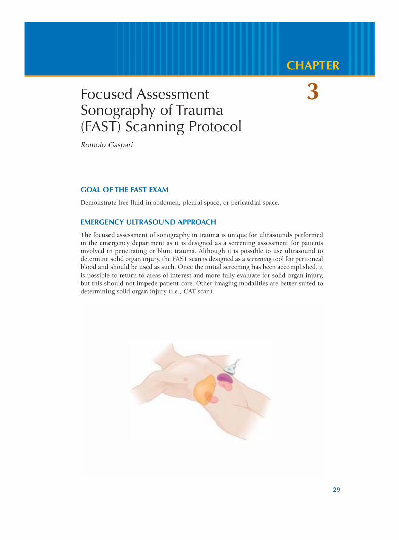
The focused assessment of sonography in trauma is unique for ultrasounds performedin the emergency department as it is designed as a screening assessment for patientsinvolved in penetrating or blunt trauma. Although it is possible to use ultrasound todetermine solid organ injury, the FAST scan is designed as a screening tool for peritonealblood and should be used as such. Once the initial screening has been accomplished, itis possible to return to areas of interest and more fully evaluate for solid organ injury,but this should not impede patient care. Other imaging modalities are better suited todetermining solid organ injury (i.e., CAT scan).
CHAPTER
3Focused AssessmentSonography of Trauma(FAST) Scanning ProtocolRomolo Gaspari
29
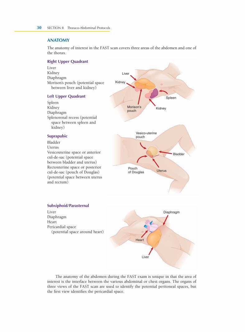
ANATOMY
The anatomy of interest in the FAST scan covers three areas of the abdomen and one ofthe thorax.
Right Upper Quadrant
LiverKidneyDiaphragmMorison’s pouch (potential space
between liver and kidney)
Left Upper Quadrant
SpleenKidneyDiaphragmSplenorenal recess (potential
space between spleen andkidney)
Suprapubic
BladderUterusVesicouterine space or anteriorcul-de-sac (potential spacebetween bladder and uterus)Rectouterine space or posteriorcul-de-sac (pouch of Douglas)(potential space between uterusand rectum)
Subxiphoid/Parasternal
LiverDiaphragmHeartPericardial space
(potential space around heart)
The anatomy of the abdomen during the FAST exam is unique in that the area ofinterest is the interface between the various abdominal or chest organs. The organs ofthree views of the FAST scan are used to identify the potential peritoneal spaces, butthe first view identifies the pericardial space.
SECTION II Thoraco-Abdominal Protocols30
Spleen
Kidney
Kidney
Liver
Morison'spouch
Bladder
UterusPouchof Douglas
Vesico-uterinepouch
Heart
Liver
Diaphragm
PATIENT POSITION
Supine
As a majority of patients are scanned following trauma, these patients will be “boardedand collared” with cervical spine and long board immobilization. There may be limitedopportunity to reposition the patient secondary to concerns for cervical spine injury. Inother words, the patient will be flat on his or her back and cannot be moved.
TRANSDUCER
3.0 to 5.0 MHz
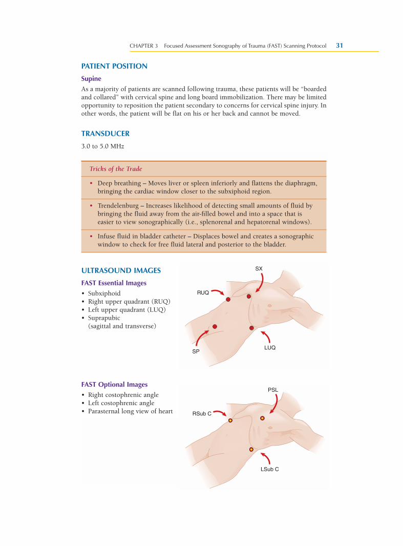
ULTRASOUND IMAGES
FAST Essential Images
• Subxiphoid• Right upper quadrant (RUQ)• Left upper quadrant (LUQ)• Suprapubic
(sagittal and transverse)
FAST Optional Images
• Right costophrenic angle• Left costophrenic angle• Parasternal long view of heart
CHAPTER 3 Focused Assessment Sonography of Trauma (FAST) Scanning Protocol 31
Tricks of the Trade
• Deep breathing – Moves liver or spleen inferiorly and flattens the diaphragm,bringing the cardiac window closer to the subxiphoid region.
• Trendelenburg – Increases likelihood of detecting small amounts of fluid bybringing the fluid away from the air-filled bowel and into a space that iseasier to view sonographically (i.e., splenorenal and hepatorenal windows).
• Infuse fluid in bladder catheter – Displaces bowel and creates a sonographicwindow to check for free fluid lateral and posterior to the bladder.
SX
SP
RUQ
LUQ
PSL
RSub C
LSub C
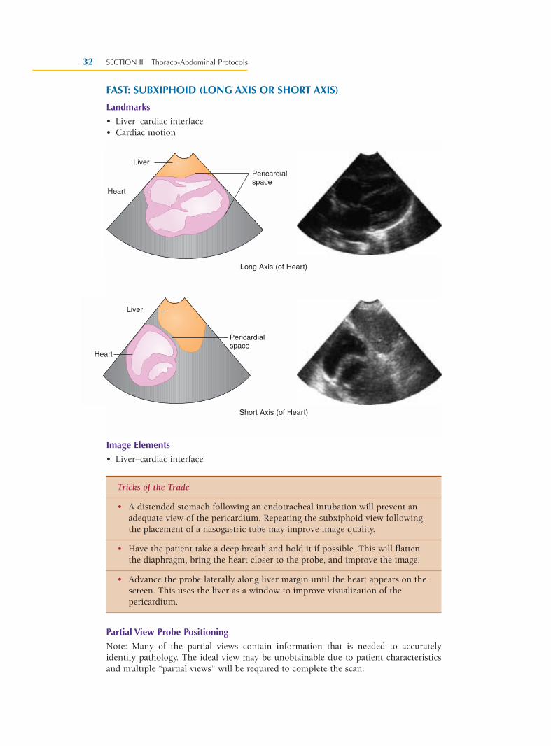
FAST: SUBXIPHOID (LONG AXIS OR SHORT AXIS)
Landmarks
• Liver–cardiac interface• Cardiac motion
Image Elements
• Liver–cardiac interface
Partial View Probe Positioning
Note: Many of the partial views contain information that is needed to accuratelyidentify pathology. The ideal view may be unobtainable due to patient characteristicsand multiple “partial views” will be required to complete the scan.
SECTION II Thoraco-Abdominal Protocols32
Long Axis (of Heart)
Short Axis (of Heart)
Tricks of the Trade
• A distended stomach following an endotracheal intubation will prevent anadequate view of the pericardium. Repeating the subxiphoid view followingthe placement of a nasogastric tube may improve image quality.
• Have the patient take a deep breath and hold it if possible. This will flattenthe diaphragm, bring the heart closer to the probe, and improve the image.
• Advance the probe laterally along liver margin until the heart appears on thescreen. This uses the liver as a window to improve visualization of thepericardium.
Liver
Heart
Pericardialspace
Liver
Heart
Pericardialspace
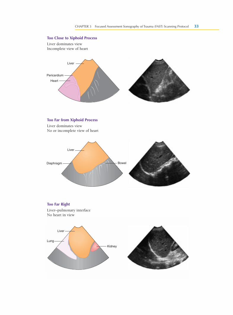
Too Close to Xiphoid Process
Liver dominates viewIncomplete view of heart
Too Far from Xiphoid Process
Liver dominates viewNo or incomplete view of heart
Too Far Right
Liver–pulmonary interfaceNo heart in view
CHAPTER 3 Focused Assessment Sonography of Trauma (FAST) Scanning Protocol 33
Liver
Heart
Pericardium
Liver
Diaphragm Bowel
Kidney
Liver
Lung
Too Far Left
No liver in imageNo heart in viewStomach gas obscures view
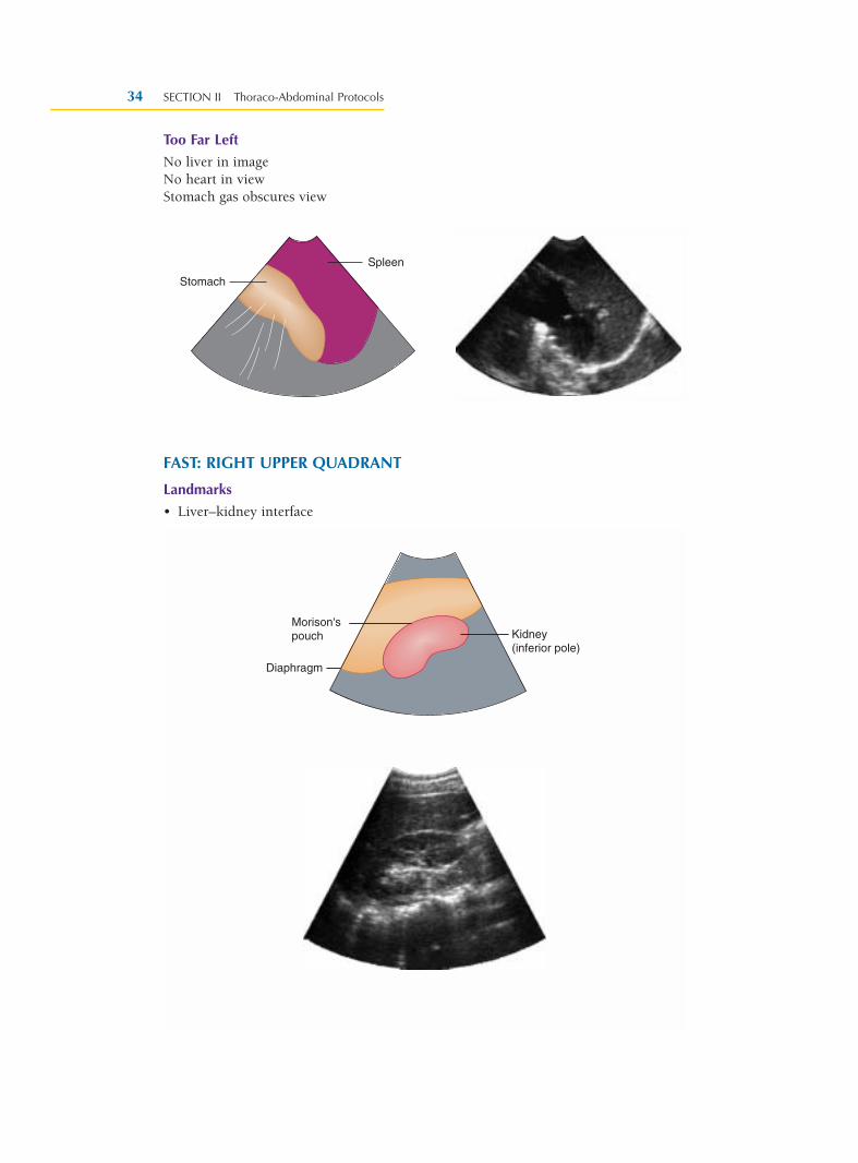
FAST: RIGHT UPPER QUADRANT
Landmarks
• Liver–kidney interface
SECTION II Thoraco-Abdominal Protocols34
Spleen
Stomach
Morison'spouch Kidney
(inferior pole)
Diaphragm
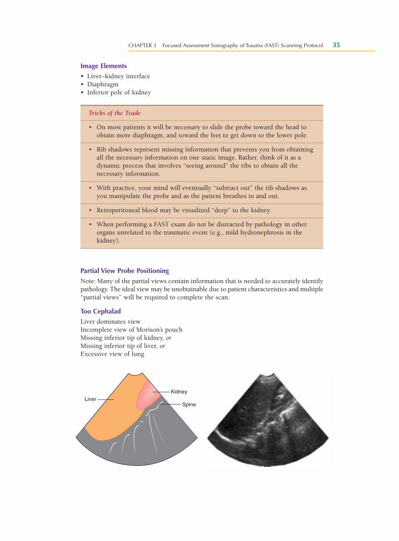
Image Elements
• Liver–kidney interface• Diaphragm• Inferior pole of kidney
Partial View Probe Positioning
Note: Many of the partial views contain information that is needed to accurately identifypathology. The ideal view may be unobtainable due to patient characteristics and multiple“partial views” will be required to complete the scan.
Too Cephalad
Liver dominates viewIncomplete view of Morison’s pouchMissing inferior tip of kidney, orMissing inferior tip of liver, orExcessive view of lung
CHAPTER 3 Focused Assessment Sonography of Trauma (FAST) Scanning Protocol 35
Tricks of the Trade
• On most patients it will be necessary to slide the probe toward the head toobtain more diaphragm, and toward the feet to get down to the lower pole.
• Rib shadows represent missing information that prevents you from obtainingall the necessary information on one static image. Rather, think of it as adynamic process that involves “seeing around” the ribs to obtain all thenecessary information.
• With practice, your mind will eventually “subtract out” the rib shadows asyou manipulate the probe and as the patient breathes in and out.
• Retroperitoneal blood may be visualized “deep” to the kidney.
• When performing a FAST exam do not be distracted by pathology in otherorgans unrelated to the traumatic event (e.g., mild hydronephrosis in thekidney).
KidneyLiver
Spine
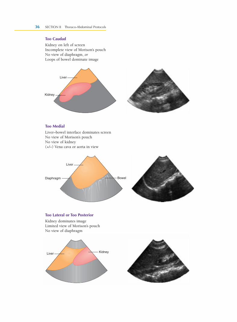
Too Caudad
Kidney on left of screenIncomplete view of Morison’s pouchNo view of diaphragm, orLoops of bowel dominate image
Too Medial
Liver–bowel interface dominates screenNo view of Morison’s pouchNo view of kidney(+/–) Vena cava or aorta in view
Too Lateral or Too Posterior
Kidney dominates imageLimited view of Morison’s pouchNo view of diaphragm
SECTION II Thoraco-Abdominal Protocols36
Kidney
Liver
Liver
Diaphragm Bowel
KidneyLiver
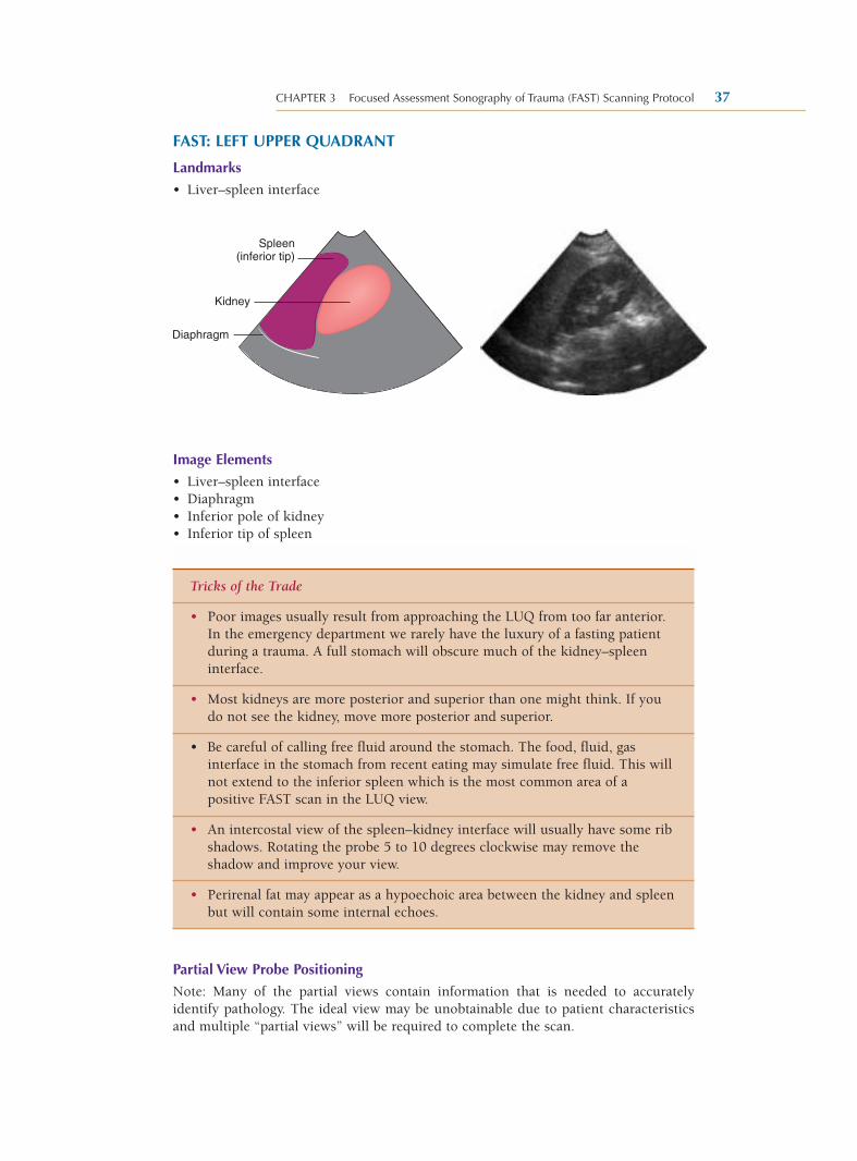
FAST: LEFT UPPER QUADRANT
Landmarks
• Liver–spleen interface
Image Elements
• Liver–spleen interface• Diaphragm• Inferior pole of kidney• Inferior tip of spleen
Partial View Probe Positioning
Note: Many of the partial views contain information that is needed to accuratelyidentify pathology. The ideal view may be unobtainable due to patient characteristicsand multiple “partial views” will be required to complete the scan.
CHAPTER 3 Focused Assessment Sonography of Trauma (FAST) Scanning Protocol 37
Tricks of the Trade
• Poor images usually result from approaching the LUQ from too far anterior.In the emergency department we rarely have the luxury of a fasting patientduring a trauma. A full stomach will obscure much of the kidney–spleeninterface.
• Most kidneys are more posterior and superior than one might think. If youdo not see the kidney, move more posterior and superior.
• Be careful of calling free fluid around the stomach. The food, fluid, gasinterface in the stomach from recent eating may simulate free fluid. This willnot extend to the inferior spleen which is the most common area of apositive FAST scan in the LUQ view.
• An intercostal view of the spleen–kidney interface will usually have some ribshadows. Rotating the probe 5 to 10 degrees clockwise may remove theshadow and improve your view.
• Perirenal fat may appear as a hypoechoic area between the kidney and spleenbut will contain some internal echoes.
Spleen(inferior tip)
Kidney
Diaphragm
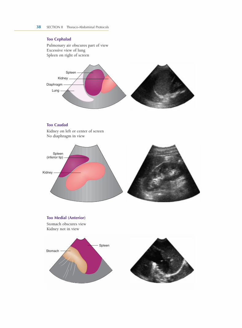
Too Cephalad
Pulmonary air obscures part of viewExcessive view of lungSpleen on right of screen
Too Caudad
Kidney on left or center of screenNo diaphragm in view
Too Medial (Anterior)
Stomach obscures viewKidney not in view
SECTION II Thoraco-Abdominal Protocols38
Spleen
Kidney
Diaphragm
Lung
Spleen(inferior tip)
Kidney
Spleen
Stomach
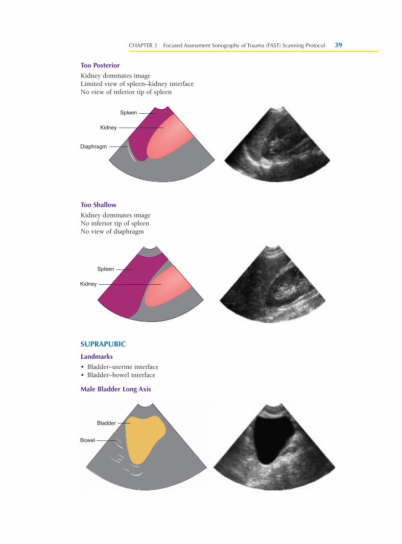
Too Posterior
Kidney dominates imageLimited view of spleen–kidney interfaceNo view of inferior tip of spleen
Too Shallow
Kidney dominates imageNo inferior tip of spleenNo view of diaphragm
SUPRAPUBIC
Landmarks
• Bladder–uterine interface• Bladder–bowel interface
Male Bladder Long Axis
CHAPTER 3 Focused Assessment Sonography of Trauma (FAST) Scanning Protocol 39
Spleen
Kidney
Diaphragm
Spleen
Kidney
Bladder
Bowel
Bladder
Bowel
Bladder
Bowel
Bladder
Bowel
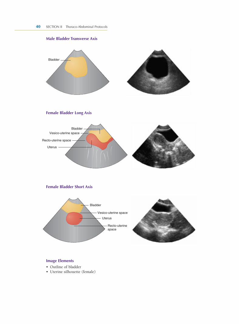
Male Bladder Transverse Axis
Female Bladder Long Axis
Female Bladder Short Axis
Image Elements
• Outline of bladder• Uterine silhouette (female)
SECTION II Thoraco-Abdominal Protocols40
Bladder
BladderVesico-uterine space
Uterus
Recto-uterine space
Bladder
Vesico-uterine space
Uterus
Recto-uterinespace
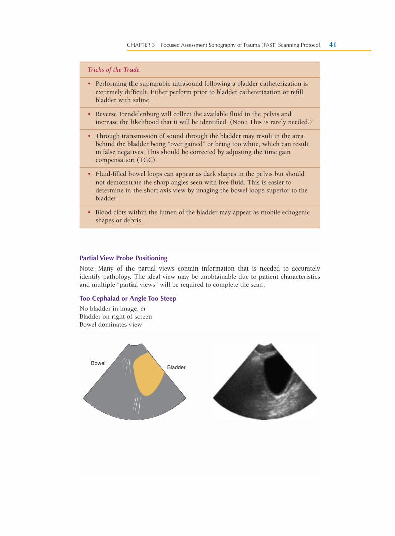
Partial View Probe Positioning
Note: Many of the partial views contain information that is needed to accuratelyidentify pathology. The ideal view may be unobtainable due to patient characteristicsand multiple “partial views” will be required to complete the scan.
Too Cephalad or Angle Too Steep
No bladder in image, orBladder on right of screenBowel dominates view
CHAPTER 3 Focused Assessment Sonography of Trauma (FAST) Scanning Protocol 41
Tricks of the Trade
• Performing the suprapubic ultrasound following a bladder catheterization isextremely difficult. Either perform prior to bladder catheterization or refillbladder with saline.
• Reverse Trendelenburg will collect the available fluid in the pelvis andincrease the likelihood that it will be identified. (Note: This is rarely needed.)
• Through transmission of sound through the bladder may result in the areabehind the bladder being “over gained” or being too white, which can resultin false negatives. This should be corrected by adjusting the time gaincompensation (TGC).
• Fluid-filled bowel loops can appear as dark shapes in the pelvis but shouldnot demonstrate the sharp angles seen with free fluid. This is easier todetermine in the short axis view by imaging the bowel loops superior to thebladder.
• Blood clots within the lumen of the bladder may appear as mobile echogenicshapes or debris.
BladderBowel
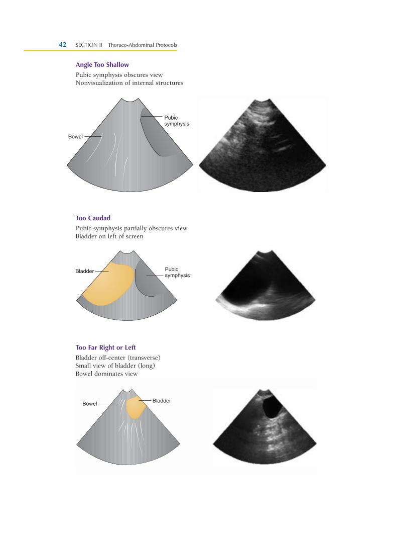
Angle Too Shallow
Pubic symphysis obscures viewNonvisualization of internal structures
Too Caudad
Pubic symphysis partially obscures viewBladder on left of screen
Too Far Right or Left
Bladder off-center (transverse)Small view of bladder (long)Bowel dominates view
SECTION II Thoraco-Abdominal Protocols42
Pubicsymphysis
Bowel
Pubicsymphysis
Bladder
BladderBowel
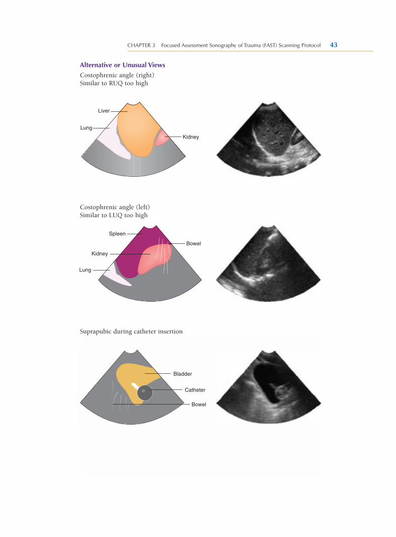
Alternative or Unusual Views
Costophrenic angle (right)Similar to RUQ too high
Costophrenic angle (left)Similar to LUQ too high
Suprapubic during catheter insertion
CHAPTER 3 Focused Assessment Sonography of Trauma (FAST) Scanning Protocol 43
Kidney
Liver
Lung
Spleen
Kidney
Lung
Bowel
Bladder
Catheter
Bowel
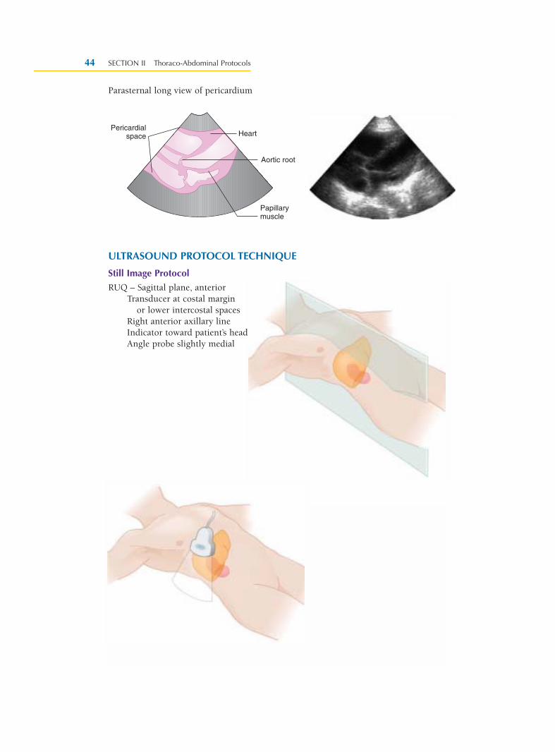
Parasternal long view of pericardium
ULTRASOUND PROTOCOL TECHNIQUE
Still Image Protocol
RUQ – Sagittal plane, anteriorTransducer at costal margin
or lower intercostal spacesRight anterior axillary lineIndicator toward patient’s headAngle probe slightly medial
SECTION II Thoraco-Abdominal Protocols44
Pericardialspace Heart
Aortic root
Papillarymuscle

RUQ – Coronal plane, lateralTransducer at costal margin or lower intercostalRight middle axillary lineIndicator toward patient’s headAngle probe slightly posterior
CHAPTER 3 Focused Assessment Sonography of Trauma (FAST) Scanning Protocol 45
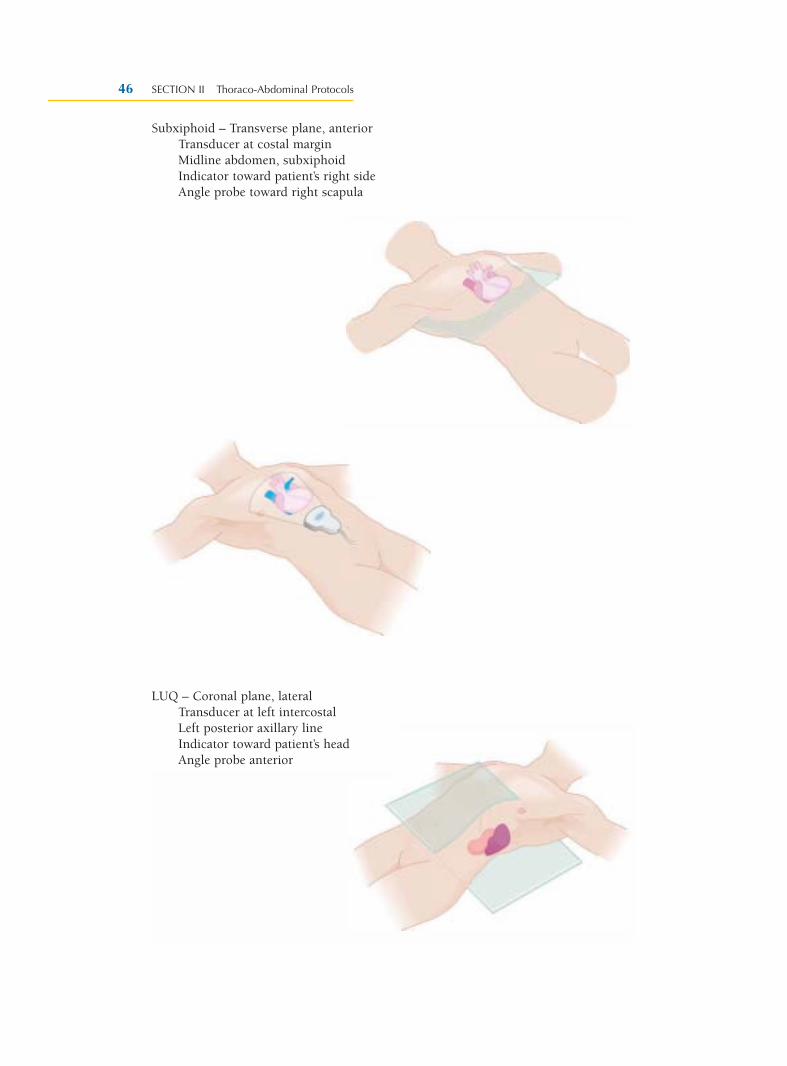
Subxiphoid – Transverse plane, anteriorTransducer at costal marginMidline abdomen, subxiphoidIndicator toward patient’s right sideAngle probe toward right scapula
LUQ – Coronal plane, lateralTransducer at left intercostalLeft posterior axillary lineIndicator toward patient’s headAngle probe anterior
SECTION II Thoraco-Abdominal Protocols46
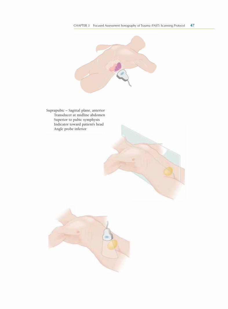
Suprapubic – Sagittal plane, anteriorTransducer at midline abdomenSuperior to pubic symphysisIndicator toward patient’s headAngle probe inferior
CHAPTER 3 Focused Assessment Sonography of Trauma (FAST) Scanning Protocol 47
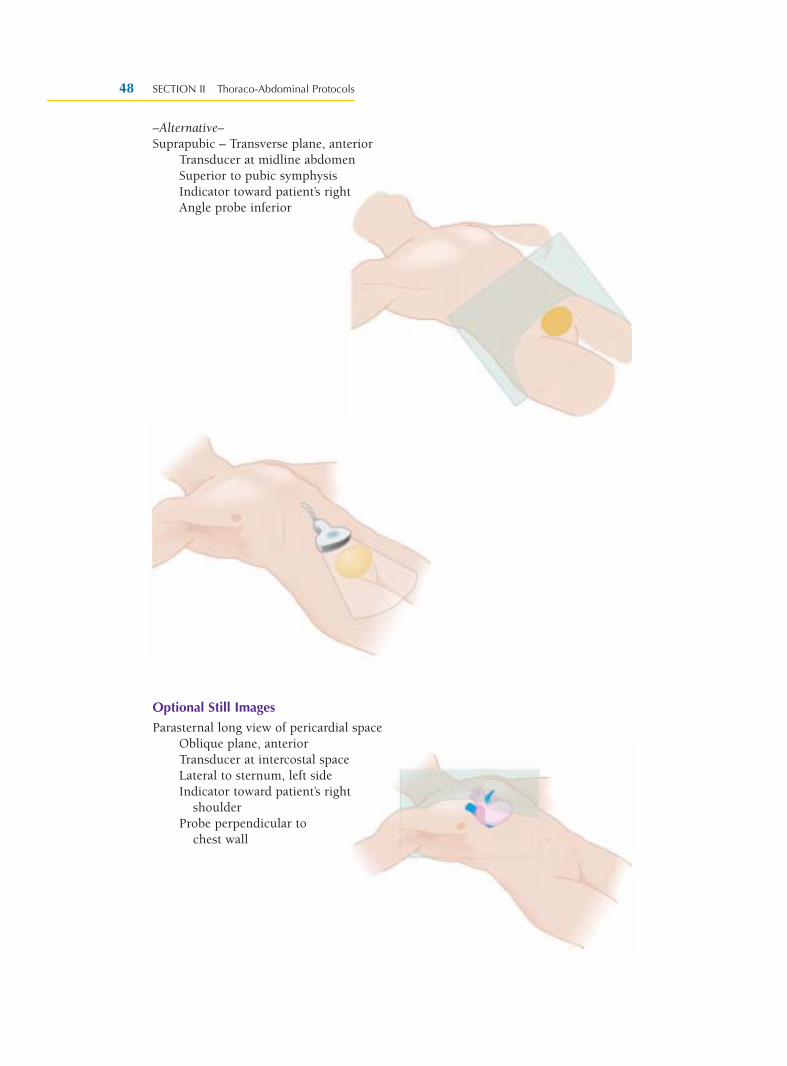
–Alternative–Suprapubic – Transverse plane, anterior
Transducer at midline abdomenSuperior to pubic symphysisIndicator toward patient’s rightAngle probe inferior
Optional Still Images
Parasternal long view of pericardial spaceOblique plane, anteriorTransducer at intercostal spaceLateral to sternum, left sideIndicator toward patient’s right
shoulderProbe perpendicular to
chest wall
SECTION II Thoraco-Abdominal Protocols48
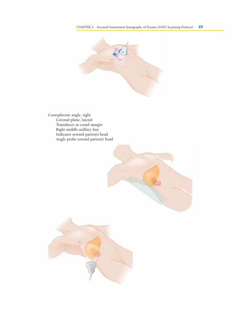
Costophrenic angle, rightCoronal plane, lateralTransducer at costal marginRight middle axillary lineIndicator toward patient’s headAngle probe toward patient’s head
CHAPTER 3 Focused Assessment Sonography of Trauma (FAST) Scanning Protocol 49
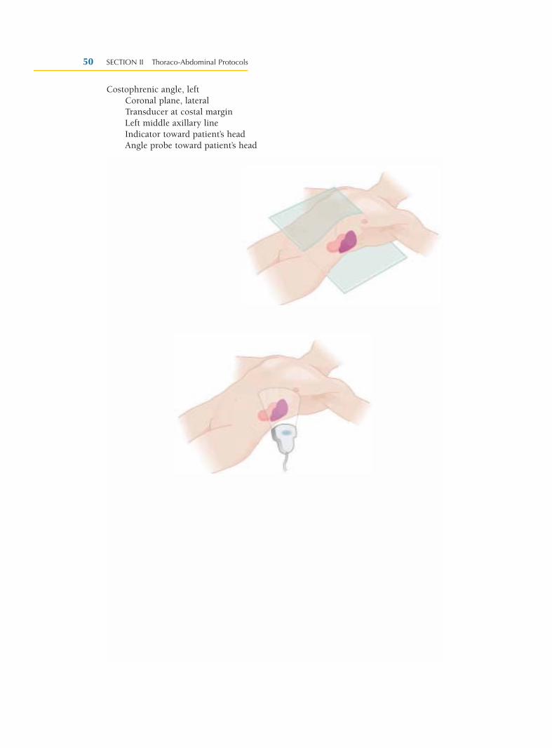
Costophrenic angle, leftCoronal plane, lateralTransducer at costal marginLeft middle axillary lineIndicator toward patient’s headAngle probe toward patient’s head
SECTION II Thoraco-Abdominal Protocols50
ULTRASOUND PROTOCOL TECHNIQUE
Videotape Protocol
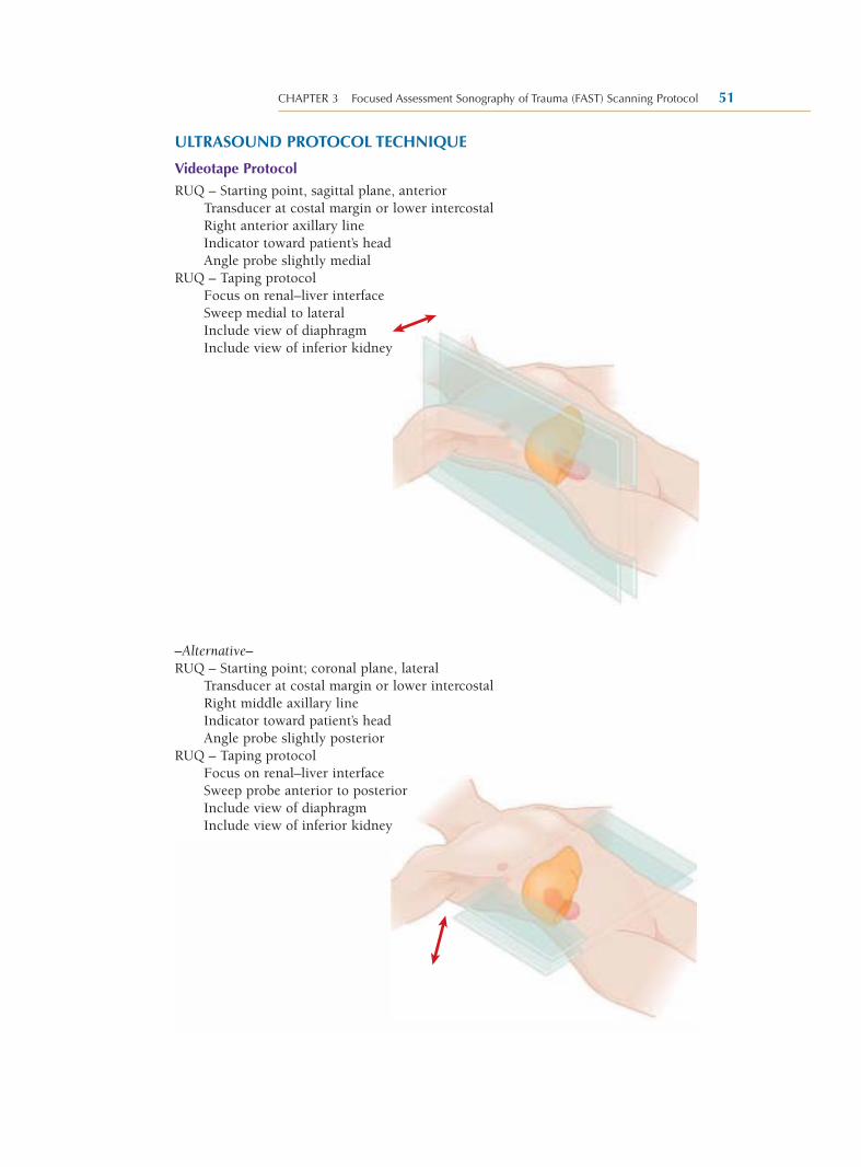
RUQ – Starting point, sagittal plane, anteriorTransducer at costal margin or lower intercostalRight anterior axillary lineIndicator toward patient’s headAngle probe slightly medial
RUQ – Taping protocolFocus on renal–liver interfaceSweep medial to lateralInclude view of diaphragmInclude view of inferior kidney
–Alternative–RUQ – Starting point; coronal plane, lateral
Transducer at costal margin or lower intercostalRight middle axillary lineIndicator toward patient’s headAngle probe slightly posterior
RUQ – Taping protocolFocus on renal–liver interfaceSweep probe anterior to posteriorInclude view of diaphragmInclude view of inferior kidney
CHAPTER 3 Focused Assessment Sonography of Trauma (FAST) Scanning Protocol 51
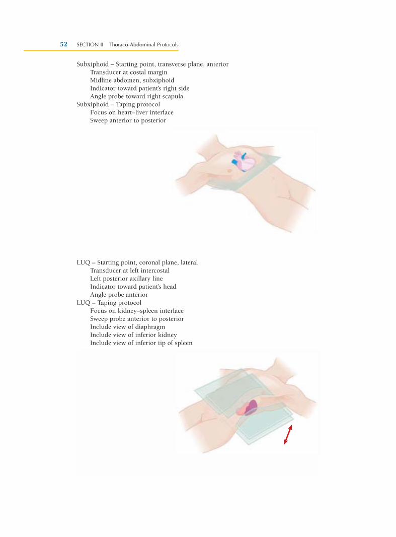
Subxiphoid – Starting point, transverse plane, anteriorTransducer at costal marginMidline abdomen, subxiphoidIndicator toward patient’s right sideAngle probe toward right scapula
Subxiphoid – Taping protocolFocus on heart–liver interfaceSweep anterior to posterior
LUQ – Starting point, coronal plane, lateralTransducer at left intercostalLeft posterior axillary lineIndicator toward patient’s headAngle probe anterior
LUQ – Taping protocolFocus on kidney–spleen interfaceSweep probe anterior to posteriorInclude view of diaphragmInclude view of inferior kidneyInclude view of inferior tip of spleen
SECTION II Thoraco-Abdominal Protocols52
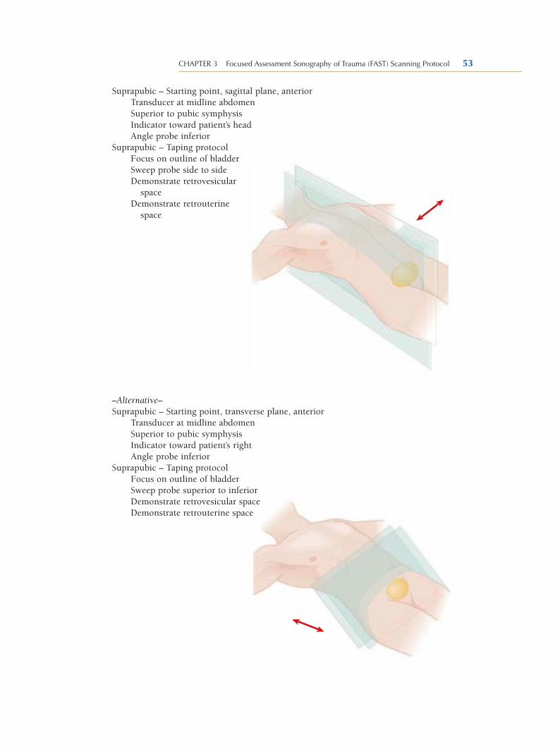
Suprapubic – Starting point, sagittal plane, anteriorTransducer at midline abdomenSuperior to pubic symphysisIndicator toward patient’s headAngle probe inferior
Suprapubic – Taping protocolFocus on outline of bladderSweep probe side to sideDemonstrate retrovesicular
spaceDemonstrate retrouterine
space
–Alternative–Suprapubic – Starting point, transverse plane, anterior
Transducer at midline abdomenSuperior to pubic symphysisIndicator toward patient’s rightAngle probe inferior
Suprapubic – Taping protocolFocus on outline of bladderSweep probe superior to inferiorDemonstrate retrovesicular spaceDemonstrate retrouterine space
CHAPTER 3 Focused Assessment Sonography of Trauma (FAST) Scanning Protocol 53
Optional Video Images
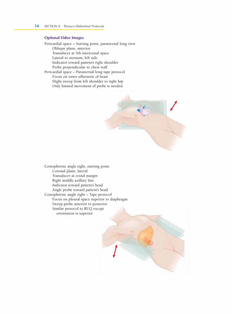
Pericardial space – Starting point, parasternal long viewOblique plane, anteriorTransducer at 5th intercostal spaceLateral to sternum, left sideIndicator toward patient’s right shoulderProbe perpendicular to chest wall
Pericardial space – Parasternal long tape protocolFocus on outer silhouette of heartSlight sweep from left shoulder to right hipOnly limited movement of probe is needed
Costophrenic angle right, starting pointCoronal plane, lateralTransducer at costal marginRight middle axillary lineIndicator toward patient’s headAngle probe toward patient’s head
Costophrenic angle right – Tape protocolFocus on pleural space superior to diaphragmSweep probe anterior to posteriorSimilar protocol to RUQ except
orientation is superior
SECTION II Thoraco-Abdominal Protocols54
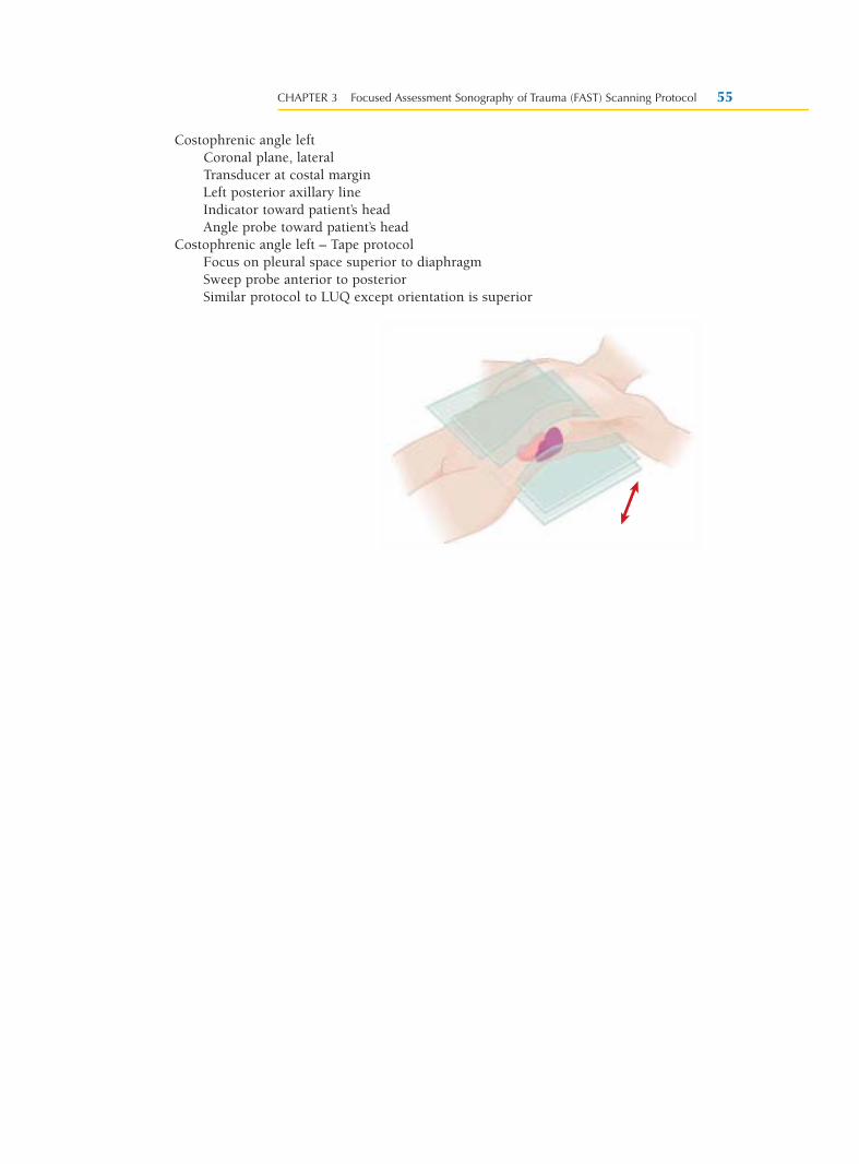
Costophrenic angle leftCoronal plane, lateralTransducer at costal marginLeft posterior axillary lineIndicator toward patient’s headAngle probe toward patient’s head
Costophrenic angle left – Tape protocolFocus on pleural space superior to diaphragmSweep probe anterior to posteriorSimilar protocol to LUQ except orientation is superior
CHAPTER 3 Focused Assessment Sonography of Trauma (FAST) Scanning Protocol 55