Embed Size (px)
Citation preview

The Rayne Institute, 4th Floor, Lambeth Wing
King’s College London
St. Thomas’ Hospital, Westminster Bridge Road-
London, SE1 7EH, UK .
RX114 – HeartFlow Technical Evaluation
08
08
Fall
The Rayne Institute, 4th Floor, Lambeth Wing
King’s College London
St. Thomas’ Hospital, Westminster Bridge Road-
London, SE1 7EH, UK .
1
Table of Contents
Table of Figures ....................................................................................... 2
Project Details ......................................................................................... 3
Abbreviations ......................................................................................... 4
1. Summary .......................................................................................... 5
2. Intended Use .................................................................................... 6
3. Scanners and Scanning Guidelines .................................................... 7
4. Software Version .............................................................................. 8
5. Analysis Workflow ............................................................................ 9
5.1 Data Upload 9 5.2 Quality Check 9 5.3 Anatomy Extraction 9 5.4 Identification of Regions of Un-Interpretability 10 5.5 Plaque Removal 11 5.6 Lumen Extraction 11 5.7 Case preparation 11 5.8 Review 12
6. Reproducibility ............................................................................... 13
7. Analyst Training and Workload ....................................................... 15
8. Risk Analysis ................................................................................... 16
9. Security .......................................................................................... 17
References ............................................................................................ 19
Appendix 1 ............................................................................................ 20
Appendix 2 ............................................................................................ 22

The Rayne Institute, 4th Floor, Lambeth Wing
King’s College London
St. Thomas’ Hospital, Westminster Bridge Road-
London, SE1 7EH, UK .
2
Table of Figures
Figure 1: Review process ............................................................................................... 12
Figure 2: Case turnaround time on a monthly basis ..................................................... 16
Figure 3: Number of cases analysed ............................................................................. 16

The Rayne Institute, 4th Floor, Lambeth Wing
King’s College London
St. Thomas’ Hospital, Westminster Bridge Road-
London, SE1 7EH, UK .
3
Project Details
Work package reference RX114
Work package name HeartFlow Technical Evaluation
Produced by KiTEC - King's Technology Evaluation Centre
King's College London
4th Floor, Lambeth Wing
St. Thomas’ Hospital
Westminster Bridge Road
London, SE1 7EH, UK
phone: +44 (0) 207 188 3052
Authors (alphabetical) Chalkidou, Anastasia
Keevil, Stephen
Lewis, Cornelius
McMillan, Viktoria
Correspondence to Viktoria McMillan, [email protected]
Date report completed 29th May 2015
Version 2.2
The Rayne Institute, 4th Floor, Lambeth Wing
King’s College London
St. Thomas’ Hospital, Westminster Bridge Road-
London, SE1 7EH, UK .
4
Abbreviations
BPM Beats per minute
CCTA Coronary Computed Tomography Angiography
CI Confidence Intervals
CT Computed Tomography
CTQ Computed tomography Quality
DICOM Digital Imaging and Communication in Medicine
DOB Date of Birth
FFR Fractional Flow Reserve
FFRCT Device name
FOV Field of vision
HDP High Density Plaque
HU Hounsfield Unit
IDP Intermediate Density Plaque
IFU Instructions for Use
LAD Left Anterior Descending
LCX Left Circumflex artery
LM Left Main artery
LDP Low Density Plaque
LOA Limits of agreement
MTAC Medical Technologies Advisory Committee
NIST National Institute of Standards and Technology
PACS Picture Archiving and Communication System
PHI Protected Health Information
R&R Repeatability & Reproducibility
RCA Right Coronary Artery
ROU Region of Un-Interpretability
SCCT Society of Cardiovascular Computed Tomography
SD Standard Deviation
SOP Standard Operating Procedure
QA/QC Quality Assurance/Quality Control

The Rayne Institute, 4th Floor, Lambeth Wing
King’s College London
St. Thomas’ Hospital, Westminster Bridge Road-
London, SE1 7EH, UK .
5
1. Summary
HeartFlow FFRCT was selected by the NICE MTAC as a technology suitable for guidance
production on the 18th of December 2014. The MTAC, however, raised the following concerns
about the technology.
The reproducibility of the results. Specifically, this related to the role of a Case Analyst in
the mathematical modelling on which the HeartFlow FFRCT results are based.
Information governance arrangements for the remote processing of data.
In response to these concerns, KiTEC, in collaboration with the sponsor, has provided an outline
of the procedure and listed available evidence on reproducibility. Furthermore, using publicly
available information and information provided by the sponsor, issues related to Analyst training
and workload, risk analysis and security have been addressed. KiTEC concluded the following.
There is a QA/QC process in place that ensures only data that fulfil the quality
requirements are processed. To further increase the quality assurance and minimise
risk, different members of the team are responsible for performing different parts of the
analysis.
The majority of the analysis is automated; however, the Analyst can manually edit any
part of the analysis. These edits can affect FFRCT estimation. Despite this, according to
Gaur et al. (2014), who published the only available data on FFRCT reproducibility, the
complete process results in acceptable 95% CI limits of agreement of -0.06 to 0.08.
Lumen extraction reproducibility, one step in the process of FFRCT computation,
decreases in the distal portion of the vessel (Gage R&R = 29.4%). This could be a result of
multiple variables including lower contrast perfusion/CT quality at the distal end of the
vessel, lower CT resolution, smaller vessel diameter, and disease burden. FFRCT
reproducibility was found to be equivalent to invasive FFR reproducibility.
According to published evidence, FFRCT slightly underestimates values in comparison
with FFR (Koo et al. 2011, Min et al. 2012, Norgaard et al. 2014).
As part of a continuous monitoring program, HeartFlow monitors FFRCT reproducibility
by re-processing 5% of its commercial case volume on a weekly basis (currently 206
vessels from 60 cases are processed twice). According to the sponsor, reproducibility is
consistent with the published literature (Gaur et al. 2014).

The Rayne Institute, 4th Floor, Lambeth Wing
King’s College London
St. Thomas’ Hospital, Westminster Bridge Road-
London, SE1 7EH, UK .
6
The sponsor fulfils all the requirements for protecting data confidentiality and integrity
for remote processing. Furthermore, the ability to process fully anonymised DICOM data
enables NHS customers to adopt this approach for extra security.
2. Intended Use
HeartFlow FFRCT is a post-processing image analysis software package that non-invasively
estimates FFR using previously acquired CCTA studies for clinically stable symptomatic patients
with coronary artery disease. The safety and effectiveness of the FFRCT analysis has not been
evaluated for the following populations (HeartFlow 2015).
Suspicion of acute coronary syndrome
Myocardial infarction within the last month (30 days)
Complex congenital heart disease
Prior coronary artery bypass graft surgery
Patients with a Body Mass Index >35
Patients who require emergency procedures or have any evidence of ongoing or active
clinical instability, including acute chest pain (sudden onset), cardiogenic shock, unstable
blood pressure with systolic blood pressure <90 mmHg, severe congestive heart failure
(New York Heart Association III or IV) or acute pulmonary oedema
In addition, due to the potential for artefacts in the CT data or degradation of CT data quality,
the safety and effectiveness of the FFRCT analysis has not been evaluated for the following
populations (HeartFlow 2015).
Patients with intracoronary metallic stents
Patients with prior pacemaker or internal defibrillator lead implantation
Patients with prosthetic heart valves
Patients with significant arrhythmias or tachycardia (uncontrolled by medication) that
would preclude CT acquisition
Coronary vessels with excessive calcification

The Rayne Institute, 4th Floor, Lambeth Wing
King’s College London
St. Thomas’ Hospital, Westminster Bridge Road-
London, SE1 7EH, UK .
7
3. Scanners and Scanning Guidelines
The CCTA imaging data for HeartFlow analysis must be acquired by scanners designed for
coronary imaging applications (≥64 slices). Scanners from all major vendors including GE,
Siemens, Phillips and Toshiba, have been successfully used for HeartFlow analysis. HeartFlow’s
scanning protocol follows the SCCT guideline (Abbara et al. 2009). According to this guideline the
CCTA acquisition parameters are as follows.
1. Patient specific inclusion: Heart Rate, < 65 bpm, ideally < 60 bpm
2. Coronary CT acquisition
Minimum number of slices: 64,
Tube voltage: 100-120kV,
Tube current: adjust for patient size/weight and desired image noise,
Minimum slice thickness: < 1mm,
Minimum axial resolution: 0.35mm or 14.2 lp/cm,
Cardiac gating
i. Prospective,
ii. Retrospective,
iii. Flash
Scan range: tracheal bifurcation or the mid-level of the left pulmonary artery to
just below the lower cardiac border.
3. Image reconstruction and post processing
Reconstruction kernel: FFRCT was validated with datasets from multiple CT
platform vendors and reconstruction algorithms and techniques, including
iterative reconstruction algorithms. HeartFlow has no recommendation on
reconstruction algorithms or techniques. HeartFlow is able to generate
anatomic models in all cases where there is sufficient contrast-to-noise such
that lumen boundaries can be visualized, irrespective of specific parameters.
Cardiac phase
i. Diastolic 100%
ii. Systolic
FOV: < 25.0 cm
4. Pre-acquisition medications
Beta blockers
The Rayne Institute, 4th Floor, Lambeth Wing
King’s College London
St. Thomas’ Hospital, Westminster Bridge Road-
London, SE1 7EH, UK .
8
i. Administration: administer to achieve short-term heart rate reduction
for the purpose of CCTA
ii. Route: oral and intravenous
Nitrates
i. Dosage / route: 400–800mg (1–2 tablets, and preferably the latter) of
sublingual nitroglycerin a few minutes before the initiation of the scan
protocol.
4. Software Version
The original CE mark was granted for FFRCT version 1.x. The technical file has been updated to
support subsequent releases including HeartFlow’s most recent commercial release, version 1.7.
According to the sponsor there were only minor differences between versions, all of which were
intended to address usability and support issues. The sponsor claims that none of these changes
impacted upon the intended use or principles of operation. However, the EAC notes that
different versions of the software can have a significant impact on diagnostic accuracy. For
example in the NXT trial (Norgaard et al. 2014) changes in the automated image processing
methods to more accurately identify the luminal boundary and changes in the physiological
models (models of microcirculatory resistance) were implemented. This resulted in a substantial
improvement in diagnostic performance when evaluated retrospectively by using data from the
DISCOVER-FLOW (Koo et al. 2011) and DeFACTO studies (Min et al. 2012).
*******************************************************************************
*******************************************************************************
********************************************************************************
********************************************************************************
********************************************************************************
********************************************************************************
*******************************************************************************
********************************************************************************
********************************************************************************
*******************************************************************************
*******************************************************************************

The Rayne Institute, 4th Floor, Lambeth Wing
King’s College London
St. Thomas’ Hospital, Westminster Bridge Road-
London, SE1 7EH, UK .
9
*******************************************************************************
****************************************
5. Analysis Workflow
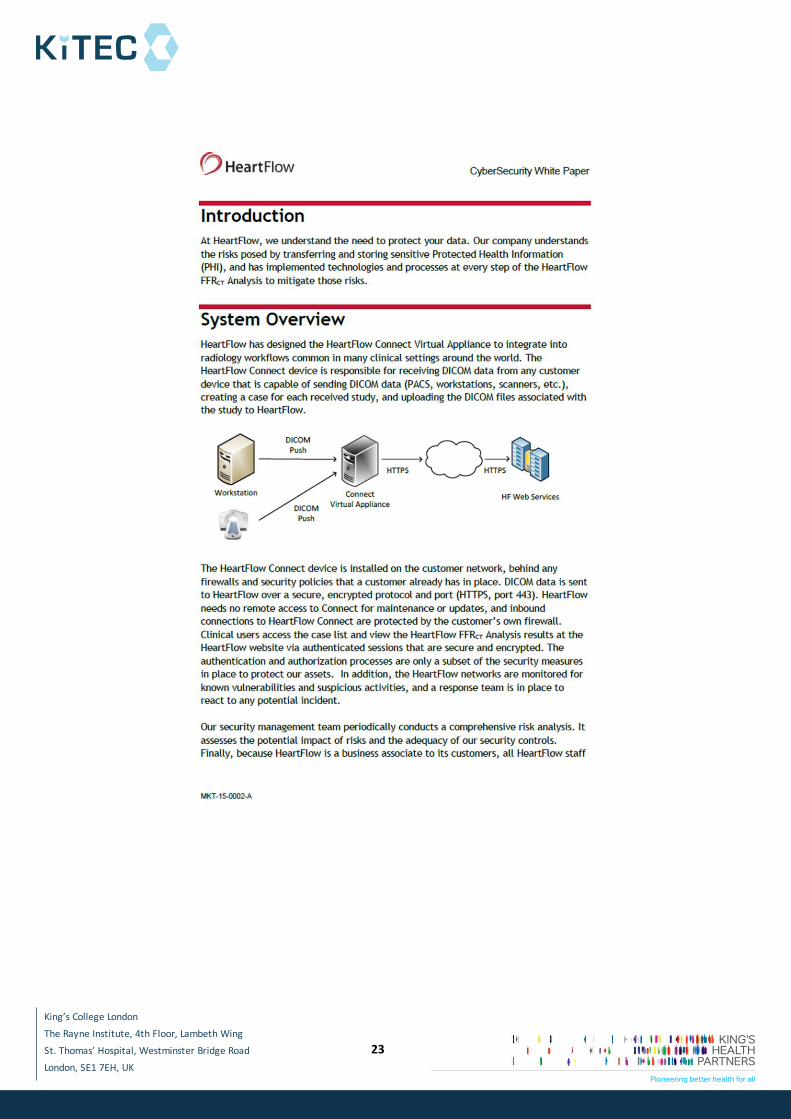
5.1 Data Upload The clinician uploads the data on HeartFlow servers using HeartFlow Connect, a network
application that enables the CCTA data to be transmitted via a secure connection to the
HeartFlow core laboratory for analysis. Data can be uploaded from any device capable of sending
DICOM data including PACS, workstations or directly from CT scanners.
5.2 Quality Check
*******************************************************************************
*******************************************************************************
*******************************************************************************
*******************************************************************************
*******************************************************************************
*******************************************************************************
*******************************************************************************
*******************************************************************************
*******************************************************************************
*******************************************************************************
*******************************************************************************
*******************************************************************************
*******************************************************************************
*******************************************************************************
*******************************************************************************
*******************************************************************************
******************
5.3 Anatomy Extraction

The Rayne Institute, 4th Floor, Lambeth Wing
King’s College London
St. Thomas’ Hospital, Westminster Bridge Road-
London, SE1 7EH, UK .
10
***************************************************************************************************************************************************************************************************************************************************************************************************************************************************************************************************************************************************************************************************************************************************************************************************************************************************************************************************************************************************************************************************************************************************************************************************************************************************************************************************************************************************************************************************************************************************************************************************************1****************************************************************************************************************************************************************************************************************************************************1******************************************************
**********************************
*
*
*
*
*
*
**************************
*
*
*
********************************************************************
*
*
*
***************************************************************************
***************************************************************************
****************************************************************************
***************
*
*
*
****************************************************************************
***************************************************************************
***************************************************************************
**************
5.4 Identification of Regions of Un-Interpretability *******************************************************************************
*******************************************************************************
The Rayne Institute, 4th Floor, Lambeth Wing
King’s College London
St. Thomas’ Hospital, Westminster Bridge Road-
London, SE1 7EH, UK .
11
*******************************************************************************
*******************************************************************************
*********************************
5.5 Plaque Removal
*******************************************************************************
*******************************************************************************
*******************************************************************************
*******************************************************************************
*******************************************************************************
**********************************************
5.6 Lumen Extraction
***********************************************************************
***********************************************************************
***********************************************************************
****************************************************************
*********************************************************************
********
********
**********
*******************************************************************************
*******************************************************************************
*******************************************************************************
*******************************************************************************
**************************************************************
5.7 Case preparation
*******************************************************************************
*******************************************************************************

The Rayne Institute, 4th Floor, Lambeth Wing
King’s College London
St. Thomas’ Hospital, Westminster Bridge Road-
London, SE1 7EH, UK .
12
*******************************************************************************
******************************************
5.8 Review
*************************************************************************************************************************************************************************************************************************************************************************************************************************************************************************************************************************************************************************************************************1**********************************************************************************************1**************************
*******************************************************************************
*******************************************************************************
*******************************************************************************

The Rayne Institute, 4th Floor, Lambeth Wing
King’s College London
St. Thomas’ Hospital, Westminster Bridge Road-
London, SE1 7EH, UK .
13
***************************************************************************2***
*********************************************************
********* ******* **********
*************** * *
**************** * *
*************** * *
************************* * *
********************* * *
************** * *
********** * *
**************** * *
**************** * *
********* * *
****************** * *
6. Reproducibility
*******************************************************************************
********************************************************************************
*******************************************************************************
*******************************************************************
Table 3: Summary of reproducibility assessments performed by the manufacturer
************* **************************
***************** ****************************************************
**************** **************************************************
******* **************************************************
*********************
*********************
*
*******************************************************
**************************************************
****************** ****************************************************
*********************
********
*******************************************************
*******************************************************

The Rayne Institute, 4th Floor, Lambeth Wing
King’s College London
St. Thomas’ Hospital, Westminster Bridge Road-
London, SE1 7EH, UK .
14
*******************************************************************************
*******************************************************************************
*******************************************************************************
*******************************************************************************
*******************************************************************************
*******************************************************************************
*******************************************************************************
*******************************************************************************
********************************************************
The EAC retrieved one study which discussed the reproducibility of the HeartFlow analysis (Gaur
et al. 2014). The data provided in this publication was the initial reproducibility data submitted to
the FDA and was not required for CE mark approval. The variation of repeated FFRCT analyses was
shown to be non-inferior to the variation of repeated FFR measurements (Gaur et al. 2014).
However, the FFRCT measurements were performed in patients with mean FFR of 0.89
(SD=0.067), well above the cut-off of 0.8 that is considered as diagnostic of lesion-specific
ischaemia (Tonino et al. 2009, Serruys et al. 2012). FFR was ≤0.80 in only 12 out of 58 vessel
measurements (21%). As highlighted by the authors, because of the relatively small sample size
in this study, they were not able to determine the reproducibility of FFRCT analyses in vessels
with FFR in a narrower and more clinically relevant range (e.g. 0.75–0.85).
********************************************************************************
*******************************************************************************
*******************************************************************************
*******************************************************************************
*******************************************************************************
*******************************************************************************
*******************************************************************************
********************************************************************************
*******************************************************************************
*******************************************************************************
*******************************************************************************
*****************************************************

The Rayne Institute, 4th Floor, Lambeth Wing
King’s College London
St. Thomas’ Hospital, Westminster Bridge Road-
London, SE1 7EH, UK .
15
According to published evidence, there is good correlation between FFRCT and FFR; however,
FFRCT systematically underestimates FFR values, as outlined below (Koo et al. 2011, Min et al.
2012, Norgaard et al. 2014).
Mean difference ±SD 0.022 ±0.116, p=0.016 (Koo et al. 2011)
Mean difference 0.058; 95% CI, 0.05-0.07 (Min et al. 2012)
Mean difference ±SD 0.03±0.074, p<0.001 (Norgaard et al. 2014)
In addition a recent conference abstract by (Gaur et al. 2015) has shown moderate agreement
between FFRCT and FFR).
Finally, as noted in the sponsor’s IFU, because of the variability in FFRCT results, they should be
reviewed by an appropriately trained clinician alongside clinical data, including the patient’s
original CT images, clinical history, symptoms and other diagnostic tests, before any decisions
about treatment are made.
7. Analyst Training and Workload
*******************************************************************************
*******************************************************************************
*******************************************************************************
*******************************************************************************
*******************************************************************************
***************** Prior to requesting this information from HeartFlow, the EAC informally
approached a cardiologist to ascertain the time they thought would be required to train
someone in basic cardiac anatomy to the level required of a Case Analyst. They confirmed that 3-
4 months would be adequate.
*******************************************************************************
*******************************************************************************
*******************************************************************************
*******************************************************************************
********************************************************************Error!
Reference source not found.*****
The Rayne Institute, 4th Floor, Lambeth Wing
King’s College London
St. Thomas’ Hospital, Westminster Bridge Road-
London, SE1 7EH, UK .
16
*******3*********************************************************2*************
****************************
Figure redacted
*******3***************************
Figure removed
8. Risk Analysis
According to the sponsor, risk analysis is conducted continuously, throughout the process
lifecycle. For risks where software error is mitigated through design, the applicable design
requirement(s) is referenced in the risk mitigation.
*******************************************************************************
*******************************************************************************
********************************************************************************
*******************************************************************************
*******************************************************************************

The Rayne Institute, 4th Floor, Lambeth Wing
King’s College London
St. Thomas’ Hospital, Westminster Bridge Road-
London, SE1 7EH, UK .
17
*******************************************************************************
*******************************************************************************
*******************************************************************************
*******************************************************************************
Table ***
Table 4: A summary of the tasks each individual can perform for each case
**********
**
***********
**
******
*
*****************
***
*******
*
***************
***
* *
***************
***
* *
***************
***
* *
***************
***
* *
***************
***
* *
***************
***
* *
***************
***
* *
*************************************************************
9. Security
The DICOM data used for HeartFlow analysis often contain PHI (such as patient name, NHS
number, DOB etc. HeartFlow can also process completely anonymised data, if this is preferred by
the customer site, without compromising the results. If PHI is included, controls are
implemented to ensure data protection. Data confidentiality measures are addressed through
business associate agreements for PHI data.
Access to HeartFlow’s data centre in San Jose, California is very restricted and HeartFlow
accesses this data using password protected workstations. To ensure data integrity, all data is
validated upon receipt at HeartFlow using a checksum. To avoid malicious attacks HeartFlow
The Rayne Institute, 4th Floor, Lambeth Wing
King’s College London
St. Thomas’ Hospital, Westminster Bridge Road-
London, SE1 7EH, UK .
18
performs constant monitoring and load balancing. To gain FDA approval (FDA 2013), the sponsor
submitted platform, application and procedure controls to address the following considerations.
data confidentiality
data integrity
data availability
denial of service attacks
malware
A number of security protocols have been implemented to ensure the security of data that is
sent to HeartFlow. All FDA and CE questions regarding data security were addressed to their
satisfaction, resulting in FDA product clearance and TUV CE-mark. The security protocols include
the following.
HeartFlow web service architecture has been designed to provide business continuity,
with limited exposure. According to the sponsor the service does not have any single
point of failure
All data transmission is encrypted (SSL, TLS1.0, 1.1, 1.2)
A firewall server controls all incoming traffic
All data is stored at data centres with restricted and audited access
The NIST cybersecurity guidance is followed1
A disaster recovery plan is in place
System and application logs are collected
The sponsor informed KiTEC that HeartFlow has employed a 3rd party to monitor cyber-security.
This group is performing a risk analysis and will attempt to hack into HeartFlow’s system to
ensure all security measures are addressed. The sponsor has provided a white paper on
cybersecurity that provides additional details on the security protocols used by HeartFlow
(Appendix 2).
1 http://www.nist.gov/cyberframework/upload/cybersecurity-framework-021214.pdf

The Rayne Institute, 4th Floor, Lambeth Wing
King’s College London
St. Thomas’ Hospital, Westminster Bridge Road-
London, SE1 7EH, UK .
19
References
Abbara, S., A. Arbab-Zadeh, T. Q. Callister, et al. (2009). "SCCT guidelines for performance of coronary computed tomographic angiography: a report of the Society of Cardiovascular Computed Tomography Guidelines Committee." J Cardiovasc Comput Tomogr 3(3): 190-204. FDA (2013). DE NOVO CLASSIFICATION REQUEST FOR FFRCT V. 1.4. C. f. D. a. R. Health.

The Rayne Institute, 4th Floor, Lambeth Wing
King’s College London
St. Thomas’ Hospital, Westminster Bridge Road-
London, SE1 7EH, UK .
20
Gaur, S., H. G. Bezerra, J. F. Lassen, et al. (2014). "Fractional flow reserve derived from coronary CT angiography: Variation of repeated analyses." Journal of Cardiovascular Computed Tomography 8(4): 307-14. Gaur, S., J. Jensen, H. E. Botker, et al. (2015). "NON-INVASIVE FRACTIONAL FLOW RESERVE DERIVED FROM CORONARY COMPUTED TOMOGRAPHY ANGIOGRAPHY: EXPERIENCES FROM REAL-WORLD CLINICAL PRACTICE." Journal of the American College of Cardiology 65(10_S). HeartFlow (2015). FFRCT v1.7.0 Instructions for Use. Koo, B. K., A. Erglis, J. H. Doh, et al. (2011). "Diagnosis of ischemia-causing coronary stenoses by noninvasive fractional flow reserve computed from coronary computed tomographic angiograms: Results from the prospective multicenter DISCOVER-FLOW (Diagnosis of Ischemia-Causing Stenoses Obtained Via Noninvasive Fractional Flow Reserve) study." Journal of the American College of Cardiology 58(19): 1989-97. Min, J. K., J. Leipsic, M. J. Pencina, et al. (2012). "Diagnostic accuracy of fractional flow reserve from anatomic CT angiography." JAMA 308(12): 1237-45. Norgaard, B. L., J. Leipsic, S. Gaur, et al. (2014). "Diagnostic performance of noninvasive fractional flow reserve derived from coronary computed tomography angiography in suspected coronary artery disease: the NXT trial (Analysis of Coronary Blood Flow Using CT Angiography: Next Steps)." Journal of the American College of Cardiology 63(12): 1145-55. Serruys, P. W., C. Girasis, S.-L. Papadopoulou, et al. (2012). "Non-invasive fractional flow reserve: scientific basis, methods and perspectives." EuroIntervention 8(4): 511-19. Tonino, P. A. L., B. De Bruyne, N. H. J. Pijls, et al. (2009). "Fractional Flow Reserve versus Angiography for Guiding Percutaneous Coronary Intervention." New England Journal of Medicine 360(3): 213-24.
Appendix 1
********************************************************************************
*******************************************************************************
*******************************************************************************
*******************************************************************************
********************************************************************************

The Rayne Institute, 4th Floor, Lambeth Wing
King’s College London
St. Thomas’ Hospital, Westminster Bridge Road-
London, SE1 7EH, UK .
21
*******************************************************************************
*******************************************************************************
*******************************************************************************
*******************************************************************************
********************************************************************************
*******************************************************************************
*******************************************************************************
*******************************************************************************
********************************************************************************
*******************************************************************************
********************************************************************************
*******************************************************************************
*******************************************************************************
*******************************************************************************
*******************************************************************************
********************************************************************************
*******************************************************************************
*******************************************************************************
*****************
********************************************************************************
*******************************************************************************
********************************************************************************
*******************************************************************************
*******************************************************************************
********************************************************************************
The Rayne Institute, 4th Floor, Lambeth Wing
King’s College London
St. Thomas’ Hospital, Westminster Bridge Road-
London, SE1 7EH, UK .
22
*******************************************************************************
*******************************************************************************
*******************************************************************************
********************************************************************************
*****************************************************************
Appendix 2
The Rayne Institute, 4th Floor, Lambeth Wing
King’s College London
St. Thomas’ Hospital, Westminster Bridge Road-
London, SE1 7EH, UK .
23

The Rayne Institute, 4th Floor, Lambeth Wing
King’s College London
St. Thomas’ Hospital, Westminster Bridge Road-
London, SE1 7EH, UK .
24